K. Yonenobu · K. Nakamura · Y. Toyama (Eds.)
OPLL Ossification of the Posterior Longitudinal Ligament 2nd Edition
K. ...
120 downloads
789 Views
11MB Size
Report
This content was uploaded by our users and we assume good faith they have the permission to share this book. If you own the copyright to this book and it is wrongfully on our website, we offer a simple DMCA procedure to remove your content from our site. Start by pressing the button below!
Report copyright / DMCA form
K. Yonenobu · K. Nakamura · Y. Toyama (Eds.)
OPLL Ossification of the Posterior Longitudinal Ligament 2nd Edition
K. Yonenobu • K. Nakamura • Y. Toyama (Eds.)
OPLL
Ossification of the Posterior Longitudinal Ligament 2nd Edition
With 280 Figures
Kazuo Yonenobu, M.D., D.Med.Sc. Vice-Director National Hospital Organization Osaka-Minami Medical Center 2-1 Kidohigashi, Kawachinagano, Osaka 586-8521, Japan Kozo Nakamura, M.D., Ph.D. Professor and Chairman Department of Orthopaedic Surgery Faculty of Medicine, The University of Tokyo 7-3-1 Hongo, Bunkyo-ku, Tokyo 113-8655, Japan Yoshiaki Toyama, M.D., Ph.D. Professor and Chairman Department of Orthopedic Surgery School of Medicine, Keio University 35 Shinanomachi, Shinjuku-ku, Tokyo 160-8582, Japan
ISBN-10 4-431-32561-1 Springer Tokyo Berlin Heidelberg New York ISBN-13 978-4-431-32561-1 Springer Tokyo Berlin Heidelberg New York Library of Congress Control Number: 2006925431 Printed on acid-free paper © Springer 2006, 1997 Printed in Japan This work is subject to copyright. All rights are reserved, whether the whole or part of the material is concerned, specifically the rights of translation, reprinting, reuse of illustrations, recitation, broadcasting, reproduction on microfilms or in other ways, and storage in data banks. The use of registered names, trademarks, etc. in this publication does not imply, even in the absence of a specific statement, that such names are exempt from the relevant protective laws and regulations and therefore free for general use. Product liability: The publisher can give no guarantee for information about drug dosage and application thereof contained in this book. In every individual case the respective user must check its accuracy by consulting other pharmaceutical literature. Typesetting: SNP Best-set Typesetter Ltd., Hong Kong Printing and binding: Hicom, Japan
Preface to the Second Edition
Ossification of the posterior longitudinal ligament (OPLL) is no longer only a Japanese disease. In 2004, 18 papers on OPLL and related conditions were published, and 7 of those were from countries other than Japan. Major textbooks on spine surgery, such as The Spine, The Cervical Spine, and Spine Surgery, have devoted chapters to OPLL. Although OPLL has been recognized as a distinct spinal disease entity, several questions regarding etiology and treatment have remained unanswered. In 2002, the Committee for Study of Ossification of Spinal Ligaments, subsidized by the Ministry of Health, Labour and Welfare and chaired by Professor K. Nakamura, decided to systematically review papers on OPLL and related conditions. The purposes were to direct the research activities of the committee more effectively and to provide more certain knowledge about OPLL, in the form of clinical practice guidelines, for general practitioners and for patients suffering from the condition. A committee for this task was formed in cooperation with the Japanese Orthopaedic Association, and clinical practice guidelines for OPLL, consisting of 4 chapters and 75 research questions, were developed after almost 3 years. Unfortunately, the guidelines have been published only in Japanese as of this writing. Therefore, 4 chapters of this book (“Overview of Epidemiology and Genetics,” “Overview of Etiology and Pathogenesis,” “Diagnosis of OPLL and OYL,” and “Overview of Treatment for Ossification of the Longitudinal Ligament and the Ligamentum Flavum”) were included as summaries of the 4 chapters of the guidelines. One of the important issues that arose during the development of the guidelines was that of diagnostic criteria. OPLL was discovered before computerized tomography had been devised; therefore, OPLL was diagnosed on the basis of clinical and roentgenographic findings from conventional imaging techniques such as plain roentgenography or tomography. However, with an increase in the diversity of medical professionals who take care of patients with spinal disease and with advances in imaging technology such as computerized tomography, a small ossified lesion that usually would not grow to compress the spinal cord is sometimes diagnosed as OPLL, which confuses patients. Diagnosis of OPLL has been made based on tactical knowledge—that is, knowledge held by a closed society made up of experts in the field. This is not a rare example. Several common spinal diseases, such as cervical spondylotic myelopathy, lumbar disc herniation, and lumbar canal stenosis, are diagnosed in this manner. The committee has set tentative diagnostic criteria for OPLL until more definite criteria can be established scientifically, and those tentative diagnostic criteria for OPLL are included in this book. Since 1997, when the first edition of OPLL was published, research on OPLL has progressed steadily in genetics and bone cell physiology. Genetic studies using a variety of approaches, supported by nationwide collaboration, seem to be narrowing in on a disease-related gene. The process of ossification in this condition has been elucidated by studies using techniques of bone cell physiology. Clinical studies using imaging and electrophysiological modalities have clarified the pathophysiology of the spinal cord in OPLL. Follow-up studies have revealed long-term (more than 10 years) results of surgical treatment of both posterior and anterior approaches. All the chapters have been updated with these findings.
V
VI
Preface to the Second Edition
As clearly shown in this new edition, many facts regarding hyperostotic conditions of the spine have been cleared up through research activities over the past 30 years, mainly by the successive committees on OPLL. However, many important questions in basic and clinical research have not yet been clarified. Among the basic ones: What are the causative genes? What mechanisms work in hypertrophy and ossification of spinal ligaments in the condition? And there are others. In the clinical area, surgical decompression of the spinal cord in thoracic OPLL and ossification of the yellow ligament (OYL) is still a challenging subject. Although arguments over choice of surgical procedure have been settling down, criteria for the surgical technique for individual patients have not yet been established. Progress in basic research of OPLL may largely depend on advances in the basic sciences of genetics, bone cell physiology, and related fields. In surgery, assiduous efforts by surgeons to devise a technique for better and safer results are mandatory. Such improvement in treatment for any surgical condition is general. Additionally, for relatively rare conditions such as OPLL, establishment of a system of clinical trials is important to substantiate the significance of a new treatment, and this is the surgeon’s task. Through the challenge posed by this disease, we Japanese spine surgeons have learned many things about bone biology and about the spine and spinal cord as well, and we have developed various surgical techniques to conquer the condition. OPLL, however, still confronts us, and to surmount this refractory hyperostotic condition of the spine, we must expand our research into new fields. Development of drugs to control bone formation is one example—drugs that not only prevent progression of ossification of the spinal ligament but that also preserve spinal mobility, which we spine surgeons sometimes have neglected. Another example is repair or regeneration of the injured spinal cord. I hope that these goals will be achieved with further study of OPLL. The editors express their sincere thanks to the members of the Committee on Clinical Practice Guidelines of OPLL: Drs. K. Yonenobu (Chairperson), M. Iwasaki, K. Satomi, T. Taguchi, M. Tanaka, Y. Toyama, and S. Matsunaga; and to the members of the working group for reviewing papers: Drs. H. Aono, Y. Itoh, S. Okuda, K. Kato, K. Kaneko, J. Kouno, K. Takeuchi, K. Toyoda, K. Hayashi, and A. Miyauchi. Without their perseverance, the guidelines as well as this book would not have been completed. Kazuo Yonenobu
Preface to the First Edition
“Man can see only what he knows.” Goethe Ossification of the Posterior Longitudinal Ligament (OPLL) has long been a challenge to orthopedic spinal surgeons in Japan, and their struggle to meet that challenge has marked a turning point in the history of spinal surgery. Investigation of the etiology and treatment of the condition has taught surgeons to see diseases of the spine and their surgical treatment in a new perspective. It was truly a surprise to learn that the posterior longitudinal ligament could become a thick, bony plate in the cervical spine and impinge on the spinal cord, leading to paralysis. Even more amazing, however, is that innumerable roentgenological findings of such thick, bony lesions could be overlooked for decades before OPLL became well recognized by physicians in Japan. Progress in diagnostic imaging technology, first in computed tomography (CT) and then in magnetic resonance imaging (MRI), has helped in diagnosis and evaluation of the disease and in deciding therapeutic modalities. There is no better tool than CT and CT myelography for demonstrating the real threat of OPLL to the cervical spinal cord. MRI, however, provides more information on widespread ossified lesions from the cervical to lumbar regions, and on the intramedullary changes caused by chronic compression. It is not a great exaggeration to say that OPLL is one of the leading reasons for the enthusiastic expansion in the market for the newest diagnostic imaging tools in Japan. Despite Tukiyama’s autopsy report of OPLL in 1960, the etiology of OPLL remained unclear and its symptoms and characteristics were unfamiliar until 1975, when the Investigation Committee for OPLL moved toward better patient care and research of the etiology of the disease. Under the auspices of the Ministry of Health and Welfare, diagnostic criteria for OPLL scoring both of physical manifestations and of roentgenological findings were first established. With dissatisfied patients who failed to recover after conventional laminectomy, a new technique of decompression had to be developed. Failure of surgical decompression was thought to be due to careless methods of laminectomy in which the thick rongeur blade or Kerrison punch was introduced into an extremely narrow spinal canal. Anterior diskectomy and interbody fusion by either the Smith-Robinson or Cloward method often caused paraplegia. In the 1960s, patients with OPLL thus remained unhappy even after surgery; it was a dreary time for spinal surgeons in this country. Then came the introduction of a high-speed surgical drill for laminectomy, along with technical developments such as expansive laminoplasty (Hirabayashi, 1981) and anterior decompression by the floating of OPLL (Yamaura, 1983), which ensured decompression of the spinal cord without excision. Considered as a systemic disease, OPLL was recognized rather early to occur with high frequency in patients suffering from diabetes mellitus. There were also a few reports on metabolic and endocrine disorders in close relationship with OPLL: hypophosphatemic rickets or hypoparathyroidism. OPLL is not simple calcification, however, but ossification of the ligaments; the etiological relationship between these disorders of calcium metabolism and ectopic ossification was explored in vain. As
VII
VIII
Preface to the First Edition
precise pathological study progressed, the real harm of the lesion proved to be hyperplasia or growth of the ligament leading to occupation of the spinal canal. Fibrocartilagenous cell proliferation and matrix hyperplasia and subsequent ossification were found to be the essential processes of OPLL. What mechanism, then, stimulates the growth of the ligament in a middle-aged or older person? Various growth factors or cytokines were found to be present in the growing front of OPLL, but the mechanism that releases or regulates them has yet to be clarified. A metabolic or endocrine system abnormality may influence this renewed growth. Predisposition to OPLL has been examined in familial surveys including studies of twins, and in the future, HLA gene analysis may be able to identify those at high risk od OPLL. Finally, we consider what impact surgery for OPLL has on traditional spinal surgery. Decompression with spinal stability unimpaired, expansion of developmental canal stenosis without laminectomy, or sufficient decompression without excision of a lesion mass—these have been developed to treat paralysis due to OPLL. They have been made possible through the enthusiastic research and practice of Japanese spinal surgeons, and are now widely applicable to all sorts of diseases of the spine, without being limited to the cervical spine. This monograph should be dedicated to those patients who were destined to suffer pain and paralysis without benefit of the current achievements in spinal surgery. Keiro Ono
Contents
Preface to the Second Edition . . . . . . . . . . . . . . . . . . . . . . . . . . . . . . . . . . . . . . . . . . . Preface to the First Edition. . . . . . . . . . . . . . . . . . . . . . . . . . . . . . . . . . . . . . . . . . . . . . Contributors . . . . . . . . . . . . . . . . . . . . . . . . . . . . . . . . . . . . . . . . . . . . . . . . . . . . . . . . .
V VII XIII
1. Introduction History of Research K. Nakamura . . . . . . . . . . . . . . . . . . . . . . . . . . . . . . . . . . . . . . . . . . . . . . . . . . . . . . . .
3
2. Epidemiology Overview of Epidemiology and Genetics S. Matsunaga and T. Sakou . . . . . . . . . . . . . . . . . . . . . . . . . . . . . . . . . . . . . . . . . . .
7
OPLL: Disease Entity, Incidence, Literature Search, and Prognosis S. Matsunaga and T. Sakou . . . . . . . . . . . . . . . . . . . . . . . . . . . . . . . . . . . . . . . . . . .
11
Genetic Susceptibility to OPLL I. Inoue . . . . . . . . . . . . . . . . . . . . . . . . . . . . . . . . . . . . . . . . . . . . . . . . . . . . . . . . . . . . . .
19
3. Pathology and Pathogenesis Overview of Etiology and Pathogenesis T. Taguchi . . . . . . . . . . . . . . . . . . . . . . . . . . . . . . . . . . . . . . . . . . . . . . . . . . . . . . . . . . .
29
Etiology and Pathogenesis T. Taguchi . . . . . . . . . . . . . . . . . . . . . . . . . . . . . . . . . . . . . . . . . . . . . . . . . . . . . . . . . . .
33
Contribution of Metabolic Conditions to Ossification of the Posterior Longitudinal Ligament of the Spine H. Kawaguchi, T. Akune, N. Ogata, A. Seichi, K. Takeshita, and K. Nakamura . . . . . . . . . . . . . . . . . . . . . . . . . . . . . . . . . . . . . . . . . . . . . . . . . . . . . . . .
37
Review of Histopathological Studies on OPLL of the Cervical Spine, with Insights into the Mechanism N. Tsuzuki . . . . . . . . . . . . . . . . . . . . . . . . . . . . . . . . . . . . . . . . . . . . . . . . . . . . . . . . . . .
41
Pathology of Ossification of the Ligamentum Flavum M. Yoshida . . . . . . . . . . . . . . . . . . . . . . . . . . . . . . . . . . . . . . . . . . . . . . . . . . . . . . . . . . .
49
Possible Roles of Bone Morphogenetic Proteins and Transforming Growth Factor-βs in the Pathogenesis of OPLL and OLF H. Yoshikawa . . . . . . . . . . . . . . . . . . . . . . . . . . . . . . . . . . . . . . . . . . . . . . . . . . . . . . . .
59
Pathology of Spinal Cord Lesions Caused by Ossification of the Posterior Longitudinal Ligament Y. Hashizume, T. Kameyama, J. Mizuno, H. Nakagawa, T. Yanagi, and M. Yoshida . . . . . . . . . . . . . . . . . . . . . . . . . . . . . . . . . . . . . . . . . . . . . . . . . . . . . . .
65
IX
X
Contents
Tiptoe Walking (ttw) Mouse S. Ikegawa . . . . . . . . . . . . . . . . . . . . . . . . . . . . . . . . . . . . . . . . . . . . . . . . . . . . . . . . . . .
71
Study of Ligament Ossification and Abnormal Glucose Tolerance in the Zucker Fatty Rat K. Yamamoto and K. Kubo. . . . . . . . . . . . . . . . . . . . . . . . . . . . . . . . . . . . . . . . . . . . .
77
Experimental Murine Model of Ossification of Spinal Ligaments Induced by Bone Morphogenetic Protein-2 K. Hoshi . . . . . . . . . . . . . . . . . . . . . . . . . . . . . . . . . . . . . . . . . . . . . . . . . . . . . . . . . . . . .
93
Spinal Cord Lesions in Spinal Hyperostotic Mouse (twy/twy) Simulating Ossification of the Posterior Longitudinal Ligament of the Cervical Spine H. Baba, K. Uchida, H. Nakajima, Y. Kokubo, R. Sato, T. Yayama, S. Kobayashi, T. Inukai, and M. Kimura. . . . . . . . . . . . . . . . . . . . . . . . . . . . . . . . .
101
4. Diagnosis of OPLL and OYL Diagnosis of OPLL and OYL: Overview M. Tanaka, A. Kanazawa, and K. Yonenobu. . . . . . . . . . . . . . . . . . . . . . . . . . . . .
111
Clinical Manifestation of Cervical OPLL K. Kaneko . . . . . . . . . . . . . . . . . . . . . . . . . . . . . . . . . . . . . . . . . . . . . . . . . . . . . . . . . . . .
115
Clinical Manifestations of Thoracic OPLL and OLF M. Matsumoto, K. Chiba, and Y. Toyama . . . . . . . . . . . . . . . . . . . . . . . . . . . . . . . .
121
Diagnostic Imaging of Cervical Ossification of the Posterior Longitudinal Ligament K. Nagata and K. Sato . . . . . . . . . . . . . . . . . . . . . . . . . . . . . . . . . . . . . . . . . . . . . . . .
127
Imaging Diagnosis of Thoracic OPLL and OLF I. Kikkawa and Y. Hoshino . . . . . . . . . . . . . . . . . . . . . . . . . . . . . . . . . . . . . . . . . . . .
145
Electrophysiological Diagnosis of Cervical OPLL Myelopathy K. Shinomiya, S. Tomizawa, and S. Kawabata . . . . . . . . . . . . . . . . . . . . . . . . . . .
151
5. Treatment of OPLL and OLF Overview of Treatment for Ossification of the Longitudinal Ligament and the Ligamentum Flavum M. Iwasaki . . . . . . . . . . . . . . . . . . . . . . . . . . . . . . . . . . . . . . . . . . . . . . . . . . . . . . . . . .
165
Pharmacotherapy for Ossification of the Spinal Ligaments: Clinical Trial of Disodium (1-Hydroxyethylidene) Diphosphonate to Inhibit Progression of Ossification of the Posterior Longitudinal Ligament in the Cervical Spine after Posterior Decompression Surgery K. Yonenobu, K. Nagata, K. Abumi, Y. Toyama, and S. Kawai . . . . . . . . . . . . . .
169
Conservative Treatment of Ossification of the Posterior Longitudinal Ligament in the Cervical Spine M. Sumi, M. Doita, and K. Nishida . . . . . . . . . . . . . . . . . . . . . . . . . . . . . . . . . . . . .
177
Choice of Surgical Procedure M. Iwasaki and K. Yonenobu . . . . . . . . . . . . . . . . . . . . . . . . . . . . . . . . . . . . . . . . . .
181
Expansive Laminoplasty Y. Toyama and K. Chiba . . . . . . . . . . . . . . . . . . . . . . . . . . . . . . . . . . . . . . . . . . . . . . .
187
Expansive Open-door Laminoplasty for Ossification of the Posterior Longitudinal Ligament of the Cervical Spine: Surgical Indications, Technique, and Outcomes K. Chiba, Y. Ogawa, M. Matsumoto, and Y. Toyama . . . . . . . . . . . . . . . . . . . . . .
193
Contents
XI
Double-door Laminoplasty by Splitting Spinous Processes A. Seichi, K. Takeshita, H. Kawaguchi, and K. Nakamura . . . . . . . . . . . . . . .
201
Anterior Cervical Decompression for Cervical Myelopathy Caused by Ossification of the Posterior Longitudinal Ligament K. Shinomiya, T. Matsuoka, Y. Kurosa, S. Shindo, O. Nakai, and M. Takahashi . . . . . . . . . . . . . . . . . . . . . . . . . . . . . . . . . . . . . . . . . . . . . . . . . . . . . . .
209
Treatment of OPLL and OLF of the Cervical Spine: Long-Term Results Y. Kawaguchi . . . . . . . . . . . . . . . . . . . . . . . . . . . . . . . . . . . . . . . . . . . . . . . . . . . . . . . .
219
Choice of Surgical Procedures for Thoracic Ossification of the Posterior Longitudinal Ligament A. Seichi, K. Takeshita, and K. Nakamura . . . . . . . . . . . . . . . . . . . . . . . . . . . . .
225
Anterior Decompression and Fusion for Ossification of the Posterior Longitudinal Ligament of the Thoracic Spine: Procedure and Clinical Outcomes of Transthoracic and Transsternal Approaches K. Ohnishi, K. Miyamoto, H. Hosoe, and K. Shimizu . . . . . . . . . . . . . . . . . . . . .
231
Circumspinal Decompression with Dekyphosis Stabilization for Thoracic Myelopathy due to Ossification of the Posterior Longitudinal Ligament N. Kawahara, K. Tomita, H. Murakami, S. Demura, Y. Sekino, W. Nasu, and Y. Fujimaki . . . . . . . . . . . . . . . . . . . . . . . . . . . . . . . . . . . . . . . . . . . . . .
235
Posterior Extensive Cervicothoracic Laminoplasty Y. Nakagawa and M. Yoshida . . . . . . . . . . . . . . . . . . . . . . . . . . . . . . . . . . . . . . . . .
241
Anterior Decompression Through Posterior Approach for Thoracic Myelopathy Caused by OPLL: Ohtsuka Procedure K. Abumi, M. Ito, and A. Minami . . . . . . . . . . . . . . . . . . . . . . . . . . . . . . . . . . . . . . .
249
Surgical Treatment for Ossification of the Posterior Longitudinal Ligament of the Thoracic Spine: Outcomes of One-Stage Posterior Decompression with Corrective Fusion Surgery Y. Matsuyama, H. Yoshihara, T. Tsuji, Y. Sakai, H. Nakamura, Y. Katayama, and N. Ishiguro. . . . . . . . . . . . . . . . . . . . . . . . . . . . . . . . . . . . . . . . . .
259
Surgery for Ossification of the Ligamentum Flavum Y. Tanaka, T. Sato, and T. Aizawa . . . . . . . . . . . . . . . . . . . . . . . . . . . . . . . . . . . . . .
265
Computer-Aided Surgery for Ossification of the Spinal Ligaments A. Seichi and K. Nakamura . . . . . . . . . . . . . . . . . . . . . . . . . . . . . . . . . . . . . . . . . . .
271
Surgical Treatment of Thoracic Ossification of the Posterior Longitudinal Ligament: Intraoperative Spinal Cord Monitoring Y. Matsuyama, T. Tsuji, H. Yoshihara, Y. Sakai, H. Nakamura, and N. Ishiguro . . . . . . . . . . . . . . . . . . . . . . . . . . . . . . . . . . . . . . . . . . . . . . . . . . . . . . . . . .
279
Intraoperative Ultrasonography for Patients with Ossification of the Posterior Longitudinal Ligament Y. Tokuhashi and H. Matsuzaki . . . . . . . . . . . . . . . . . . . . . . . . . . . . . . . . . . . . . . .
287
Appendix: Diagnostic Criteria for OPLL and Diagnosis and Treatment Algorithm . . . . . . . . . . . . . . . . . . . . . . . . . . . . . . . . . . . . . . . . . . . . . . . . . . .
299
Subject Index . . . . . . . . . . . . . . . . . . . . . . . . . . . . . . . . . . . . . . . . . . . . . . . . . . . . . . . . .
303
Contributors
Abumi, K. Health Administration Center, Hokkaido University, N8 W5, Kita-ku, Sapporo 0600808, Japan Aizawa, T. Department of Orthopaedic Surgery, Tohoku University Graduate School of Medicine, 1-1 Seiryoumachi, Aoba-ku, Sendai 980-8574, Japan Akune, T. Department of Orthopaedic Surgery, Faculty of Medicine, The University of Tokyo, 7-3-1 Hongo, Bunkyo-ku, Tokyo 113-8655, Japan Baba, H. Division of Orthopaedics and Rehabilitation Medicine, Department of Surgery, Fukui University School of Medicine, Matsuoka, Fukui 910-1193, Japan Chiba, K. Department of Orthopaedic Surgery, Keio University, 35 Shinanomachi, Shinjuku-ku, Tokyo 160-8582, Japan Demura, S. Department of Orthopaedic Surgery, School of Medicine, Kanazawa University, 13-1 Takaramachi, Kanazawa 920-8641, Japan Doita, M. Department of Orthopaedic Surgery, Kobe University Graduate School of Medicine, 7-5-1 Kusunoki-cho, Chuo-ku, Kobe 650-0017, Japan Fujimaki, Y. Department of Orthopaedic Surgery, School of Medicine, Kanazawa University, 13-1 Takaramachi, Kanazawa 920-8641, Japan Hashizume, Y. Institute for Medical Science of Aging, Aichi Medical University, Nagakute-cho, Aichigun, Aichi 480-1195, Japan Hoshi, K. Department of “Fuji Soft ABC” Cartilage & Bone Regeneration, Graduate School of Medicine, The University of Tokyo, Hongo 7-3-1, Bunkyo-ku, Tokyo 113-8655, Japan Hoshino, Y. Jichi Medical University School of Medicine, 3311-1 Yakushiji, Shimotsuke, Tochigi 329-0498, Japan Hosoe, H. Department of Orthopedic Surgery, Gifu University School of Medicine, 1-1 Yanagido, Gifu 501-1193, Japan
XIII
XIV
Contributors
Ikegawa, S. Laboratory for Bone and Joint Diseases, SNP Research Center, RIKEN, 4-6-1 Shirokanedai, Minato-ku, Tokyo 108-8639, Japan Inoue, I. Division of Genetic Diagnosis, The Institute of Medical Science, The University of Tokyo, 4-6-1 Shirokanedai, Minato-ku, Tokyo 108-8639, Japan Inukai, T. Division of Orthopaedics and Rehabilitation Medicine, Department of Surgery, Fukui University School of Medicine, Matsuoka, Fukui 910-1193, Japan Ishiguro, N. Department of Orthopaedic Surgery, Nagoya University School of Medicine, 65 Tsuruma-cho, Showa-ku, Nagoya 466-8550, Japan Ito, M. Department of Orthopaedic Surgery, Hokkaido University Graduate School of Medicine, N15 W7, Kita-ku, Sapporo 060-8638, Japan Iwasaki, M. Department of Orthopaedic Surgery, Osaka University Graduate School of Medicine, 2-2 Yamadaoka, Suita, Osaka 565-0871, Japan Kameyama, T. Department of Neurology, Gifu Prefectural Tajimi Hospital, 5 Maehata-cho, Tajimi, Gifu 507-8522, Japan Kanazawa, A. Department of Orthopaedic and Rheumatic Surgery, National Hospital Organization, Osaka-Minami Medical Center, 2-1 Kidohigashi, Kawachinagano, Osaka 586-8521, Japan Kaneko, K. Department of Orthopaedic Surgery, Yamaguchi University School of Medicine, 1-1-1 Minami-Kogushi, Ube 755-8505, Yamaguchi, Japan Katayama, Y. Department of Orthopaedic Surgery, Nagoya University School of Medicine, 65 Tsuruma-cho, Showa-ku, Nagoya 466-8550, Japan Kawabata, S. Department of Spinal and Orthopaedic Surgery, Graduate School, Tokyo Medical and Dental University, 1-5-45 Yushima, Bunkyo-ku, Tokyo 113-8510, Japan Kawaguchi, H. Department of Orthopaedic Surgery, Faculty of Medicine, The University of Tokyo, 7-3-1 Hongo, Bunkyo-ku, Tokyo 113-8655, Japan Kawaguchi, Y. Department of Orthopaedic Surgery, Toyama Medical and Pharmaceutical University, 2630 Sugitani, Toyama 930-0194, Japan Kawahara, N. Department of Orthopaedic Surgery, School of Medicine, Kanazawa University, 13-1 Takaramachi, Kanazawa 920-8641, Japan Kawai, S. Department of Orthopedic Surgery, Yamaguchi University School of Medicine, Yamaguchi, Japan Kikkawa, I. Jichi Medical University School of Medicine, 3311-1 Yakushiji, Shimotsuke, Tochigi 329-0498, Japan
Contributors
XV
Kimura, M. Division of Orthopaedics and Rehabilitation Medicine, Department of Surgery, Fukui University School of Medicine, Matsuoka, Fukui 910-1193, Japan Kobayashi, S. Division of Orthopaedics and Rehabilitation Medicine, Department of Surgery, Fukui University School of Medicine, Matsuoka, Fukui 910-1193, Japan Kokubo, Y. Division of Orthopaedics and Rehabilitation Medicine, Department of Surgery, Fukui University School of Medicine, Matsuoka, Fukui 910-1193, Japan Kubo, K. Department of Orthopedic Surgery, Tokyo Medical University, 6-7-1 Nishi-Shinjuku, Shinjuku-ku, Tokyo 160-0023, Japan Kurosa, Y. Department of Orthopaedic and Spinal Surgery, Tokyo Medical and Dental University, School of Medicine, 1-5-45 Yushima, Bunkyo-ku, Tokyo 113-8519, Japan Matsumoto, M. Department of Musculoskeletal Reconstruction and Regeneration Surgery, Keio University, 35 Shinanomachi, Shinjuku-ku, Tokyo 160-8582, Japan Matsunaga, S. Department of Orthopaedic Surgery, Graduate School of Medical and Dental Sciences, Kagoshima University, Sakuragaoka, Kagoshima 890-8520, Japan Matsuoka, T. Department of Orthopaedic and Spinal Surgery, Tokyo Medical and Dental University, School of Medicine, 1-5-45 Yushima, Bunkyo-ku, Tokyo 113-8519, Japan Matsuyama, Y. Department of Orthopaedic Surgery, Nagoya University School of Medicine, 65 Tsuruma-cho, Showa-ku, Nagoya 466-8550, Japan Matsuzaki, H. Department of Orthopaedic Surgery, Surugadai Nihon University Hospital, Tokyo, Japan Minami, A. Department of Orthopaedic Surgery, Hokkaido University Graduate School of Medicine, N15 W7, Kita-ku, Sapporo 060-8638, Japan Miyamoto, K. Department of Orthopedic Surgery, Gifu University School of Medicine, 1-1 Yanagido, Gifu 501-1193, Japan Mizuno, J. Department of Neurosurgery, Aichi Medical University, Nagakute-cho, Aichi-gun, Aichi 480-1195, Japan Murakami, H. Department of Orthopaedic Surgery, School of Medicine, Kanazawa University, 13-1 Takaramachi, Kanazawa 920-8641, Japan Nagata, K. Department of Orthopaedic Surgery, Kurume University School of Medicine, 67Asahimachi, Kurume 830-0011, Japan Nakagawa, H. Department of Neurosurgery, Aichi Medical University, Nagakute-cho, Aichi-gun, Aichi 480-1195, Japan
XVI
Contributors
Nakagawa, Y. Department of Orthopedic Surgery, Wakayama Medical University, 811-1 Kimiidera, Wakayama 641-8510, Japan Nakai, O. Department of Orthopaedic and Spinal Surgery, Tokyo Medical and Dental University, School of Medicine, 1-5-45 Yushima, Bunkyo-ku, Tokyo 113-8519, Japan Nakajima, H. Division of Orthopaedics and Rehabilitation Medicine, Department of Surgery, Fukui University School of Medicine, Matsuoka, Fukui 910-1193, Japan Nakamura, H. Department of Orthopaedic Surgery, Nagoya University School of Medicine, 65 Tsuruma-cho, Showa-ku, Nagoya 466-8550, Japan Nakamura, K. Department of Orthopaedic Surgery, Faculty of Medicine, The University of Tokyo, 7-3-1 Hongo, Bunkyo-ku, Tokyo 113-8655, Japan Nasu, W. Department of Orthopaedic Surgery, School of Medicine, Kanazawa University, 13-1 Takaramachi, Kanazawa 920-8641, Japan Nishida, K. Department of Orthopaedic Surgery, Kobe University Graduate School of Medicine, 7-5-1 Kusunoki-cho, Chuo-ku, Kobe 650-0017, Japan Ogata, N. Department of Orthopaedic Surgery, Faculty of Medicine, The University of Tokyo, 7-3-1 Hongo, Bunkyo-ku, Tokyo 113-8655, Japan Ogawa, Y. Department of Orthopaedic Surgery, Keio University, 35 Shinanomachi, Shinjuku-ku, Tokyo 160-8582, Japan Ohnishi, K. Department of Orthopedic Surgery, Hirano General Hospital, 176-5 Kurono, Gifu 5011192, Japan Sakai, Y. Department of Orthopaedic Surgery, Nagoya University School of Medicine, 65 Tsuruma-cho, Showa-ku, Nagoya 466-8550, Japan Sakou, T. Sakou Orthopaedic Clinic, 1-30 Yamanokuchi, Kagoshima 892-0844, Japan Sato, K. Department of Orthopaedic Surgery, Kurume University School of Medicine, 67Asahimachi, Kurume 830-0011, Japan Sato, R. Division of Orthopaedics and Rehabilitation Medicine, Department of Surgery, Fukui University School of Medicine, Matsuoka, Fukui 910-1193, Japan Sato, T. Department of Orthopaedic Surgery, Sendai Orthopaedic Hospital, Sendai, Japan Seichi, A. Department of Orthopaedic Surgery, Faculty of Medicine, The University of Tokyo, 7-3-1 Hongo, Bunkyo-ku, Tokyo 113-8655, Japan Sekino, Y. Department of Orthopaedic Surgery, School of Medicine, Kanazawa University, 13-1 Takaramachi, Kanazawa 920-8641, Japan
Contributors
XVII
Shimizu, K. Department of Orthopedic Surgery, Gifu University School of Medicine, 1-1 Yanagido, Gifu 501-1193, Japan Shindo, S. Department of Orthopaedic and Spinal Surgery, Tokyo Medical and Dental University, School of Medicine, 1-5-45 Yushima, Bunkyo-ku, Tokyo 113-8519, Japan Shinomiya, K. Department of Orthopaedic and Spinal Surgery, Tokyo Medical and Dental University, School of Medicine, 1-5-45 Yushima, Bunkyo-ku, Tokyo 113-8519, Japan Sumi, M. Department of Orthopaedic Surgery, Japan Labour Health and Welfare Organization, Kobe Rosai Hospital, 4-1-23 Kagoike-dori, Chuo-ku, Kobe 651-0053, Japan Taguchi, T. Department of Orthopedic Surgery, Yamaguchi University School of Medicine, 1-1 Minami Kogushi, 1-Chome, Ube 755-8505, Yamaguchi, Japan Takahashi, M. Department of Orthopaedic and Spinal Surgery, Tokyo Medical and Dental University, School of Medicine, 1-5-45 Yushima, Bunkyo-ku, Tokyo 113-8519, Japan Takeshita, K. Department of Orthopaedic Surgery, Faculty of Medicine, The University of Tokyo, 7-3-1 Hongo, Bunkyo-ku, Tokyo 113-8655, Japan Tanaka, M. Department of Orthopaedic Surgery, Okayama University Medical School, 5-1 Shikatacho, 2-Chome, Okayama 700-8558, Japan Tanaka, Y. Department of Orthopaedic Surgery, Tohoku University Graduate School of Medicine, 1-1 Seiryoumachi, Aoba-ku, Sendai 980-8574, Japan Tokuhashi, Y. Department of Orthopaedic Surgery, Nihon University School of Medicine, 30-1 Oyaguchi-kamimachi, Itabashi-ku, Tokyo 173-8610, Japan Tomita, K. Department of Orthopaedic Surgery, School of Medicine, Kanazawa University, 13-1 Takaramachi, Kanazawa 920-8641, Japan Tomizawa, S. Department of Spinal and Orthopaedic Surgery, Graduate School, Tokyo Medical and Dental University, 1-5-45 Yushima, Bunkyo-ku, Tokyo 113-8519, Japan Toyama, Y. Department of Orthopaedic Surgery, School of Medicine, Keio University, 35 Shinanomachi, Shinjuku-ku, Tokyo 160-8582, Japan Tsuji, T. Department of Orthopaedic Surgery, Nagoya University School of Medicine, 65 Tsuruma-cho, Showa-ku, Nagoya 466-8550, Japan Tsuzuki, N. Toyama Ken Koshi Rehabilitation Hospital, 36 Shimo-iino, Toyama 931-8517, Japan Uchida, K. Division of Orthopaedics and Rehabilitation Medicine, Department of Surgery, Fukui University School of Medicine, Matsuoka, Fukui 910-1193, Japan
XVIII
Contributors
Yamamoto, K. Department of Orthopedic Surgery, Tokyo Medical University, 6-7-1 Nishi-Shinjuku, Tokyo 160-0023, Japan Yanagi, T. Department of Neurology, Nagoya Daini Red Cross Hospital, Aichi, 2-9 Myoken-cho, Showa-ku, Nagoya 466-8650, Japan Yayama, T. Division of Orthopaedics and Rehabilitation Medicine, Department of Surgery, Fukui University School of Medicine, Matsuoka, Fukui 910-1193, Japan Yonenobu, K. National Hospital Organization, Osaka-Minami Medical Center, 2-1 Kidohigashi, Kawachinagano, Osaka 586-8521, Japan Yoshida, M. Department of Orthopedic Surgery, Wakayama Medical University, 811-1 Kimiidera, Wakayama 641-8510, Japan Yoshida, M. Institute for Medical Science of Aging, Aichi Medical University, Nagakute-cho, Aichigun, Aichi 480-1195, Japan Yoshihara, H. Department of Orthopaedic Surgery, Nagoya University School of Medicine, 65 Tsuruma-cho, Showa-ku, Nagoya 466-8550, Japan Yoshikawa, H. Department of Orthopaedic Surgery, Osaka University Graduate School of Medicine, 2-2 Yamadaoka, Suita 565-0871, Japan
1.
Introduction
History of Research Kozo Nakamura
The first report of ossification of the posterior longitudinal ligament (OPLL) is credited to Key [1], who described it during the mid-nineteenth century. Oppenheimer [2] later reported 18 cases with calcification or ossification of the anterior and posterior ligaments. In 1960, Tsukimoto [3] found OPLL during an autopsy. Following publication of this article, many Japanese surgeons reexamined their patients’ radiographs, which resulted in the discovery of numerous missed OPLLs. In 1975, the Japanese Ministry of Public Health and Welfare (the present Ministry of Health, Labor, and Welfare) added OPLL as one of the specified diseases for clarification of etiology, epidemiology, and pathogenesis as well as establishment of criteria for diagnosis and treatment regimens. The Investigation Committee on Ossification of the Posterior Longitudinal Ligament has taken the lead in the clinical and basic research of OPLL in Japan. The first chairman, Tsuyama, conducted epidemiological, radiographic, pathological, and clinical studies. He was followed by the second chairman, Terayama, in 1981. A nationwide family study [4] of 347 cases was performed. Kurokawa took the third chairmanship in 1988, and he organized the studies into four subgroups: (1) genetic research; (2) study of the hypertrophy and progression of OPLL; (3) the pathogenesis of myelopathy caused by OPLL; and (4) diagnostic criteria. Sakou took over in 1992, and biomolecular and genetic research [5] have been energetically conducted ever since. Harata, who became the fifth chairman in 1996, conducted an epidemiological study [6] in China and assessed the quality of life in patients with OPLL [7]. In 2002, Nakamura became the sixth chairman and developed guidelines regarding OPLL for physicians [8]. Morbidity associated with OPLL in Japan is estimated to be 1.9%–3.2%. In other Asian and Eastern countries, the morbidity is equivalent or lower: 3.0% in Taiwan, 1.6%–1.8% in China, and 0.95% in Korea. In the Western world, it is 0.12% in the United States and 0.1% in Department of Orthopaedic Surgery, Faculty of Medicine, The University of Tokyo, 7-3-1 Hongo, Bunkyo-ku, Tokyo 113-8655, Japan
Germany [9]. Several members have investigated the relation between OPLL and calcium metabolism [10], diabetes, estrogen, and vitamin D. Diabetic patients were found to have a tendency to develop OPLL, and an abnormal secretion pattern of insulin by blood glucose was implicated [11]. Two animal models, the Zucker fatty rat [12] and the ttw (tip toe walking) mouse [13], were found to have ossification of spinal ligaments, and their study has contributed significantly to our understanding of OPLL. The ossification and compressed neural elements from surgical specimens or cadavers have been studied histologically [14]. Ossification of the spinal ligament is ectopic, with hypertrophy of the ligament and proliferation of cartilaginous cells in the ligament and cytokines related to bone formation (bone morphogenetic protein and transforming growth factor-β) appearing during the ossifying process. In recent years, genetic analysis has been intensively performed in humans and animal models. Various candidate genes were reported; three genes—COL11A2 [5], NPPS [13], and TGFb1—are most promising. OPLL sometimes appears as long and multiple lesions and sometimes as ossification of the dura. Patients with severe myelopathy and thoracic OPLL are most vulnerable to neurological deterioration. The treatment of thoracic OPLL is still controversial. Surgical treatment of OPLL always demands perfect planning and masterful surgical skills. Recording somatosensory evoked potentials (SEPs) was the first modern monitoring method to detect spinal nerve dysfunction during surgical maneuvers. Recently, multimodal spinal cord monitoring by SEPs and motor-evoked potentials (MEPs) [15] was proposed to find any subtle abnormality in the spinal cord. As for OPLL in the cervical spine, several new surgical procedures have been developed after experiences with postlaminectomy kyphosis and subsequent neurological deterioration. Kirita and colleagues developed a new laminectomy procedure that was substantially a new fusion technique, from which laminoplasty by Hattori evolved. Most popular is the open-door (unilateral) laminoplasty by Hirabayashi [16] and Frenchdoor (bilateral) laminoplasty of Kurokawa [17]. For the anterior approach, wide adhesion of OPLL to the dura
3
4
K. Nakamura
or ossification of the dura demands that the spinal surgeon do meticulous, skillful decompression. Yamaura proposed the anterior floating technique, in which the ossification is trimmed by a burr, promoting a shift ventrally [18]. The Investigation Committee on Ossification of the Posterior Longitudinal Ligament functions as the hub of the basic and clinical research, which has made significant contribution to our understanding of this disease and to the development of spinbal surgery.
References 1. Key CA (1838) Paraplegia depending on the ligament of the spine. Guys Hosp Rep 3:173–174 2. Oppenheimer A (1942) Calcification and ossification of vertebral ligaments (spondylitis ossificans ligamentosa): roentgen study of pathogenesis and clinical significance. Radiology 38:160–173 3. Tsukimoto H (1960) A case report: autopsy of the syndrome of compression of the spinal cord owing to ossification within the spinal canal of the cervical spine (in Japanese). Nihon Geka Hokan (Arch Jpn Chir) 29: 1003–1007 4. Terayama K, Ohtsuka K, Wada K, et al (1986) Crosssectional study—family study of the ossification of the posterior longitudinal ligament (in Japanese). In: Investigation committee’s 1986 report on the ossification of the spinal ligaments of the Japanese Ministry of Public Health and Welfare, Tokyo, pp 19–21 5. Koga H, Sakou T, Taketomi E, Hayashi K, Numaawa T, Harata S, Yone K, Matsunaga S, Otterud B, Inoue I, Leppert M (1998) Genetic mapping of ossification of the posterior longitudinal ligament of the spine. Am J Hum Genet 62:1460–1467 6. Ueyama K, Harata S, Okada M, et al (2001) The ossification of the posterior longitudinal ligament in the cervical spine in China (in Japanese). In: Investigation committee’s 2001 report on the ossification of the spinal ligaments of the Japanese Ministry of Public Health and Welfare, Tokyo, pp 157–160 7. Fujiwara K, Kawai S, Harata S, et al (2002) Problems in patients with the posterior longitudinal ligament (in Japanese). In: Investigation committee’s 2002 report on the ossification of the spinal ligaments of the Japanese Ministry of Public Health and Welfare, Tokyo, pp 169–187
8. Yonenobu K (2004) Research on development of the ossification of the posterior longitudinal ligament in the cervical spine (in Japanese). In: Investigation committee’s 2004 report on the ossification of the spinal ligaments of the Japanese Ministry of Public Health and Welfare, Tokyo, pp 87–88, 147–315 9. Sakou T, Matsunaga S (1996) Historical review: ossification of the posterior longitudinal ligament (in Japanese). Nihon Sekitsui-geka Gakkai-shi 7:437–448 10. Seichi A, Hoshino Y, Ohnishi I, Kurokawa T (1992) The role of calcium metabolism abnormalities in the development of ossification of the posterior longitudinal ligament of the cervical spine. Spine 17(S3):S30–S32 11. Akune T, Ogata N, Seichi A, Ohnishi I, Nakamura K, Kawaguchi H (2001) Insulin secretory response is positively associated with the extent of ossification of the posterior longitudinal ligament of the spine. J Bone Joint Surg Am 83:1537–1544 12. Okano T, Ishidou Y, Kato M, Imamura T, Yonemori K, Origuchi N, Matsunaga S, Yoshida H, ten Dijke P, Sakou T (1997) Orthotopic ossification of the spinal ligaments of Zucker fatty rats: a possible animal model for ossification of the human posterior longitudinal ligament. J Orthop Res 15:820–829 13. Okawa A, Ikegawa S, Nakamura I, Goto S, Moriya H, Nakamura Y (1998) Mapping of a gene responsible for twy (tip-toe walking Yoshimura), a mouse model of ossification of the posterior longitudinal ligament of the spine (OPLL). Mamm Genome 9(2):155–156 14. Ono K, Yonenobu K, Miyamoto S, Okada K (1999) Pathology of ossification of the posterior longitudinal ligament and ligamentum flavum. Clin Orthop 359:18–26 15. Matsuyama Y, Tsuji T, Yoshihara H, et al (2004) Surgical treatment of thoracic ossification of posterior longitudinal ligament: intraoperative spinal cord monitoring (in Japanese). Bessatsu Seikei Geka 45:110–119 16. Ogawa Y, Toyama Y, Chiba K, Matsumoto M, Nakamura M, Takaishi H, Hirabayashi H, Hirabayashi K (2004) Long-term results of expansive open-door laminoplasty for ossification of the posterior longitudinal ligament of the cervical spine. J Neurosurg Spine 1:168–174 17. Seichi A, Takeshita K, Ohishi I, Kawaguchi H, Akune T, Anamizu Y, Kitagawa T, Nakamura K (2001) Long-term results of double-door laminoplasty for cervical stenotic myelopathy. Spine 26:479–487 18. Matsuoka T, Yamaura I, Kurosa Y, Nakai O, Shindo S, Shinomiya K (2001) Long-term results of the anterior floating method for cervical myelopathy caused by ossification of the posterior longitudinal ligament. Spine 26:241–248
2.
Epidemiology
Overview of Epidemiology and Genetics Shunji Matsunaga1 and Takashi Sakou2
In 1838, Key [1] reported that ossification of the spinal ligaments could be responsible for spinal cord paralysis. After Tsukimoto [2] reported a postmortem examination of a Japanese patient in whom severe spinal cord symptoms had been caused by ossification of the posterior longitudinal ligament in 1960, this condition attracted attention as a disease causing neurological symptoms as well as restriction of spinal movement. Onji et al. [3], Minagi and Gronner [4], and Nagashima [5] reported in non-Japanese journals that this condition could induce spinal cord symptoms. The condition was previously called “calcification of the posterior longitudinal ligament.” After a pathology study showed that this condition involves ossified tissue, it began to be called “ossification of the posterior longitudinal ligament,” as proposed by Terayama et al. [6]. Resnick and Niwayama [7] suggested that this condition was a subtype of diffuse idiopathic skeletal hyperostosis (DISH) on the grounds that ossification is seen in some other ligaments as well as the spinal ligaments. According to epidemiological reports on ossification of spinal ligaments published to date, some patients had symptoms attributable to ossification, whereas others were symptom-free but showed ossification on radiographs or computed tomography (CT) scans. In this chapter, the term “ossification of the posterior longitudinal ligament of the cervical spine” (OPLL) is used to indicate cases presenting with clinical symptoms attributable to ossification of this ligament; the term “ossified posterior longitudinal ligament of the cervical spine” (asymptomatic OPLL) is used for cases where no clinical symptoms are noted. In Japan, epidemiological studies of OPLL have been performed primarily within the framework of the Ministry of Health and Welfare (MHW) study group on specific diseases, which was formed in 1975. A number of Japanese epidemiological studies of this disease have been published in Japan, but few such studies have been 1 Department of Orthopaedic Surgery, Graduate School of Medical and Dental Sciences, Kagoshima University, Sakuragaoka, Kagoshima 890-8520, Japan 2 Sakou Orthopaedic Clinic, 1–30 Yamanokuchi, Kagoshima 892-0844, Japan
reported in other countries. Epidemiological studies have shown that OPLL is seen relatively frequently among Japanese people, that it occurs about twice as often in men as in women, and that it develops predominantly during middle age. Although the exact etiology of OPLL is unknown, involvement of genetic factors has been suggested, as some patients have a positive familial history. Attempts to identify a gene responsible for OPLL have been unsuccessful. This chapter outlines the evidence related to epidemiology and genetics derived from guidelines concerning the diagnosis and treatment of OPLL. When the incidence of OPLL is compared among different countries, the incidence is higher for the Japanese population than for Western populations. Most reports on OPLL published to date have originated from Japan, with only a few such reports from Western countries—OPLL has been considered a disease specific to the Japanese [8,9]. The incidence of OPLL among Japanese people is reported to be about 3% (1.8%–4.1%) [10], which is higher than the incidence reported for Chinese (0.2%–1.8%) [11,12], Koreans (0.95%) [13], Americans (0.12%) [13], or Germans (0.10%) [13]. However, some investigators have reported an incidence of OPLL among Italians (1.8%) [14] and Taiwanese (3.0%) [15] comparable to that of the Japanese population. The diagnostic criteria for OPLL differ among countries, and no published report has definitively demonstrated that the incidence of OPLL is significantly higher for Japanese people than for other countries’ people. No evidence of regional difference in the incidence of OPLL within Japan has been observed [10]. According to nationwide MHW statistics reported in 1975, the male/female ratio for patients diagnosed as having OPLL was 1.96 [16] (the ratio has been 1.1–3.0 in many reports). Although the MHW data were not derived from cross-sectional epidemiological surveys, the sex ratio for OPLL is estimated to be about 2 : 1. A Japanese survey in Yachiho, a village in Nagano Prefecture, revealed a male/female ratio of 1.79 in regard to the incidence of OPLL (4.3% in men and 2.4% in women) [17]. According to surveys of 2529 employees in three cities of China (Beijing, Changchun, and Chifeng), the incidence is 1.67% for men and 1.04% for
7
8
S. Matsunaga and T. Sakou
women, with a male/female ratio of 1.6 : 1 : 0 [18]. In a survey of Italians [14], no marked sex-related difference was noted in the incidence of OPLL between men (1.9%) and women (1.75%), with a male/female ratio of 1.08 : 1.00. OPLL often develops during middle age. Its incidence is particularly high near the age of 50 years. Reports from the MHW study group for 1976 [16] and 1986 [19] showed that the disease had a high incidence at about age 50. No conclusions have yet been drawn as to whether the incidence of OPLL has been changing over time. According to a nationwide survey conducted in 1975 by the MHW study group on intractable diseases [16] 2142 OPLL patients had been registered, and the number of OPLL patients per one million population was estimated at 19.8. In the MHW study group survey conducted in 1985, the number of registered OPLL patients had increased to 5818, and the number of OPLL patients per one million population was estimated at 63.3 [19]. Although these reports by the MHW study group suggest an increase in the number of registered OPLL patients, the figures shown in their reports do not seem to reflect the actual number of patients. We cannot be sure that the OPLL incidence has been increasing. The results of pedigree surveys, twin surveys, HLA haplotype analyses, and genetic analyses supported the involvement of genetic factors in the onset of OPLL. In a nationwide pedigree survey of OPLL in Japan [20], radiographic evidence of OPLL was seen in 23% of all blood relatives and in 29% of brothers of OPLL patients. In a twin-pair survey [21], OPLL was seen in both twins in 85% of all monozygotic twins investigated. However, the inheritance of OPLL was not identified by pedigree or twin surveys. In a survey of the HLA haplotype, conducted primarily in Kagoshima [22], the HLA haplotype coincidence rate was significantly high between OPLL patients and their brothers, endorsing the view that OPLL has some genetic background. The coincidence of the HLA haplotype was also demonstrated in an analysis conducted in Sapporo [23]. A mutation of type 11 collagen A2 gene on the short arm of chromosome 6 [24] and polymorphism of the nucleotide pyrophosphatase (NPPS) gene [25] have been reported to be possibly responsible for OPLL. More recently, a mutation of type 6 collagen A1 gene on chromosome 21 was suggested by genome-wide chain analysis to be a gene possibly involved in OPLL [26]. However, none of these genes has been established as a factor responsible for the onset of OPLL.
References 1. Key CA (1838) On paraplegia depending on the ligaments of the spine. Guys Hosp Rep 3:17–34
2. Tsukimoto H (1960) A case report-autopsy of syndrome of compression of spinal cord owing to ossification within spinal canal of cervical spines. Arch Jpn Chir 29:1003– 1007 (in Japanese) 3. Onji Y, Akiyama H, Shimomura Y, Ono K, Fukuda S, Mizuno S (1967) Posterior paravertebral ossification causing cervical myelopathy: a report of eighteen cases. J Bone Joint Surg Am 49:1314–1328 4. Minagi H, Gronner AT (1969) Calcification of the posterior longitudinal ligament: a cause of cervical myelopathy. Am J Roentgenol Radium Ther Nucl Med 105:365–369 5. Nagashima C (1972) Cervical myelopathy due to ossification of the posterior longitudinal ligament. J Neurosurg 37:653–660 6. Terayama K, Maruyama S, Miyashita R, Yakubukuro K, Kinoshita M, Shimizu Y, Mochizuki I (1964) Ossification of the posterior longitudinal ligament in the cervical spine. Seikeigeka 15:1083–1095 (in Japanese) 7. Resnick D, Niwayama G (1976) Radiographic and pathologic features of spinal involvement in diffuse idiopathic skeletal hyperostosis (DISH). Radiology 119:559–568 8. Breidahl P (1969) Ossification of the posterior longitudinal ligament of the spine. “The Japanese disease” occurring in patients of British descent. Aust Radiol 13:311– 313 9. Dietemann JL, Dirheimer Y, Babin E, Edel L, Dosch JC, Hirsch E, Wackenheim A (1985) Ossification of the posterior longitudinal ligament (Japanese disease): a radiological study in 12 cases. J Neuroradiol 12:212–222 10. Sakou T, Matsunaga S (1996) Ossification of the posterior longitudinal ligament. J Jpn Spine Res Soc 7:437–448 (in Japanese) 11. Liu K (1990) Epidemiological study on ossification of the posterior longitudinal ligament (OPLL) in the cervical spine: comparison of prevalence between Japanese and Taiwanese. J Jpn Orthop Assoc 64:401–408 (in Japanese) 12. Tomita T, Harata S, Ueyama K, Araki T, Ito J, Sato T, Sannohe A, Tian W, Yamada S, Sonoda S, Rong G, Jia Y, Dang GT, Cai Q, Liu S (1994) Epidemiological study of ossification of the posterior longitudinal ligament (OPLL) of cervical spine and cervical spondylotic changes in China. Investigation Committee 1993 report on the ossification of the spinal ligaments of the Japanese Ministry of Public Health and Welfare, pp 101–105 (in Japanese) 13. Izawa K (1980) Comparative radiographic study on the incidence of ossification of the cervical spine among Japanese, Koreans, Americans, and Germans. J Jpn Orthop Assoc 54:461–474 (in Japanese) 14. Terayama K, Ohtsuka K (1984) Epidemiological study of OPLL in Bologna, Italy. Investigation Committee 1983 report on the ossification of the spinal ligaments of the Japanese Ministry of Public Health and Welfare, pp 55–62 (in Japanese) 15. Kurokawa T (1978) Prevalence of ossification of the posterior longitudinal ligament of the cervical spine in Taiwan, Hong Kong, and Singapore. Investigation Committee 1977 report on the ossification of the spinal ligaments of the Japanese Ministry of Public Health and Welfare, pp 26–27 (in Japanese) 16. Terayama K, Kurokawa T, Seki H (1976) National survey of ossification of the posterior longitudinal ligament. Investigation Committee 1975 report on the ossification of the spinal ligaments of the Japanese Ministry of Public Health and Welfare, pp 8–33 (in Japanese)
Overview of Epidemiology and Genetics 17. Ohtsuka K, Terayama K, Yanagihara M, Wada K, Kasuga K, Machida T, Matsushima S (1987) A radiological population study on the ossification of the posterior longitudinal ligament in the spine. Arch Orthop Trauma Surg 1987;106:89–93 18. Tomita T, Harata S, Ueyama K, Araki T, Ito J, Sato T, Sannohe A, Tian W, Yamada S, Sonoda S, Rong G, Jia Y, Dang GT, Cai Q, Liu S (1994) Epidemiological study of ossification of the posterior longitudinal ligament (OPLL) of cervical spine and cervical spondylotic changes in China. Investigation Committee 1993 report on the ossification of the spinal ligaments of the Japanese Ministry of Public Health and Welfare, pp 101–105 (in Japanese) 19. Sasaki R, Aoki K, Mizuno S, Asano A, Katsuta N, Terayama K, Ohtsuka Y (1986) National survey of ossification of the spinal ligament. Investigation Committee 1985 report on the ossification of the spinal ligaments of the Japanese Ministry of Public Health and Welfare, pp 43–48 (in Japanese) 20. Terayama K (1989) Genetic studies on ossification of the posterior longitudinal ligament of the spine. Spine 14:1184–1191 21. Miura Y, Kawai K (1993) Genetic studies of OPLL: analysis of twins. Seikeigeka 44:993–998 (in Japanese)
9
22. Sakou T, Taketomi E, Matsunaga S, Yamaguchi M, Sonoda S, Yashiki S (1991) Genetic study of ossification of the posterior longitudinal ligament in the cervical spine with human leukocyte antigen haplotype. Spine 16:1249–1252 23. Sugawara O, Suematsu N, Naka T (1990) Ossification of the posterior longitudinal ligament of the cervical spine and HLA. Bessatu Seikeigaka 18:186–189 (in Japanese) 24. Koga H, Sakou T, Taketomi E, Hayashi K, Numasawa T, Harata S, Yone K, Matsunaga S, Otterud B, Inoue I (1998) Genetic mapping of ossification of the posterior longitudinal ligament of the spine. Am J Hum Genet 62:1460– 1467 25. Nakamura I, Ikegawa S, Okawa A, Okuda S, Koshizuka Y, Kawaguchi H, Nakamura K, Koyama T, Goto S, Toguchida J, Matsushita M, Ochi T, Takaoka K, Nakamura Y (1999) Association of the human NPPS gene with ossification of the posterior longitudinal ligament of the spine (OPLL). Hum Genet 104:492–497 26. Tanaka T, Ikari K, Furushima K, Okada A, Tanaka H, Furukawa K, Yoshida K, Ikeda T, Ikegawa S, Hunt SC, Takeda J, Toh S, Harata S, Nakajima T, Inoue I (2003) Genomewide linkage and linkage disequilibrium analyses identify COL6A1 on chromosome 21, as the locus for ossification of the posterior longitudinal ligament of the spine. Am J Hum Genet 73:812–822
OPLL: Disease Entity, Incidence, Literature Search, and Prognosis Shunji Matsunaga1 and Takashi Sakou2
Disease Entity Ossification of the posterior longitudinal ligament (OPLL) is a hyperostotic condition of the spine associated with severe neurological deficit [1–5]. The disease was first reported more than a century and a half ago [6]. OPLL was previously considered specific to Asian peoples [7] and did not attract attention in Europe or the United States. However, because of reports that this disease occurs in Caucasians [8–14] and that about half of the patients with diffuse idiopathic skeletal hyperostosis (DISH), which is well known in Europe and the United States, had OPLL, this disease has come to be recognized as a subtype of DISH [15,16]. Resnick et al. [15] reported DISH to be a common disorder characterized by bone proliferation in axial and extraaxial sites. The most characteristic abnormalities in this condition are ligamentous calcification and ossification along the vertebral body [16]. Changes in extraspinal locations are also frequent, including ligament and tendon calcification and ossification, paraarticular osteophytes, and bony excrescence at sites of ligament and tendon attachment to bone. In their study of a group of 74 patients with DISH, 37 (50%) patients had concomitant OPLL on cervical radiographs [17]. Whereas DISH is a fairly common disease among the general population of Caucasians more than 50 years of age, its frequent association with OPLL suggests that OPLL itself cannot be a rare disease in Caucasians. In 1992, Epstein proposed a new concept for OPLL. Epstein examined computed tomography (CT) scans of the cervical spine in Caucasians and noted hypertrophy of the posterior longitudinal ligament with punctuate calcification. This finding was described as ossification of the posterior longitudinal ligament in evolution (OEV) [18]. Epstein emphasized that the prevalence of OPLL among Caucasians with cervical myelopathy has recently increased from 2% to 25% [19]. All epidemio1 Department of Orthopaedic Surgery, Graduate School of Medical and Dental Sciences, Kagoshima University, Sakuragaoka, Kagoshima 890-8520, Japan 2 Sakou Orthopaedic Clinic, 1–30 Yamanokuchi, Kagoshima 892-0844, Japan
logical surveys of OPLL by Japanese researchers were conducted using plain radiography of the cervical spine for OPLL diagnosis. Most Japanese researchers did not include OEV in the OPLL survey. There is controversy between Japanese and North American researchers regarding the definition of OPLL.
Incidence OPLL was found to occur in 1.5%–2.4% [20–27] of adult outpatients with cervical disorders at several university hospitals in Japan (Table 1). In the same survey of foreign countries, the prevalence of OPLL was 0.4%– 3.0% in Asian countries [28–32]. In a review of plain cervical spine films by Yamauchi and colleagues [28,33] and Izawa [27], the incidence of OPLL among Japanese patients was 2.1% (143/6994), 1.0% in Koreans, 0.1% in North Americans, and 0.1% in Germans. A survey in Italy in 1984 by Terayama and Ohtsuka [34], however, revealed a high incidence of OPLL in Italy (Table 2). Our overseas survey of OPLL at the Utah University Hospital in the United States [35] revealed 8 (1.3%) cases of OPLL in the cervical spine among 599 subjects. To determine the incidence of OPLL in various countries around the world, epidemiological studies among the general population were sought. The incidence of OPLL in the general Japanese population was reported to be 1.9%–4.3% [36–41] among people more than 30 years of age (Table 3). However, few studies have been conducted on the general population in other countries. We performed a study in Taiwan on 1004 Chinese and 529 Takasago Tribe people who were more than 30 years of age [42,43]. The incidence of OPLL was 0.2% for the Chinese and 0.4% for the Takasago Tribe population, figures that are lower than those for the Japanese population. Recently, Tomita et al. [44] carried out an epidemiological study of OPLL in China that involved 2029 Chinese and 500 Mongolian subjects. According to that study, the prevalence of OPLL was 1.6% among the Chinese and 1.8% among the Mongolians.
11
12
S. Matsunaga and T. Sakou
Table 1. OPLL in outpatient clinic for cervical disorders in Japan Study
Year
Location of survey
Subjects (no.)
Age of subject (years)
OPLL (no.)
Okamoto [20] Yanagi [21] Onji [22] Shinoda [23] Harata [24] Sakou [25] Kurihara [26] Izawa [27]
1967 1967 1967 1971 1976 1978 1978 1980
Okayama Nagoya Osaka Sapporo Hirosaki Okinawa Kobe Tokyo
1000 1300 1800 3747 2275 1969 9349 6944
ND >20 ND >10 ND >30 >15 >20
21 37 31 55 33 30 183 143
Incidence of OPLL (%) 2.1 2.8 1.7 1.5 1.5 1.5 2.0 2.1
ND, not detailed
Table 2. OPLL in outpatient clinics worldwide Study
Year
Country
Subjects (no.)
Age of subject (years)
OPLL (no.)
Asia Yamauchi [28] Kurokawa [29]
1978 1978
Yamaura [30] Tezuka [31] Lee [32]
1978 1980 1991
Europe and USA Yamauchi [33] Terayama [34] Izawa [27]
1979 1984 1980
Firoozmia [12] Ijiri [35]
1982 1996
Incidence of OPLL (%)
Korea Taiwan Hong Kong Philippines Taiwan Singapore
529 395 498 332 661 5167
>20 >40 >40 ND >20 >30
5 12 2 5 14 43
1.0 3.0 0.8 1.5 2.1 0.8
West Germany Italy USA (Minnesota) USA (Hawaii) USA (New York) USA (Utah)
1060 1258 840 490 1000 599
>27 >35 >30 >20 >20 >30
1 22 1 3 7 8
0.1 1.7 0.1 0.6 0.7 1.3
Table 3. Incidence of OPLL among general population in Japan Study
Year
Location of survey
Subjects (M/F)
Age of subjects (years)
OPLL (no.)
Ikata [36] Ohtani [37] Yamauchi [38]
1979 1980 1982
Sakou [39] Ohtsuka [40] Ikata [41]
1982 1984 1985
Tokushima Yaeyama Kamogawa Kofu Kagoshima Yachiho Tokushima
705 (330/366) 1046 (578/468) 788 (408/379) 383 (169/214) 585 (195/390) 1058 (440/618) 415 (122/293)
>20 >20 >40 >40 >30 >50 >30
21 21 20 13 11 34 18
Literature Search Several studies [4,5,45–47] on the clinical characteristics of OPLL have been published. The clinical characteristics of patients with OPLL in articles from Japanese researchers and those from other countries have been similar. Terayama, a member of the Investigation Committee on Ossification of the Spinal Ligaments of the Japanese Ministry of Public Health and Welfare, performed the first national survey of OPLL in 1975 [46]. A total of 880 hospitals, including university hospitals,
Incidence of OPLL (%) 2.0 2.0 2.5 3.4 1.9 3.2 4.3
were asked to participate in this survey, and 2142 OPLL patients were registered. The results of the survey indicated that OPLL typically develops in patients older than 40 years of age and has a male predominance of 2 : 1 to 3 : 1. The average age of onset was 51.2 years in male patients and 48.9 years in female patients. Altogether, 67% of patients were 45–65 years old. A total of 95% of the patients had some clinical symptoms, with the other 5% symptom-free. The initial complaints typically consisted of cervical discomfort in conjunction with numbness of the upper extremity.
OPLL: Disease Entity, Incidence, Historical Research, Prognosis
The typically recognized symptoms of OPLL are as follows: sensory and motor dysfunction of the upper and lower extremities, hyperreflexia of the tendon reflex, pathological reflex, and bladder dysfunction. In all, 16.8% of the patients in the survey needed help with activities of daily living; 5.4% of patients exhibited rapid aggravation of symptoms, and 11.4% had chronic aggravation. Symptoms appeared spontaneously and continued to progress. Initial complaints typically consisted of cervical discomfort in conjunction with numbness or myeloradiculopathy usually characterized by symmetrical upper and lower extremity findings. Commonly, if quadriparesis evolves rapidly, sphincteric dysfunction may also be noted [47]. Altogether, 9.7% of the survey patients had diabetes mellitus. As for the glucose tolerance test, 29% of the patients exhibited a diabetes mellitus pattern, an incidence significantly higher than that (5%) of an age-equivalent group without OPLL. About one-fourth (23%) of the patients had a history of trauma to the cervical region. Trauma to the cervical spine may have precipitated the onset of symptoms, including quadriparesis [48–50]. However, the incidence of trauma that caused symptoms was only 15% [46]. A genetic survey of OPLL patients has revealed a high rate of occurrence among families [51,52]. The nationwide survey of 347 families of OPLL evaluated by Terayama revealed that OPLL was detected radiographically in 24% of the second-degree or closer blood relatives and 30% of OPLL patients’ siblings. The authors looked at another 220 of the second-degree or closer blood relatives of 72 patients with OPLL and determined that 32 families (44%) were indeed predisposed to this condition [53]. A nationwide study was conducted by the Committee; it included 10 sets of twins (eight monozygotic twin-pairs and two dizygotic twinpairs) who exhibited OPLL[54]. Six of the eight monozygotic twin-pairs had OPLL, suggesting or indicating that a genetic factor contributes to the frequency of this disease among twins. A human leukocyte antigen (HLA) haplotype analysis provides a useful means for studying the genetic background of diseases, and it has been performed in patients with OPLL [55]. A specific HLA haplotype for OPLL was not found in this study, although an interesting finding was that if a sibling had the same two haplotypes as the proband, the incidence of OPLL was much higher than if the sibling had only one haplotype that was the same as that of the proband [56]. If neither haplotype was seen in the proband, the occurrence was almost nil (Table 4). The HLA gene is located on the short arm of chromosome 6. DNA analysis was therefore performed in the region of HLA genes on chromosome 6. Genetic linkage evidence of the genetic susceptibility of OPLL mapped to the HLA complex of chromosome 6 by a nonparametric genetic linkage
13
Table 4. Relation between the share of identical HLA haplotypes and existence of OPLL in 61 siblings No. of identical strands Two (n = 19) One (n = 21) None (n = 21)
No. of siblings with OPLL 10 (53%) 5 (24%) 1 (5%)
HLA, human leukocyte antigen The percentages represent the proportion of siblings with OPLL, a seen on roetgenograms and CT scans in each group. The percentage of OPLL in the two-strands identical group is significantly higher than in the other two groups (P < 0.05)
study with 91 affected sib-pairs with OPLL revealed that collagen α2(XI) is a candidate gene for OPLL [57,58].
Prognosis Few studies have evaluated the progression of OPLL in a prospective fashion. Altogether, 112 patients with OPLL who had been treated conservatively were studied (75 men, 37 women) [59]. They ranged in age from 27 to 78 years (mean 54.5 years), and they were followed 1.0–16.9 years. Progression of ossification (length and thickness) was demonstrated in these patients (24% increased length, 13% increased thickness) over a 5year follow-up. However, the amount of progression was small. At 10 years the maximum progression in length was 43 mm (equivalent to the height of two vertebral bodies) and 3.4 mm in thickness in one case of continuous OPLL. During ossification progression, the type of ossification changed in some instances. The continuous type changed to the mixed type in three cases. The segmental type changed to the mixed type in three cases and to the continuous type in three cases, and the mixed type changed to the continuous type in one instance. In our biomechanical study, progression of OPLL was recognized at the site of increased strain in the intervertebral disc [60]. Progression of ossification did not always lead to exacerbation of symptoms, although there were some instances of worsening. The course of the ossification in 94 patients who underwent surgery was carefully followed. There were 75 men and 19 women in this cohort, whose ages ranged from 23 to 79 years (mean 54.8 years). Follow-up periods varied from 8.9 years for anterior decompressions and fusions, to 2.5 years for laminoplasties, and to 6.6 years for laminectomy. Ossification progressed markedly and at a higher rate in laminectomy (40%) and laminoplasty (35%) patients and appeared at relatively shorter intervals following these surgical procedures (i.e., earliest within 2 months after surgery and most often within 6 months). The frequency of the ossification progression
14
S. Matsunaga and T. Sakou
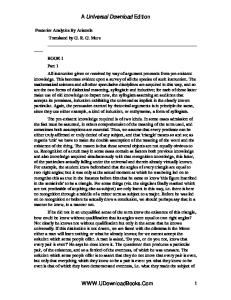
was shown to be higher in laminectomy and laminoplasty patients when compared with conservatively treated individuals [61,62]. Possible explanations include (1) mechanical stress increasing in the cervical spine because of destruction of the posterior supportive elements and (2) biological stimulation produced by the laminoplasty or laminectomy. The prognosis of patients with OPLL has generally been thought to be disappointing. We examined the natural course of this disease [63]. In our recent study [64], a total of 450 patients, average age 74.6 years at last evaluation, were prospectively followed neurologically for an average of 17.6 years (10–30 years) to discern the “natural history” of the disease progression. Myelopathy was originally recognized in 127 patients, 91 of whom were managed surgically. The remaining 36 myelopathic patients were treated conservatively, with increased myelopathy being observed in 23 (65%) of these individuals. For the 323 patients without original myelopathy, 64 (20%) became myelopathic during the follow-up interval. The Kaplan-Meier estimates [65] of myelopathy-free survival among patients without myelopathy at the first visit was 71% at 30 years of follow-up (Fig. 1). The 45 patients with more than 60% of the spinal canal compromised by OPLL were all myelopathic. As a dynamic factor, range of motion (ROM) of the cervical spine was calculated by dynamic X-ray radiography. The relation between the presence or absence of myelopathy and ROM was determined in 204 patients with a minimum space available—spinal canal (SAC) diameters of 6 mm to less than 14 mm. The total ROM in the group with myelopathy was significantly greater than in the group without myelopathy (Table 5). Although myelopathy was recognized in all patients with more than 60% of the spinal canal compromised by OPLL, minimal OPLL at first examination rarely developed to OPLL with more than 60% stenosis during the follow-up. Therefore, one cannot simply say that
myelopathy develops with OPLL. Rather, dynamic factors (e.g., ROM) appear to be more important for the evolution of myelopathy in patients with less than 60% of the canal compromised by OPLL [66]. Findings in this long-term prospective analysis of OPLL patients revealed that the cumulative myelopathy-free survival rate among patients without myelopathy at the first visit was 71% after 30 years. A longitudinal cohort study of 216 elderly patients with OPLL for an average of 12.6 years was performed to determine the quality of life (QOL) of the patients after treatment [67]. The cumulative survival rate of patients with (Nurick) grade 5 severe myelopathy before treatment was 20% at 70 years of age, whereas that of patients without myelopathy or with grade 1, 2 , 3, or 4 myelopathy before treatment was 80%. Patients were statistically more likely to live independent of assistance for activities of daily living when they underwent surgical therapy for grade 3 or 4 myelopathy than those with similar degrees of myelopathy who underwent conservative therapy. For patients with grade 5 myelopathy at the first examination, the final QOL was poor regardless of the therapeutic method. The prevalence of fractures in patients with OPLL was 1.4% for men and 8.6% for women. The bone mineral density in these patients without myelopathy was significantly higher than that in healthy subjects of the same age. These data
Table 5. Range of motion of the cervical spine in patients with a minimum spinal canal diameter of ≥6 mm but <14 mm Presence of myelopathy
ROM of cervical spine 51.0° ± 17.5° 39.0° ± 9.5°
Yes No Rom, range of motion Results are expressed as the mean ± SD P < 0.01 between groups
% 100 90 80 70 60 50 40 30 20 10 0 0
5
10
15
20
25
Time from the first visit (years)
30
Fig. 1. Kaplan-Meier estimate of myelopathy-free rate among patients who did not exhibit myelopathy at the first examination
OPLL: Disease Entity, Incidence, Historical Research, Prognosis
suggest that surgical treatment should be chosen for patients exhibiting moderate myelopathy to obtain satisfactory QOL for a long period of time. Severe myelopathy can be induced by minor cervical trauma in patients with OPLL. Results of surgical treatment for this condition are far from satisfactory. Some advocate preventive surgery prior to the onset of myelopathy for patients with OPLL and potential spinal stenosis due to ossified ligaments. However, a rationale for preventive surgery for patients with OPLL who do not exhibit myelopathy has not been established. In our prospective investigation of 368 patients who did not have myelopathy at the time of the initial consultation, only 6 (2%) patients subsequently developed myelopathy induced by trauma [68]. Ossification types in patients who developed myelopathy induced by trauma were mainly the mixed type. Preventive surgery prior to the onset of myelopathy is unnecessary for most patients with OPLL. Acknowledgments. The studies presented here were supported in part by a grant-in-aid from the Investigation Committee on the Ossification of the Spinal Ligaments of the Japanese Ministry of Public Health and Welfare.
12.
13.
14.
15.
16.
17.
18.
References
19.
1. Bakay L, Cares HL, Smith RJ (1970) Ossification in the region of the posterior longitudinal ligament as a cause of cervical myelopathy. J Neurol Neurosurg Psychiatry 33:263–268 2. Minagi H, Gronner AT (19699 Calcification of the posterior longitudinal ligament: a cause of cervical myelopathy. AJR Am J Roentgenol 105:365–369 3. Nagashima C (1972) Cervical myelopathy due to ossification of the posterior longitudinal ligament. J Neurosurg 37:653–660 4. Ono K, Ota H, Tada K, Hamada H, Takaoka K (1977) Ossified posterior longitudinal ligament: a clinicopathologic study. Spine 2:126–138 5. Tsuyama N (1984) Ossification of the posterior longitudinal ligament of the spine. Clin Orthop 184:71–84 6. Key GA (1838) On paraplegia depending on the ligament of the spine. Guys Hosp Rep 3:17–34 7. Matsunaga S, Sakou T (1997) Epidemiology of ossification of the posterior longitudinal ligament. In: Yonenobu K, Sakou T, Ono K (eds) OPLL. Springer, Tokyo, pp 3–17 8. Hanna M, Watt I (1979) Posterior longitudinal ligament calcification of the cervical spine. Br J Radiol 52:901–905 9. Wennekes MJ, Anten HWM, Korten JJ (1984) Ossification of the posterior longitudinal ligament. Clin Neurol Neurosurg 87:297–302 10. Lecky BFR, Britton JA (1984) Cervical myelopathy due to ossification of the posterior longitudinal ligament. J Neurol Neurosurg Psychiatry 47:1355–1361 11. Trojan DA, Pokrupa R, Ford RM, Adamsbaum C, Hill RO, Esdaile JM (1992) Diagnosis and treatment of ossification
20.
21.
22.
23.
24.
25.
26.
15
of the posterior longitudinal ligament of the spine: report of eight cases and literature review. Am J Med 92:296–306 Firooznia H, Benjamin VM, Pinto RS, Olimbu C, Rafll M, Leitman BS, McCauley DI (1982) Calcification and ossification of posterior longitudinal ligament of spine: its role in secondary narrowing of spinal canal and cord compression. NY State J Med 82:1193–1198 Klara PM, McDonnel DE (1986) Ossification of the posterior longitudinal ligament in Caucasians: diagnosis and surgical intervention. Neurosurgery 19:212–217 McAfee PC, Regan JJ, Bohlman HH (1987) Cervical cord compression from ossification of the posterior longitudinal ligament in non-Orientals. J Bone Joint Surg Br 69:569–573 Resnick D, Shaul SR, Robinsons JM (1975) Diffuse idiopathic skeletal hyperostosis (DISH): Forestier’s disease with extraspinal manifestations. Radiology 115: 513–524 Resnick D, Guerra J Jr, Robinson CA, Vint VC (1978) Association of diffuse idiopathic skeletal hyperostosis (DISH) and calcification and ossification of the posterior longitudinal ligament. AJR Am J Roentgenol 131:1049– 1053 Resnick D, Niwayama G (1976) Radiographic and pathologic features of spinal involvement in diffuse idiopathic skeletal hyperostosis (DISH) Radiology 119:559– 568. Epstein NE (1994) Ossification of the posterior longitudinal ligament in evolution in 12 patients. Spine 19:673– 681 Epstein NE (1994) The surgical management of ossification of the posterior longitudinal ligament in 43 North Americans. Spine 19:664–672 Okamoto Y (1967) Ossification of the posterior longitudinal ligament of cervical spine with or without myelopathy. J Jpn Orthop Assoc 40:1349–1360 Yanagi T, Yamamura Y, Andou K, Sofue I (1967) Ossification of the posterior longitudinal ligament in the cervical spine: a clinical and radiological analysis of thirty-seven cases. Rinsho Shinkei 7:727–735 (in Japanese) Onji Y, Akiyama H, Shimomura Y, Ono K, Fukuda S, Mizuno S (1967) Posterior paravertebral ossification causing cervical myelopathy: a report of eighteen cases. J Bone Joint Surg Am 49:1314–1328 Shinoda Y, Hanzawa S, Nonaka K, Oowada O (1971) Ossification of the posterior longitudinal ligament. Seikeigeka 22:383–391 (in Japanese) Hrata S (1976) Research report on ossification of the posterior longitudinal ligament: Investigation Committee 1975 report on the ossification of the spinal ligaments of the Japanese Ministry of Public Health and Welfare, pp 43–48 (in Japanese) Sakou T, Tomimura K, Maehara T, Kawamura H, Morizono Y, Nagamine T (1978) Epidemiological study of ossification of the posterior longitudinal ligament in the cervical spine in Okinawa Prefecture. Investigation Committee 1977 report on the ossification of the spinal ligaments of the Japanese Ministry of Public Health and Welfare, pp 172–173 (in Japanese) Kurihara A, Kataoka O, Maeda A, Kawai K (1978) Clinical picture and course of the ossification of posterior longitudinal ligament of the cervical spines. Seikeigeka 29:745– 751 (Japanese)
16
S. Matsunaga and T. Sakou
27. Izawa K (1980) Comparative radiographic study on the incidence of ossification of the cervical spine among Japanese, Koreans, Americans, and Germans. J Jpn Orthop Assoc 54:461–474 (in Japanese) 28. Yamauchi H (1978) Epidemiological and pathological study of ossification of the posterior longitudinal ligament of the cervical spine. Investigation Committee 1977 report on the ossification of the spinal ligaments of the Japanese Ministry of Public Health and Welfare, pp 21–25 (in Japanese) 29. Kurokawa T (1978) Prevalence of ossification of the posterior longitudinal ligament of the cervical spine in Taiwan, Hong Kong, and Singapore. Investigation Committee 1977 report on the ossification of the spinal ligaments of the Japanese Ministry of Public Health and Welfare, pp 8–9 (in Japanese) 30. Yamaura I, Kamikozuru M, Shinomiya K (1978) Therapeutic modalities and epidemiological study of ossification of the posterior longitudinal ligament of the cervical spine. Investigation Committee 1977 report on the ossification of the spinal ligaments of the Japanese Ministry of Public Health and Welfare, pp 18–20 (in Japanese) 31. Tezuka S (1980) Epidemiological study of ossification of the posterior longitudinal ligament of the cervical spine in Taiwan. Investigation Committee 1979 report on the ossification of the spinal ligaments of the Japanese Ministry of Public Health and Welfare, pp 19–23 (in Japanese) 32. Lee T, Chacha PB, Orth MC, Khoo J (1991) Ossification of posterior longitudinal ligament of the cervical spine in non-Japanese Asians. Surg Neurol 35:40–44 33. Yamauchi H, Izawa K, Sasaki K, Noromoto T, Honda H, Kusue K (1979) Radiological examination by plain film of the cervical spine in West Germany. Investigation Committee 1978 report on the ossification of the spinal ligaments of the Japanese Ministry of Public Health and Welfare, pp 22–23 (in Japanese) 34. Terayama K, Ohtsuka Y (1984) Epidemiological study of ossification of the posterior longitudinal ligament on Bologna in Italy. Investigation Committee 1983 report on the ossification of the spinal ligaments of the Japanese Ministry of Public Health and Welfare, pp 55–62 (in Japanese) 35. Ijiri K, Sakou T, Taketomi E, Matsunaga S (1996) Epidemiological study of ossification of posterior longitudinal ligament in Utah. Investigation Committee 1995 report on the ossification of the spinal ligaments of the Japanese Ministry of Public Health and Welfare, pp 24–25 (in Japanese) 36. Ikata T, Tezuka S (1979) Epidemiological study on the prevalence of ossification of the posterior longitudinal ligament. Investigation Committee 1978 report on the ossification of the spinal ligaments of the Japanese Ministry of Public Health and Welfare, pp 24–27 (in Japanese) 37. Ohtani K, Higuchi M, Watanabe T, Nakai S, Fujimura S, Manzoku S, Kosaka M, Shibazaki T, Tufuhisa M, Saito T (1980) Epidemiological study of ossification of the posterior longitudinal ligament of the cervical spine in Yaeyama Islands of Okinawa. Investigation Committee 1979 report on the ossification of the spinal ligaments of the Japanese Ministry of Public Health and Welfare, pp 17–18 (in Japanese)
38. Yamauchi H, Issei K, Endou A, Kameta I, Kondou A, Yamaguchi T (1982) Comparative study on the prevalence of OPLL by plain X-ray film and heavy metal content of hair between Chiba and Yamanashi. Investigation Committee 1981 report on the ossification of the spinal ligaments of the Japanese Ministry of Public Health and Welfare, pp 15–19 (in Japanese) 39. Sakou T, Morimoto N (1982) Epidemiological study of the cervical OPLL on islands of Kagoshima. Investigation Committee 1981 report on the ossification of the spinal ligaments of the Japanese Ministry of Public Health and Welfare, pp 20–23 (in Japanese) 40. Ohtsuka Y, Terayama K, Wada K, Kasuga K, Matsushima S, Machida T, Furukawa K (1984) Epidemiological study of ossification of the spinal ligament on Yachiho in Nagano Prefecture. Investigation Committee 1983 report on the ossification of the spinal ligaments of the Japanese Ministry of Public Health and Welfare, pp 63–67 (in Japanese) 41. Ikata T, Takada K, Murase M, Kashiwaguchi S (1985) Epidemiological study of ossification of the posterior longitudinal ligament of the cervical spine. Investigation Committee 1984 report on the ossification of the spinal ligaments of the Japanese Ministry of Public Health and Welfare, pp 61–65 (in Japanese) 42. Sakou T, Morimoto N, Wan S, Ryu K (1985) Epidemiological study of ossification of the posterior longitudinal ligament of the cervical spine in general population in Taiwan. Investigation Committee 1984 report on the ossification of the spinal ligaments of the Japanese Ministry of Public Health and Welfare, pp 66–70 (in Japanese) 43. Sakou T, Taketomi E, Sameshima T (1988) Epidemiological study of ossification of the posterior longitudinal ligament of the cervical spine on Takasago-tribe in Taiwan. Investigation Committee 1987 report on the ossification of the spinal ligaments of the Japanese Ministry of Public Health and Welfare, pp 8–9 (in Japanese) 44. Tomita T, Harata S, Ueyama K, Araki T, Ito J, Sato T, Sannohe A, Tian W, Yamada S, Sonoda S, Rong G, Jia Y, Dang GT, Cai Q, Liu S (1994) Epidemiological study of ossification of the posterior longitudinal ligament (OPLL) of cervical spine and cervical spondylotic changes in China. Investigation Committee 1993 report on the ossification of the spinal ligaments of the Japanese Ministry of Public Health and Welfare, pp 101–105 (in Japanese) 45. Yanagi T (1970) Ossification of the posterior longitudinal ligament: a clinical and radiological analysis of forty-six cases. Brain Nerve 22:909–921 (in Japanese) 46. Terayama K, Kurokawa T, Seki H (1975) National survey of ossification of the posterior longitudinal ligament. Investigation Committee 1975 report on the ossification of the spinal ligaments of the Japanese Ministry of Public Health and Welfare, pp 8–33 (in Japanese) 47. Tsuyama N (1984) Ossification of the posterior longitudinal ligament of the spine. Clin Orthop 184:71–84 48. Takeda T, Arima T (1972) A case report of ossification of posterior longitudinal ligament with tetrapalsy by mild trauma. Rinsho Seikei Geka 7:949–953 (in Japanese) 49. Katoh S, Ikata T, Hirai N, Okada Y, Nakauchi K (1995) Influence of minor trauma to the neck on the neurological outcome in patients with ossification of the posterior longitudinal ligament (OPLL) of the cervical spine. Paraplegia 33:330–333
OPLL: Disease Entity, Incidence, Historical Research, Prognosis 50. Fujimura Y, Nakamura M, Toyama Y (1998) Influence of minor trauma on surgical results in patients with cervical OPLL. J Spinal Disord 11:16–20 51. Terayama K (1987) Family study of ossification of the posterior longitudinal ligament. Investigation Committee 1986 report on the ossification of the spinal ligaments of the Japanese Ministry of Public Health and Welfare, pp 10–11 (in Japanese) 52. Terayama K (1989) Genetic studies on ossification of the posterior longitudinal ligament of the spine. Spine 14:1184–1191 53. Uehara H, Sakou T, Taketomi K, Matsunaga S, Uamaguchi Y (1994) Familial study of hereditary factor for the ossification of the posterior longitudinal ligament in the cervical spine. Seikeigeka 45:1341–1345 (in Japanese) 54. Miura Y, Furusho T, Ibaraki K, Takemitsu Y (1992) Genetic studies for OPLL: analysis of twins. Investigation Committee 1991 report on the ossification of the spinal ligaments of the Japanese Ministry of Public Health and Welfare, pp 5–7 (in Japanese) 55. Sakou T, Taketomi E, Matsunaga S, Yamaguchi Y, Sonoda S, Yashiki S (1991) Genetic study of ossification of the posterior longitudinal ligament in the cervical spine with human leukocyte antigen haplotype. Spine 6:1249–1252 56. Matsunaga S, Yamaguchi M, Hayashi K, Sakou T (1999) Genetic analysis of ossification of the posterior longitudinal ligament. Spine 24:937–938 57. Koga H, Sakou T, Hayashi K, Numazawa T, Harata S, Yone K, Matsunaga S, Otterud B, Inoue I (1998) Genetic mapping os ossification of the posterior longitudinal ligament of the spine. Am J Genet 62:1460–1467 58. Maeda S, Koga H, Matsunaga S, Numazawa T, Ikari K, Furushima K, Harata S, Takeda J, Sakou T, Inoue I (2001) Gender-specific haplotype association of collagen α2(X1) gene in ossification of the posterior longitudinal ligament of the spine. J Hum Genet 46:1–4 59. Taketomi E (1997) Progression of ossification of the posterior longitudinal ligament in the cervical spine. J Jpn Spine Res Soc 8:359–366
17
60. Matsunaga S, Sakou T, Taketomi E, Nakanishi K (1996) Effects of strain distribution in the intervertebral discs on the progression of ossification of the posterior longitudinal ligaments. Spine 21:184–189 61. Ichimoto H, Kawai S, Oda H, Saika M, Taguchi T, Hiura Y (1991) Postoperative progression pattern of ossification of the posterior longitudinal ligament in cervical spine. Investigation Committee 1990 report on the ossification of the spinal ligaments of the Japanese Ministry of Public Health and Welfare, pp 199–200 (in Japanese) 62. Miyazaki K, Hirofuji E, Onozaki A, Okada N, Tada H, Mizuno Y (1993) Follow-up studies on the development of ossification of the posterior longitudinal ligament in the cervical region after simultaneous multisegmental laminectomy. Spine Spinal Cord 6:905–910 (in Japanese) 63. Matsunaga S, Sakou T, Taketomi E, Yamaguchi M, Okano T (1994) The natural course of myelopathy caused by ossification of the posterior longitudinal ligament in the cervical spine. Clin Orthop 305:168–177 64. Matsunaga S, Sakou T, Taketomi E, Komiya S (2004) Clinical course of patients with ossification of the posterior longitudinal ligament: a minimum 10-year cohort study. J Neurosurg (Spine) 100:245–248 65. Kaplan EL, Meier P (1958) Nonparametric estimation from incomplete observations. J Am Stat Assoc 53:457–481 66. Matrsunaga S, Kukita M, Hayashi K, Shinkura R, Koriyama C, Sakou T, Komiya S (2002) Pathogenesis of myelopathy of patients with ossification of the posterior longitudinal ligament. J Neurosurg Spine 96:168–172 67. Matsunaga S, Sakou T, Arishima Y, Koga H, Hayashi K, Komiya S (2001) Quality of life in elderly patients with ossification of the posterior longitudinal ligament. Spine 26:494–498 68. Matsunaga S, Sakou T, Hayashi K, Ishidou Y, Hirotsu M, Komiya S (2002) Trauma-induced myelopathy in patients with ossification of the posterior longitudinal ligament. J Neurosurg (Spine) 97:172–175
Genetic Susceptibility to OPLL Ituro Inoue
Introduction Ossification of the posterior longitudinal ligament of the spine (OPLL) is a pathological condition of the paravertebral ligament that causes ectopic bone formation possibly through an endochondral ossification process. OPLL is a common disorder among Japanese and throughout Asian populations but is uncommon in Caucasian populations. The incidence of this disorder in Japan is reported to be 2%–4% in the general population over 30 years of age (see Chapter 4). Although multiple etiologies for OPLL must be considered, evidence from studies of twins and siblings strongly indicate that a certain proportion of the cases of this common disorder are genetically determined. OPLL has long been recognized as a disease of unknown etiology, but identification of genetic susceptibilities would clarify the biometabolic pathway that leads to the development of ectopic bone formation, thereby eventually leading to a full understanding of OPLL. Since the first edition of this book was published, there has been significant progress in the identification of genetic susceptibilities to OPLL by several research groups. In this chapter, I discuss how we can approach the genetic factors of OPLL and how and to what extent the susceptibility genes are involved in the etiology of OPLL. Various research groups have applied different strategies to approach the question of genetic susceptibility, such as genetic linkage analysis and positional cloning, an association study with candidate genes, and gene discovery from an OPLL mouse model (Fig. 1) [1]. Each approach has advantages and disadvantages. For the genetic linkage-based approach, family recruitment is the major difficulty because numerous members of the nuclear family are needed to perform a robust nonparametric linkage analysis for such a complex disease as OPLL. However, if a certain genetic component exists, the linkage-based approach followed by positional (candidate) cloning is the standard and most
Division of Genetic Diagnosis, The Institute of Medical Science, The University of Tokyo, 4-6-1 Shirokanedai, Minatoku, Tokyo 108-8639, Japan
promising method. The candidate gene association approach is a direct method for detecting the causality. In general, the biopathway related to a complex disease cannot be explicitly determined; therefore, selecting a candidate gene depends on the researcher’s knowledge, and its success relies on luck. Naturally occurring animal models would be applied not only for detecting the causality in the animal but also for detecting the causality in the human counterpart. The results of this approach should be highly probable if the model animal shares a common etiological background with the human disease, which is indeed difficult to predict. In fact, despite enormous efforts, most of the causalities of animal models do not correlate with those of the human diseases. Because of the complexities of OPLL, continuous efforts by cooperative groups also supported by the Investigation Committee on Ossification of the Spinal Ligaments are needed and may provide complete understanding of OPLL in the near future.
Materials and Methods Disease Criteria and Subjects OPLL was diagnosed based on the presence of ectopic bone formation in the posterior longitudinal ligament, as seen by radiography or computed tomography (CT) examinations of the cervical, thoracic, or lumbar portions of the spine. The diagnosis of diffuse idiopathic hyperostosis (DISH) was based on the criteria of Utsinger [2] established through radiography or CT examinations of the cervical, thoracic, and lumbar portions of the spine. The siblings of OPLL patients were screened for spinal bone formation by radiography, and affected siblings (including those who were asymptomatic) were recruited [3,4]. A total of 142 affected sib-pairs from 70 Japanese families were recruited. In total, 98 pairs were from Kagoshima University, 40 pairs from Hirosaki, 2 pairs from Asahikawa, and 1 pair each from Wakayama and Okinawa. The family structure was as follows: 51 affected pairs, 12 affected trios, 5 affected quartets, 1 affected quintet, and 1 affected sextet. The affected siblings
19
20
I. Inoue
Genetic Linkage Study
loci
Fig. 1. Approaches to identifying genetic susceptibility of complex disease
Positional Genes
Association Study
Candidate Genes
Case-control comparison with chi-square test
Animal Model
Causal Gene Human counterpart Identification of Disease Genes
included 76 women and 93 men. For the allelic association study, 342 unrelated OPLL patients (73 familial cases including 70 probands utilized for the linkage study and 269 sporadic cases) and 298 unrelated nonOPLL subjects were recruited at the Kagoshima and Hirosaki Universities. All the non-OPLL subjects were more than 60 years of age with no signs of spinal ossification (examined by standard radiography), thereby excluding most unmanifested disease. The DNA samples for the association study were 97 DISH patients and 298 controls for the Japanese population and 96 DISH patients and 96 controls for the Czech population.
Microsatellite Genotyping and Affected Sib-pair Linkage Analysis Multiplex fluorescent genotyping was performed using the ABI Linkage Mapping Set version 2 (Applied Biosystems, Tokyo, Japan), which covers the entire human genome with 404 polymorphic markers. Microsatellite genotyping was performed as described previously [4]. GeneHunter-Plus [5] was used to analyze the data on affected sib-pairs. Multipoint analysis of the data from the genome-wide scan and from fine mapping was performed by weighting each family equally with GeneHunter-Plus. Allele frequencies of microsatellite markers for references were estimated in 64 unrelated Japanese subjects.
SNP Genotyping Gene-based single nucleotide polymorphisms (SNPs) were selected at 3- to 10-kb intervals to cover the gene from the two public databases: NCBI dbSNP (http://www.ncbi.nlm.nih.gov/SNP/) and IMS-JST JSNP (http://snp.ims.u-tokyo.ac.jp/). For candidate genes, direct sequencing was performed to identify SNPs. SNPs were genotyped using either the pyrosequencing method, real-time pyrophosphate DNA sequencing on a PSQ96 Instrument (Pyrosequencing AB, Uppsala, Sweden), or direct sequencing using BigDye Termina-
tor cycle sequencing on an ABI PRISM 3700 DNA analyzer (Applied Biosystems).
Other Statistical Analyses Allelic frequencies of SNPs between cases and controls were compared using a contingency χ2 test. Haplotype frequencies for multiple SNPs were estimated using the expectation-maximization method with the Arlequin program (available at the Arlequin web site) or the SNPAlyze program (Dynacom, Mobara, Japan). We calculated the LD between pairs of SNPs using the standard definitions of D″ and r2 as described elsewhere [6].
Isolation and Culture of Spinal Ligament Cells and Reverse Transcription Interspinous ligament tissues at the cervical spine were aseptically obtained during surgical operation using the expansive laminoplasty technique with the posterior approach from 10 OPLL and 10 non-OPLL (cervical spondylotic myelopathy) patients. The ligament tissues were washed with phosphate-buffered saline (PBS) several times and then minced into about 1 mm2 pieces. The explant cells were cultured in Dulbecco’s modified Eagle’s medium supplemented with 10% fetal bovine serum and 0.2 mM L-ascorbic acid 2-phosphate. Total RNA was extracted from cultured cells with Trizol reagent according to the manufacturer’s protocol (GIBCO, Rockville, MD, USA). A 1-µg aliquot of total RNA was reverse-transcribed with an oligo (dT)12–18 primer using M-MLV reverse transcriptase (GIBCO) at 37°C for 1 h.
Results HLA Association and Genetic Linkage Study An important first step in understanding the role of genetic factors of OPLL is the 1991 report by Sakou
Genetic Susceptibility to OPLL
et al., who demonstrated an allelic association of HLA markers DR2 and Bw62 with OPLL (P < 0.01) [7]. In addition, the siblings in the report who had HLA haplotypes identical to those of the probands showed a high incidence of OPLL. In the case of OPLL, the mode of inheritance has not been clearly determined, and ascertainment of a large pedigree with multiple generations is not practical because of the late onset of the disease. Therefore, affected sib-pairs would be a useful resource for approaching the question of genetic susceptibility to OPLL. According to the report of Sakou et al. on HLA association, Koga et al. applied nonparametric genetic linkage methods—independent of the transmission model, prior disease frequency, and penetrance—to 91 affected sib-pairs from 53 families to test the hypothesis that a gene predisposed to the OPLL phenotype resides on chromosome 6p [3]. Seven highly informative dinucleotide markers at the HLA complex were selected for the linkage study. Strong evidence of genetic linkage was detected with the marker D6S276 (P = 0.0000059), which is located distal to the HLA locus. In addition, weaker linkages with other neighboring markers were observed. The HLA association and linkage results predict that a genetic susceptibility to OPLL is likely in the HLA or nearby region.
Candidate Genes, COL11A2 and RXRb, in the Linkage Region Two genes in the region proximal to HLA class II between the markers D6S276 and D6S291—collagen α2(XI) (COL11A2) and retinoic X receptor β (RXRb) genes—are considered likely candidates for OPLL, and the patients have been extensively screened for molecular variants. Because collagen α2(XI) encoded by COL11A2 is a fibril-forming minor collagen of chondrocytes and endochondral ossification is the key pathological finding in the ectopic ossified lesion of OPLL, COL11A2 may reasonably be related to OPLL. RXRb, adjacent to COL11A2, was also investigated for its potential involvement in OPLL. OPLL occurrence has been frequently observed in patients with dermal disorders who had been treated by long-term administration of vitamin A, the precursor of retinoic acid. In COL11A2, 66 exons and 1300 bp of the promoter region were screened, and 19 distinct molecular variants were identified; there was strong evidence of an allelic association with OPLL, with a T to A nucleotide substitution at position −4 from the acceptor site in intron 6 [denoting intron 6 (−4A)], suggesting a functional role of this polymorphism in the pathogenesis of OPLL (Fig. 2A)[3]. In RXRb, three polymorphisms were detected and two of them locating 3′UTR, 3′End (+140) and 3′End (+561) polymorphisms, showed positive associations with OPLL (P = 0.0028 and 0.034, respectively) (Fig. 2B) [8].
21
However, these polymorphisms were in linkage disequilibrium with the intron 6 (−4A) of the COL11A2, and showed less significant allelic association to OPLL than did intron 6 (−4A). Taken together, it is strongly thought that intron 6 (−4A) of COL11A2 constitutes a genetic susceptibility to OPLL. In addition, a haplotype including intron 6 (−4A) was strongly associated in male patients (P = 0.0003) and not evidently associated in female patients (P = 0.21) [9]. Because of an incidence difference by gender (male/female ratio 2 : 1), the gender-specific association would be an interesting finding to use in a further investigation of the etiological differences by gender. Because intron 6 (−4A) was found to be more frequent in non-OPLL subjects than in OPLL patients, it is possible that the intron 6 (−4A) allele is protective in the ossification process. Despite these genetic findings, determining the involvement of the COL11A2 in the etiology of OPLL is hampered by a lack of evidence regarding the functional significance of the COL11A2 variants.
Functional Impact of SNPs in COL11A2 In the case of common diseases, even if the genetic susceptibilities are determined by current molecular genetics the daunting task of examining these lifelong genetically determined disturbances by practical experimental procedures remains. Strong evidence of an allelic association with OPLL was observed with the T to A nucleotide substitution at position −4 from the acceptor site in intron 6 [intron 6 (−4A)] in COL11A2, suggesting a functional role of the polymorphism in the pathogenesis of OPLL. Because the region containing exons 6–8 of the COL11A2 transcript exhibits a complicated splicing pattern, as previously described in humans and the mouse, intron 6 (−4A) might affect splicing of COL11A2 mRNA. The cervical interspinous ligament tissues from 10 OPLL patients and 10 controls were subjected to primary culture; and the total RNA prepared from cultured cells was subjected to reverse transcription-polymerase chain reaction (RT-PCR) analysis to determine whether the splicing alterations of COL11A2 mRNA are a result of the genetic variation [10]. PCR primers and oligonucleotide probes were designed to cover exons 6–8 (Fig. 3). Differences in the splicing pattern due to the genetic variation at intron 6, despite the disease status, were observed. Altogether, 1 of 20 samples obtained was homozygous for intron 6 (−4A), which showed mainly three bands (Fig. 3, lane 3). The intron 6 (−4A) allele is associated with an increased amount of E5789 (lacking exon 6) and E579 (lacking exons 6 and 8). E59 (lacking exons 6, 7, and 8), observed predominantly in cartilage tissue, was almost absent in the cells of the intron 6 (−4A) homozygote. The intron 6 (−4T) homozygote contains mainly three bands: E56789 (all of the
22
I. Inoue
A. COL11A2 Exon 27(+21) Intron 35(+3) Intron 1(+1910) Intron 9(+10) Intron 22(+70) Exon 36(+42) Exon 32(+36) Intron 6(+608) Intron 20(+32) Exon 32(+42) Exon 54(+40) Exon 1(+24) Exon 64(+49) Intron 6(+353) Exon 20(+5)
Exon 43 (+24) A-G (P1058P) ( p = 0.021)
Promoter (-182) A-C ( p = 0.024) Exon 6 (+28) (E272K) ( p = 0.002)
Exon 46 (+18) C-T (P1128P) ( p = 0.0333)
Fig. 2. Molecular variants for COL11A2 and RXRB. A Twenty distinct molecular variants were identified for COL11A2, and association results with OPLL are shown. B Three variants were identified in RXRB, and association results with OPLL are shown
Intron 6 (-4) ( p = 0.0004)
B. RXRB Exon 10 (+378) T-A ( p = 0.3098)
RXRβ
3’ End (+561) C-CC ( p = 0.0344)
COL 11A2 3’ End (+140) A-T ( p = 0.0028)
Probe Ex 5/9
COL11A2 mRNA Exon 5 Probe :
6
7
Ex 6
Ex 5
8
Ex 7
9
Ex 8
5F
9R
non-OPLL
Exon combination MW
TT
TA
OPLL AA
E 5 6 7 8 9 (498bp) E 5 7 8 9 (420bp) E 5 6 7 9 (318bp) E 5 7 9 (240bp) E 5 9 (177bp)
GAPDH
exons are present), E5679 (lacking exon 8), and E59 (Fig. 3, lanes 1 and 6). As expected, mixed splicing patterns were observed in the heterozygous samples (Fig. 3, lanes 2, 4, and 5) owing to the participation of both alleles. In summary, the functional impact of the intron 6 polymorphism of COL11A2 that results in altered splicing in the region containing exons 6–8 was demonstrated. These exons encode an acidic subdomain, also called a variable region because of the complicated alternative splicing that depends on cell type or developmental stage. The consequence of the altered splicing due to the intron 6 (−4A), especially the existence of exon 7, may be part of the biological mechanism by which individual differences of COL11A2 are protective in the development of ectopic ossification in the cervical ligament.
TA
TA
TT
Fig. 3. Alternative splicing of exons 6–8 of COL11A2 mRNA depend on the intron 6 genotype. A RNAs from cultured ligament cells with distinct genotypes at intron 6 (−4) were subjected to reverse transcription-polymerase chain reaction (RT-PCR) analyses with the primer set (Ex5/9) that covers the exons 6–8 region. B PCR products were separated on agarose gel and visualized. The splicing pattern (shown on the left) depends on the intron 6 (−4) genotype (top) and was estimated by size and confirmed by Southern blot analysis
TWY (ttw) Mouse Model for OPLL and NPSS as the Susceptibility The tiptoe Yoshimura (TWY or ttw) mouse, which has been considered an experimental model for OPLL, has hyperostosis of the ligaments similar to that seen with OPLL and shows autosomal dominance. Identifying the genetic determinant of the animal model might lead to elucidation of the metabolic pathway involved in OPLL; moreover, the determinant might be responsible for the genetic susceptibility to human OPLL. Using positional candidate gene analysis, a nonsense mutation (Gly568stop) in the nucleotide pyrophosphatase gene (Npps) was identified as the genetic causality in mice [11]. The human counterpart, NPPS, locating on chromosome 6q, was then naturally considered a candidate gene for
Genetic Susceptibility to OPLL
OPLL, and the gene was extensively screened for genetic susceptibility. NPPS spanned about 80 kb of the genomic DNA and consisted of 25 exons; all of the exons, including exon-intron boundaries and about 1.5 kb of the promoter region, were screened for nucleotide variations. Among 10 molecular variants, a base deletion in intron 20 (IVS20-11delT) was significantly associated with OPLL (65.3% for OPLL and 53.9% for the control; χ2 = 8.85, P = 0.0029) [12]. Details of the NPPS study are described elsewhere (see Chapter 2). Although the association result of IVS20-11delT could not be confirmed by the following studies, a novel rare variant (IVS1514T→C) might be associated with severity and young onset of OPLL [3,13]. However, by subgrouping, the sample size of a group was markedly decreased, so a larger scale association design would be needed.
23
showed evidence of linkage (defined by Zlr > 2.2) [14]. Because the best evidence of linkage was observed with markers on chromosome 21q, high-resolution mapping was performed by adding eight markers. The fine mapping on chromosome 21 indicated that the linkage peak (Zlr = 3.97) was close to the q terminus near marker D21S1903. These results strongly indicate that the genetic susceptibility to OPLL localizes on the q terminus of chromosome 21.
Screening of Genes on the Linkage Region of Chromosome 21: Identification of COL6A1
To understand the whole picture of OPLL susceptibility, a genome-wide linkage study in 70 Japanese nuclear families comprising 169 affected subjects and 142 sib-pairs was performed. Using the GeneHunter-Plus program, multipoint Zlr scores for all chromosomes (except the Y chromosome) are computed as shown in Fig. 4. The most prominent evidence of linkage was on chromosome 21q22, with a maximum Zlr score of 3.09, near marker D21S266 [14]. In addition to chromosome 21, five loci on chromosomes (1p, 6p, 11q, 14q, 16q)
To pinpoint the locus for OPLL on chromosome 21, linkage disequilibrium mapping was performed using gene-based SNPs in the linkage region (Zlr > 2.2) spanning approximately 30 cM. First, 600 SNPs of 150 genes in the linkage region with 96 selected patients and 96 controls were genotyped. Allelic association was carried out, and statistical significance was assessed by χ2 analysis with a contingency table. Altogether, 74 SNPs of 24 genes exhibited significant allelic associations (P < 0.05). For the second screening, 74 SNPs in a larger number of OPLL patients (n = 280) and non-OPLL controls (n = 210) were genotyped. Furthermore, 14 SNPs of seven genes had allelic associations (P < 0.01) with OPLL. The most significant association with OPLL was observed with SNPs of COL6A1, which is located 1.2 Mb from the peak linkage marker D21S1903. Because the four genes (COL18A1, PCBP3, COL6A1, COL6A2) that
Fig. 4. Genome-wide linkage study of OPLL among 142 sibpairs. The framework genome scan for OPLL susceptibility loci is depicted (top). Linkage results were expressed as Zlr
scores, which were calculated with GeneHunter-Plus. Several linkage regions are summarized showing the position, peak Zlr, and markers in the region (bottom)
Genome-wide Linkage Study: Affected Sib-pair Linkage Analysis
I. Inoue N - terminal globular domain
triple helix domain
256 amino acid residues
C - terminal globular domain
337 amino acid residues
436 amino acid residues
Intron 4 (+13) Intron 4 (+20) Intron 4 (+37) Intron 8 (-208) Intron 9 (+62) Intron 9 (+188) Intron 9 (-19) Exon 15 (+39) Intron 15 (+39) Intron 18 (-138)
5‘UTR (-5) Intron 2 (+651) Intron 2 (+758) Intron 3 (+1326)
26/27
Association study 342 OPLL patients 298 non-OPLL
P=0.0008
35
Intron 21 (+18) Intron 21 (-36) Intron 22 (+19) Intron 32 (-29) Intron 33 (+15) Intron 33 (+20) Intron 33 (+55) Intron 33 (+88) Exon 35 (+85) Exon 35 (+203) Exon 35 (+205) Exon 35 (+332)
8/9
1
Intron 19 (+27) Intron 19 (+116) Intron 19 (+361) Intron 19 (-152) Intron 20 (+745) Intron 20 (-32)
24
P=0.0009 P=0.000003 P=0.00005
Fig. 5. Genetic structure of COL6A1, location of single nucleotide polymorphisms SNPs), and the association results
showed positive associations with OPLL are clustered within a 750-kb region in the vicinity of the marker D21S1903, extensive SNP and pair-wise linkage disequilibrium analyses were performed in the region. SNPs of COL6A1 that had the strongest association with OPLL were in a gene-specific linkage disequilibrium block; therefore, COL6A1was examined in more detail as the most likely candidate gene for OPLL. A total of 32 distinct SNPs, including the 4 SNPs used for the first screening, 25 additional dbSNPs, and 7 non-dbSNPs identified by direct sequencing were genotyped in all the patients (n = 342) and controls (n = 298). Of the 32 SNPs, 4 had a particularly strong association (P < 0.0001) (Fig. 5). The most significant allelic association with OPLL was observed with a T to C substitution at intron 32 (−29) [χ2 = 21.99, df = 1, P = 0.000003, odds ratio = 1.82 with 95% confidence interval (CI) = 1.60– 2.35] [14]. COL6A1 encodes the α1 chain of type VI collagen, which is an extracellular matrix protein constituted by a short central triple helix flanked by two large globular domains (Fig. 5). The most strongly associated SNP with OPLL, the T to C substitution at intron 32 (−29), is near the branch site of the intron, so the SNP possibly affects the lariat-shaped structure, causing aberrant splicing. Thus far, we have not detected a splicing variant of COL6A1 using cultured ligament cells harboring intron 32 (−29) variant. The functional impact of the polymorphisms of COL6A1 is uncertain, but COL6A1 might lead to increased bone mass. Two genes, COL6A1 and COL11A2, both encode extracellular
matrix proteins, which might provide a scaffold for (pre)osteoblastic cells or chondrocytes that subsequently proceed to membranous or endochondral ossification [15]. Therefore, molecular variants of the extracellular matrix proteins might be implicated in the ectopic ossification observed with OPLL.
Genetic Approach to Diffuse Idiopathic Skeletal Hyperostosis Diffuse idiopathic skeletal hyperostosis is a common skeletal disease in patients with frequencies of 17.6% in men and 11.7% in women over the age of 60 years in Western countries. It has clinical characteristics of ligamentous ossification of the anterolateral aspect of the spinal column, occasionally leading to bony ankylosis [16,17]. Because the common clinical and metabolic features of OPLL and DISH have been well described with less information about the extent of their overlap, and DISH can be diverse among populations [18], DISH subjects from Japanese and Czech populations were genotyped for the SNPs of COL6A1, and an allelic association study was performed to define the genetic etiology [19]. The intron 32 (−29) polymorphism of COL6A1 was associated in the Japanese DISH patients (32.8% in patients vs. 21.6% in controls, P = 0.022), demonstrating that DISH and OPLL shared the same genetic etiology. This also implies that the intron 32 (−29) SNP is most likely associated with the hyperostotic state. However, the SNP of COL6A1 was not significantly
Genetic Susceptibility to OPLL
associated with DISH in the Czech population (35.4% in patients vs. 36.5% in controls, P = 0.825), suggesting that the contribution of COL6A1 to DISH varies from population to population [19] because, other than COL6A1, additional genetic and environmental factors may also have some effects on the disease.
8.
9.
Conclusions It is our consensus that elucidation of the genetic factors involved in OPLL is a key issue for fully understanding the pathophysiology of OPLL in the search for future therapeutic methods. For a number of years efforts have been made to identify genes responsible for OPLL. Extracellular matrix genes such as COL11A2 and COL6A1 were identified as conveying genetic susceptibility, and involvement of NPPS was reported as well. Thus far, however, the results have not been consistent. Evidently, a much larger association study is required to clarify this issue. Also genetic studies in populations other than the Japanese is needed to investigate etiological identity or differences in distinct populations. Understanding the role of genetic factors in the etiology of OPLL allows more precise definition of nongenetic factors, such as environmental and lifestyle-oriented factors, which may help improve the patients’ status.
10.
11.
12.
13.
14.
References 1. Lander ES, Schork N-J (1994) Genetic dissection of complex traits. Science 265:2037–2048 2. Utsinger PD (1985) Diffuse idiopathic skeletal hyperostosis. Clin Rheum Dis 11:325–351 3. Koga H, Sakou T, Taketomi E, Hayashi K, Numasawa T, Harata S, Yone K, Matsunaga S, Otterud B, Inoue I, Leppert M (1998) Genetic mapping of ossification of the posterior longitudinal ligament of the spine. Am J Hum Genet 62:1460–1467 4. Furushima K, Shimo-Onoda K, Maeda S, Nobukuni T, Ikari K, Koga H, Komiya S, Nakajima T, Harata S, Inoue I (2002) Large scale screening for candidate genes of ossification of the posterior longitudinal ligament of the spine. J Bone Miner Res 17:128–137 5. Kong A, Cox NJ (1997) Allele-sharing models: LOD scores and accurate linkage tests. Am J Hum Genet 61:1179– 1188 6. Nakajima T, Jorde LB, Ishigami T, Umemura S, Emi M, Lalouel JM, Inoue I (2002) Nucleotide diversity and haplotype structure of the human angiotensinogen gene in two populations. Am J Hum Genet 70:108–123 7. Sakou T, Taketomi E, Matsunaga S, Yamaguchi M, Sonoda S, Yashiki S (1991) Genetic study of ossification
15.
16. 17.
18.
19.
25
of the posterior longitudinal ligament in the cervical spine with human leukocyte antigen haplotype. Spine 16:1249–1252 Numasawa T, Koga H, Ueyama K, Maeda S, Sakou T, Harata S, Leppert M, Inoue I (1999) Human retinoic receptor β: complete genomic sequence and mutation search for ossification of posterior longitudinal ligament of the spine. J Bone Miner Res 14:500–508 Maeda S, Koga H, Matsunaga S, Numasawa T, Takeda J, Harata S, Sakou T, Inoue I (2001) Gender-specific haplotype association of collagen α2(Xl) gene in ossification of the posterior longitudinal ligament of the spine. J Hum Genet 46:1–4 Maeda S, Ishidou Y, Koga H, Taketomi E, Ikari K, Komiya S, Takeda J, Sakou T, Inoue I (2001) Functional impact of human collagen α2(Xl) gene polymorphism in pathogenesis of ossification of the posterior longitudinal ligament of the spine. J Bone Miner Res 16:948–957 Okawa A, Nakamura I, Goto S, Moriya H, Nakamura Y, Ikegawa S (1998) Mutation in Npps in a mouse model of ossification of the posterior longitudinal ligament of the spine. Nat Genet 19:271–273 Nakamura I, Ikegawa S, Okawa A, Okuda S, Koshizuka Y, Kawaguchi H, Nakamura K, Koyama T, Goto S, Toguchida J, Matsushita M, Ochi T, Takaoka K, Nakamura Y (1999) Association of the human NPPS gene with ossification of the posterior longitudinal ligament of the spine (OPLL). Hum Genet 104:492–497 Koshizuka Y, Kawaguchi H, Ogata N, Ikeda T, Mabuchi A, Seichi A, Nakamura Y, Nakamura K, Ikegawa S (2002) Nucleotide pyrophosphatase gene polymorphism associated with ossification of the posterior longitudinal ligament of the spine. J Bone Miner Res 17:138–144 Tanaka T, Ikari K, Furushima K, Okada A, Tanaka H, Furukawa K, Yoshida K, Ikeda T, Ikegawa S, Hunt S, Takeda J, Toh S, Harata S, Nakajima T, Inoue I (2003) Genomewide linkage and linkage disequilibrium analyses identify COL6A1, on chromosome 21, as the locus for ossification of the posterior longitudinal ligament of the spine. Am J Hum Genet 73:812–822 Zhang Y, Chen Q (2000) Changes of matrilin forms during endochondral ossification: molecular basis of oligomeric assembly. J Biol Chem 275:32628–32634 Meyer PR Jr (1999) Diffuse idiopathic skeletal hyperostosis in the cervical spine. Clin Orthop 359:49–57 Trojan DA, Pouchot J, Pokrupa R, Ford RM, Adamsbaum C, Hill RO, Esdaile JM (1992) Diagnosis and treatment of ossification of the posterior longitudinal ligament of the spine: report of eight cases and literature review. Am J Med 92:296–306 Weinfeld RM, Olson PN, Maki DD, Griffiths HJ (1997) The prevalence of diffuse idiopathic skeletal hyperostosis (DISH) in two large American Midwest metropolitan hospital populations. Skeletal Radiol 26:222–225 Tsukahara S, Miyazawa N, Akagawa H, Forejtova S, Pavelka K, Tanaka T, Toh S, Tajima A, Akiyama I, Inoue I (2005, in press) COL6A1, the candidate gene for ossification of posterior longitudinal ligament, is associated with diffuse idiopathic skeletal hyperostosis in Japanese. Spine
Overview of Etiology and Pathogenesis Toshihiko Taguchi
The posterior longitudinal ligament extends from the skull to the sacrum. This ligament is more firmly attached to the discs than to the vertebral bodies. It displays two strata of fibers. The superficial, longer strands form a distinct strap whose filaments bridge several vertebral bodies. A second, deeper stratum spans only two vertebral bodies and forms a lateral curving extension of fibers that pass along the dorsum of the disc and out through the intervertebral foramen. The posterior longitudinal ligament differs considerably from the anterior longitudinal ligament with respect to the clinical significance of its relation to the neural elements, including the spinal cord and nerve roots [1]. Ossification of the posterior longitudinal ligament (OPLL) was first diagnosed in an autopsy case reported by Tsukimoto in 1960 [2]. Since then there have been many reports from Japan in which OPLL causes myelopathy [3–5]. At first, this disease was called calcification of the longitudinal ligament. After pathological examination revealed that the longitudinal ligament is ossified in these cases, the disease was renamed OPLL. As there were only a few reports of OPLL from countries other than Japan, OPLL was once known as a “Japanese disease.” The disease came to be recognized as the conditions associated with ankylosing skeletal hyperostosis reported by Forestier and Lagier [6]. In 1976 Resnick and Niwayama [7] described the entity diffuse idiopathic skeletal hyperostosis (DISH) and regarded OPLL as a type of DISH. Its clinical features were reported in detail by Nakanishi et al. [8] and Ono et al. [9]. Researchers have not yet fully clarified why the longitudinal ligament becomes ossified. Over the past decades a considerable number of studies have been conducted regarding the factors related to the occurrence and development of OPLL, including many environmental, systemic, and local factors. One environmental factor is the diet. An example of systemic factors is the genetic background, with the metabolic or endocrinological characteristics Department of Orthopedic Surgery, Yamaguchi University School of Medicine, 1-1 Minami Kogushi, 1-Chome, Ube 7558505, Yamaguchi, Japan
of an individual (i.e., hereditary transmission, hormonal abnormality, abnormal calcium metabolism, an association with diabetes mellitus). Common local factors include mechanical stress to the cervical spine (a form of local dynamic stress) or chronic trauma to the cervical spine. It is clear that OPLL has a genetic background [10– 14]. This is supported by family studies, twin studies, and HLA haplotype analysis. The pathological gene for OPLL has not yet been determined. Identifying this gene probably requires the study of families with OPLL using linkage analysis. In a previous family study, the incidence of OPLL in blood relatives in second-degree relationships was 23.2%. Because of this high incidence, it is easier to collect sibling pairs with OPLL, investigating many such pairs using nonparametric linkage analysis. As there are racial differences in the incidence of OPLL, one of the causes of OPLL is thought to be life environment, especially the diet. There are reports that patients with OPLL prefer vegetable protein to animal protein, in contrast to controls [15,16], but this hypothesis has not been confirmed by well-designed control studies. The relationship between vitamin A and OPLL has been investigated [17,18]. It is possible that taking an excess of vitamin A puts one at risk for OPLL, but there is no conclusive proof of this. A high incidence of OPLL has been reported in patients with metabolic and endocrinological disorders. Such disorders include calcium metabolic abnormality, hypoparathyroidism, vitamin D-resistant hypophosphatemic rickets, disturbances in glucose metabolism, and growth hormone secretion or action. There have been many studies of the correlation between calcium metabolic abnormality and OPLL [19–22]. These study results suggest that calcium metabolic abnormality is related to the occurrence and development of OPLL. In an investigation of the relation between OPLL and hypoparathyroidism, many patients with hypoparathyroidism were found to have OPLL [23]. There is no general agreement concerning the relation between OPLL and hypoparathyroidism.
29
30
T. Taguchi
Vitamin D-resistant hypophosphatemic rickets [24] is well known to be associated with OPLL. The occurrence of OPLL is suspected to be related to a derangement in calcium and phosphate metabolism, but the incidence of OPLL combined with vitamin D-resistant hypophosphatemic rickets is obscure because the sample of patients is small. A considerable number of studies have been conducted regarding the relation between OPLL and diabetes mellitus [23,25–28]. Summarizing these studies, we concluded that diabetes mellitus does not directly take part in osteogenesis, but obesity and disturbances in glucose metabolism do induce OPLL. Acromegaly is sometimes reported to accompany OPLL. It is possible that changes in growth hormone secretion or its actions influence the development of OPLL [19]. The above-mentioned factors related to OPLL are not fully supported by high-grade evidence because most studies on the etiology and pathogenesis of OPLL were experimental studies. Such studies are difficult to perform as randomized controlled trials (RCTs). Pathology studies of OPLL indicate that the damaged parts of the spinal cord show tissue softening and necrosis. Myelopathy is induced by static compression of the spinal cord by an ossified mass. Ono et al. [9] reported that Japanese patients with an anteroposterior (AP) dimension of the cervical canal that has decreased by more than 40% on cervical spine films can develop severe spinal cord symptoms. Kawaguchi et al. [29] reported that some patients have slight symptoms with the AP dimension decreased by more than 40%. An ossified mass, however, does not always correlate with the severity of the myelopathy. Some reports indicate that severe myelopathy can be induced by minor cervical trauma in patients with OPLL [24,30]. These findings show that cervical myelopathy due to OPLL results from dynamic factors in the spinal cord as well as static factors.
References 1. Rothman RH, Simeone FA (1992) The spine. Saunders, Philadelphia, pp 50–51 2. Tsukimoto H (1960) A case report: autopsy of syndrome of compression of spinal cord owing to ossification within spinal canal of cervical spines (in Japanese). Nihon Geka Hokan (Arch Jpn Chir) 29:1003–1007 3. Onji Y, Akiyam H, Shimomura Y, Ono K, Hukuda S, Mizuno S (1967) Posterior paravertebral ossification causing cervical myelopathy: a report of eighteen cases. J Bone Joint Surg Am 49:1314–1328 4. Minagi H, Gronner AT (1969) Calcification of the posterior longitudinal ligament: a cause of cervical myelopathy. Am J Roentgenol Radium Ther Nucl Med 105:365–369
5. Nagashima C (1972) Cervical myelopathy due to ossification of the posterior longitudinal ligament. J Neurosurg 37:653–660 6. Forestier J, Lagier R (1971) Ankylosing hyperostosis of the spine. Clin Orthop 74:65–83 7. Resnick D, Niwayama G (1976) Radiographic and pathologic features of spinal involvement in diffuse idiopathic skeletal hyperostosis (DISH). Radiology 119:559–568 8. Nakanishi T, Mannen T, Toyokura Y, Sakaguchi R, Tsuyama N (1974) Symptomatic ossification of the posterior longitudinal ligament of the cervical spine: clinical findings. Neurology 24:1139–1143 9. Ono K, Ota H, Tada K, Hamada H, Takaoka K (1977) Ossified posterior longitudinal ligament: a clinicopathologic study. Spine 2:126–132 10. Sakou T, Taketomi E, Matsunaga S, Yamaguchi M, Sonoda S, Yashiki S (1991) Genetic study of ossification of the posterior longitudinal ligament in the cervical spine with human leukocyte antigen haplotype. Spine 6:1249– 1252 11. Terayam K, Wada K, Ohtsuka K, Tsuyam N, Kurokawa T, Ohtani K, Yamauchi H, Yamaura I, Miura S, Kaneda S, Harata S, Kokubun S, Inoue S, Motegi M, Miyazaki K, Ono K, Kataoka O, Ikata T, Hattori S, Sakou T, Furusho T (1984) Genetic study of the family of patients with ossification of the posterior longitudinal ligament in the cervical spine (in Japanese). In: Investigation committee 1983 report on the ossification of the spinal ligament. Japanese Ministry of Public Health and Welfare, Tokyo, pp 17–23 12. Terayam K (1987) Family study of ossification of the posterior longitudinal ligament (in Japanese) In: Investigation committee 1986 report on the ossification of the spinal ligament. Japanese Ministry of Public Health and Welfare, Tokyo, pp 10–11 13. Terayam K (1989) Genetic study on ossification of the posterior longitudinal ligament of the spine. Spine 14: 1184–1191 14. Matsunaga S, Sakou T, Taketomi E, Yamaguchi M, Hayasi K, Koga H (1993) Ossification of the spinal ligament and human leukocyte antigen haplotype (in Japanese). Spine Spinal Cord 6:781–785 15. Musya Y (1990) Etiological study on spinal ligament ossification with special reference to dietary habits and serum sex hormones. J Jpn Orthop Assoc 64:1059–1071 16. Morisu M (1994) Influence of foods on the posterior longitudinal ligament of the cervical spine and serum sex hormone. J Jpn Orthop Assoc 68:1056–1067 17. Tosti A, Albisinni U, Bettoli V, Merlini L, Lama L (1987) Ossification of the posterior longitudinal ligament associated with etretinate therapy. Dermatologica 175: 57–58 18. Imamura K, Sakou T, Taketomi E, Matsunaga S (1993) Retinoid induced ossification of the spinal ligament. Orthop Traumatol 42:1540–1542 19. Goto K, Yamazaki M, Tagawa M, Goto S, Kon T, Moriya H (1998) Involvement of insulin-like growth factor I in development of ossification of the posterior longitudinal ligament of the spine. Calcif Tissue Int 62:158–165 20. Mamada T, Hoshino Y, Ohnishi I, Seichi A, Saita K, Kurokawa T (1994) Bone mineral density in the whole body of patients with the ossification of the posterior longitudinal ligament of the cervical spine (in Japanese). Seikei Geka (Orthop Surg) 45:1229–1233
Overview of Etiology and Pathogenesis 21. Ikeda Y, Goto S, Yamazaki M, Nishogaki H, Nakajima H, Minami N, Ikeda O, Ogasawara A, Moriya H (1997) Study of biochemical markers and bone mineral density in types of ossification of posterior longitudinal ligament of the cervical spine. In: Investigation committee 1996 report on the ossification of the spinal ligament. Japanese Ministry of Public Health and Welfare, Tokyo, pp 17–23, 67–70 22. Tamano K, Ikata T, Katoh S, Takada S (1997) Evaluation of markers for bone formation in patients with ossification of the spinal ligament. In: Investigation committee 1996 report on the ossification of the spinal ligament. Japanese Ministry of Public Health and Welfare, Tokyo, pp 90–93 23. Okazaki T, Takuwa Y, Yamamoto M, Matsumoto T, Igarashi T, Kurokawa T, Ogata E (1984) Ossification of the paravertebral ligaments; a frequent complication of hypoparathyroidism. Metabolism 33:710–713 24. Katoh S, Ikata T, Hirai N, Okada Y, Nakauchi K (1995) Influence of minor trauma to the neck on the neurological outcome in patients with ossification of the posterior longitudinal ligament (OPLL) of the cervical spine. Paraplegia 33:330–333 25. Seiiti A, Mamada T, Hoshino Y (1993) Calcium metabolism abnormality in OPLL. Seikeigeka (Orthop Surg) 44:1012–1016
31
26. Kojima H, Tanaka S, Miyaji Y, Watanabe H, Onomura T (1990) A study on physical disposition in cervical OPLL, with special reference to generalized hyperostosis, obesity and glucose intolerance (in Japanese). Central Jpn J Orthop Surg Traumatol 33:2200–2201 27. Shingyouchi Y, Nagahama A, Niida M (1996) Ligamentous ossification of the cervical spine in the late middleaged Japanese men: its relation to body mass index and glucose metabolism. Spine 21:2474–2478 28. Akune T, Ogata N, Seichi A, Ohnishi I, Nakamura K, Kawaguchi H (2001) Insulin secretory response is positively associated with the extent of ossification of the posterior longitudinal ligament of the spine. J Bone Joint Surg Am 83:1537–1544 29. Kawaguchi H, Kurokawa T, Machida H, Hoshino Y, Hirabayashi S, Oonishi G, Ktoh M, Mamada T (1991) Roentgenological manifestation of ossification of the posterior longitudinal ligament in the cervical spine causing severe spinal canal stenosis: a group comparison with and without marked spinal cord dysfunction (in Japanese). J Jpn Orthop Assoc 65:173–180 30. Matsunaga S, Sakou T, Hayashi K, Ishidou Y, Hirotsu M, Komiya S (2002) Trauma-induced myelopathy in patients with ossification of the posterior longitudinal ligament. J Neurosurg 97:172–175
Etiology and Pathogenesis Toshihiko Taguchi
Ossification of the posterior longitudinal ligament (OPLL) in an autopsy case was first described by Tsukimoto [1] in 1960. Since then a number of clinical and pathological studies of this disease have been reported [2–6]. Increased recognition of OPLL led the Ministry of Public Health and Welfare of Japan to appoint, in 1974, a special study group, the Investigation Committee on OPLL, to make extensive studies ranging from basic research to clinical investigation. To date, various systemic and regional factors have been reported concerning the etiology and pathogenesis of OPLL. The pathogenesis of OPLL has two aspects: the pathogenesis of ossification of the posterior longitudinal ligament and the pathogenesis of the myelopathy induced when OPLL compresses the spinal cord. It is not fully understood why the posterior longitudinal ligament becomes ossified, although it is clear that the occurrence of OPLL has a genetic background [7–11]. This is supported by family studies, twin studies, and HLA haplotype analysis. The pathological gene of OPLL has not yet been identified. Details of the genetic background of OPLL are described in another part of this book. The occurrence and development of OPLL involve many environmental, systemic, and local factors. One environmental factor is dietary life. An example of a systemic factor is the metabolic or endocrinological background of an individual, and a common local factor is mechanical stress on the cervical spine. There are racial differences in the incidence of OPLL. OPLL used to be called a Japanese disease because it was rarely found in other countries. Because of this, one of the causes of OPLL is thought to be life style, especially dietary factors. There are reports that patients with OPLL prefer vegetable protein to animal protein, in comparison with controls [12,13], but this hypothesis has not been confirmed by well-designed control studies. The relation between vitamin A and OPLL has been investigated. Tosti and colleagues [14] reported a Department of Orthopedic Surgery, Yamaguchi University School of Medicine, 1-1 Minami Kogushi, 1-Chome, Ube 7558505, Yamaguchi, Japan
patient on long-term vitamin A therapy who had an increased tendency to develop hyperostosis and who developed OPLL. Imamura et al. [15] radiographically evaluated nine patients who were treated with etretinate for disorders of keratinization. Five patients showed ossification of the cervical spinal ligament and developed ossification during therapy. It is possible that taking an excess of vitamin A puts one at risk for OPLL, but there is no conclusive proof of this. A high incidence of OPLL has been reported in patients with metabolic and endocrinological disorders. Such disorders include derangements in mineral metabolism such as hypoparathyroidism and vitamin D-resistant hypophosphatemic rickets, disturbances in glucose metabolism, and growth hormone secretion or actions. There have been many studies of the correlation between calcium metabolic abnormality and OPLL. Patients with OPLL were reported to show an increase in systemic bone mineral density [16]. Bone alkaline phosphatase and osteocalcin also were investigated in OPLL patients [17,18], but the authors of these studies did not reach a definite conclusion. Seichi et al. noted that patients with OPLL showed a significantly low response to the oral calcium tolerance test. This result suggests that the development of OPLL is associated with decreased intestinal calcium absorption [19]. In an investigation of the relation between OPLL and hypoparathyroidism, Okazaki et al. reported that 9 of 12 patients with hypoparathyroidism were found to have OPLL [20], although there was a case report that a patient with hyperparathyroidism also had OPLL. There is no general agreement concerning the relation between OPLL and hypoparathyroidism. Vitamin D-resistant hypophosphatemic rickets [21] is well known to be associated with OPLL. The occurrence of OPLL is suspected to be related to a derangement in calcium and phosphate metabolism, but the incidence of OPLL combined with vitamin D-resistant hypophosphatemic rickets is obscure because the sample of patients is small. These study results suggest that calcium metabolic abnormality is related to the occurrence and development of OPLL.
33
34
T. Taguchi
A considerable number of studies have been conducted regarding the relation between OPLL and diabetes mellitus. Diabetes mellitus patients have been reported to have a high incidence of OPLL of the cervical spine compared with the general population. Kojima et al. investigated 97 persons found to have OPLL on cervical radiography [22]; they found that more than half of them were obese, and 92% had disturbed glucose metabolism. Similar results were reported by other authors as well [19,20]. Miyamoto [23] reported that 16% of 74 patients with OPLL had disturbed glucose metabolism. Shingyouchi et al. [24] examined lateral cervical radiographs, 75-g oral glucose tolerance test results, and the bone mass index (BMI) of 4802 Japanese men and found that obesity and glucose intolerance were risk factors for OPLL. A total of 100 patients with OPLL were investigated with regard to indices of glucose metabolism: fasting plasma glucose and serum insulin level, hemoglobin A1c level, insulinogenic index. They found that the indices as well as age and BMI correlated with the extent of ossification [25]. The extent of OPLL was not correlated with the fasting plasma glucose or hemoglobin A1c level, but it was correlated with age, BMI, and the insulinogenic index. Summarizing these studies, we concluded that diabetes mellitus does not directly take part in osteogenesis, but obesity and disturbances in glucose metabolism do induce OPLL. Acromegaly is sometimes reported to accompany OPLL. There is a possibility that changes in growth hormone secretion or actions influence the development of OPLL [26]. At present, none of these derangements has been directly shown to play a causative role in the development of OPLL. Hence it is necessary to accumulate evidence that these derangements have such an influence. OPLL is not always related to a clinical symptom. Observation of the natural course of the disease has revealed that the development of OPLL does not always lead to myelopathy, and some patients have been shown to have OPLL with no symptoms. It is not clear why these persons have no symptoms under chronic compression of the spinal cord. Okano et al. [27] reported that 16% of OPLL sufferers without myelopathy eventually develop it. Therefore, physicians should discriminate between persons who have OPLL with no symptoms and OPLL patients with symptoms. Neck trauma is the cause of myelopathy in 13% of persons with OPLL [28]. However, persons with OPLL can reduce the risk to 2% when they are aware of their OPLL and are careful to avoid neck trauma in daily life [29]. Informed consent concerning the possibility of developing traumainduced myelopathy may be important for OPLL patients without myelopathy. Families with OPLL patients should be informed about the hereditary nature of this disease. It would be better not to emphasize too
much that OPLL is related to cervical myelopathy, so families with OPLL patients do not feel anxiety about the disease. There are reports that the occurrence of myelopathy is related directly to the narrowing ratio of the spinal canal by OPLL. Myelopathy always occurs if the narrowing ratio is greater than 60% [6], and there is a high risk of myelopathy when the ratio is greater than 50% [30]. Ono and colleagues [6] reported that Japanese patients with an anteroposterior (AP) dimension of the cervical canal that is decreased by more than 40% on cervical spine films can develop severe spinal cord symptoms. Nishiura et al. showed that 57% of patients with a narrowing ratio greater than 50% developed myelopathy [30], and Kawaguchi et al. [31] reported that some patients exhibit slight symptoms when the AP dimension is decreased by more than 40%. These findings show that cervical myelopathy due to OPLL results not only from static factors but also from dynamic factors. Persons who have discontinuous-type OPLL tend to develop myelopathy more often than persons who have continuous-type OPLL. The discontinuous site of OPLL has more movement than continuous sites of OPLL, and this movement is a dynamic factor that compresses the spinal cord and induces myelopathy [32]. The pathogenesis of OPLL cannot be fully clarified. OPLL seems to occur and develop as a result of systemic and regional factors in combination with a genetic abnormality.
References 1. Tsukimoto H (1960) A case report: autopsy of syndrome of compression of spinal cord owing to ossification within spinal canal of cervical spines (in Japanese). Nihon Geka Hokan (Arch Jpn Chir) 29:1003–1007 2. Okamoto Y, Yasuma T (1967) Ossification of the posterior longitudinal ligament of cervical spine with or without myelopathy (in Japanese). J Jpn Orthop Assoc 40: 1349–1360 3. Onji Y, Akiyam H, Shimomura Y, Ono K, Hukuda S, Mizuno S (1967) Posterior paravertebral ossification causing cervical myelopathy: a report of eighteen cases. J Bone Joint Surg Am 49:1314–1328 4. Yamaura I (1975) A clinicopathological study of the ossifying process in the cervical posterior longitudinal ligament (in Japanese). Saigaiigaku (Traumatol Med) 18:651–662 5. Tsuyama N (1984) Ossification of the posterior longitudinal ligament of the spine. Clin Orthop 184:71–84 6. Ono K, Ota H, Tada K, Hamada H, Takaoka K (1977) Ossified posterior longitudinal ligament: a clinicopathologic study. Spine 2:126–132 7. Sakou T, Taketomi E, Matsunaga S, Yamaguchi M, Sonoda S, Yashiki S (1991) Genetic study of ossification of the posterior longitudinal ligament in the cervical
Etiology and Pathogenesis
8.
9.
10.
11.
12.
13.
14.
15.
16.
17.
18.
19.
spine with human leukocyte antigen haplotype. Spine 6:1249–1252 Terayam K, Wada K, Ohtsuka K, Tsuyam N, Kurokawa T, Ohtani K, Yamauchi H, Yamaura I, Miura S, Kaneda S, Harata S, Kokubun S, Inoue S, Motegi M, Miyazaki K, Ono K, Kataoka O, Ikata T, Hattori S, Sakou T, Furusho T (1984) Genetic study of the family of patients with ossification of the posterior longitudinal ligament in the cervical spine (in Japanese). In: Investigation committee 1983 report on the ossification of the spinal ligament. Japanese Ministry of Public Health and Welfare, Tokyo, pp 17–23 Terayam K (1987) Family study of ossification of the posterior longitudinal ligament (in Japanese). In: Investigation committee 1986 report on the ossification of the spinal ligament. Japanese Ministry of Public Health and Welfare, Tokyo, pp 10–11 Terayam K (1989) Genetic study on ossification of the posterior longitudinal ligament of the spine. Spine 14:1184–1191 Matsunaga S, Sakou T, Taketomi E, Yamaguchi M, Hayasi K, Koga H (1993) Ossification of the spinal ligament and human leukocyte antigen haplotype (in Japanese). Spine Spinal Cord 6:781–785 Musya Y (1990) Etiological study on spinal ligament ossification with special reference to dietary habits and serum sex hormones. J Jpn Orthop Assoc 64:1059–1071 Morisu M (1994) Influence of foods on the posterior longitudinal ligament of the cervical spine and serum sex hormone. J Jpn Orthop Assoc 68:1056–1067 Tosti A, Albisinni U, Bettoli V, Merlini L, Lama L (1987) Ossification of the posterior longitudinal ligament associated with etretinate therapy. Dermatologica 175:57–58 Imamura K, Sakou T, Taketomi E, Matsunaga S (1993) Retinoid induced ossification of the spinal ligament. Orthop Traumatol 42:1540–1542 Mamada T, Hoshino Y, Ohnishi I, Seichi A, Saita K, Kurokawa T (1994) Bone mineral density in the whole body of patients with the ossification of the posterior longitudinal ligament of the cervical spine (in Japanese). Seikei Geka (Orthop Surg) 45:1229–1233 Ikeda Y, Goto S, Yamazaki M, Nishogaki H, Nakajima H, Minami N, Ikeda O, Ogasawara A, Moriya H (1997) Study of biochemical markers and bone mineral density in types of ossification of posterior longitudinal ligament of the cervical spine. In: Investigation committee 1996 report on the ossification of the spinal ligament. Japanese Ministry of Public Health and Welfare, Tokyo, pp 67–70 Tamano K, Ikata T, Katoh S, Takada S (1997) Evaluation of markers for bone formation in patients with ossification of the spinal ligament. In: Investigation committee 1996 report on the ossification of the spinal ligament. Japanese Ministry of Public Health and Welfare, Tokyo, pp 90–93 Seichi A, Mamada T, Hoshino Y (1993) Calcium metabolism abnormality in OPLL. Seikeigeka (Orthop Surg) 44:1012–1016
35
20. Okazaki T, Takuwa Y, Yamamoto M, Matsumoto T, Igarashi T, Kurokawa T, Ogata E (1984) Ossification of the paravertebral ligaments; a frequent complication of hypoparathyroidism. Metabolism 33:710–713 21. Kitajima I, Une F, Kuriyama M, Nakashima H, Igata A (1885) Mother and child cases of familial vitamin D resistant rickets: pathogenesis of ankylosing spinal hyperostosis. J Jpn Soc Intern Med 74:447–451 22. Kojima H, Tanaka S, Miyaji Y, Watanabe H, Onomura T (1990) A study on physical disposition in cervical OPLL, with special reference to generalized hyperostosis, obesity and glucose intolerance (in Japanese). Central Jpn J Orthop Surg Traumatol 33:2200–2201 23. Miyamoto M, Takemitsu Y, Harada Y (1990) Serum insulin level of patients with ossification of the posterior longitudinal ligament. J East Jpt. Clin Orthop 2:251–253 24. Shingyouchi Y, Nagahama A, Niida M (1996) Ligamentous ossification of the cervical spine in the late middleaged Japanese men: its relation to body mass index and glucose metabolism. Spine 21:2474–2478 25. Akune T, Ogata N, Seichi A, Ohnishi I, Nakamura K, Kawaguchi H (2001) Insulin secretory response is positively associated with the extent of ossification of the posterior longitudinal ligament of the spine. J Bone Joint Surg Am 83:1537–1544 26. Goto K, Yamazaki M, Tagawa M, Goto S, Kon T, Moriya H, Fujimura S (1998) Involvement of insulin-like growth factor I in development of ossification of the posterior longitudinal ligament of the spine. Calcif Tissue Int 62:158–165 27. Okano T, Sakou T, Taketomi E, Matsunaga S, Ijiri K, Iwao S (1994) Natural history of ossification of posterior longitudinal ligament (in Japanese). J West Jpn Res Soc Spine 20:83–86 28. Matsunaga S, Sakoh T, Taketomi E (1993) The natural course of spinal cord symptoms of ossification of the posterior longitudinal ligament (in Japanese). Seikeigeka (Orthop Surg) 44:1127–1131 29. Matsunaga S, Sakou T, Hayashi K, Ishidou Y, Hirotsu M, Komiya S (2002) Trauma-induced myelopathy in patients with ossification of the posterior longitudinal ligament. J Neurosurg 97:172–175 30. Nishiura I, Koyama M, Handa H (1994) Clinical study on 182 cases with OPLL. Spine Spinal Cord 7:126–138 31. Kawaguchi H, Kurokawa T, Machida H, Hoshino Y, Hirabayashi S, Oonishi G, Katoh M, Mamada T (1991) Roentgenological manifestation of ossification of the posterior longitudinal ligament in the cervical spine causing severe spinal canal stenosis: a group comparison with and without marked spinal cord dysfunction (in Japanese). J Jpn Orthop Assoc 65:173–180 32. Jayakumar PN, Kolluri VR, Vasudev MK, Srikanth SG (1996) Ossification of the posterior longitudinal ligament of the cervical spine in Asian Indians: a multiracial comparison. Clin Neurol Neurosurg 98:142–148
Contribution of Metabolic Conditions to Ossification of the Posterior Longitudinal Ligament of the Spine Hiroshi Kawaguchi, Toru Akune, Naoshi Ogata, Atsushi Seichi, Katsushi Takeshita, and Kozo Nakamura
Introduction Ossification of the posterior longitudinal ligament (OPLL) is a disease that involves ectopic ossification in the ligament of the spine. There is a prevalence of 2%– 4% in Asian countries mostly in patients older than 40 years of age, and a lower prevalence has been reported in non-Asian countries. Patients with OPLL have greater systemic bone mineral density [1] and sometimes have endocrinological disorders [2–14]. The latter include disturbances in glucose metabolism, derangements in mineral metabolism such as hypoparathyroidism and vitamin D-resistant hypophosphatemic rickets/osteomalacia, and alterations in sex hormone and growth hormone secretion or action. Because one of the major, serious complications of OPLL is compression myelopathy, prevention of the incidence and progression of ossification by controlling metabolic abnormalities should have significant clinical importance.
Insulin and OPLL Patients with OPLL and diffuse idiopathic skeletal hyperostosis (DISH) have been reported to be obese and have glucose intolerance as well [2–5]. The prevalence of OPLL is high in patients with noninsulin-dependent diabetes mellitus (NIDDM) [3,5]. Because patients with obesity and NIDDM often exhibit impaired action and increased secretion of insulin, there is a possibility that changes in the secretion or action of insulin may play a role in the progression of the disease. Our recent investigation examining the relation between glucose intolerance and the extent of ossification in OPLL patients revealed that the severity of glucose intolerance was not associated with the extent of ossification, but the insulin secretory response was [6] (Fig. 1). It is therefore speculated that the upDepartment of Orthopaedic Surgery, Faculty of Medicine, The University of Tokyo, 7-3-1 Hongo, Bunkyo-ku, Tokyo 1138655, Japan
regulation of insulin production due to impaired insulin action may stimulate osteoprogenitor cells in the ligament to induce ossification. Insulin initiates cellular responses by binding to its cell-surface receptor tyrosine kinase receptor, which then activates essential adaptor molecule insulin receptor substrates (IRSs) followed by downstream signaling pathways such as phosphatidylinositol-3 kinase (PI3K)/ Akt and mitogen-activated protein kinases (MAPKs). The mammalian IRS family contains at least four members: ubiquitous IRS-1 and IRS-2, adipose tissuepredominant IRS-3, and IRS-4, which is expressed in the thymus, brain, and kidney. We previously reported that IRS-1 and IRS-2 are expressed in bone [14,15]. Although IRS-1 and IRS-2 are known to be essential for intracellular signaling of insulin, these two adaptor molecules have distinct biological roles and are differentially expressed in a variety of cells. Our previous studies revealed that only IRS-1 (not IRS-2) is expressed in the cartilage of the growth plate or the fracture callus, so skeletal growth and fracture healing were impaired in IRS-1−/− mice but were normal in IRS-2−/− mice [16,17]. In bone, IRS-1 is expressed solely in cells of osteoblast lineage, whereas IRS-2 is expressed in cells of both osteoblast and osteoclast lineages [14,15]. These knockout mice exhibited severe osteopenia with distinct mechanisms: IRS-1−/− mice exhibited low bone turnover in which both bone formation and resorption were decreased [14,18], whereas IRS-2−/− mice exhibited an uncoupling status, with decreased bone formation and increased bone resorption [15]. It therefore seems that IRS-1 is important for maintaining bone turnover, and IRS-2 is important for retaining the predominance of anabolic function over catabolic function of osteoblasts (Fig. 2).
Calciotropic Hormones and OPLL In 1977, Adams and Davis reported an OPLL patient with idiopathic hypoparathyroidism; and they found four additional cases with such an association in the literature [7]. However, no further efforts have been
37
38
H. Kawaguchi et al.
Fig. 1. Correlation between OPLL extent and diabetes-related factors: fasting plasma glucose (FPG) level, hemoglobin A1c (HbA1c) level, fasting serum insulin level (Fasting insulin), and insulinogenic index (a ratio of the increment of serum
insulin level to that of glucose) in OPLL patients (n = 52). OPLL was positively correlated with parameters of insulin production, but not with those of diabetes
Fig. 2. Mechanism of bone formation by insulin through insulin receptor substrates (IRS-1 and IRS-2) signals
made to examine the relation between the two disorders. A radiological survey of 17 patients with hypoparathyroidism revealed that more than half of the patients with hypoparathyroidism (9 cases, 53%) had OPLL or other forms of paravertebral ossification [8]. Interestingly, the incidence of ossification was correlated with the duration of the untreated period. These studies were unable to determine if a reduction in parathyroid hormone action itself, the presence of hypocalcemia with hyperphosphatemia, a reduction in the active vitamin D level, or a combination of these abnormalities affected the progression of paravertebral
ossification. Nevertheless, the results suggested that early diagnosis of hypoparathyroidism and correction of the metabolic abnormalities by treatment with active vitamin D metabolites can prevent aggravation of OPLL and the development of compression myelopathy. To clarify the relation between OPLL and parathyroid hormone–vitamin D axis even further, detailed examinations of mineral metabolism were performed in patients with OPLL. The baseline calcium levels in blood and urine in OPLL patients did not differ from those in control subjects [10]. However, many of the OPLL patients exhibited a blunted calciuric response to
Metabolic Background of OPLL
an oral Ca load. OPLL patients with a low calciuric response showed lower serum Ca and phosphate levels with higher nephrogenous cyclic AMP than those with a normal calciuric response, indicating the presence of secondary hyperparathyroidism. Serum levels of 25hydroxyvitamin D [25(OH)D] and 1,25-dihydroxyvitamin D [1,25(OH)2D] were within the normal range in both groups. Because the calciuric response mostly represents intestinal Ca absorption, and intestinal Ca absorption is regulated by 1,25(OH)2D, it was suggested that the actions of 1,25(OH)2D may be impaired in OPLL patients with a reduced calciuric response. Seichi et al. subsequently studied the difference in the progression of OPLL between patients with a low calciuric response and those with a normal calciuric response [9] and found that OPLL patients with a low calciuric response had higher rates of OPLL progression (88% of patients) than did those with a normal calciuric response (31%). These results along with the previous observations suggested that OPLL progression is associated with a reduction in vitamin D action. However, it is not known whether impairment of vitamin D action in the intestine or reduced vitamin D action in bone is related to OPLL progression. The possibility should also be considered that the reduced calciuric response to an oral Ca load does not reflect a reduction in intestinal Ca absorption but may represent an increase in the apposition of absorbed Ca into bone or the ossified tissues. Further studies are needed to clarify these issues. Vitamin D-resistant hypophosphatemic rickets/ osteomalacia is another example of disorders of mineral metabolism that are known to be associated with OPLL [11]. This disorder is characterized by a reduction in renal phosphate reabsorption and serum phosphate level with rickets/osteomalacia. High doses of active vitamin D are required to treat the hypophosphatemia and rickets/osteomalacia. It is not known whether treatment with active vitamin D can reduce the incidence or progression of OPLL, and the mechanism whereby OPLL frequently accompanies this disorder remains to be clarified.
Growth Hormone and OPLL Growth hormone (GH) is essential for skeletal growth. Acromegaly and gigantism caused by excessive GH secretion sometimes accompany OPLL, and skeletal abnormalities in DISH patients resemble those in acromegalic patients [2]. Most of the actions of GH in bone and cartilage are mediated by insulin-like growth factor (IGF-I), and an increase in GH secretion is associated with an elevation in systemic and local concentrations of IGF-I in bone and cartilage. Based on these observations, it has been postulated that changes in GH secre-
39
tion or action may also play some role in the progression of OPLL. However, serum GH levels were not elevated in OPLL or other spinal hyperostotic diseases. Ikegawa et al. compared serum levels of GH, GH-binding protein (GHBP), IGF-I, and IGF-II in OPLL patients with those in cervical spondylotic patients and found that only GHBP levels were elevated in OPLL patients [12]. Because GHBP may be derived from the membrane receptor for GH by truncation of the GH-binding portion of the receptor, there is a possibility that the increase in GHBP reflects an increase in GH receptors in OPLL patients.
Sex Hormones and OPLL Estrogen and androgen play an important role in bone metabolism, and deficiency of these sex hormones causes bone loss. Motegi et al. reported that serum total estrogen levels are elevated in male OPLL patients, whereas serum levels of 5α-dihydrotestosterone are low [13]. They also studied estrogen receptors in surgical specimens of OPLL tissues using a receptor-binding assay and found that estrogen receptors in cells from the posterior longitudinal ligaments of OPLL patients had higher affinities than those from non-OPLL patients. Because estrogen is shown to stimulate the synthesis of growth factors such as IGF-I [19] and transforming growth factor-β (TGFβ) [20] in osteoblastic cells, the observed changes in serum estrogen and tissue estrogen receptors may be associated with increased production of these growth factors in OPLL tissues. However, these results cannot explain why there are many more male than female patients with OPLL.
Discussion OPLL is a common disease whose etiology involves a multiplicity of genetic and environmental factors. The prevalence of OPLL increases with age, implying the involvement of environmental factors that accumulate with aging. However, accumulated evidence clearly demonstrated that in patients with OPLL there exists a subgroup with early onset, obesity, and severe ossification. It is likely that genetic backgrounds contribute much more strongly to the ossification in this subgroup of patients than in other OPLL patients with later onset and mild ossification. However, the contribution of several genes is not definite, and these case-control association studies are able to investigate the relation only with the existence of OPLL, not with the severity of the disease. Because the ossification type is variable in OPLL patients, the etiology of OPLL likely involves a variety of genetic and environmental factors, and it
40
H. Kawaguchi et al.
is possible that the progression of OPLL may be regulated by factors different from those regulating its incidence. Knowledge of the metabolic background of OPLL may possibly be applied to the diagnosis and treatment of the disease. The postoperative course of patients with OPLL is not always satisfactory, one of the reasons for which is the recurrence or progression of ossification around the operated site. Hence, some diagnostic marker to predict OPLL progression may be useful when choosing the surgical method and the range of decompression. For example, the insulinogenic index may be a serum marker for this condition. In addition, there is no treatment to prevent progression of OPLL. Some drugs to regulate the metabolic status could possibly be a novel treatment.
References 1. Mamada T, Nakamura K, Hoshino Y, Saita K, Kurokawa T (1997) Bone mineral density in patients with ossification of the posterior longitudinal ligament: minimal decrease of bone mineral density with aging. Spine 22:2388–2392 2. Julkunen H, Karava R, Viljanen V (1966) Hyperostosis of the spine in diabetes and acromegaly. Diabetologia 2:123–126 3. Kawagishi T, Harata M (1979) Studies of the prevalence of the ossification of the posterior longitudinal ligaments of the cervical spine in diabetic patients (in Japanese). Clin Orthop Surg 14:718–722 4. Shingyouchi Y, Nagahama A, Niida M (1996) Ligamentous ossification of the cervical spine in the late middleaged Japanese men: its relation to body mass index and glucose metabolism. Spine 21:2474–2478 5. Takeuchi Y, Matsumoto T, Takuwa Y, Hoshino Y, Kurokawa T, Shibuya N, Ogata E (1989) High incidence of obesity and elevated serum immunoreactive insulin level in patients with paravertebral ligamentous ossification; a relationship to the development of ectopic ossification. J Bone Miner Metab 7:17–21 6. Akune T, Ogata N, Seichi A, Ohnishi I, Nakamura K, Kawaguchi H (2001) Insulin secretory response is positively associated with the extent of ossification of the posterior longitudinal ligament of the spine. J Bone Joint Surg Am 83:1537–1544 7. Adams JE, Davis M (1977) Paravertebral and peripheral ligamentous ossification: an unusual association of hypoparathyroidism. Postgrad Med J 53:167–172 8. Okazaki T, Takuwa Y, Yamamoto M, Matsumoto T, Igarashi T, Kurokawa T, Ogata E (1984) Ossification of the paravertebral ligaments; a frequent complication of hypoparathyroidism. Metabolism 33:710–713
9. Seichi A, Hoshino Y, Ohnishi I, Kurokawa T (1992) The role of calcium metabolism abnormality in the development of ossification of the posterior longitudinal ligament of the cervical spine. Spine 17(Suppl):30–32 10. Takuwa Y, Matsumoto T, Kurokawa T, Iizuka M, Hoshino Y, Hata K, Ogata E (1985) Calcium metabolism in paravertebral ligamentous ossification. Acta Endocrinol (Copenh) 109:428–432 11. Rasmussen H, Anast C (1983) Familiar hypophosphatemic rickets and vitamin D-dependent rickets. In: Stanbury JB, Wyngaarden JB, Fredrickson DS, et al (eds) The metabolic basis of inherited diseases. McGraw-Hill, New York, pp 1743–1773 12. Ikegawa S, Kurokawa T, Hizuka N, Hoshino Y, Ohnishi I, Shizume K (1993) Increase of serum growth hormonebinding protein in patients with ossification of the posterior longitudinal ligament of the spine. Spine 18:1757– 1760 13. Motegi M, Musha Y, Morisu M, Wada A, Furufu T (1993) Etiological study on spinal ligament ossification with special reference to dietary habits and serum sex hormones (in Japanese). Seikeigeka 44:1017–1026 14. Ogata N, Chikazu D, Kubota N, Terauchi Y, Tobe K, Azuma Y, Ohta T, Kadowaki T, Nakamura K, Kawaguchi H (2000) Insulin receptor substrate-1 in osteoblast is indispensable for maintaining bone turnover. J Clin Invest 105:935–943 15. Akune T, Ogata N, Hoshi K, Kubota N, Terauchi Y, Tobe K, Takagi H, Azuma Y, Kadowaki T, Nakamura K, Kawaguchi H (2002) Insulin receptor substrate-2 maintains predominance of anabolic function over catabolic function of osteoblasts. J Cell Biol 159:147–156 16. Hoshi K, Ogata N, Shimoaka T, Terauchi Y, Kadowaki T, Kenmotsu S, Chung U, Ozawa H, Nakamura K, Kawaguchi H (2004) Deficiency of insulin receptor substrate-1 impairs skeletal growth through early closure of epiphyseal cartilage. J Bone Miner Res 19:214–223 17. Shimoaka T, Kamekura S, Chikuda H, Hoshi K, Chung U, Akune T, Maruyama Z, Komori T, Matsumoto M, Ogawa W, Terauchi Y, Kadowaki T, Nakamura K, Kawaguchi H (2004) Impairment of bone healing by insulin receptor substrate-1 deficiency. J Biol Chem 279:15314–15322 18. Yamaguchi M, Ogata N, Shinoda Y, Akune T, Kamekura S, Terauchi Y, Kadowaki T, Hoshi K, Chung UI, Nakamura K, Kawaguchi H (2005) Insulin receptor substrate-1 is required for bone anabolic function of parathyroid hormone in mice. Endocrinology 146:2620–2628 19. Ernst M, Rodan GA (1991) Estradiol regulation of insulinlike growth factor-I expression in osteoblastic cells: evidence for transcriptional control. Mol Endocrinol 5:1081–1089 20. Komm BS, Terpening CM, Benz DJ, Graeme KA, Gallegos A, Korg M, Greene GL, O’Malley BW, Haussler MR (1988) Estrogen binding, receptor mRNA, and biological response in osteoblast-like osteosarcoma cells. Science 241: 81–83.
Review of Histopathological Studies on OPLL of the Cervical Spine, with Insights into the Mechanism Nobuyuki Tsuzuki
Introduction Since the first report on ossification of the posterior longitudinal ligament (OPLL) by Tsukimoto in 1960 [1], extensive studies of OPLL have been performed from various aspects. Microscopic histopathological investigations of OPLL specimens, obtained during surgery or from postmortem examinations, including those of OPLL-related deaths, revealed the fundamental aspects of OPLL pathology [2–14]. This, in turn, stimulated studies on OPLL in various related fields [13]. This chapter describes the important histopathological findings of OPLL and their contribution to resolving OPLLrelated difficulties.
Anatomical Structure of the Posterior Longitudinal Ligament The PLL is composed of two layers: deep and superficial. OPLL has been observed to originate at the site of attachment of the deep PLL layer to the posterior corner of the vertebral body (PCVB). OPLL then progresses to the superficial and deep layers as well as downward and upward over the intervertebral space. Macroscopically, the PLL extends downward from C1 to the sacrum, thus covering the spine posteriorly. Just above the C1 level, the PLL combines with the membrana tectoria (occipitoaxial ligament), which is thought to be an upward prolongation of the PLL. At the C1 level, the cruciate ligament of the atlas is situated anterior to the PLL. At each vertebral level below C3, the deep layer of the PLL is attached firmly to the PCVB. Toward the center the point of attachment is some distance from the posterior edge, whereas the deep layer is relatively loosely connected to the posterior vertebral wall. Toyama Ken Koshi Rehabilitation Hospital, 36 Shimo-iino, Toyama 931-8517, Japan
With regard to the periosteum on the posterior surface of vertebral bodies, Honda [8] clarified that an apparent periosteum was observed at a younger age, and it was different from the deep layer of the PLL. However, after age 30 it disappeared, and the deep layer of the PLL was seen to be directly attached to the posterior vertebral wall. Sasaki [3] stated that the periosteum and the deep layer of the PLL fused and became attached to the posterior vertebral wall. These studies reveal that, in regard to bone-forming ability, the deep layer of the PLL might have the potential to form the periosteum. A vascular network surrounds the PLL, with small vascular plexuses present between the superficial and deep PLL layers.
Radiological Studies of OPLL Prevalence of OPLL at Postmortem Examination Tsuzuki [7,12] investigated the cervical spines of 350 autopsied Japanese patients roentgenographically and histopathologically. They found a 20% incidence of OPLL among patients older than 60 years of age regardless of clinical evidence of OPLL.
OPLL: Types, Occurrence, Progression Based on their configurations, well-developed cervical OPLLs were divided into segmental, continuous, mixed, and other types [15]. Based on the grade of bone proliferation, they were classified into a low-grade proliferating type (thin type) and a high-grade proliferating type (thick type). Yamaura [2] called the former the spondylotic type and the latter the hyperostotic type. With regard to the initial site of OPLL, it has been agreed that segmental OPLL begins at the PCVB. However, there were two differing opinions with regard to the initial sites of continuous OPLL. The first line of thought stated that it was an extension from the segmental type. The other speculated that multiple small
41
42
N. Tsuzuki
OPLLs, which were formed in the preossifying hypertrophic PLL (HPLL), fused to form a continuous OPLL, and it was not an extended form of segmental OPLL [2]. Although all observed continuous OPLLs had one or more osseous connections with the PCVB, similar to segmental OPLL, there was no report on the sequential radiographic observation of continuous OPLL development; and the time of development of osseous continuity with the PCVB was unclear. Therefore, these two theories have remained speculations. During its development, the incipient small OPLLs might exhibit many variant forms, such as rod-shaped, hook-shaped, cortical-protruded, and so on. Depending on the origin of OPLL, Tsuzuki et al. [6] reported a vertebral marginal type and a posterior wall type of OPLL, although the latter is rare (Fig. 1). Tsuzuki et al. [6] also demonstrated coexistence of the ossifying process in the deep (periosteal) and superficial layers of the PLL (Fig. 2), which might fuse during their development to form a thick OPLL or may remain separate. In the series of Tsuzuki et al., which mainly comprised short segmental OPLLs presumably belonging to the early stage of development, deep and superficial ossification processes were seen to coexist in most cases. Cervical OPLLs have been known to show accelerated growth after cervical laminectomy [9], suggesting that
an external force works as a triggering factor for OPLL development.
Changes in Neighboring Tissues: Dura, Ligaments, and Vertebrae The dural membrane is sometimes involved in ossification [16,17]. Although the precise mechanism of dural ossification (DO) has not been elucidated, it was assumed that DO is due to extension of the boneforming process from the OPLL into the dura because DO has always been shown to be in contact with the OPLL. Ossification of the cruciate ligament of the atlas has not been reported, even when it is in contact with the OPLL. Goto [18] reported an increase in bone mass in the posterior half of the vertebral body neighboring the OPLL.
a
a
b b Fig. 1. Two types of ossification processes in the superficial layer. See text for details. a Marginal type, C4, in a 57-year-old man. b Posterior wall type, C3, in a 55-year-old man. (From Tsuzuki et al. [6], with permission)
Fig. 2. Coexistence of ossification processes in the deep and superficial layers, C4, in a 69-year-old man. White arrow in a indicates the level in b. See text for details. Note the differences in the radiographic densities of the ossification of the deep layer between lateral and cross views. (From Tsuzuki et al. [6], with permission)
OPLL Pathology
43
Role of Disc Degeneration in OPLL There were two opinions regarding the role of intervertebral discs in the development of OPLL. The first opinion was that degeneration and protrusion of the disc played active roles in OPLL via two methods: (1) producing a condition that creates mechanical stress on the PLL and (2) secreting humoral growth factors. The other opinion denied these speculations [4,5,19,20]. With regard to the disc space height, the height was either reduced or maintained. The former was observed frequently in segmental OPLL accompanied by spondylosis, whereas normal disc height was frequently observed in the continuous hyperostotic type of OPLL. In the latter case, the disc substance neighboring the OPLL showed proliferation of the cartilage cells and the ground substance (Fig. 3). The mechanism of proliferation of the disc components remained unclear.
Fig. 3. Normal disc height with proliferation of cartilage-like cells and ground substances in the posterior portion of an intervertebral disc in a case of continuous ossification of the posterior longitudinal ligament (OPLL) in a 63-year-old man. See text for details. Histopathological preparation of the sagittal section of the posterior portion of the C3-C4 disc. A white mark at the inlet indicates the location. (H&E, ×200) (From Tsuzuki et al. [14], with permission)
Microscopic Findings of OPLL Two ossification processes have been observed: endochondral ossification and intramembranous ossification. In all reported cases of OPLL, the endochondral ossification process was always observed, irrespective of the type and thickness of the OPLL (Fig. 4), whereas the intramembranous ossification process has been observed less frequently at the center or periphery of a massive OPLL (Fig. 5). All researchers have agreed that the endochondral ossification process plays a major role in OPLL; however, during the development of hyperostotic OPLL, the intramembranous ossification process might also play some role. These two processes might differ in terms of the speed of bone formation; that is, bone formation may be slow by endochondral ossification and rapid by intramembranous ossification. Tsuzuki et al. [6] found a small amount of ossification in the attachment of the deep layer of PCVB in a patient with segmental OPLL on the other vertebrae (Fig. 6). A question arose as to whether the small amount of bone formation at the PCVB was the initial OPLL or the initial spondylotic spur. Tanaka et al. [10] investigated the differences between the initial stages of spondylotic spur formation and OPLL and suggested that even during the initial stage the two could be differentiated from each other. Spondylotic spurs show continuity with the posterior edge of the vertebra even during the initial stage and are accompanied by a high degree of degeneration of the neighboring discs. A mass of disc substance resembling nucleus pulposus exists between the spur and the attachment of the PLL to the PCVB. In contrast, the initial OPLL is localized in the attachment of the PLL, which is at some distance from
a
b Fig. 4. Enchondral ossification process with proliferation of small vessels in a 61-year-old man. See text for details. a Enchondral ossification process at the tip of continuous OPLL at the C4 level; the location is indicated by a small white arrow (inset) at the inlet. The large black arrow indicates the location of b. b Enlarged view of the proliferation of small blood vessels along with the appearance of small round cells and matrix hyperplasia. a, b H&E, a ×25, b ×200) (From Tsuzuki et al. [14], with permission)
44
N. Tsuzuki
a
a
b
b Fig. 5. Intramembranous ossification process at the periphery of massive OPLL in a 65-year-old man with C2-C5 continuous OPLL See text for details. a Cross section at C4. Arrowhead indicates the location of b. In b, an intramembranous ossification process along with proliferation of small vessels is observed at the anterior aspect of massive OPLL. (H&E, ×200) (From Tsuzuki [12], with permission)
the posterior vertebral edge toward the center of the posterior wall and shows no osseous continuity with the vertebral body. Considering these discriminating points, it is probable that the small ossification process seen in Fig. 6 is the early stage of OPLL. In Fig. 6, the following changes were observed from the PLL to the vertebral body: (1) small vessel proliferation (Fig. 6a,c); (2) ligamentous hypertrophy (Fig. 6a); (3) proliferation of chondroblastic cells (Fig. 6a); (4) small ossified PLL (Fig. 6a); and (5) proliferation of fibroblast-like cells occurring widely in the attachment of the deep layer of the PLL to the PCVB. Distinct differences were observed between this part and the other nearby parts of the PLL (Fig. 6b). These processes appeared to be in agreement with the endochondral ossification process. In this ossification process, the attachment of the deep layer showing proliferation of fibroblastic-like cells could be assumed to be the site where the mesenchymal cells were changed to osteoblastic or chondroblastic cells by the action of bone morphogenetic proteins (BMPs) [21]. The center of ossification in the preossifying area may be determined by many exogenous and endogenous factors, including
c Fig. 6. Small ossification processes in the attachment of the deep layer of the PLL to the posterior corner of the vertebral body (PCVB) in a 46-year-old man. See text for details. a General view of the upper posterior corner of C6. Thin and thick arrows indicate the locations of b and c, respectively. b Enlarged view of a small ossification process in the attachment of the deep layer of the PLL to C6-PCVB. c Thick arrow indicates the boundary between the nonhypertrophied and hypertrophied portions of the deep layer of the PLL; thin arrow indicates the proliferation of small vessels in the nonhypertrophied portion. (a–c H&E, a ×5, b ×25, c ×100). (From Tsuzuki et al. [6], with permission)
a local mechanical factor. Similar preossifying areas that were continuous with the surface of the nearby bone were observed in many other cases. In a patient with mixed-type massive OPLL, a short but thick OPLL with similar configuration of the early (initial) stage of segmental OPLL (Fig. 1a) was also found
OPLL Pathology
at the PCVB (Fig. 7). This finding implies that continuous OPLL could be an extension of segmental OPLL. The presence of proliferation of small vessels prior to ossification at the apex or in the center of OPLL (Figs. 4,5) is suggestive of an active ossification process. If the vascular proliferation occurs at the apex of the OPLL, the external force might be one of the provoking causes. The PCVB may play a role in the development of OPLL by providing a site of sustained external force to the PLL. Furthermore, although controversial, the PCVB may provide bone-inducing substances to the PLL from the adjacent disc or vertebral body. Two types of HPLL were observed. In the first type, HPLL occurred over a short distance; it preceded OPLL and was soon followed by the ossification process. In the second type, there was extensive hypertrophy of the PLL, with no ossification apparent on plain radiography or MRI. The latter type is shown in Fig. 8. The patient
45
Fig. 7. Short but thick OPLL at the PCVB in a 63-year-old man with mixed-type OPLL from C2 to C5. See text for details. Short but thick OPLL is present at the upper PCVB of C6. The location is indicated by a small white arrow (inset) at the inlet. (H&E, ×25) (From Tsuzuki et al. [6], with permission)
a-2
Fig. 8. Hypertrophy of the posterior longitudinal ligament (HPLL) in a 60-year-old man. See text for details. a Plain tomography and MRI of the cervical spine (a-1). On CT (a-2), OPLL or calcification in the PLL is observed at the C4 level but not at the C5 level (a-3). b Lateral MRI (b1) and soft radiography findings in the resected specimen (b-2). (From Iizuka et al. [23], with permission)
a-1
a-3
b-1
b-2
46
N. Tsuzuki
had an extensive, thick PLL 3–6 mm from C3-C4 to C6C7. The HPLL from C5 to C6-C7 showed no OPLL apparent on lateral plain radiography including a tomogram; however, the soft radiographic examination revealed the presence of multiple small calcifications or ossifications inside the HPLL. This appeared to be a case of low ossifying potential; however, whether this type of HPLL might develop into continuous OPLL remains controversial [22,23]. In an electron microscopic study, Kubota et al. [24] reported that OPLL did not show any particular findings other than the common calcifying process in normal or pathological calcified tissues. OPLL was composed of bundles of collagen fibers intermingled with occasional fibroblasts and rare blood vessels. Some ligaments contained matrix vesicles in the vicinity of degenerated cells. Hydroxyapatite crystals were frequently precipitated in the matrix vesicles.
Surgical Pathology As mentioned before, in OPLL the ossification extended to the adjacent dura. During decompressive surgery for excising OPLL, the presence of DO is an important problem. Wide excision of the dura along with the OPLL causes leakage of cerebrospinal fluid, sometimes leading to catastrophic results. If the DO is separated from the OPLL, it could be detected by appropriate roentgenographic methods prior to surgery. However, if it is merged with OPLL, its detection becomes difficult. Mizuno et al. [17] conducted a detailed study on DO in cervical OPLL. From a total of 111 excised OPLLs, 17 DOs were detected (15.3%); among them, 10 were detected in 17 nonsegmental OPLLs (continuous and mixed). Depending on their characteristic roentgenographic findings, DOs were grouped into three basic types: isolated, double-layered, and en bloc. In the en bloc type, the DO was merged with OPLL, and it occurred at a frequency of 1/94 segmental OPLLs (1.1%) and 3/17 nonsegmental OPLLs (17.6%). In the case of thoracic OPLL, no detailed reports on DO are available, probably because of the infrequent occurrence of thoracic OPLL.
Conclusions Pathological investigations have disclosed the following fundamental findings regarding OPLL. 1. Presence of two types of ossification processes, namely, endochondral and intramembranous, in OPLL formation and the presence of a wide preossifying area 2. Importance of PCVB as the initial site of OPLL
3. Presence of separate ossification processes in the deep and superficial layers of PLL, at least during the early stage of OPLL development 4. Involvement of three posterior components at the same time: PLL, posterior aspect of the vertebral body, and intervertebral disc 5. Presence of inducing and growth-promoting factors for OPLL The frequent association of OPLL with ossification of other paraspinal ligaments suggests that OPLL may be a manifestation of a generalized condition that provokes ossification of the ligamentous tissue. Presumably, endogenous factors might play a major role in determining the degree of OPLL development. The reason for the predilection of OPLL and the calcification of ligamentum flavum at the cervical spine and the ossification of ligamentum flavum at the thoracic spine are yet to be elucidated. The anatomical characteristics of these local structures that contribute to ossification or calcification remained unclear. Investigation of endogenous factors (e.g., growth and genetic factors) might elucidate the reason for the racial predominance of OPLL, which in turn may lead to the development of endogenous therapy to eradicate OPLL. The results of histopathological investigations performed in the past might be useful for investigating the effect of endogenous therapy if it is realized in the future.
References 1. Tsukimoto H (1960) On an autopsied case of compression myelopathy with callus formation in the cervical canal. Arch Jpn Chir 29:1003–1007 2. Yamaura I (1975) A clinico-pathological study of ossifying process in cervical posterior longitudinal ligament. Orthop Surg Traumatol 18:651–662 3. Sasaki T (1975) Considerations on the nature of the ossification of posterior longitudinal ligamentum of the cervical spine. Orthop Surg Traumatol 18:663–669 4. Ono K, Ota H, Tada K, Hamada H, Takaoka K (1977) Ossified posterior longitudinal ligament: a clinicopathological study. Spine 2:126–138 5. Hashizume Y (1980) Pathological studies on the ossification of the posterior longitudinal ligament (OPLL). Acta Pathol Jpn 30:255–273 6. Tsuzuki N, Imai T, Hotta Y (1981) Histopathological findings of the ossification of the posterior longitudinal ligament of the cervical spine and their significance. J Jpn Orthop Assoc 55:387–397 7. Investigation Committee on OPLL of the Japanese Ministry of Public Health and Welfare (1981) The ossification of the posterior longitudinal ligament of the spine (OPLL). J Jpn Orthop Assoc 55:425–440 8. Honda H (1984) Histopathological study of the aging of the posterior portion of the human cervical vertebral bodies and discs with special reference to the early
OPLL Pathology
9. 10.
11.
12.
13.
14.
15.
16.
17.
ossification of the posterior longitudinal ligament. J Jpn Orthop Assoc 57:1881–1893 Tsuyama N (1984) Ossification of the posterior longitudinal ligament. Clin Orthop 184:71–84 Tanaka N, Tsuchiya T, Shiokawa A, Tashiro K, Yamazaki T, Yoshiki S (1986) Histopathological studies of the osteophytes and ossification of the posterior longitudinal ligament (OPLL) in the cervical spine. J Jpn Orthop Assoc 60:323–336 Saika M (1987) A morphological study of the etiology and growth of ossification of the posterior longitudinal ligament of the spine. J Jpn Orthop Assoc 61:1059–1072 Tsuzuki N (1987) Ossification of the posterior longitudinal ligament (OPLL) of the cervical spine: its incidence and histopathology. Jpn Dtsch Med Berichte 32(1):11–22 Tsuzuki N (1996) Ten-years history of the Japan Spine Research Society: study of ossification of spinal ligaments. Orthop Surg 47:1227–1236 Tsuzuki N, Hotta Y, Tsuyama N, Kurokawa T, Kuribayashi Y (1982) Histopathological study on the appearance and progression of the ossification of the posterior longitudinal ligament of the cervical spine: annual report (1982) of the investigation committee on OPLL. Japanese Ministry of Health and Welfare, Tokyo, pp 89–99 Seki H, Tsuyama N, Hayashi K, Kurokawa T, Imai S, Yamabe N, Nakajima M (1974) Clinical study on the 185 patients with ossification of the longitudinal ligament of the cervical spine. Orthop Surg 25:704–710 Epstein NE (2001) Identification of ossification of the posterior longitudinal ligament extending through the dura on preoperative computed tomographic examinations of the cervical spine. Spine 26:182–186 Mizuno J, Nakagawa H, Matsuno N, Song J (2005) Dural ossification associated with cervical ossification of the
18.
19. 20.
21.
22.
23.
24.
47
posterior longitudinal ligament: frequency of dural ossification and comparison of neuroimaging modalities in ability to identify the disease. J Neurosurg Spine 2:425–430 Goto S (1994) Pathology on ossification of posterior longitudinal ligament in the cervical spine. Rinsho Seikei Geka 14:1175–1184 Yamaura I (1993) Regional factor provoking OPLL related to disc degeneration. Spine Spinal Cord 6:795–801 Hanakita J, Suwa H, Namura S, Mizuno M, Ootsuka T, Asahi M (1994) The significance of the cervical soft disc herniation in the ossification of the posterior longitudinal ligament. Spine 19:412–418 Yonemori K, Imaura T, Ishidou Y, Okano T, Matsunaga S, Yoshida H, Kato M, Sampath TK, Miyazono K, Dijke PT, Sakou T (1997) Bone morphogenetic protein receptors and activin receptors are highly expressed in ossified ligament tissue of patients with ossification of the posterior longitudinal ligament. Am J Pathol 150:1335–1347 Yoshizawa T, Naitoh Y, Yoshida M, Yabuki T, Kanazawa I (1991) Cervical myelopathy due to hypertrophy of the posterior longitudinal ligament (HPLL)—a case report. Rinsho Shinkeigaku 31:720–724 Iizuka T, Tsuzuki N, Takahashi K, Abe R, Saiki K, Sugawara I, Suyama N (1995) Assessment of cervical myelopathy with hypertrophy of the posterior longitudinal ligament: third report with immunohistological analysis: annual report (1995) of the investigation committee on OPLL. Japanese Ministry of Health and Welfare, Tokyo, pp 256–262 Kubota T, Sato K, Kawano H, Yamamoto S, Hirano A, Hashizume S (1984) Ultrastructure of the early calcification in cervical ossification of the posterior longitudinal ligament. J Neurosurg 61:131–135
Pathology of Ossification of the Ligamentum Flavum Munehito Yoshida
Introduction Ossification of the ligamentum flavum (OLF) is a pathological condition that causes myelopathy, radiculopathy, or both [1,2]. Reports have shown that it is relatively common in the Japanese population compared to that in American or European populations [3,4]. The etiology of hypertrophy and ossification of the ligamentum flavum is still not fully understood, but an association with ossification of the posterior longitudinal ligament (OPLL), or diffuse idiopathic skeletal hyperostosis, has been found. OLF can be diagnosed on lateral radiographs, manifesting as ossification of the spinal foramen. When comparing the narrowing of the spinal canal as seen by computed tomography (CT) or magnetic resonance imaging (MRI), the CT scan may provide information superior to that of MRI because it shows precisely the areas where there is protruding ossification from the posterior to the anterior aspect of the spinal canal [3–6]. Historically, OLF was first observed on lateral radiographs and reported by Polgar in 1920 [7]. In 1938, Anzai [1] described the first case with neurological symptoms and identified OLF in a specimen removed during the operation. Oppenheimer [8] also observed OLF on plain radiographs in diffuse idiopathic skeletal hyperostosis and ankylosing spondylitis. He speculated that such ossification might be responsible for a radicular neuropathy. In 1960 Yamaguchi et al. [2] reported an operative case with severe myelopathy; Koizumi [9], Yanagi [10], and Nagashima [11] subsequently reported similar cases. Most cases of OLF occur in the thoracic spine, especially the lower third of the thoracic or the thoracolumbar spine; OLF rarely occurs in the cervical spine [12,13]. Because thoracic spinal canal stenosis resulting in thoracic myelopathy or radiculopathy has been noted recently [14,15], OLF is now recognized as a clinical entity causing thoracic myelopathy manifesting as Department of Orthopaedic Surgery, Wakayama Medical University, 811-1 Kimiidera, Wakayama City, Wakayama 6418510, Japan
OPLL and spondylosis [12,16–19]. When OLF was considered a contributing factor in patients with herniated thoracic discs, the surgical results were poorer than those in patients without OLF [20]. However, outside Japan, unlike OPLL in the cervical spine, thoracic myelopathy secondary to OLF is sometimes overlooked or misdiagnosed as degenerative overgrowth by the posterior spinal element consisting of the superior articular processes [21–24]. This error results from a lack of knowledge about this pathological condition. OLF has been recognized as a composite lesion because the combination of ossification of the spinal ligaments with hyperostotic changes is frequently encountered [10,12,25,26]. Small degrees of OLF may be considered a degenerative change, as its incidence in radiographic studies of the spinal columns of aged persons has ranged from 4.5% [27] to 25.0% [13]. It has been suggested that the mechanism of hypertrophy, overgrowth, and progression of ossification of the ligaments plays an important role in the pathological process of myelopathy. This chapter describes the pathology of OLF by comparing autopsy specimens with typical specimens obtained during surgery. It also discusses the pathogenesis of OLF and the recent literature about the contribution of growth factors.
Development of OLF In most OLF cases, the initial changes in the ligamentum flavum occur at the site of attachment of the caudal portions (Fig. 1a), and ossification extends from the lateral aspect to the center along the superficial layer of the hypertrophied ligamentum flavum and then above to the anterior parts of cephalic portions [28]. In a small number of OLF cases, the initial changes begin at the central or both central and lateral portions [29]. Ossification of the cephalic portions progresses to the caudal portions, and hyperostosis of the pedicle occurs, resulting in nodular formations [11]. However, the cephalic and caudal parts of OLF never unite completely in the intervening space, even in specimens with thickened nodular OLF in the fibrocartilaginous matrix (Fig. 1b,c)
49
50
M. Yoshida
a,b
c
Fig. 1. Softex radiographs show the development of ossification of the ligamentum flavum (OLF). a Initial ossification at the attachment of the caudal portion. b Nodular-type OLF.
c Final stage of OLF. Both the cephalic and caudal portions of OLF were fused but never united completely in the intervening space
[5]. In the surrounding bony structures, the articular processes and laminae are also thickened with compact lamellar bones but do not directly compress the spinal cord. OLF develops on them, leading to thoracic spinal canal stenosis with consequent thoracic myelopathy. Thickened nodular OLF was most commonly found in patients with spinal hyperostosis that depended on the degree of the ossifying diathesis [5,30–32].
an ample and specialized nerve supply, and a few scattered fibrocartilage cells with reserved activity, among other structures [37]. With aging, small osteophytes develop in the ligamentum flavum at the ligamentosseous junction (enthesis), which shows marked intraligamentous calcification, swelling and hyalinization of the collagen fibers, the appearance of fibrocartilagenous cells, and a reduction in the elastic fibers (Fig. 2). It is thought that this small OLF is a degenerative enthesophyte that developed from the enthesis [32,38].
Histopathology of the Ligamentum Flavum Anatomically, the ligamentum flavum exists in the interlaminar space and supporting tissue, forming part of the posterior wall of the spinal canal. The ligamentum flavum has two portions at each intervertebral disc level: the central (interlaminar) and lateral (capsular) portions [33,34]. Its fibers are attached tightly to the lamina, superior articular process, and pedicle of the next vertebra. The average composition of the fibers is 80% elastin and 20% collagen, as described by YongHing et al. [35]. This composition changes with age, however, and it has been reported that collagen increases in relation to decreasing elastin. The bony attachment of the ligamentum flavum is a four-layered structure [36], the enthesis, as described by Niepel and Sitaj [37]. The four layers are the ossification layer, calcified cartilage, nonmineralized cartilage, and ligament. The elastic fibers run almost cephalocaudally in the interlaminar portion and obliquely in the capsular portion; they then continue into bone as perforating fibers. The enthesis also occupies a key position in the pathological process of the diseases, or so-called enthesopathy. It is well known that the enthesis has a rich vascular supply, highly active metabolism,
Differentiation Between Degenerative Osteophytes and OLF To understand the cause of the overgrowth of cartilaginous tissue that precedes the development of OLF, we investigated the changes in the enthesis of the ligamentum flavum immunohistochemically using typespecific human monoclonal anti-collagen antibodies I–VI [36]. The specimens were obtained during surgery from 10 patients with OLF; specimens from 23 autopsy cases were used as controls. The average age was 55 years for the OLF patients and 60 years for the controls. Collagen types I, III, and VI were found in the unossified ligaments. Type II collagen was demonstrated only in the ossified cartilage and nonmineralized cartilage layers of the enthesis (Fig. 3a). The width of each layer with positively stained type II collagen was measured with a micrometer (Table 1). There was no significant difference in the width of the ossified cartilage layer, but the difference in the width of the nonmineralized layer between the OLF group and the controls was significant (Fig. 3b). As the enthesis differentiated from fibrocartilage, the cells proliferated toward the degenerating ligament and gradually changed their structural
51
Pathology of OLF
a,b
c Fig. 2. a Softex radiograph shows small osteophytes in the ligamentum flavum in an autopsy cadaver specimen. b Gross sagittal section. Elastica-van Gieson (EVG) stain (left); arrows show ossification projecting horizontally from the superior
a
border of the lower lamina. Toluidine blue (TB) stain (right); arrows show metachromasia at the enthesis. c Elastic fibers run almost cephalocaudally and are attached directly to the bone. EVG, ×100
c
b Fig. 3. a Photomicrograph is positive for type II collagen only in the ossified cartilage and unmineralized cartilage layers of the enthesis. B, bone; MC, mineralized cartilage layer; L, ligament. b Photomicrograph shows a significant difference in the
type II collagen between controls and OLF surgical specimens. ×25. c Photomicrograph of fibrocartilaginous cell at the enthesis that proliferated toward the degenerating ligament. AzanMallory, ×50
52
M. Yoshida
OFL, ossification of the ligamentum flavum Data are means ± SD expressed as micrometers (µm) *P < 0.001
numerous fibrocartilaginous cells with abundant matrices including type II collagen were seen more abundantly in OLF than in OPLL (Fig. 4) [30,31,38]. At the transitional areas adjacent to the ossified areas, there were various morphological phenomena: irregular arrangement of the fibrous structures; abundant collagen fibers; irregular, ruptured, and fewer elastic fibers; numerous cartilage cells; calcified tissues; premature ostens; and proliferating vessels (Fig. 5a) [32]. These characteristic histological findings suggest that numerous fibrocartilaginous cells existed in the abundant collagen fibers and produced a large amount of type II collagen (Fig. 5b) [40,41]. There are two theories [36,42] regarding the origin of these cartilaginous cells: Either the chondrocytes at the enthesis of the ligament extended to the ligament side, or the fibroblasts that already existed in the ligament changed to chondrocytes via metaplasia. The region adjacent to the bone overgrowth had a complicated appearance and showed an enthesis-like calcified front that was formed by calcification of the matrix of the nonmineralized cartilaginous layers. Thus, the developmental mode of OLF was confirmed to be mainly endochondral ossification (Fig. 6) [5,30,31,40,41]. The accompanying hypertrophic cartilaginous proliferation, however, showed additional
Fig. 4. Thicked nodular type of OLF in a 57-year-old woman. a Lateral radiograph shows multilevel OLF at the lower thoracic spine (arrows) that is maximum at T11-T12 (black arrow). b Computed tomography (CT) scan shows that the thoracic spinal canal is markedly stenotic because of OLF. c Specimen (c1) obtained during surgery; Softex radiograph (c2) of the same specimen shows the thickened nodular type of OLF with an intervening space (arrow). d A gross sagittal
section shows OLF with an intervening space with fibrous tissue (arrow). Azan-Mallory ×5. e In the section shown in d, ossification and lamina were positive for type I collagen. The preossifying matrix at the tip of the intervening space (arrow) is also stained. f In the section shown in d, enthesis (single arrow) and preossifying matrix at the tip of the intervening space (double arrows) were also positive for type II collagen
characteristics to those of chondrocytes (Fig. 3c). Active production of type II collagen by the chondrocytes was revealed in the hyperplastic extracellular matrix. Therefore, it was thought that proliferation of type II collagen at the enthesis resulted in the formation of a hypertrophied ligament before it developed into OLF.
Pathology of Ossification of the Ligamentum Flavum The OLF extended along the superficial layer of the hypertrophied ligament, as in OPLL [39]. However, Table 1. Width of ossified and unimineralized cartilage layer at the enthesis Measurement Width of positive staining for type II collagen* Ossified cartilage layer* Unmineralized cartilage layer*
Control (n = 23)
OLF (n = 10)
440 ± 290
1280 ± 480
200 ± 200 320 ± 280
220 ± 60 1170 ± 480
Pathology of OLF
53
c
d
e Fig. 4. Continued
f
54
M. Yoshida
a
a
b b Fig. 5. a Photomicrograph shows numerous cartilage cells among increased and swollen collagen fibers; the elastic fibers were scanty and ruptured. Elastica-van Gieson stain, ×100. b Extracellular matrix, excluding elastin, was positive for type II collagen around the proliferating cartilage cells. Chondrocytes show evidence of type II collagen production. Left ×25; right ×100
c
intramembranous ossification at the margin of the thickened OLF [30]. In this region, proliferating small vessels and numerous mesenchymal cells were seen with no evidence of endochondral ossification (Fig. 7). These ossified regions had the basic multicellular unit that exists in normal cortical bone and changes to lamellar bone because of remodeling by both osteoclasts and osteoblasts [42].
Factors Related to the Development of Ossification Role of Mechanical Stress When considering the mechanism of ossification development, the theory states that both dynamic and static mechanical stresses act as local factors in the development of OLF under a general ossifying diathesis [12,43,44]. Kurakami et al. [45] and Yamazaki et al. [46]
Fig. 6. a Photomicrograph of the intervening space shows active endochondral ossification. Note the numerous proliferating cartilage cells with abundant matrices and nutritional vessels, Azan-Mallory, ×50. b Section of a transitional region from preossifing to ossified ligament stained for type II collagen, ×25. c Same section of the transitional region was positive for type I collagen. The preossifying matrix around the proliferating chondrocytes was also lightly stained. ×50
described disc degeneration and vertebral wedging acting as local factors that increase the tension of the ligamentum flavum. Ungersbock [22] also reported that disc degeneration from herniation led to hyperostotic changes, mainly in the articular processes. Otani et al. [47] found OLF in 58% of 29 adult patients with kyphosis. They therefore indicated that localized mechanical stress that affected the ligamentum flavum was a contributing factor to ossification development. Anatomically, the ligamentum flavum in the thoracic region is subjected to static stress continuously, and it is greater
55
Pathology of OLF
a
b Fig. 7. a Active stage of the intramembranous ossification. Note the presence of proliferating small vessels and numerous mesenchymal cells around these vessels. H&E, ×50. b Same
section shows type IV collagen (upper panel) in the area of proliferating small vessels and type V collagen (lower panel) around the proliferating mesenchymal cells. ×25
in flexion than in extension [48–50]. As for local dynamic factors, some have reported that the relation between tension and ossification in the thoracic spine is due to the same mechanism as that in a traction spur. Therefore, it is thought that the development of OLF depends on mechanical stress [51,52]. However, formation of the ossified tissue at the enthesis (enthesopathy) is self-limited, and massive ossification is uncommon. OLF is therefore due to something more than enesopathy.
A recent study also showed differentiation of spinal ligament fibroblasts into chondrocytes as a result of induction by BMP-2. Expression and localization of BMPs and their receptors in OLF further suggest their role in the promotion of endochondral ossification at the ectopic ossification site. On the other hand, Ono et al. [56] examined the appearance and localization of TGFβ1, fibronectin, and bone alkaline phosphatase in OLF lesions from four patients. Fibronectin is essential to physiologic endochondral ossification and bone induction by BMPs [57]. Based on these results, it is believed that TGFβ1 and fibronectin may contribute to the hypertrophy and ossification of the ligamentum flavum; moreover, OLF may develop through endochondral ossification at the base of the lesion and membranous ossification at the top of the lesion. Recently, a key molecule regulating cartilage formation was identified. The molecule is called cartilagederived morphogenetic protein (CDMP)-1 and has been identified as a member of the TGFβ superfamily. Thus, CDMP-1 appears to be a key molecule in physiologic chondrogenesis in humans. Nakase et al. [58] reported that CDMP-1 was immunolocalized in spindleshaped cells distant from the ossification front. Chondrocytes in the intermediate zone and ossification front also showed positive immunoreactivity for anti-CDMP1 antibody. These findings indicate a close relation between the appearance of BMPs and TGFβs and the development and growth of ossificaton of the ligament.
Role of Growth Factors Based on the findings described above, the role of growth factors that can initiate and stimulate production of new cartilaginous tissue and bone formation has been studied during the past decade. Growth factors are believed to be important in the pathogenesis of the ossification of both the posterior longitudinal ligament and the ligamentum flavum. Studies have shown that numerous growth factors regulate the development, growth, and maintenance of cartilage and bone tissues. Among them, bone morphogenetic proteins (BMPs) and transforming growth factor-β (TGFβ) may have important roles in the pathogenesis of OPLL and OLF [53–55]: BMPs initiate cartilage and bone differentiation and induce new cartilage and bone formation in vivo, whereas TGFβ stimulates cartilage and bone formation via determined chondroprogenitor and osteoprogenitor cells in vivo.
56
M. Yoshida
Conclusions Microscopic findings in OLF specimens showed an overgrowth of type II collagen preceding the development of ossification. There was also a reduction in the amount of elastin. OLF was confirmed to be mainly endochondral ossification. Additional intramembranous ossification was, however, seen at the tip of the nodule-shaped ossification. Ossification extended along the superficial layer of the hypertrophied ligament, as in OPLL. It was suggested that the mechanism of OLF development depends intimately not only on dynamic and static mechanical stresses but also on the role of some growth factors as well.
References 1. Anzai N (1938) Four cases with radiculomyelopathy due to hypertrophic changes of the ligamentum flavum (in Japanese). J Jpn Orthop Assoc 13:305–316 2. Yamaguchi H, Tamagake S, Fujita S (1960) A case of the ossification of the ligamentum flavum with spinal cord tumor symptoms (in Japanese). Seikei Geka 11:951–956 3. Yonenobu K, Ebara S, Fujiwara K, Yamashita K, Ono K, Yamamoto T, Harada N, Ogino H, Ojima S (1987) Thoracic myelopathy secondary to ossification of the spinal ligament. J Neurosurg 66:511–518 4. Okada K, Oka S, Tohge K, Ono K, Yonenobu K, Hosoya T (1991) Thoracic myelopathy caused by ossification of the ligamentum flavum: clinicopathologic study and surgical treatment. Spine 16:280–287 5. Hattori A, Endoh H, Suzuki K, Kaneda M (1976) Ossification of the thoracic ligamentum flavum with compression of the spinal cord: a report of six cases. J Jpn Orthop Assoc 50:1141–1146 6. Yoshizawa H, Ohiwa T, Iwata H, Nishizawa K, Nakamura H (1988) High thoracic myelopathy due to ossification of the ligamentum flavum. Neuroorthopaedics 5:36–44 7. Polgar F (1929) Uber interarkuelle Wirbelverkalkung. Fortschr Geb Rontgen 40:292–298 8. Oppenheimer A (1942) Calcification and ossification of vertebral ligaments (spondylitis ossificans ligamentosa): roentogen study of pathogenesis and clinical significance. Radiology 38:160–173 9. Koizumi M (1962) Three cases of spinal cord paralysis proved by ligamenta flava ossification (in Japanese). Rinsho Geka 17:1181–1188 10. Yanagi T, Kato H, Shiozawa Z (1972) Ossification of ligamenta flava of the thoracic spine assosiation with radiculomyelopathy. Clin Neurol 12:571–577 11. Nagashima C (1975) Myelopathy due to ossification of the posterior longitudinal and the yellow ligament (in Japanese). Saigai Igaku 18:671–683 12. Kaneda K, Sato E, Higuchi M, Nohara H, Kokuma T, Honma N, Kohzaki A, Fujitani N (1981) Thoracic spinal canal stenosis due to ossification of the spinal canal ligaments (in Japanese). Rinsyo Seikeigeka 16:63– 74
13. Saiki K, Hattori S, Kawai S (1981) The ossification of the yellow ligament in the thoracic spine (in Japanese). Seikei Geka 24:191–198 14. Govoni AF (1971) Developmental stenosis of a thoracic vertebra resulting in narrowing of the spinal canal. AJR Am J Roentgenol 112:401–404 15. Barnett GH, Hardy RW Jr, Little JR, Bay JW, Sypert GW (1987) Thoracic spinal canal stenosis. J Neurosurg 66:338–344 16. Miyasaka K, Kaneda K, Ito T, Takei H, Sugimoto S, Tsuru M (1982) Ossification of spinal ligaments causing thoracic radiculomyelopathy. Radiology 143:463–468 17. Omojola MF, Cardoso ER, Fox AJ, Drake CG, Durward QJ (1982) Thoracic myelopathy secondary to ossified ligamentum flavum. J Neurosurg 56:448–450 18. Yamamoto I, Matsumae M, Ikeda A, Shibuya N, Sato O, Nakamura K (1988) Thoracic spinal stenosis: experience with seven cases. J Neurosurg 68:37–40 19. Williams DM, Gabrielsen TO, Latack JT (1982) Ossification in the caudal attachments of the ligamentum flavum. Radiology 45:693–697 20. Otani K, Yoshida M, Fujii E, Nakai S, Shibasaki K (1988) Thoracic disc herniation: surgical treatment in 23 patients. Spine 13:1262–1267 21. Marzluff JM, Hungerford GD, Kempe LG, Rawe SE, Trevor R, Perot PL Jr (1979) Thoracic myelopathy caused by osteophytes of the articular processes: thoracic spondylosis. J Neurosurg 50:779–783 22. Ungersbock K, Perneczky A, Korn A (1987) Thoracic vertebrostenosis combined with thoracic disc herniation: case report and review of the literature. Spine 12:612–615 23. Johnsson KE, Petersson H, Wollheim FA, Saveland H (1983) Diffuse idiopathic skeletal hyperostosis (DISH) causing spinal stenosis and sudden paraplegia. J Rheumatol 10:784–789 24. Smith DE, Godersky JC (1987) Thoracic spondylosis: an unusual cause of myelopathy. Neurosurgery 20:569–593 25. Machida H, Kurokawa T, Tanaka H, Kobayashi M, Nakamura K, Iizuka T (1981) The relationship between ossification of posterior longitudinal ligament and ossification of thoracic yellow ligament (in Japanese). Seikei Saigaigeka 16:185–189 26. Sakou T, Morizono Y, Yoshinaga K, Yone K, Wakimaru K (1985) Results of surgical treatment for the ossification of yellow ligament and posterior longitudinal ligament; thoracic and thoracolumbar spine (in Japanese). Seikeigeka 36:1367–1375 27. Ohtsuka K, Yanaguhara M (1987) Epidermic and statistic study on the ossification of the spinal ligament (in Japanese). Seikeigeka mook no. 50, ossification of the spinal ligament, pp 12–25 28. Sakou T, Tomimura K, Maehara T (1977) Pathophysiological study of the ossification of the ligamentum flavum (in Japanese). Rinsho Seikei Geka 12:368–376 29. Yanagi T, Naito A, Yashuda T (1985) CT scan and pathological study of the ossification of the ligamentum flavum (in Japanese). Investigation Committee on Ossification of the Spinal Ligament, Japanese Ministry of Public Health and Welfare, Tokyo, pp 126–134 30. Ono K, Okada K, Touge K, Yonenobu K (1980) Pathological study on the ossification of the ligamentum flavum (in Japanese). Investigation Committee on Ossification of the Spinal Ligament, Japanese Ministry of Public Health and Welfare, Tokyo, pp 131–139
Pathology of OLF 31. Nakatani S, Kimura H, Kataoka O (1982) Investigation of the ossification of the spinal ligament: fourth report (in Japanese). Investigation Committee on Ossification of the Spinal Ligament, Japanese Ministry of Public Health and Welfare, Tokyo, pp 81P–87P 32. Hotta Y (1985) Anatomical study of the yellow ligament of the spine with special reference to its ossification. J Jpn Orthop Assoc 59:311–325 33. Naffziger HC, Inman V, Saunders JBdeCM (1988) Lesions of the intervertebral disc and ligamenta flava: clinical and anatomical studies. Surg Gynecol Obstet 66:288–299 34. Hiraoka S (1955) The ossification of the ligamentum flavum of the spinal foramina (in Japanese). Geka No Ryoiki 3:6–11 35. Yong-Hing K, Reilly J, Kirkaldy-Willis WH (1976) The ligamentum flavum. Spine 1:226–234 36. Yoshida M, Oura H, Shima K, Iwahashi T, Natsumi K, Ohshima A (1990) Immunohistochemical study on the enthesis of the ligamentum flavum in the thoracic spine (in Japanese). Bessatsu Seikeigeka 18:75–80 37. Niepel GA, Sitaj S (1979) Enthesopathy. Clin Rheum Dis 5:857–871 38. Resnick D, Niwayama G (1983) Entheses and enthesopathy: anatomical, pathological, and radiological correlation. Radiology 146:1–9 39. Ono K, Ota H, Tada K, Hamada H, Takaoka K (1977) Ossified posterior longitudinal ligament: a clinicopathological study. Spine 2:126–138 40. Yoshida M, Tamaki T, Iwahashi T, Natsumi K, Ohshima A (1989) Immunohistochemical localization of collagens of the ossified ligamentum flavum in the thoracic spine (in Japanese). Central Jpn J Orthop Surg Traumatol 32:124–126 41. Yoshida M, Shima K, Taniguchi Y, Tamaki T, Tanaka T (1992) Hypertrophied ligamentum flavum in lumbar spinal canal stenosis- pathogenesis and morphologic and immunohistochemical observation. Spine 17:1353–1360 42. Miyamoto S, Yonenobu K, Fujiwara K, Ono K (1991) Pathologic study of ossification of the ligamentum flavum (in Japanese). Spine Spinal Cord 4:523–526 43. Resnik D, Niwayama G (1976) Radiographic and pathologic features of spinal involvement in diffuse idiopathic skeletal hyperosteosis (DISH). Radiology 119:559–566 44. Yamaoka K, Igata T, Yonezawa M (1982) Roentgenographic study of dynamic factor in the thoracic myelopathy with OYL. Central Jpn J Orthop Surg Traumatol 25: 961–963 45. Kurakami C, Kaneda K, Abumi K, Hashimoto T, Shirato O, Takahashi H, Takeda N, Fujitani N (1988) Study on
46.
47.
48.
49. 50. 51.
52. 53.
54.
55.
56.
57.
58.
57
pathology of ossification of the ligamentum flavum of the thoracolumbar spine (in Japanese). Rinsyo Seikeigeka 23:441–448 Yamazaki A, Homma T, Uchiyama S, Takahashi H (1995) Ossification of the ligamentum flavum and disc degeneration: spinal disorders in growth and aging. Springer, Tokyo, pp 249–253 Otani K, Aihara T, Tanaka A, Shibasaki K (1986) Ossification of the ligamentum flavum of the thoracic spine. Int Orthop 10:135–139 Stoltmann HF, Blackwood W (1964) The role of ligamenta flava in the pathogenesis of myelopathy in cervical spondylosis. Brain 86:45–54 Herzog W (1950) Zur Morphologie und Pathologie des Ligamentum Flavum. Frankfurt Z Pathol 61:250–267 White AA III, Panjabi MM (1978) The basic kinematics of the human spine. Spine 3:12–20 Hotta Y (1988) Histopathologic studies of the enthesis with emphasis on ossification of the yellow ligament in spine (in Japanese). Orthop Surg Traumatol 31:499–506 Macnab I (1970) The traction spur. J Bone Joint Surg Am S3:663–670 Miyamoto S, Yonenobu K, Ono K (1993) Immunohistochemical demonstration of transforming growth factor-β, fibronectin and alkaline phosphatase in the ossified spinal ligaments. Seikeigeka 44:1048–1055 Hayashi K, Ishidou Y, Yonemori K, Nagamine T, Origuchi N, Maeda S, Imamura T, Kato M, Yoshida H, Sampath TK, ten Dijke P, Sakou T (1997) Expression and localization of bone morphogenetic proteins (BMPs) and BMP receptors in ossification of the ligamentum flavum. Bone 21:23–30 Hoshi K, Amizuka N, Sakou T, Kurokawa T, Ozawa H (1997) Fibroblasts of spinal ligaments pathologically differentiate into chondrocytes induced by human recombinant bone morphogenic protein-2: morphological examination of spinal ligaments. Bone 21:23–30 Ono K, Yonenobu K, Miyamoto S, Ikada K (1999) Pathology of ossification of the posterior longitudinal ligament and ligamentum flavum. Clin Orthop Relat Res 359:18–26 Miyamoto S, Yonenobu K, Ono K (1993) Elevated plasma fibronectin concentrations in patients with ossification of the posterior longitudinal ligament and ossification of the ligamentum flavum. Spine 18:2267–2270 Nakase T, Ariga K, Yonenobu K, Tsumaki N, Luyten FP, Mukai Y, Sato I, Yoshikawa H (2001) Activation and localization of cartilage-derived morphogenetic protein-1 at the site of ossification of the ligamentum flavum Eur Spine J 10:289–294
Possible Roles of Bone Morphogenetic Proteins and Transforming Growth Factor-bs in the Pathogenesis of OPLL and OLF Hideki Yoshikawa
Introduction Ossification of the posterior longitudinal ligament (OPLL) and the ligamentum flavum (OLF) is a pathological condition in the spinal ligament, with heterotopic bone mainly through endochondral ossification. Bone morphogenetic proteins (BMPs) and transforming growth factor-βs, which belongs to the transforming growth factor-β superfamily (TGFβs), have been responsible for new bone and cartilage formation in vivo. The participating regulatory factors in the complex process (e.g., ligands of the TGF-β superfamily and responsive cell types that express their specific receptors) may resemble those that lead to pathological ectopic bone formation. Therefore, they might be causative factors in the pathogenesis of OPLL and OLF. Possible mechanisms are as follows: (1) systemic overexpression of BMPs/TGFβs, their receptors, or both in the patients, such as BMP-4 overexpression in fibrodysplasia ossificans progressiva [1]; (2) local overexpression of BMPs/TGFβs, their receptors, or both around or in the spinal ligaments; and (3) enhancement of responsiveness to BMPs/TGFβs in the mesenchymal cells around or in the ligaments. Systemic overexpression of BMPs/TGFβs in OPLL or OLF patients has never been reported, but there have been several reports of local overexpression of BMPs/TGFβs, their receptors, or both and of enhancement of responsiveness to BMPs/TGFβs. These data suggest that BMPs/TGFβs may play a significant role in the pathogenesis of OPLL and OLF. This chapter reviews the accumulated information on BMPs/TGFβs in OPLL and OLF and discusses the biological and clinical significance of BMPs/TGFβs.
Department of Orthopaedic Surgery, Osaka University Graduate School of Medicine, 2-2 Yamadaoka, Suita 565-0871, Japan
Bone Morphogenetic Proteins and Transforming Growth Factor-bs Bone morphogenetic proteins (BMPs) were originally identified as proteins capable of inducing ectopic cartilage and bone formation when implanted subcutaneously or in muscle pouches. This ectopic cartilage/bone formation recapitulates the entire sequence of events that occurs during endochondral bone development in limb buds, where there is a sequential cascade of events: chemotaxis of mesenchymal cells; mesenchymal cell condensation and proliferation and their differentiation into chondrocytes that produce cartilage matrix; angiogenesis and vascular invasion; absorption of cartilage with the appearance of osteoblasts that deposit bone matrix; and finally mineralization and remodeling. In 1988, Wozney et al. [2] cloned the BMP-1–4 genes, and subsequent studies have revealed that BMPs comprise a large subfamily of the TGFβ superfamily. Abnormality of the gene coding for BMPs has never been reported in OPLL and OLF patients, but overexpression of BMP-4 in lymphocytes from patients with fibrodysplasia ossificans progressiva [1] suggests the possibility of some abnormality of BMP genes in patients with spinal ligament ossification. BMPs bind to BMP receptors on the cell surface, and their signals are transduced intracellularly by Smad proteins [3]. Recent studies of tissue-specific activation and inactivation of BMP signals have revealed that BMP signals control proliferation and differentiation of chondrocytes, differentiation of osteoblasts, and bone quality [4]. BMPs are now used for regeneration of bone during fracture healing and spinal fusions as well as in dental tissue engineering. BMPs 2 and 7 are currently approved by the U.S. Food and Drug Administration (FDA) for clinical use. On the other hand, TGFβs were identified as a group of molecules that mediate many key events during normal development and growth of diverse tissues, including cartilage and bone [5]. Daily injections of TGFβ into the periosteum of parietal bones or long bones of neonatal rats resulted in localized cartilage and bone formation [6]. The TGFβs seem to stimulate
59
60
H. Yoshikawa
new cartilage and bone formation by the periosteal chondroprogenitor and osteoprogenitor cells. Although they have no inductive activity of new cartilage or bone formation at extraskeletal sites, their biological effects suggest that TGFβs may contribute to the development, growth, and maintenance of ectopic bone formation, including OPLL and OLF. Several studies have been conducted to examine the relation between TGFβ polymorphism (T → C transition in the signal sequence) and OPLL. The T869 → C polymorphism of the TGFβ gene is a genetic determinant of a predisposition to OPLL, with the C allele representing a risk factor for genetic susceptibility to OPLL in Japanese subjects [7]. The polymorphism was not a factor associated with the occurrence of OPLL but, rather, a factor related to the area of the ossified lesion. The “C” allele might be a risk factor for patients with OPLL in other areas as well as the cervical lesion [8].
process of enchondral ossification; and (4) the ossified ligamentum flavum increases in size with time and causes gradual compression and deformation of the spinal cord, resulting in the pathological changes. Saito et al. studied chronic cord compression with OLF induced by a crude BMP fraction in rabbits [10]. The ossification pattern was similar, and the histopathological changes in the spinal cord due to BMP-induced OLF seemed to be an early pathologic condition caused by chronic cord compression.
Experimental and Clinical OLF Induced by BMP Some reports have demonstrated that local implantation of BMPs could induce OLF in animals [9–11]. Miyamoto et al. reported that OLF and secondary spinal cord compression were induced in mice by implantation of partially purified BMP [9]. A BMP–collagen composite was implanted into the posterolateral epidural space. After 4 weeks, radiologic examination showed that the beak-like calcified prominences arose from the laminae and protruded into the spinal canal, as seen on a lateral radiograph (Fig. 1). Histological examination of the ligamenta flava showed that the ligamentum flavum became hypertrophied and the newly formed bony prominences protruded into the spinal canal at 4 weeks; the endochondral ossification then became more advanced and matured at 8 weeks (Fig. 2). The spinal cords exhibited deformation secondary to compression by the protruded ossified ligament. In those specimens with severe deformation of the spinal cord, both the white and gray matter had degenerated, and demyelination in the posterior and lateral white columns was detected. The study indicates that OLF can be experimentally induced by BMP, and that mesenchymal cells able to respond to BMP and differentiate into chondrocytes/osteoblasts do exist in or around the ligamentum flavum of mice. Another important outcome was that the pathological findings showed a close resemblance to those reported in clinicopathological studies: (1) ossification is accompanied by degeneration and hypertrophy of the ligament; (2) the bony prominences arise from the superficial surface of the laminae and extend along the superficial layer of the hypertrophied ligament; (3) the ossified ligament develops through a
Fig. 1. Lateral radiologic picture of a lumber spine 8 weeks after bone morphogenetic protein (BMP) implantation. Beaklike bony prominences (arrow) are discernible
Fig. 2. Histological picture of a sagittal section of the ligamentum flavum 4 weeks after BMP implantation. The ligament is hypertrophic and bony prominences (arrowheads) protrude into the spinal canal from the laminae (L) and compress the spinal cord (SC)
BMP/TGF-β in OPLL and OLF
Hoshi et al. injected an aqueous solution containing recombinant human BMP-2 into murine ligamenta flava, and the ossification process was analyzed morphologically [11]. During the week immediately following injection of BMP-2, ligamentous fibroblasts began to proliferate, differentiating into alkaline phosphatasepositive chondrocytes surrounded by an extracellular matrix composed of type I and II collagen. By the second week, differentiated chondrocytes at various stages were observed in type II collagen-rich matrix. These chondrocytes showed an abundance of BMP receptors type IA and II. By the third week BMP-induced ossification had compressed the spinal cord, and by the sixth week the ligamentous tissue had been almost completely replaced by bone. BMP receptors were upregulated during chondrification of ligamentous fibroblasts induced by exogenous BMP-2, suggesting that BMPs play an important role in the ossification of spinal ligaments. Clinically, there is an interesting phenomenon of OLF induced by BMP-producing osteoblastoma. Okuda et al. reported three cases of spinal osteoblastoma with OLF adjacent to the tumor [12]. Computed tomography (CT) demonstrated a typical radiolucent nidus in the spinal pedicle/lamina with a dense sclerotic rim. In addition, ectopic bone formation at the insertion point of the ligamentum flavum adjacent to the tumor was clearly illustrated. Histological examination revealed endochondral OLF, which is quite unusual for normal young adults. Immunohistochemical assays demonstrated that BMP-2/4 was expressed in the osteoblastic tumor cells. The report raises the possibility that BMPs secreted from the tumor cells triggered ectopic ossification in the spinal ligament.
Expression of BMPs, TGFbs, and Their Receptors in OPLL and OLF In 1992, Kawaguchi et al. reported expression and localization of BMP-2 and TGFβs at the ossification site of the posterior longitudinal ligament of the cervical spine [13]. The immunohistochemical analysis showed that both BMP-2 and TGFβ were present in the ossified matrices and chondrocytes of adjacent cartilaginous areas of OPLL. Immunostaining for BMP-2 was also positive in mesenchymal cells with fibroblastic features in contact with the cartilaginous areas, but the staining for TGFβ was negative in those cells. Expression of BMP-2 and TGFβ was limited to the ossified ligament, but no expression for BMP-2 or TGFβ was detected in the nonossified areas of the posterior longitudinal ligament from the same patients. These data suggested that BMP-2 and TGFβ play important roles in the development of OPLL. BMP-2 may be responsible for initiating
61
the formation of OPLL by differentiating mesenchymal cells into chondrocytes or osteoblasts, and TGFβ may stimulate bone formation at a later stage during ossification. In 1997, Yonemori et al. reported expression of OP1/BMP-7 and their receptors (BMPR-IA, BMPR-IB, BMPR-II), as seen by immunohistochemistry, in ossified ligament tissues of patients with OPLL and control ligament tissues from patients with cervical disc herniation [14]. OP-1/BMP-7 was expressed in chondrocytes near the calcified zone. Expression of BMPRs was elevated in OPLL patients compared with controls. BMPRIA, BMPR-IB, and BMPR-II expression was observed not only in chondrocytes in fibrocartilage tissue around the calcified zone but also in fibroblast-like spindle cells of the nonossified ligament. OP1/BMP-7 was not detected in the control cases, and the BMPRs were only weakly expressed in fibrocartilage tissue at the site of ligament attachments to bone. Enhanced expression of BMPRs at the nonossified ligament in OPLL patients suggests that these cells have a greater potential to differentiate into osteogenic cells than ligament cells from non-OPLL patients. As for OLF, Hayashi et al. examined immunohistochemically the expression and localization of bone morphogenetic proteins (BMP-2/4 and OP1/BMP-7) and their receptors (BMPR-IA, BMPR-IB, BMPR-II) in the ligamentum flavum of five patients with OLF [15]. BMP-2/4 and OP-1/BMP-7 were co-localized in OLF patients. The BMPRs appeared extensively in mature and immature chondrocytes around the calcified zone and in spindle-shaped cells and round cells in a part remote from ossified foci in examined OLF tissue. In the control cases, expression of BMPs and BMPRs was reduced and was observed only around the calcified zone at the insertion of the ligamentum flavum to the bone. In summary, the expression profile of BMPs and BMPRs in OPLL and OLF patients was entirely different from that of the control patients, suggesting that BMPs may be involved in promoting endochondral ossification at ectopic ossification sites in patients with OPLL and OLF.
Analyses of Spinal Ligament Cells from OPLL and OYL Patients Various cells derived from spinal ligaments and the surrounding tissues have been cultured and characterized, and the effects of BMPs and TGFβs on those cells have been examined in vitro [16,17]. The ligament cell lines obtained from nonossified sites in patients with OPLL were found to have several phenotypic characteristics for osteoblasts: high alkaline phosphatase (ALP) activity, parathyroid hormone (PTH)- and prostaglan-
62
H. Yoshikawa
din E2 (PGE2)-stimulated increases in cyclic AMP, and responses to both calcitonin (CT) and 1,25-dihydroxyvitamin D3 [1,25-(OH)2D3], suggesting that many cells with osteoblast-like characteristics are present [17]. ALP activity was high in the cultured cells of OPLL patients. Exogenous TGFβ inhibited proliferation in the OPLL cells but promoted proliferation in control cells, suggesting that the spinal ligaments of OPLL patients have an osteogenic predisposition and that TFGβ may play a role in the ossification [18]. Kon et al. isolated spinal ligament cells from OPLL and non-OPLL patients and analyzed the effects of BMP2 on these cells [19]. BMP-2 caused a significant increase in ALP activity in four OPLL cell lines, whereas the activity did not change in the non-OPLL cells. Among OPLL cells, BMP-2 stimulated DNA synthesis in four cell lines and procollagen type I carboxyl-terminal peptide (PICP) synthesis in five cell lines, indicating that BMP-2 preferentially induces osteogenic differentiation in OPLL cells versus non-OPLL cells. OPLL cells exhibited a response to BMP-2 different from that of the non-OPLL cells, suggesting that the expression of BMP receptor(s) and the signal transduction initiated by BMP-2 in the spinal ligament cells of OPLL patients somewhat deviate from those in normal spinal ligament cells. More recently, Tanno et al. reported the effects of in vitro sinusoidal cyclic stretch on cultured spinal ligament cells derived from OPLL and non-OPLL patients [20]. The mRNA expressions of BMP-2, BMP4, and BMP receptors as well as ALP activity in cell layers and production of BMPs in the conditioned medium were significantly increased by cyclic stretch in OPLL cells, whereas no change was observed in non-OPLL cells. Mechanical stress seemed to play a role in the progression of OPLL through the induction of osteogenic differentiation in spinal ligament cells and promotion of the autocrine/paracrine mechanism of BMPs.
References 1. Shafritz AB, Shore EM, Gannon FH, Zasloff MA, Taub R, Muenke M, Kaplan FS (1996) Overexpression of an osteogenic morphogen in fibrodysplasia ossificans progressiva. N Engl J Med, 335:555–561 2. Wozney JM, Rosen V, Celeste AJ, Mitsock LM, Whitters MJ, Kriz RW, Hewick RM, Wang EA (1988) Novel regulators of bone formation: molecular clones and activities. Science 242:1528–1534 3. Miyazono K, Maeda S, Imamura T (2005) BMP receptor signaling: transcriptional targets, regulation of signals, and signaling cross-talk. Cytokine Growth Factor Rev 16:251–263 4. Tsumaki N, Yoshikawa H (2005) The role of bone morphogenetic proteins in endochondral bone formation. Cytokine Growth Factor Rev 16:279–285
5. Joyce ME, Roberts AB, Sporn MB, Bolander ME (1990) Transforming growth factor-beta and the initiation of chondrogenesis and osteogenesis in the rat femur. J Cell Biol 110:2195–2207 6. Noda M, Camilliere JJ (1989) In vivo stimulation of bone formation by transforming growth factor-β. Endocrinology 124:2991–2994 7. Kamiya M, Harada A, Mizuno M, Iwata H, Yamada Y (2001) Association between a polymorphism of the transforming growth factor-beta1 gene and genetic susceptibility to ossification of the posterior longitudinal ligament in Japanese patients. Spine 26:1264–1266 8. Kawaguchi Y, Furushima K, Sugimori K, Inoue I, Kimura T (2003) Association between polymorphism of the transforming growth factor-beta1 gene with the radiologic characteristic and ossification of the posterior longitudinal ligament. Spine 28:1424–1426 9. Miyamoto S, Takaoka K, Yonenobu K, Ono K (1992) Ossification of the ligamentum flavum induced by bone morphogenetic protein: an experimental study in mice. J Bone Joint Surg Br 74:279–283 10. Saito H, Mimatsu K, Sato K, Hashizume Y (1992) Histopathologic and morphometric study of spinal cord lesion in a chronic cord compression model using bone morphogenetic protein in rabbits. Spine 17:1368–1374 11. Hoshi K, Amizuka N, Sakou T, Kurokawa T, Ozawa H (1997) Fibroblasts of spinal ligaments pathologically differentiate into chondrocytes induced by recombinant human bone morphogenetic protein-2: morphological examinations for ossification of spinal ligaments. Bone 21:155–162 12. Okuda S, Myoui A, Nakase T, Wada E, Yonenobu K, Yoshikawa H (2001) Ossification of the ligamentum flavum associated with osteoblastoma: a report of three cases. Skeletal Radiol 30:402–406 13. Kawaguchi H, Kurokawa T, Hoshino Y, Kawahara H, Ogata E, Matsumoto T (1992) Immunohistochemical demonstration of bone morphogenetic protein-2 and transforming growth factor-β in the ossification of the posterior longitudinal ligament of the cervical spine. Spine 17:S33–S36 14. Yonemori K, Imamura T, Ishidou Y, Okano T, Matsunaga S, Yoshida H, Kato M, Sampath TK, Miyazono K, ten Dijke P, Sakou T (1997) Bone morphogenetic protein receptors and activin receptors are highly expressed in ossified ligament tissues of patients with ossification of the posterior longitudinal ligament. Am J Pathol 150:1335–1347 15. Hayashi K, Ishidou Y, Yonemori K, Nagamine T, Origuchi N, Maeda S, Imamura T, Kato M, Yoshida H, Sampath TK, ten Dijke P, Sakou T (1997) Expression and localization of bone morphogenetic proteins (BMPs) and BMP receptors in ossification of the ligamentum flavum. Bone 21:23–30 16. Ishida Y, Kawai S (1993) Characterization of cultured cells derived from ossification of the posterior longitudinal ligament of the spine. Bone 14:85–91 17. Ishida Y, Kawai S (1993) Effects of bone-seeking hormones on DNA synthesis, cyclic AMP level, and alkaline phosphatase activity in cultured cells from human posterior longitudinal ligament of the spine. J Bone Miner Res 8:1291–1300 18. Inaba K, Matsunaga S, Ishidou Y, Imamura T, Yoshida H (1996) Effect of transforming growth factor-beta on fibro-
BMP/TGF-β in OPLL and OLF blasts in ossification of the posterior longitudinal ligament. In Vivo 10:445–449 19. Kon T, Yamazaki M, Tagawa M, Goto S, Terakado A, Moriya H, Fujimura S (1997) Bone morphogenetic protein2 stimulates differentiation of cultured spinal ligament cells from patients with ossification of the posterior longitudinal ligament. Calcif Tissue Int 60:291–296
63
20. Tanno M, Furukawa KI, Ueyama K, Harata S, Motomura S (2003) Uniaxial cyclic stretch induces osteogenic differentiation and synthesis of bone morphogenetic proteins of spinal ligament cells derived from patients with ossification of the posterior longitudinal ligaments. Bone 33:475–484
3.
Pathology and Pathogenesis
Pathology of Spinal Cord Lesions Caused by Ossification of the Posterior Longitudinal Ligament Yoshio Hashizume1, Takashi Kameyama2, Junichi Mizuno3, Hiroshi Nakagawa3, Tsutomu Yanagi4, and Mari Yoshida1
Introduction Ossification of the posterior longitudinal ligament (OPLL) causing spinal cord compression was first reported in 1838 by Key [1]; this was followed by only two reports [2,3] in the first part of this century. Ossification of the posterior longitudinal ligament was not recognized as definite clinical entity until 1960. In Japan, Tsukimoto [4] first described this pathological condition following autopsy findings in 1960. Since then, a number of reports have appeared [5–8]. Increased recognition of OPLL led the Ministry of Public Health and Welfare of Japan to appoint a special study group, the Investigation Committee on OPLL, in 1974, and extensive studies ranging from basic research to clinical investigations have been conducted. Although OPLL is rare among Caucasians, it is a significant cause of myelopathy in middle-aged and older Japanese adults. The patients initially suffer from numbness and pain in the upper extremities and the neck. The disease progresses gradually and results in gait disturbance, spastic paralysis, and bladder disturbance. OPLL-induced myelopathy can be said to be characterized by a chronic, gradually enhanced, persistent compression of the spinal cord. Detailed pathological investigations of the spinal cord in OPLL are limited [4,9–13]. This chapter is concerned with the pathological changes of the spinal cord as observed in autopsied cases.
1 Institute for Medical Science of Aging, Aichi Medical University, Nagakute-cho, Aichi-gun, Aichi 480-1195, Japan 2 Department of Neurology, Gifu Prefectural Tajimi Hospital, 5 Maehata-cho, Tajimi, Gifu 507-8522, Japan 3 Department of Neurosurgery, Aichi Medical University, Nagakute-cho, Aichi-gun, Aichi 480-1195, Japan 4 Department of Neurology, Nagoya Daini Red Cross Hospital, Aichi, 2–9 Myoken-cho, Showa-ku, Nagoya 466-8650, Japan
Pathological Characteristics of Spinal Cord Damage Caused by OPLL The spinal cord was markedly flattened by compression caused by the ossified mass, and the compression was most severe at the level of the intervertebral disk. The diameter of the spinal cord in transverse section was about 2 mm (Fig. 1). The posterior protrusion of the intervertebral cartilage played an important role in damage to the spinal cord; damage by OPLL was correlated to spinal canal stenosis. At the upper cervical level, the spinal canal is wider, so marked cord compression did not develop despite a large ossified mass (Fig. 2). On the other hand, in the lower cervical level, severe spinal cord damage was brought about by ossification because the spinal canal is narrow (Fig. 3).
Pathological Changes of Gray Matter The anterior horns were moderately flattened (Fig. 4) and showed loss of nerve cells (Fig. 5) and proliferation of glial cells. Fibrous gliosis of the anterior horns was found even at the level where no marked deformity of the spinal cord was found. The spinal cord damage was pathologically classified into four categories on the basis of the degree of destruction (stage 0–3) [14] (Table 1). In stage 0 and stage 1, major pathological changes in the gray matter and the degree of compression of the spinal cord were well correlated to deformity of the anterior horn. In stage 2 and stage 3, neurons were almost completely obliterated and necrosis with cavitation was frequently observed. Destruction of the spinal cord in stage 2 and stage 3 is considered to be irreversible; therefore, surgical treatment is recommended at stage 1.
Pathological Changes of White Matter In the white matter, myelin destruction and loss of axons were seen, with status spongiosus. The anterior
65
66
Y. Hashizume et al.
Fig. 1. The spinal cord was markedly flattened by compression caused by the ossified mass. The diameter of the spinal cord in transverse section was about 2 mm. Klüver-Barrera (K-B) stain, ×9.3
Fig. 4. The anterior horns were moderately flattened. K-B stain, ×10
Fig. 5. Loss of nerve cells in the anterior horn. K-B stain, ×35 Fig. 2. At the upper cervical level, the spinal canal was wider so marked cord compression did not develop although there was a large ossified mass. K-B stain, ×5.4
Table 1. Stage of spinal cord damage caused by ossification of the posterior longitudinal ligament (OPLL) Stage 0 1 2 3
Fig. 3. At the lower cervical level, severe spinal cord damage was brought about by ossification because of the narrow spinal canal. K-B stain, ×5.4
Description Normal or mild compression of anterior horn without neuronal loss Mild compression of anterior horn with partial neuronal loss Marked deformity of anterior horn with severe neuronal loss Severe spinal cord damage with cystic cavity
column was damaged by the direct compression caused by the ossified mass, but damage in the anterior column was slighter than that in the lateral and posterior column. The main pathological changes in the spinal cord caused by OPLL were found in the gray matter rather than the white matter. The most intensively damaged part of the spinal cord extended from the middle part of the gray matter to the ventral part of the posterior column with cavity formation. This distribu-
Pathology of OPLL Spinal Cord Lesions
tion of spinal cord lesions caused by OPLL has been reported by Inoue [9], Murakami et al. [11], and Yamaura et al. [15].
Nerve Root Damage by OPLL In addition to myelopathy, radiculopathy also presents important clinical neurological signs and symptoms in this disorder. The anterior nerve roots are injured by direct compression caused by the ossification where the roots emerge from the spinal cord. The nerve roots are also injured at the place where the roots penetrate the dura mater by being displaced and stretched posteriorly by the ossified mass. Histological examination showed marked loss of nerve fibers in both anterior and posterior roots at the levels of the severely compressed cord.
67
spinal cord was greater than 60% of normal in most of the cases in which the cord had a boomerang shape, but it was reduced to less than 60% of normal in more than one segment in the cases with a triangular cord shape. In conclusion, a triangular-shaped spinal cord with a transverse area less than 60% of normal in more than one segment appeared to be associated with severe and irreversible pathological changes in cases of OPLL.
Mechanism of Spinal Cord Damage by OPLL
The cross-sectional shape of the spinal cord at the most severely affected segment was classified into two categories: boomerang (convex lateral surfaces and concave anterior surface) and triangular (angular lateral surfaces and flat anterior surface) [16]. In cases with a boomerang shape, even when the compression was severe major pathological changes were restricted to the gray matter, and the white matter was relatively well preserved (Fig. 6). In cases with a triangular shape, pathological changes were more severe: both white matter and gray matter were involved, and only the anterior column was free of pathological changes (Fig. 7). There were severe pathological changes over more than one segment, and both descending and ascending degeneration were observed. The transverse area of the
Not only direct compression that is mechanically induced by ossification but also the secondary circulatory disturbance should be taken into consideration as important factors in the damage to the spinal cord. Mair and Druckman [17] considered, from pathological examination of cervical spondylosis, that the lesions had resulted from compression of the anterior spinal artery and its branches by the protruded disk. Yamazaki et al. [18] reported an autopsy case of OPLL showing occlusion of the anterior spinal artery. Braig et al. [19] reported, from a microangiographic study, that narrowing of extraspinal arteries by contact with spondylotic ridges contributed to the reduction of blood supply to the cervical cord in cervical spondylosis. However, we considered that circulatory disturbance of the venous system was a more important factor than that of the arterial system. Kameyama et al. [20] showed that the location of cysts on a transverse plane commonly extended from the intermediate zone and the posterior horn to the lateral aspects of the posterior column bilaterally in the cervical cord of OPLL (Fig. 8) and cervical spondylosis. There were prominent thick-walled vessels, predominantly venules, within and around the cysts accompanied by dilated perivascular spaces (Fig. 9). Kameyama
Fig. 6. In cases in which the cross section of the spinal cord had a boomerang shape, major pathological changes were restricted to the gray matter, and the white matter was relatively well preserved. K-B stain, ×10
Fig. 7. In cases in which the cord acquired a triangular shape, pathological changes were more severe: both white matter and gray matter were involved, and only the anterior column was free of pathological changes. K-B stain, ×10
The Relationship Between Morphology and Pathology of the Spinal Cord in OPLL
68
Y. Hashizume et al.
Fig. 8. The location of cysts extended from the intermediate zone and the posterior horns to lateral aspects of the posterior column bilaterally. K-B stain, ×10
Fig. 9. Prominent thick-walled vessels, predominantly venules, were seen within and around the cysts and were accompanied by dilated perviascular spaces. Hematoxylin and eosin (H-E) stain, ×85
et al. considered that venous congestion and susequent necrosis play a significant role in the pathogenesis of spinal cord cysts secondary to chronic compression. Coalescence of dilated perviascular spaces seemed to also contribute to formation of the cysts.
Other Pathological Findings in OPLL Hypertrophy of the Posterior Longitudinal Ligament and Ossification of the Dura Mater As a cause of compression myelopathy, hypertrophy of the posterior longitudinal ligament (PLL) is also important. Hypertrophy of the PLL is more rare than OPLL, and there is a controversy about its nature and pathology as to whether this is the early stage of OPLL. Hypertrophy of the PLL was mainly composed of chondroid tissue without formation of a remarkable ossified mass as in OPLL [21] (Fig. 10). In OPLL, ossification takes place not only in the ligament but also in the dura mater. Considering that the dura mater plays an important role in protecting the spinal cord against damage, ossification of the dura mater may be an important factor in damage to the spinal cord.
Aberrant Peripheral Nerve Bundles and Remyelination of Peripheral-Type Myelin Aberrant peripheral nerve bundles (APNB) are defined as masses or bundles of nerve fibers without perineural sheath cells ranging from 30 to 200 µm in diameter [22]. They are found exclusively in the intraspinal perivascular space and in the spinal subarachnoid space. Bodian and Luxol Fast Blue (LFB) stains showed that most of
Fig. 10. Hypertrophy of the posterior longitudinal ligament was mainly composed of chondroid tissue without forming a remarkable ossified mass as in OPLL. H-E, ×5.4
Pathology of OPLL Spinal Cord Lesions
69
Fig. 11. Many aberrant nerve bundles (APNB) were distributed throughout the posterior and lateral column. The posterior subarachnoid space was filled with APNB. K-B stain, ×8.8
Fig. 12. Descending degeneration of the pyramidal tract and comma tract in the caudal segments below the most seriously damaged part of the spinal cord. KB stain, ×10
the constituent nerve fibers in APNB were myelinated. APNB have been found in syringomyelia, spinal cord injury, prolapsed intervertebral disk disease, neurofibromatosis, and familial amyloidotic polyneuropathy. In our examined cases of OPLL, many APNB were distributed throughout the posterior and lateral column, and the posterior subarachnoid space was filled with APNB (Fig. 11). They are considered to be reactive and regenerative in the damaged spinal cord. In the severely damaged cord, peripheral-type myelin, which was stained dark blue by LFB plus periodic acid-Schiff (LFB + PAS) stain, was observed at the posterior and lateral column near the entrance zone of the posterior roots. Remyelination by peripheral-type myelin has been described also in cases of multiple sclerosis, spinal trauma, and vascular disorder, and is considered to consist of remyelination around the preserved axons by Schwann cells originating from the posterior roots.
Summary
Waller Degeneration of the Spinal Cord in OPLL When recording histological findings of spinal cord damage caused by OPLL, we have to distinguish primary spinal cord lesions from secondary changes of Wallerian degeneration. We could observe descending degeneration of the pyramidal tract and comma tract in the caudal segments below the most seriously damaged part (Fig. 12) and the posterior column and spinocerebellar tract in the oral segments by means of a detailed histological examination of preparations of transverse sections of each segemntal level of the spinal cord.
Compression of the spinal cord was marked at the level of the intervertebral disk, and the spinal cord was strikingly flattened anteroposteriorly. Intensive damage was seen in the gray matter as compared to the white matter. The white matter showed demyelination and axon loss with status spongiosus, which was more marked in the posterolateral than in the anterior column. The crosssectional shape of the spinal cord at the most severely affected segment was classified into two categories: boomerang and triangular. A triangular-shaped spinal cord with a transverse area less than 60% of normal in more than one segment appeared to be associated with severe and irreversible pathological changes in cases of OPLL. The most seriously damaged parts of the spinal cord showed tissue necrosis and cavity formation that extended from the central parts of the gray matter to the ventral parts of the posterior column. Venous congestion and subsequent necrosis seemed to play a significant role in the pathogenesis of spinal cord cysts secondary to chronic compression. Spinal cord damage was pathologically classified in four categories as to the degree of destruction (stage 0–stage 3). Destruction of the spinal cord in stage 2 and stage 3 is considered to be irreversible; therefore, surgical treatment is recommended at stage 1. The spinal nerve roots that showed marked demyelination and axon loss were damaged by ossification where the anterior nerve roots emerge from the spinal cord and where the roots penetrate the dura. As causes of compression myelopathy, hypertrophy of the PLL and ossification of the dura mater were also important. Aberrant peripheral nerve bundles and peripheral-type remyelination were observed in severely damaged parts of the spinal cord.
70
Y. Hashizume et al.
References 1. Key CA (1838) On paraplegia, depending on disease of the ligaments of the spine. Guy’s Hosp Rep 3:17–34 2. Knaggs RL (1925) Spondylitis deformans. Br J Surg 12:524–526 3. Oppenheimer A (1942) Calcification and ossification of vertebral ligaments (spondylitis ossificans ligamentosa): roentgen study of pathogenesis and clinical significance. Radiology 38:160–173 4. Tsukimoto H (1960) A case report: autopsy of syndrome of compression of spinal cord owing to ossfication within spinal canal of cervical spines (in Japanese). Nippon Geka Hokan (Kyoto) (Arch Jpn Chir) 29:1003–1007 5. Koizumi M (1962) Three cases of spinal cord paralysis proved by ligamenta flava ossification (in Japanese). Rinsho Geka (Tokyo) (J Clin Surg) 17:1181–1188 6. Suzuki K, Udagawa E, Nagano M, Takada S (1962) Ectopic calcification in the cervical epidural space and its clinical significance (in Japanese). Nippon Seikeigeka Gakkai Zasshi (J Jpn Orthop Assoc) 36:256 7. Terayama K, Maruyama S, Miyashita R, Minai K, Kinoshita M, Shimizu Y (1964) On the ossification of ligament longitudinal posterior in the cervical spine (in Japanese). Seikeigeka (Tokyo) (Clin Orthop Surg) 15:1083– 1095 8. Onji Y, Akiyama H, Shimomura Y, Ono K, Hukuda S, Mizuno S (1967) Posterior paravertebral ossification causing cervical myelopathy: a report of eighteen cases. J Bone Jt Surg 49A:1314–1328 9. Inoue K, Mannen T, Nakanishi T, ToyoKura Y, Nagashima K (1976) An autopsy case of ossification of the posterior longitudinal ligament of the cervical spine (in Japanese). Shinkei Kenkyu No Shinpo (Tokyo) (Adv Neurol Sci) 20:425–433 10. Ono K, Ota H, Tada K, Hamada H, Takaoka K (1977) Ossified posterior longitudinal ligament: a clinicopathologic study. Spine 2:126–138 11. Murakami N, Muroga T, Sobue I (1978) Cervical myelopathy due to ossification of the posterior longitudinal ligament: a clinicopathological study. Arch Neurol 35: 33–36
12. Hashizume Y (1980) Pathological studies on the ossification of the posterior longitudinal ligament. Acta Pathol Jpn 30:255–273 13. Hashizume Y, Iijima S, Kishimoto H, Yanagi T (1984) Pathology of spinal cord lesions caused by ossification of the posterior longitudinal ligament. Acta Neuropathol (Berl) 63:123–130 14. Mizuno J, Nakagawa H, Iwata K, Hashizume Y (1992) Pathology of spinal cord lesions caused by ossification of the posterior longitudinal ligament, with special reference to reversibility of the spinal cord lesion. Neurol Res 14:312–314 15. Yamaura I, Fujii K, Saito T, Isobe Y, Furuya K (1973) Ossification of the posterior longitudinal ligament in cervical spine, a necropsy case added (in Japanese). Kantouseisaishi (Kantov J Jpn Orthop Traomatol) 4:268–273 16. Kameyama T, Hashizume Y, Ando T, Takahashi A, Yanagi T, Mizuno J (1995) Spinal cord morphology and pathology in ossification of the posterior longitudinal ligament. Brain 118:263–278 17. Mair WGP, Druckman R (1953) The pathology of spinal cord lesions and their relation to the clinical features in protrusions of cervical intervertebral discs. Brain 76:70–91 18. Yamzaki Y, Toda N, Nubotani T, Yamada K, Fukuda Y (1972) Anterior spinal artery syndrome by the ossification of the posterior longitudinal ligament in cervical spine, an autopsy case. Chubuseisaishi (••) 15:849–858 19. Braig A, Turnbull I, Hassler O (1966) Effects of mechanical stresses on the spinal cord in cervical spondylosis. Acta Radiol 4:45–56 20. Kameyama T, Hahsizume Y, Sobue G (1996) Spinal cord cysts in compressive cervical myelopathy: a neuropathological study. In: Investigation committee report on the ossification of the cervical ligaments of the Japanese Ministry of Public Health and Welfare, Tokyo, June 1996 21. Mizuno J, Nakagawa H, Iwata K, Hashizume Y (1992) Compression myelopathy due to hypertrophy of the posterior longitudinal ligament associated with herniated intervertebral discs. Neuro-Orthopedics 13:113–120 22. Kamiya M, Hahsizume Y (1989) Pathological studies of aberrant peripheral nerve bundles of spinal cords. Acta Neuropathol 79:18–22
Tiptoe Walking (ttw) Mouse Shiro Ikegawa
Ectopic Ossification: A Critical Problem Ectopic ossification causes a variety of serious clinical consequences. It often occurs as a severe clinical complication after soft tissue damage, including neurologic trauma, muscle injury, and joint arthroplasty. Ectopic ossification also presents itself as fibrodysplasia ossificans progressiva, an autosomal dominant disorder (MIM #135100), where severe and progressive ectopic ossification often leads to premature death. Ectopic ossification of spinal ligaments occasionally results in ossification of the posterior longitudinal ligament of the spine (OPLL) and ossification of the yellow ligament of the spine (OYL), common causes of myelopathy. Thus, ectopic ossification is a critical problem, but its pathogenesis remains largely obscure.
ttw: A Good Model for Ectopic Ossification To clarify its pathogenesis and develop new treatments to combat ectopic ossification, reliable animal models are necessary. ttw (tiptoe walking) previously called twy (tiptoe walking of Yoshimura)—is an excellent model for ectopic ossification [1,2]. ttw is a naturally occurring mutant mouse that originated from ICR (jcl) strain. The mode of inheritance is autosomal recessive with complete penetrance. Progression of ectopic ossification in the mice is easily monitored by the contracture of the limb joints (ankylosis at the terminal stage), which leads to characteristic “tiptoe” walking. ttw exhibits ossification of various soft tissues such as tendons, cartilage, and ligaments in the extremities and spine (Fig. 1). A histological study showed that the ossification of the spinal ligaments produced is similar to that seen with human OPLL [3]. The ossification occurs
Laboratory for Bone and Joint Diseases, SNP Research Center, RIKEN, 4-6-1 Shirokanedai, Minato-ku, Tokyo 108-8639, Japan
immediately after weaning and progresses within a short period of time. Therefore, ttw is a convenient model that could reproduce the early events associated with pathogenesis of ectopic ossification in vivo. Notably, despite marked ectopic ossification, decreased intrinsic bone was found in ttw (Fig. 2). The mechanism of the paradoxical decrease of bone remains to be determined, although it may result from a “steal” phenomenon. ttw may thus be an interesting tool for studying bone metabolism as well.
Molecular Pathology of ttw Through positional cloning using a parametric linkage analysis followed by a candidate gene approach, we have determined that ttw is caused by a nonsense mutation [c.1813G>T (Gly568stop)] of a gene encoding nucleotide pyrophosphatase (NPPS) [2,4]. NPPS (alias PDNP, or phosphodiesterase nucleotide pyrophosphatase-I) is a membrane-bound ectoenzyme with both alkaline phosphodiesterase-I and nucleotide pyrophosphatase activities [5]. The ttw mutation is predicted to delete more than one-third of the NPPS protein (Fig. 3). Therefore, loss of function of the mutant protein is the most likely consequence. Actually, we found that NPPS activity in serum and osteoblastic cells is decreased in the mouse (Fig. 4, Table 1). These genetic and biological characterizations enable us to utilize ttw as a prospective model for ectopic ossification to estimate the effects of early intervention and the protective effects of drugs.
NPPS: Implicated in Ectopic Ossification NPPS generates inorganic phosphate and pyrophosphate (PPi). PPi is a major inhibitor of physiologic and pathologic calcification and ossification. It inhibits precipitation of calcium phosphate [6] and organization of hydroxyapatite to bone matrix [7]. These lines of evidence suggest that PPi metabolism associated with
71
72
S. Ikegawa Table 1. NPPS activity and PPi in vesicles isolated from conditioned media of cultured osteoblastic cells from ttw Phenotype
NPPS (pmol/h/µg protein)
PPi (pmol/h/µg protein)
363 ± 69 142 ± 22
0.15 ± 0.06 0.11 ± 0.04
wt ttw
The wt mice are littermates of ttw with genotypes not homozygous for the ttw mutation NPPS, nucleotide pyrophosphatase; PPi, inorganic phosphate and pyrophosphate From Masuda I, Ikegawa S, Hirose J, Ryan LM. Trans Orthop Res Soc 2001 (abstract)
Fig. 1. Ectopic ossification in ttw. Lateral radiograph of the knee joint of ttw shows marked ectopic ossification of soft tissues around the joint (meniscus, capsule, and ligament)
ttw a
b
Control
c
d
Fig. 2. Paradoxical decrease of eutopic bone in ttw. Sagittal (a, c) and horizontal (b, d) sections of proximal tibias
Cytoplasmic tail transmembrane domain
extracellular portion 905
568
1
COOH
H2 N
≠
≠
≠
≠
≠
≠ EF-hand calcium-biding domain
cysteinerich region
H2 N
COOH
S S H2 N
S S
S S COOH
Fig. 3. Structure of the mouse nucleotide pyrophosphatase (NPPS) protein. Top NPPS peptide. Bottom Dimeric structure. The ttw mutation (Gly568stop) deletes more than one-third of the NPPS protein. Arrows, N-linked glycosylation site; double vertical line, position of truncation mutation
U/L(plasma) /g (protein)
ttw (Tiptoe Walking)
73
To clarify the mechanism of ectopic ossification in ttw, we examined whether dietary Ca and Pi could modulate the ttw phenotype. We tested combinations of high and low contents of Ca and Pi in the diet and found that a high-Pi diet markedly accelerated ectopic ossification [12]. Increased influx of Pi can be a strong drive for ectopic ossification in ttw in which NPPS is deficient.
10 8 6 4 2 0
ttw / ttw
w /ttw
w/w
(n=8)
(n=11)
(n=8)
Fig. 4. Decreased NPPS activity in ttw. The plasma phosphodiesterase activity of ttw and its unaffected littermates (w/w and ttw/w) where w indicates the wild type) were measured by a thymidine monophosphate paranitrophenylester (TMPNP) assay as previously described [5]. Absorbance at 405 nm was determined by a BioRad model 550-microtiter reader. Total protein was measured by a protein assay kit (Biorad). Measurements were done in duplicate. The phosphodiesterase activity is presented as units per plasma per protein: 1 U is the amount of phosphodiesterase activity needed to catalyze 1 mole of substrate per hour. The activity is decreased according to the number of ttw alleles, being almost absent in ttw. Bars indicate the means, and vertical lines the SD. The numbers of samples are in parentheses
NPPS plays an important role in regulating ectopic ossification (Fig. 5). Notably, ank (ankylosis), another murine model of ectopic ossification, is also caused by abnormal PPi metabolism. It results from the nonsense mutation of the membrane transporter of PPi [8], resulting in decreased extracellular PPi. These in vivo models underscore the importance of extracellular PPi metabolism in ectopic ossification.
Phosphate and Pyrophosphate Metabolism NPPS–PPi metabolism is closely related to abnormal calcium (Ca)–phosphate (Pi) metabolism. PPi is degraded to Pi by alkaline phosphatase (ALP). Hypophosphatasia, ALP deficiency (MIM #146300, #241500), presents with a marked decrease of ossification due to increased PPi. Abnormal Pi metabolism has been found in patients with OPLL and its associated disease [9]. Patients with hypophosphatemic rickets/osteomalacia (MIM #+307800) and hypoparathyroidism frequently develop ectopic ossification of the tendons and ligaments of the spine and limbs [10,11]. Therefore, NPPS may be involved with regulation of ectopic ossification by controlling Pi metabolism.
Early Change of ttw : Calcification Precedes Ossification To understand the mechanism of ectopic ossification in ttw, early radiographic and histological changes of ttw were examined. As early as 3 weeks after birth, calcification was observed in tendons, ear cartilage, and periarticular tissues. Ectopic calcification has been observed in annulus fibrosus at 4 weeks. In the Achilles tendon, it initially presents as focal calcification in the middle of the tendon (Fig. 6a), which then extends longitudinally along the collagen fibers. There is no infiltration of inflammatory cells. The lesion progresses along the substance of the tendon and finally merges with bone (Fig. 7). Therefore, the initial event of ectopic ossification in ttw is not enthesopathy (Fig. 6b). The ectopic ossification occurs via a mechanism different from that of enchondral ossification, which is a more reasonable explanation, taking into consideration the function of NPPs.
Study of Molecular Mechanism of Ectopic Ossification Using ttw To gain insight into the molecular mechanism of ectopic ossification—specifically to identify genes involved in ectopic ossification—we examined the expression profile of genes during enhanced ossification in ttw on a high-phosphate diet using the differential display method [12]. The mice were fed a high Ca/high Pi or high Ca/low Pi diet after weaning at 3 weeks of age; the mice were then serially sacrificed until 2 weeks after the start of the diet. The ear cartilage, which showed ectopic ossification, was resected en bloc, and the total RNA was subjected to fluorescence differential display analysis using a Hieroglyph FluoroDD kit (Genomyx, Foster City, CA, USA). We identified nine mouse genes that showed differential expression: Six genes were up-regulated by the high Pi diet, and three were down-regulated; six of the nine genes were novel [12–14] (Table 2). Two of them were highly specific to cartilage, suggesting a specific role in enchondral ossification. Interestingly, one of the genes, Cystatin 10 [15], had no
74
S. Ikegawa Fig. 5. NPPS-PPi (pyrophosphate) system. NPPS regulates bone metabolism through PPi
NTP Nucleotide sugar substrate X
NPPS PPi ALP ACP
Pi
Ca-P Ca
Ca-P apatite
type I collagen matrix proteins
[intestine] : stimulatory [kidney]
food
"mineralization"
urine
: inhibitory
[bone] "resorption"
a
b
4w
4.5 w
Fig. 6. Histology of the Achilles tendon of ttw. a Middle portion of the tendon 4 weeks after birth. Focal calcification in the middle of tendon is the initial lesion. b Tendon insertion at 6 weeks. The ossified lesion (arrow) in the tendon is not continuous with the calcaneus (right)
5w
Fig. 7. Early stage of ectopic ossification in ttw. The lateral radiograph (soft X-ray) of the Achilles tendon shows progression of the ectopic calcification/ ossification (arrowheads). The lesion progresses along the substance of the tendon and merges with the bone
ttw (Tiptoe Walking)
75
Table 2. Novel mouse genes identified by differential dysplay analysis Gene symbol
Gene name
Expression in high phosphate
Human counterpart
Postulated function
UniGene ID
Prg4 RNase4 Cst10 Calm4 Wdr8 EtosI
proteoglycan4 Rnase A family, 4 Cystatin 10 Calmodulin 4 WD repeat domain 8 Ectopic ossification gene 1
Increased Increased Increased Decreased Decreased Decreased
PRG4a RNASE4 (–) (–) WDR8 (–)
ECM protein Ribonuclease Enzyme inhibitor Intra-Ca regulator Unknown Unknown
Mm.329131 Mm.202665 Mm.117117 Mm.21075 Mm.46508 Mm.296952
a
Alias: megakaryocyte-stimulating factor (MSF), articular superficial zone protein (SZP)
human counterpart, presenting more evidence for a difference between mouse and human in bone metabolism and skeletal pathology. Characterization of these genes would give novel insight into the mechanism of ectopic ossification and hence the etiology of OPLL. Ossification is a multistep process, and the initial event is most critical for its prevention and treatment. Thus, ttw is a useful tool for solving the mystery of ectopic ossification.
6. 7.
8.
9.
Ackowledgments. The author thanks Drs. Ikuko Masuda and Yusuke Nakamura for help in performing the study. I also thank the OPLL patients who participated in the study.
10.
11.
References 12. 1. Hosoda Y, Yoshimura Y, Higaki SA (1981) New breed of mouse showing multiple osteochondral lesions twy mouse. Ryumachi 21:157–164 2. Okawa A, Nakamura I, Goto S, Moriya H, Nakamura Y, Ikegawa S (1998) Mutation in Npps in a mouse model of ossification of the posterior longitudinal ligament of the spine. Nat Genet 19:271–273 3. Goto S, Yamazaki M (1997) Pathogenesis of ossification of the spinal ligaments. In: Yonenobu K, Sakou T, Ono K (eds) Ossification of the posterior longitudinal ligament. Springer, Tokyo, pp 29–37 4. Okawa A, Ikegawa S, Nakamura I, Goto S, Moriya H, Nakamura Y (1998) Mapping of a gene responsible for twy (tip-toe walking Yoshimura), a mouse model of ossification of the posterior longitudinal ligament of the spine (OPLL). Mamm Genome 9:155–156 5. Cardenal A, Masuda I, Haas AL, Ono W, McCarty DJ (1996) Identification of a nucleotide pyrophosphohydro-
13.
14.
15.
lase from articular tissues in human serum. Arthritis Rheum 39:252–256 Fleisch H (1981) Diphosphonates: history and mechanisms of action. Metab Bone Dis Relat Res 3:279–288 Meyer JL (1984) Can biological calcification occur in the presence of pyrophosphate? Arch Biochem Biophys 231:1–8 Ho AM, Johnson MD, Kingsley DM (2000) Role of the mouse ank gene in control of tissue calcification and arthritis. Science 289:265–270 Takuwa Y, Matsumoto T, Kurokawa T, Iizuka M, Hoshino Y, Hata K, Ogata E (1985) Calcium metabolism in paravertebral ligamentous ossification. Acta Endocrinol (Copenh) 109:428–432 Yoshikawa S, Shiba M, Suzuki A (1968) Spinal-cord compression in untreated adult cases of vitamin-D resistant rickets. J Bone Joint Surg Am 50:743–752 Okazaki T, Takuwa Y, Yamamoto M, Matsumoto T, Igarashi T, Kurokawa T, Ogata E (1984) Ossification of the paravertebral ligaments: a frequent complication of hypoparathyroidism. Metabolism 33:710–713 Koshizuka Y, Ikegawa S, Sano M, Nakamura K, Nakamura Y (2001) Isolation of novel mouse genes associated with ectopic ossification by differential display method using ttw, a mouse model for ectopic ossification. Cytogenet Cell Genet 94:163–168 Ikegawa S, Sano M, Koshizuka Y, Nakamura Y (2000) Isolation, characterization and mapping of the mouse and human PRG4 (proteoglycan 4) genes. Cytogenet Cell Genet 90:291–297 Koshizuka Y, Ikegawa S, Sano M, Nakamura K, Nakamura Y (2001) Isolation, characterization, and mapping of the mouse and human WDR8 genes, members of a novel wd repeat gene family. Genomics 72:252–259 Koshizuka Y, Yamada T, Hoshi K, Ogasawara T, Chung UI, Kawano H, Nakamura Y, Nakamura K, Ikegawa S, Kawaguchi H (2003) Cystatin 10, a novel chondrocytespecific protein, may promote the last steps of the chondrocyte differentiation pathway. J Biol Chem 278:48259–48266
Study of Ligament Ossification and Abnormal Glucose Tolerance in the Zucker Fatty Rat Kengo Yamamoto and Kosuke Kubo
Introduction Spinal ligament ossification has been described in obese patients with an associated mild abnormal glucose tolerance. An epidemiological study of patients with ossification of the posterior longitudinal ligament (OPLL) has shown a high rate of complications related to abnormal glucose tolerance. Although there were no significant correlations between the severity of the ligament ossification and fasting blood glucose values, or with hemoglobin A1c (an indicator of the severity of diabetes), significant correlations were found with both fasting insulin levels and increased insulin secretory responsiveness. It was also reported that the severity of OPLL was mild in the non-insulin-dependent diabetes (NIDDM) group, and a high incidence of severe ossification was observed in those with borderline-type diabetes, which is manifested by mildly abnormal glucose tolerance [1]. The Zucker fatty rat (ZFR) is an animal model of obesity identified by Zucker and colleagues as a natural spontaneous mutation among the 13M strain of hybrid rats (13C albino strain rats × M strain black rats). These rats inherit obesity through a simple recessive gene (fa), and homozygous recessive animals are infertile because of incomplete gonadal development. A 25% rate of expression is observed when they are crossed with heterozygous rats [2]. Homozygous recessive rats (fa/fa) are considered ZFRs, whereas heterozygous rats (Fa/fa) and homozygous dominant rats (Fa/Fa) rats are called Zucker lean rats. It is easy to distinguish them because the ZFRs begin to exhibit obesity around 4 weeks after birth. The phenotype of ZFRs includes abnormal glucose tolerance, obesity secondary to overeating and sympathetic nervous system hypofunction, infertility secondary to gonadal hypofunction, decreased growth hormone (GH) secretion, and severe reactive hyperleptinemia and hyperinsulinemia. They have an internal physical environment that resembles what in humans
is referred to as “obese syndrome” and “early-stage type II (non-insulin-dependent) diabetes.” We consider them extremely suitable as an experimental animal model to elucidate spinal ligament ossification, obesity, and insulin sensitivity. In addition to observing a tendency toward ossification, which is the prodromal stage of ligament ossification histologically in the anterior longitudinal ligament, the posterior longitudinal ligament, and the ligamentum flavum, it is now clear that ectopic ossification sites are observed in the periarticular ligaments, including the Achilles tendon in ZFRs, and we have been using these rats for research on spinal ligament ossification in our department since 1984 [3]. Endocrinologically, ZFRs exhibit mild abnormal glucose tolerance in addition to the characteristics already described. A decrease in insulin secretion begins to be seen at around 12 months of age, pathologically resembling the so-called borderline to mildly abnormal glucose tolerance in humans. In recent years we have conducted histological studies on spinal ligaments, which are the sites of ectopic ossification in ZFRs, and at the site of insertion of the Achilles tendon, where calcification is observed in almost all animals. We have also examined experimental animals that have secondary hyperleptinemia and abnormal glucose tolerance due to leptin resistance, a characteristic of these rats. We prepared monosodium glutamate (MSG)-treated rats with non-treated Wistar rats and Zucker lean rats (NFRs). Both the ZFRs and MSG-treated rats displayed hyperleptinemia and were the same in terms of initially manifesting hyperinsulinemia associated with leptin resistance in the arcuate nucleus of the hypothalamus. We also carried out histopathological and immunohistochemical studies focusing on the contribution of the insulin/insulin-like growth factor 1 (IGF-I) signal pathway and compared their spinal ligament sites.
Experiment 1 Department of Orthopedic Surgery, Tokyo Medical University, 6-7-1 Nishi-Shinjuku, Tokyo 160-0023, Japan
Experiment 1 was a pathological study of spinal ligament ossification and Achilles tendon ossification in
77
78
K. Yamamoto and K. Kubo
ZFRs and an assessment of factors associated with bone formation.
Experimental Animals The experimental animals used were ZFRs (fa/fa) and NFRs (fa heterozygotes and Fa homozygotes). The animals were obtained by crossing heterozygotes (Fa/ fa). A total of 100 rats were used: an experimental group and a control group of 50 ZFRs each (1–20 months of age).
Immunostaining For the Achilles tendon sites anti-IGF-I, anti-insulin receptor substrate-1 (IRS-1), anti-connective tissue growth factor (CTGF), and anti-osteocalcin were used as the primary antibodies, with staining performed by the ABC method. In addition, expression on the ventral side proximal to the site of insertion of the Achilles tendon in the calcaneus was measured as the percentage of a fixed area (0.5 × 0.5 mm) occupied by positive cells. Student’s t-test was used for statistical analysis.
Results Methods Blood Biochemistry Studies Thoracotomy was performed under inhalation anesthesia on rats at each month of age, and blood was collected from the left ventricle of the heart. Fasting blood glucose and insulin levels were measured in the serum, and differences in values were analyzed for statistical significance by Student’s t-test.
Histopathological Studies After perfusion-fixation with 4% paraformaldehyde, the entire spinal column and hind limbs were removed. After fixation for 24 h, the spinal column was decalcified with 10% ethylenediaminetetraacetic acid (EDTA) for 1 week and the Achilles tendon area for 96 h. They were then embedded in paraffin. Serial 4-µm sagittal sections were prepared and stained with hematoxylin and eosin (H&E) and toluidine blue (TB) pH 4.1.
Soft X-ray Radiography Soft X-ray images of the excised Achilles tendon sites were acquired with an ESM machine (Softex, Kanagawa, Japan) under fixed conditions of 20 KVp, 2.0 mA, and 30 s.
Slightly higher fasting blood glucose values were seen in the ZFRs than in the NFRs at every month of age, and the mean insulin values were higher in the ZFRs than in the NFRs at every month of age. At 3 months of age the mean value for the ZFRs was 285.6 ± 19.7 µU/ml, which was approximately five times higher than the 55.2 ± 7.8 µU/ml in the NFRs. At 6 months it peaked in the ZFRs at a mean value of 320.4 ± 13 µU/ml, approximately six times the mean value of 52.6 ± 10.1 µU/ml for the NFRs, decreasing in the ZFR each month thereafter (Table 1). In histopathological and immunostaining studies of the Achilles tendon sites, the cartilage matrix stained with pH 4.1 TB. In the H&E-stained sections chondrocyte-like cells were observed at the calcified sites on the ventral side proximal to the site of insertion into the calcaneus, and they proliferated and hypertrophied as the age increased (Fig. 1). The cartilage matrix of the chondrocyte-like cells observed with H&E staining also stained with TB. Immunohistological staining of the same sites in both the ZFRs and the NFRs at every month of age revealed expression of positive cells among the chondrocyte-like cells with all of the antibodies used (i.e., IGF-I, IRS-1, CTGF, osteocalcin) (Fig. 2). The proportions of the area occupied by cells that were positive for each immunostaining were signifi-
Table 1. Fasting blood glucose levels and mean insulin levels by age Parameter 3 Months Serum glucose (mg/dl) ZFR NFR Serum insulin (µU/ml) NFR ZFR
Serum levels at 3–12 months 6 Months 9 Months
12 Months
120.4 ± 3.2 * 95.0 ± 4.5
105.8 ± 4.6 ** 100.0 ± 2.5
108.2 ± 5.5 ** 105.0 ± 2.5
117.0 ± 2.3 * 102.4 ± 4.0
289.8 ± 18.0 * 55.2 ± 7.8
320.4 ± 13.0 * 52.6 ± 10.1
259.2 ± 19.9 * 57.2 ± 11.9
197.0 ± 20.2 * 67.2 ± 13.0
Slightly higher fasting blood glucose values were seen in the Zucker fatty rats (ZFRs) than in the Zucker lean rats (NFRs) at every month of age; and the mean insulin values were higher in the ZFRs than in the NFRs at every month of age *P < 0.01; **P < 0.05
Abnormal Glucose Tolerance and Ligament Ossification
H.E.stainings (× 400)
ZFR (6 months)
NFR (6 months)
79
T.B.stainings (× 400)
ZFR (6 months)
NFR (6 months)
Fig. 1. In the hematoxylin and eosin (H&E)-stained sections, chondrocyte-like cells (arrow) were observed at the calcified sites on the ventral side proximal to the site of insertion into
the calcaneus. The cartilage matrix (arrow) stained with pH 4.1 toluidine blue (TB)
cantly higher for IRS-1, CTGF, and osteocalcin in the ZFRs than in the NFRs at all ages, and the mean values for IGF-I at every month of age also tended to be higher in the ZFRs (Table 2). Soft X-ray images of the Achilles tendon site in the ZFRs showed ectopic calcification on the ventral side proximal to the site of insertion of the Achilles tendon into the calcaneus in almost every animal, and it tended to increase with age. In contrast, only slight calcification shadows were detected from 6 months of age onward in the control NFRs (Fig. 3).
sites. Moreover, CTGF has attracted interest as an important factor that promotes this overall ossification process in endochondral ossification. It promotes chondrocyte maturation during the premature stage and hypertrophy of mature chondrocytes, acts on the endothelium of intraosseous blood vessels, and promotes invasion of new blood vessels into calcified cartilage [4]. In other words, CTGF appears to be an autocrine promoting factor for endochondral ossification. The observations of increased expression of CTGF protein in the chondrocyte-like cells that proliferated Achilles tendon sites in the ZFRs suggest that endochondral ossification at the sites of chondrocyte-like cell proliferation increased during the process of ligament ossification progression in the ZFRs. CTGF is a member of the IGF-binding protein (IGFBP) superfamily and binds to IGF-I or insulin in the blood, and they enhance each other’s actions. As the animals grew older, more CTGF was produced by the chondrocytes that proliferate in Achilles tendons in ZFRs than in NFRs, and endochondral ossification was stimulated. The results of this experiment suggested that the ossification-enhancing action of insulin and IGF-I, due to the hyperinsulinemia, is involved (Fig. 4). Osteocalcin, produced by matured osteoblasts, is a differentiation marker for them. The fact that osteocalcin expression was found to have significantly increased in the Achilles tendons of the ZFRs appears to confirm that the calcification was not simply the result of calcareous deposition in degenerated, necrotic tissue but that mature osteoblasts expressed it as a result of the ossification-promoting action of IGF-I/insulin signals and CTGF.
Discussion We began our research on OPLL in Zucker fatty rats in 1984. Although we have never been able to confirm radiographically the extensive spinal ligament ossification seen in clinical cases, small foci of ossification have been detected microscopically in the anterior longitudinal ligament and posterior longitudinal ligament; and an ossification layer has been confirmed radiographically at the Achilles tendon site in every animal. IGF-I and insulin have similar metabolic activity; and because they are anabolic and the structure of their receptors is similar, they are now known to bind to each other’s receptors. The blood studies in this experiment showed that the ZFRs exhibited severe hyperinsulinemia from 4 months of age onward; and it appeared possible that the hyperinsulinemia environment becomes the ligand, exerts a cell-proliferating action mediated by IGF-I receptors, and promotes ossification at Achilles tendon
80
K. Yamamoto and K. Kubo
GF–1
ZFR
CTGF
NFR
ZFR osteocalcin
RS–1
ZFR
NFR
NFR
ZFR
NFR
(×400)
Fig. 2. Both the ZFRs and the NFRs demonstrated expression of positive cells among the chondrocyte-like cells with insulinlike growth factor-I (IGF-I), insulin receptor substrate-1 (IRS-
1), connective tissue growth factor (CTGF), and osteocalcin antibodies
Experiment 2
tinuous mechanical stress, and bone morphogenetic factors have been studied as local factors. However, until recently nothing has been elucidated in regard to the pathology and factors predisposing to ossification in the ZFR, which is a naturally occurring disease model of spinal ligament ossification. In 1996, a missense mutation in the leptin receptor gene was cloned as a genetic characteristic of ZFRs, and it was reported that as a result the ligand-binding ability of the leptin receptor is greatly reduced, leptin signaling is impaired systemically, and the animals exhibit secondary hyperleptinemia.
Experiment 2 was a study of insulin and IGF-I signaling in ZFRs, MSG-treated Wistar rats, and NFRs.
Background Involvement of endocrine abnormalities, abnormal glucose tolerance, obesity, and genetic factors have been investigated as causes of spinal ligament ossification; and the structural characteristics of the spine, con-
Abnormal Glucose Tolerance and Ligament Ossification
81
Table 2. Proportion of the of the area occupied by cells positive for immunostaining and the insulinlike growth factor-I level by age 3 Months Insulin-like growth factor-I ZFR NFR Insulin receptor substrate-1 ZFR NFR Connective tissue growth factor ZFR NFR Osteocalcin ZFR NFR
Image analysis (%), by age 6 Months 9 Months
2.56 ± 0.25 ** 2.17 ± 0.24
2.71 ± 0.20
12 Months 3.21 ± 0.50
2.39 ± 0.27
3.25 ± 0.55 ** 2.59 ± 0.21
3.32 ± 0.46 * 1.87 ± 0.30
4.23 ± 0.50 * 2.82 ± 0.47
4.02 ± 0.42 * 3.14 ± 0.38
2.62 ± 0.48 ** 2.50 ± 0.41
3.76 ± 0.44 * 1.90 ± 0.90
3.25 ± 0.57 * 0.83 ± 0.75
3.11 ± 0.60 * 0.67 ± 0.41
1.29 ± 0.58 ** 0.52 ± 0.33
3.95 ± 0.53 * 2.57 ± 0.35
4.27 ± 0.52 ** 3.20 ± 0.64
4.47 ± 0.51 * 3.55 ± 0.26
4.70 ± 0.42 ** 3.74 ± 0.53
2.71 ± 0.27
Data values are expressed as means ± SD The proportions of areas occupied by cells that were positive for immunostaining were significantly higher for insulin receptor substrate-1 (IRS-1), connective tissue growth factor (CTGF), and osteocalcin in the ZFRs than in the NFRs at all ages. The mean values for insulin-like growth factor-I (IGF-I) also tended to be higher in the ZFRs at all ages *P < 0.01; **P < 0.05
ZFR
NFR
3
6
9
months
12
3
6
9
12
months
Fig. 3. Soft X-ray radiography. ZFRs showed ectopic calcification on the ventral side proximal to the site of insertion of the Achilles tendon into the calcaneus in almost every animal, and it tended to increase with age
82
K. Yamamoto and K. Kubo
IGFBPs IGF-1
Membrane
Insulin
Insulin receptor
IGF-1 receptor -PO4
Fig. 4. CTGF is a member of the IGF binding protein (IGFBP) superfamily and binds to IGF-I or insulin in the blood, and they enhance each other’s actions
IRS-1
-PO4
IGF-independent action Bone metabolism
Glycogen storage
Leptin is an adipocytokine produced by white adipose tissue and was identified by Friedman and colleagues in 1994. Leptin has been shown to play an important role in the modulation of insulin sensitivity; and because insulin actions strengthen and weaken according to differences in sensitivity to these adipocytokines, congenital leptin resistance causes abnormal glucose tolerance [5]. Mainly by increasing insulin sensitivity, leptin– leptin receptor signals contribute to energy metabolism, including glucose and lipid metabolism. Leptin receptors are expressed in the osteoblasts of the long tubular bones and growth plate cartilage of vertebral bodies from the fetal stage and growth stage onward. In addition to having been observed to have a direct action on bone growth, they have been found to exert a differentiation-suppressing action on osteoblast differentiation and adipocytes and to modulate osteoblast differentiation from bone marrow adipocytes in a paracrine manner [6]. On the other hand, leptin has been reported to have a central bone-modulating action, and the existence of a central inhibitory pathway mediated by the leptin–hypothalamic ventromedial nucleus– sympathetic nervous system that suppresses bone mass, mediated by the β-2 receptors expressed in osteoblasts, has been described [7]. The two types of MSG-treated rat prepared in this study for comparison with ZFRs were originally reported by Olney in 1969 [8]. More specifically, administration of the l-amino acid MSG to newborn animals causes degenerative necrosis in the arcuate nucleus of the hypothalamus (the satiety center) within a short time; and by producing leptin receptor resistance in the hypothalamic arcuate nucleus, it causes the onset of hyperleptinemia and hyperinsulinemia. We administered MSG to Wistar rats and Fa/Fa rats of the same strain (NFR) and used Fa/Fa rats not given MSG as a control group. We then conducted a histopathological study of the sites of attachment of the
ZFR
MSG Wister
Control
Fig. 5. Phenotypes of rats
posterior longitudinal ligament and osteoblast differentiation in an obese environment. The next phase was to investigate immunohistochemically the involvement of insulin/IGF-I and leptin signals.
Experimental Animals In the first sub-experiment of experiment 2 (experiment 2-1), three groups of rats were used. ZFRs (ZFR group), newborn rats from the first litters of Wistar rats given consecutive daily injections of MSG (Wako Pure Chemical Industries, Tokyo, Japan), 4 mg/g body weight/day, under the skin of their backs for 5 days beginning on the day of birth (MSG-treated group). A control group of rats were given injections of the same volume of physiological saline as MSG (control group) (Fig. 5).
Abnormal Glucose Tolerance and Ligament Ossification
83
Fig. 6. Phenotypes of ZFR, NFR, and MSG-treated NFR rats
ZFR
In the second sub-experiment (experiment 2-2), only male animals were used in three groups of 20 rats each: ZFRs 6–9 months of age, NFRs as controls, and MSGtreated newborn rats (homozygous dominant Fa/Fa rats) given daily injections of MSG 4 mg/g body weight under the skin of the back for 5 consecutive days beginning on the day after birth from a cross between homozygous dominant Fa/Fa rats (Fig. 6).
Methods Experiment 2-1: Ossification of the Spinal Ligaments of the ZFRs and MSG-treated Wistar Rats Spinal ligament ossification was studied by comparing certain items in three groups of 28 animals (aged 1–12 months) each: a ZFR group, an MSG-treated group, and a control group. Thoracotomy was performed under diethyl ether inhalation anesthesia. Blood was collected from the left ventricle; and fasting blood glucose, blood leptin, blood insulin, and blood IGF-I concentrations were measured. The animals were then perfusion-fixed with 4% paraformaldehyde, and the bone mineral density (BMD) was measured by dual Xray absorptiometry (DEXA) in a 10 × 10 mm area centered over the femoral diaphysis. The data obtained were analyzed for significant differences by Student’s t-test. The spine was then excised after measuring the BMD and fixed for an additional 24 h with 4% paraformaldehyde. After decalcification for 7 days with 20% EDTA and embedding in paraffin, 4-µm serial sagittal sections were prepared and stained with H&E and toluidine blue (TB).
NFR
MSG
(10 months)
Experiment 2-2: Immunohistochemical Study of the Spinal Ligaments of ZFRs and MSG-treated NFRs Morning blood glucose, insulin, IGF-I, and leptin concentrations were measured. The animals were sacrificed at different points in time, blood was collected from the right ventricle, and the concentrations of the above substances were measured. Thoracotomy was performed under general anesthesia, and the animal was exsanguinated by collecting blood from the auricle of the right atrium. Physiological saline was perfused from the left ventricle at the same time as the exsanguination, and the animals were perfusion-fixed with 4% paraformaldehyde. The upper thoracic vertebrae, T1-T6, were excised and fixed for 48 h. Then, after delipidation processing for 24 h, decalcification with 20% EDTA was performed for approximately 3 weeks, and thin paraffin sections were prepared. Histological staining was performed with H&E and TB. The LsAB method was used for immunohistochemical staining, and cells that stained positive with the primary antibodies to insulin receptor β, IGF-I receptor β, and insulin-receptor substrates (IRS-1 and IRS-2) were counted with NIH Image. Differences in counts between groups were analyzed for significance by Student’s unpaired t-test.
Results Experiment 2-1: Bone Mineral Content and Histological Assessment of the Spinal Ligaments of ZFRs and MSG-treated Rats The results of the analyses of the blood samples in each of the groups showed slightly higher fasting blood
84
K. Yamamoto and K. Kubo
glucose values in the MSG-treated rats than in the other groups at every month of age. The blood insulin concentrations peaked at 2 months of age in the ZFR group and at 4 months of age in the MSG-treated group; they tended to decline slowly with the monthly increases in age thereafter. No clear differences in blood IGF-I concentration were observed in any of the three groups. The BMD measurements in the femur were slightly increased in the ZFR group compared to that of the control group from 3 months of age onward. At 6 months of age, the femoral BMD in the ZFRs was 180.8 ± 20.6 mg/cm2, significantly higher than the BMD of 131.3 ± 12.1 mg/cm2 in the control group. The BMD was 131.7 ± 6.3 mg/cm2 in the MSG-treated group, which is not significantly different from the BMD in the control group (Fig. 7). Histopathological examination of the H&E-stained sections showed no evidence of degeneration or bulging in the intervertebral discs in any of the three groups up to and including 3 months of age; however, from 4 months of age onward, anterior bulging of the intervertebral space on the ventral side was seen in the upper thoracic spine of the ZFRs, and structural breakdown was seen in the annulus fibrosus area. In addition, proliferation of spindle-shaped cells and chondrocyte-like cells was noted from the enthesis sites of the anterior longitudinal ligament (the initial change due to spinal ligament ossification) to the area of annulus fibrosus breakdown. The ventral bulging of the intervertebral discs increased each month until 12 months of age. In the MSG-treated group, on the other hand, the structure of the annulus fibrosus was maintained even at 12 months of age. TB staining of the same section showed staining of the matrix around the chondrocytes at the margins of the vertebral bodies in all three groups and staining in the matrix around the chondrocyte-like cells seen in the bulging area of the intervertebral discs of the ZFRs.
Experiment 2-2: Immunohistological Study of the Spinal Ligament Sites in ZFRs and MSG-treated NFRs The same blood studies were performed as in experiment 2-1, and the results obtained in the MSG-treated NFRs were similar to those in the MSG-treated Wistar rats. More specifically, from the 4-month animals onward the MSG-treated NFRs exhibited hyperleptinemia and hyperinsulinemia the same as did the ZFRs, and the increases in fasting blood glucose values fluctuated within a slightly higher range of values than in the ZFRs (Fig. 8). The soft X-ray images of the spinal ligament sites did not show any examples of clear continuous posterior longitudinal ligament (PLL) in the ZFR group, but continuous bulging on both the ventral and dorsal sides of the intervertebral discs was observed. Decreased height of the vertebral bodies and narrowing of their trabeculae were noted. There were clear changes up to 9 months of age in both the MSG-treated group and the NFR group. Staining the fifth thoracic intervertebral disc in the ZFRs with H&E revealed breakdown of the annulus fibrosus of the intervertebral disc, degeneration toward elastic fibers, the emergence of osteoblasts in both the cartilage endplates and sites of attachment of the ligaments, and calcification. The boundaries of the ligament attachment sites were indistinct in the ZFR group, and there was thickening of the cartilage endplates and an increase in hypertrophic chondrocytes, but the structure of the annulus fibrosus was maintained in the other two groups (i.e., MSG-treated group and NFR group). TB staining revealed thickening of the cartilage endplate layer and irregularity of the anterior line of calcification in the MSG-treated group; in the ZFR group there was severe calcification associated with irregularities containing many mark-
(mg/cm2)
250
ZFR 200
MSG
150
Wister
Control
100 50 0
(6 months)
Fig. 7. At 6 months of age, the femoral BMD in the ZFRs was 180.8 ± 20.6 mg/cm2, significantly higher than the BMD value of 131.3 ± 12.1 mg/cm2 in the control group
Abnormal Glucose Tolerance and Ligament Ossification
85
Fig. 8. Blood test results. Beginning with the 4-month animals and onward, the MSG-treated NFRs exhibited hyperleptinemia and hyperinsulinemia similar to the ZFRs
edly hypertrophic chondrocyte-like cells and the emergence of many osteoblasts around them at the tips of the cartilage endplates and the sites of ligament attachment (Fig. 9). There were significantly more cells that stain positive with anti-insulin receptor β antibody among the hypertrophic osteocytes of the annulus fibrosus, ligament attachment sites, and cartilage endplates in the ZFR group than in the other two groups; and an increase in the number of weakly positive cells was observed in the MSG-treated group. Staining with anti-IGF-1 receptor β antibody showed an increase in the number of positive cells in the degenerated annulus fibrosus and ligament attachment sites in the ZFR group, whereas all that was seen in the other two groups was an occasional positive cell among the chondrocytes of the annulus fibrosus (Fig. 10). Staining with anti-IRS-1 antibody revealed significantly strongly positive cells among the hypertrophic chondrocytes at the sites of degeneration in the annulus fibrosus, ligament attachment sites, and cartilage
endplates of the ZFRs. A slight increase in the number of positive cells in the annulus fibrosus was observed in the MSG-treated rats compared with those in the NFRs. Staining with anti-(IRS-2) antibody revealed the presence of positive cells in the annulus fibrosus in the NFRs but a reduced number of positive cells in the ZFRs. Although they were sparse in the MSG-treated rats, positive cells were observed in the some of the annulus fibrosus and cartilage endplate sites (Fig. 11). The number of cells in the same surface area at the same site in the ligament attachment sites in each group that were positive after the immunohistological staining described above were counted with NIH Image software and compared between pairs of groups (n = 20) by means of Student’s unpaired t-test (95% confidence intervals). The numbers of IRS-1-positive cells were as follows: NFR group, 145.9 ± 43.1 (mean ± SD); MSG-treated group, 115.9 ± 24.9; ZFR group, 279.8 ± 113.1. There was a significant increase in the number of IRS-1-positive cells in the ZFR group compared to
86
K. Yamamoto and K. Kubo
NFR
MSG
ZFR
Fibrous Ring H.E.stainings
Cartilage endplate
Fibrous Ring
T.B.stainings
Cartilage endplate Fig. 9. H&E staining of the fifth thoracic intervertebral disc in the ZFRs revealed breakdown of the annulus fibrosus of the intervertebral disc, with degeneration toward elastic fibers
the other two groups (i.e., NFR group and MSG-treated group). The number of IRS-2-positive cells were as follows: NFR group, 178.4 ± 48.6; MSG-treated group, 100.4 ± 45.1; and ZFR group, 73.6 ± 31.4. There were significant reductions in the number of positive cells in the ZFR and MSG-treated groups, in that order, compared with the NFR group, the difference between the ZFR and MSG-treated groups being also significant (Fig. 12).
Discussion Because many humoral factors may have an ossification-enhancing action in ZFRs, we destroyed the arcuate nucleus of the hypothalamus with MSG in experiment 2, and we used Wistar rats and NFRs to prepare MSG-
treated rats, which develop hyperleptinemia and hyperinsulinemia because of the absence of postnatal feeding suppression, the same as ZFRs. We also conducted studies on their spinal ligaments in a comparison with animals that develop NIDDM at an early stage. We found no clear chondrocyte-like cell proliferation that could be said to represent the prodromal stage of ligament ossification in the intervertebral discs or spinal ligaments in either of the MSG-treated groups. Similarly, no ectopic spinal ligament ossification was found in other animal models of type 2 diabetes in the literature, and the results of experiment 2 also suggested that the hyperglycemic and hyperinsulinemic conditions alone are not causative factors of ectopic ossification [9]. However, blood insulin values in humans are known to be positively correlated with bone density and have been found to affect bone metabolism independent of glucose metabolism. Moreover, resistance due to
Abnormal Glucose Tolerance and Ligament Ossification
Insulin
NFR
87
receptorb × 100
MSG
ZFR
receptor b × 100
IGF-1
NFR
MSG
ZFR
Fig. 10. Staining with anti-IGF-I receptor β antibody showed an increase in the number of positive cells in the degenerated annulus fibrosus and ligament attachment sites in the ZFR group. MSG, monosodium glutamate
IRS-1-positive cells (NIH image)
NFR
MSG
P = 0.0003
P = 0.0014
P = 0.1551
NFR
ZFR
MSG
ZFR
IRS-2-positive cells (NIH image) P = 0.0025
Fig. 11. Staining with anti-IRS-1 antibody revealed significantly strongly positive cells among the hypertrophic chondrocytes at the sites of degeneration in the annulus fibrosus, ligament attachment, and cartilage endplates of the ZFRs
NFR
MSG
P = 0.1669
P = 0.0001
NFR
ZFR
MSG
ZFR
88
K. Yamamoto and K. Kubo
IRS-1 ×100
NFR
MSG
ZFR
IRS-2 ×100
NFR
MSG
ZFR
Fig. 12. A significant increase in the number of IRS-1 positive cells was found in the ZFR group compared to the other two groups
decreased leptin sensitivity has been reported to affect the bone-forming ability of the insulin/IGF-I signal on osteoblasts that is mediated by IRS-1 and IRS-2 [10,11]. After insulin binds to the receptors, the information relay mechanism of the insulin/IGF-I signal, mediated by unique receptor substrates (IRSs), activates the P13 kinase pathway, a metabolic pathway even further downstream, and the MAP kinase pathway, a cell proliferation pathway. The receptor substrates should exert their actions, and the distribution of the receptor substrate types differs by tissue specificity. Bone formation research on gene-deficient mice in recent years has shown that IRS-1, which is found in large amounts in the musculoskeletal system and is mainly responsible for the signal transmission to MAP kinase, is chiefly responsible for supporting bone metabolic turnover, and IRS-2, which is found in large amounts in the liver and elsewhere. It is mainly responsible for signal transmission to P13 kinase and, in addition to carbohydrate metabolism, has an inhibitory action on bone resorption. Research done using IRS-2-deficient mice in recent years found that the mice have leptin resistance in the hypothalamus, liver, and blood vessel endothelial cells, among other sites, and that they possess activity that improves insulin sensitivity mediated by IRS-2
[12]. In terms of the mechanism, the leptin signal activates phosphorylation and downstream signaling systems of IRS-2, mainly mediated by the JAK2 and STAT3 pathways; and because it improves insulin resistance (increases sensitivity) [13], a common pathway has been reported for the leptin and insulin/ IGF-I signaling systems (Fig. 13). Based on the above, the difference in local sensitivity of IRS-1 and IRS-2, the insulin receptor substrates involved in bone formation, appears to have become a factor in local ectopic ossification of the spinal ligaments associated with leptin resistance. Therefore, we immunohistologically investigated the difference in the level of expression between IRS-1 and IRS-2 in the spinal ligament sites of the three groups. The results indicated that annulus fibrosus breakdown and an increase in formation of fibrous cartilage associated with an increase in number of chondrocytelike cells in the bulges of the cartilage endplates and ligament attachment sites were predominantly seen in the ZFRs. A significant increase in insulin/IGF-Ireceptor-positive cells was observed in the ZFR and MSG-treated groups, but a significant increase in IRS1-positive cells was observed in the ZFR group alone; IRS-2-positive cells were slightly reduced compared to the MSG-treated and NFR groups. An increase in IRS-
Abnormal Glucose Tolerance and Ligament Ossification Fig. 13. Cross-talk of insulin/IGF-I signals and leptin
Insulin receptor
Leptin receptor
IGF-1 receptor
JAK2 IRS-2
PI3Kp85
Negative feedback
89
receptor
IRS-1 Ash/Grb2
SOS ras
PDK1
Raf
Metabolic effects
MEK Osteoblast ↑
1-positive cells was noted in the chondrocyte-like cell proliferation sites at the ligament attachment sites in the ZFR group, but no increase in IRS-2-positive cells was detected; in addition, the immunohistological findings suggested that an increase in cell proliferation signals mediated mainly by IRS-1 is involved in the ligament ossification process in ZFRs. Similar to the observation of an increase in BMD in the femur of ZFRs, they exhibited an increase in bone mass associated with an increase in bone formation during the juvenile stage, the same as in leptin-deficient ob/ob mice and leptin receptor-deficient mice. However, abnormal bone mass was seldom observed in other rats or mice with dietary obesity. In reports on studies related to hyperinsulinemia, abnormal glucose tolerance environments, and spinal ligament ossification, no ectopic ossification was observed at the spinal ligament sites in obese-NIDDM model rats (OLETF) [9] and no clear changes were detected at the spinal ligament sites in the MSG-treated Wistar rats or NFRs in the present study. Possible mechanisms to explain these findings include the interplay between resistance due to receptor abnormalities (decreased receptors). Hyperinsulinemia is a causative factor for the increase in bone mass and increased ectopic ossification. Even though an increase in bone mass was observed during the juvenile stage, because in both our ZFRs and examples in the literature the decrease in bone mass became more marked in animals more than 50 weeks of age (when insulin secretion decreases) than in lean rats of the same strain (Fa/ fa, FA/Fa = NFR), hyperinsulinemia is deeply involved in the development of ectopic ossification. A study from this perspective is thus needed. IRS-2-positive cells in the intervertebral discs, cartilage endplates, and ligament attachment sites tended to be decreased in the ZFR group compared with the other
ERK1/2
groups, but a significant increase in IRS-1-positive cells was observed, suggesting mainly involvement of an increase in cell proliferation signals mediated by IRS-1 in the spinal ligament cells of ZFRs. However, the central bone suppressive action is deficient in ZFRs because of abnormal leptin receptors in the ventromedial nucleus of the hypothalamus. On the other hand, transmission of the bone formation and the promoting action of leptin on spinal ligament cells is impaired because of abnormal receptor function, and a decrease in phosphorylation may occur downstream from IRS-2 (Fig. 14). Because of the decrease in signals to PI3Kp85 and PDK1, the principal downstream glucose metabolism pathways of IRS-2, negative feedback to IRS-1 does not occur. Moreover, in the hyperinsulinemic environment, there may be an abnormal increase in the downstream signals below IRS-1, which are responsible for cell proliferation activity. MSG-treated rats exhibit hyperleptinemia, but central bone modulation (inhibition) occurs as a result of the bone-modulating action of the sympathetic nervous system by the leptin receptors in the ventromedial nucleus; and IRS-2 phosphorylation in the insulin-IGF-I signal transmission of the cellular portion of the spinal ligaments is thought to be influenced by the hyperleptinemia increase. It is therefore considered difficult to observe increases in bone mass and spinal ligament ossification as in dietary obese rats and other genetically obese diabetic rats. Differences in insulin/IGF-I signals at ectopic ossification sites, especially regarding the sensitivity of insulin receptor substrates that cause leptin resistance, may be deeply involved in spinal ligament ossification. It is therefore necessary to quantify the various downstream signals and investigate the impact of the modulation of bone formation by the sympathetic nervous system on the development and progression of ligament ossification.
90
K. Yamamoto and K. Kubo Nurvus Sympathius Activation ×
Hypothalamus Bone Fornation
Fig. 14. Endocrine action of leptin in ZFR Osteoblast cell b 2receptor
VMH Appetite (b 2 stimulation ×)
Arc Depression of Cell Growth (−)
Growth Effects ↑
Leptin ↑
×
IRS-2 Phosphorylation
×
b 3receptor Negative Adipocyte ↑↑ feedback (−)
Future Prospects The Zucker fatty rat has been used in research as a naturally occurring animal model of spinal ligament ossification. However, because it has genetic characteristics that closely resemble the “visceral fat syndrome,” in which insulin resistance is associated with visceral obesity in humans, it can be said to deserve attention as a highly suitable animal model from the standpoint of metabolic modulation by leptin and the sympathetic nervous system. The incidence of fractures has been reported to decrease when β-blockers are used to treat hypertension in human populations, and the decrease in sympathetic nervous system and impact on bone metabolism seen in Zucker fatty rats may represent an important pathway. In recent years, research in our laboratory has confirmed the emergence of numerous β2-receptorpositive cells among osteoblasts that coincide with ligament ossification prelesion sites of spinal ligament sites. In the future we plan to administer β2-blockers to normal rats. Moreover, as the various actions of insulin have become elucidated, the tissue distribution and actions of its specific receptor substrates, IRSs, have come to be understood. One of them, IRS-1, which contributes most to bone metabolism, is said to promote ossification, mainly by increasing bone turnover [14]. Moreover, the emergence of numerous hypertrophic chondrocytes and juvenile osteoblasts, which are sensitive to IRS-1, is seen in the parts of the ligaments of Zucker fatty rats that are in the preossification stage, suggesting a state of high-turnover bone metabolism in those areas. On the other hand, in 1997 Yamamoto,
Kawai, and colleagues of our department reported an experiment in which the osteoporosis drug EHDP was administered to Zucker fatty rats in their diet. It was found to exert an inhibitory effect on ossification in the spinal ligament area. If ossification in the Zucker fatty rat is assumed to occur as a result of increased signals downstream of IRS-1, the results of the two experiments can be said to be precisely what would be expected. We think that a difference between IRS-1 and IRS-2 sensitivity in transmission of this signal is involved in the mechanism of ossification. Hence, in the future we plan to measure proteins downstream of the signal in the spinal ligament area and other organs.
Conclusions Significant expression of insulin-sensitive cells and the IGF-I-binding protein CTGF was observed in the cellular hyperplasia areas of the spinal ligament sites and Achilles tendon ectopic ossification sites of Zucker fatty rats. We prepared MSG-treated rats, which exhibit hyperleptinemia and hyperinsulinemia from 6 months of age onward, as in ZFRs; however, as in other rats with dietary obesity, no ligament ossification changes were seen at the spinal ligament sites. Moreover, significantly more expression of IRS-1 was observed in the hypertrophic chondrocytes and juvenile osteoblasts in the cell hyperplasia areas of the spinal ligaments of the ZFRs, and there was little expression of IRS-2 in the ZFRs compared to the other groups. Therefore, a difference in sensitivity between the insulin receptor substrates (i.e., a decrease in IRS-2 and a cor-
Abnormal Glucose Tolerance and Ligament Ossification
responding increase in IRS-1) due to hyperinsulinemia and hyperleptinemia is thought to be involved, suggesting that an increase in bone metabolism mediated by IRS-1 is the cause of the ossification at the spinal ligament sites in ZFRs.
References 1. Akune T (2004) Investigation of factors associated with glucose metabolism in ossification of the posterior spinal ligament. Orthop Surg 45:19–23 2. Zucker LM, Antoniades HN (1972) Insulin and obesity in the Zucker genetically obese rat “fatty.” Endocrinology 90:1320–1330 3. Yamamoto K (2002) Spinal ligament ossification in the Zucker fatty rat. The Bone 16(3):225–233 4. Nishida T, Nakanishi T, Asano M, Shimo T, Takigawa M (2000) Effects of CTGF/Hcs24, a hypertrophic chondrocyte-specific gene product, on the proliferation and differentiation of osteoblastic cells in vitro. J Cell Physiol 184:197–206 5. Furukawa K (2002) Molecular mechanism of insulin resistance. Bunshi Kekkanbyo 3(2):15–21 6. Sugihara S (2003) Child development and leptin. Bio Clin 18(1):54–58
91
7. Takeda S, Karsenty G (2003) Central modulation of bone metabolism by leptin: mechanism mediated by the sympathetic nervous system. Mol Med 40:696–701 8. Olney JW (1969) Brain lesion, obesity and other disturbances in mice treated with monosodium glutamate. Science 164:719–721 9. Yamazaki M (2001) Analysis of bone formation (bone resorption factors) and spinal ligament tissue in obesity and NIDDM model rats (OLETF). 2000 Research reports on studies on measures to treat MHLW speciallydesignated diseases. Surveys and studies on spinal ligament ossification, pp 92–94 10. Ogata N (2000) Insulin receptor substrate-1 in osteoblasts is indispensable for maintaining bone turnover. J Clin Invest 105:935–943 11. Akune T, Ogata N, Hoshi K, Kubota N, Terauchi Y, Tobe K, Takagi H, Azuma Y, Kadowaki T, Nakamura K, Kawaguchi H (2002) Insulin receptor substrate-2 maintains predominance of anabolic function over catabolic function of osteoblasts. J Cell Biol 159:147–156 12. Hoshi K (2004) Examination of the bone-forming ability of insulin-receptor-substrate-2-deficient mice. The Bone 18(2):123–126 13. Nakajima K (1998) Signal transmission mechanism of the leptin receptor. Bio Clin 13(2):29–34 14. Kawaguchi H (2002) Molecular mechanism of osteoporosis due to aging. J Jpn Soc Bone Morphometry 12:43–50
Experimental Murine Model of Ossification of Spinal Ligaments Induced by Bone Morphogenetic Protein-2 Kazuto Hoshi1
Introduction When the spinal ligaments, including the posterior longitudinal ligament and the ligamentum flavum, ectopically ossify, the spinal cord gradually becomes compressed, which results in severe progressive paralysis. This pathological ossification of the posterior longitudinal ligament (OPLL) usually occurs in the cervical spine, whereas that of the ligamentum flavum (OLF) is often seen in the thoracic spine. Because the incidence of OPLL in Japan is reported to be rather higher than that in other countries, it is the focus of intensive study in orthopedic or neurosurgical fields in Japan. Although several hypotheses that this disease may be caused by microdamage to the ligaments following disc degeneration [1], hyperparathyroidism [2], high intake of vegetables and salt with a decrease in the serum levels of sex hormones [3], abnormal glucose tolerance and hyperinsulinemia [4], up-regulation of local factors including bone morphogenetic protein-2 (BMP-2) and transforming growth factor-β (TGFβ) [5], changes in growth hormone (GH) action mediated by GP-binding protein [6], or genetic backgrounds related to TGFβ [7], collagen type XI [8], estrogen receptor, or interleukin-1β [9] have been proposed, the detailed molecular mechanisms remain unknown. Previous clinicopathological findings reported by Ono et al. suggested that ossification occurred during an endochondral process because cartilage-like tissue has been observed between the ossification site and a normal ligament [10]. Okada et al. also reported that cartilage-like tissues formed at both ends of the ossification sites, speculating that an ossification nest was
Department of Orthopaedic Surgery, Faculty of Medicine, The University of Tokyo, 7-3-1 Hongo, Bunkyo-ku, Tokyo 113-8655, Japan Present address: 1 Department of “Fuji Soft ABC” Cartilage & Bone Regeneration, Graduate School of Medicine, The University of Tokyo, 7-3-1 Hongo, Bunkyo-ku, Tokyo 113-8655, Japan
formed by endochondral ossification [11]. Thus, ossification mainly occurs by endochondral ossification, although intramembranous ossification was also observed. In addition, because the ossified lesion extends as far as the point at which the ligament is attached to the bone (enthesis) [10,11], events during enthesis are thought to play important roles in the pathogenesis of this ectopic ossification. However, because many of the previous clinicopathological data were obtained either from autopsy or during surgery, they showed only a late stage or the end stage of the process. Virtually no documentation is available regarding the initial or developing stages of the process, and therefore an important key to elucidating the pathogenesis has not been obtained.
BMPs and OPLL Using immunohistochemical analyses, Kawaguchi et al. documented that BMP-2 and TGFβ were localized around ossified areas of the longitudinal ligaments in surgical specimens from OPLL patients [5]. BMPs had been originally defined as substances inducing new bones when transplanted subcutaneously or intramuscularly with some carriers. Thereafter, they were shown to have a variety of functions, including nonosteogenic development. TGFβ and its homologous molecules comprise a large, diverse group of morphogens, the TFGβ superfamily, to which BMPs also belong. TGFβ does not induce ectopic bone formation by itself but enhances bone formation when it is transplanted subperiosteally and so can participate in bone formation, similar to BMPs. OP-1/BMP-7, another member of the BMP family, was also expressed in chondrocytes near the calcified zone of ossified ligament tissues in patients with OPLL [12] or OLF [13]. Kawaguchi et al. also observed the localization of BMP receptors, including types IA, IB, and II, in the cells of the areas around the ossified tissues. Those findings suggested that BMPs may be involved in promoting endochondral ossification at ectopic ossification sites in OLF and OPLL.
93
94
K. Hoshi
Exogenous BMPs were shown to induce ossification of the ligamentum flavum in mice and rabbits. Miyamoto et al. surgically implanted BMP pellets (0.2 mg) that had been partially purified from murine osteosarcoma (Dunn) in the epidural spaces of lumbar spines of ICR-strain mice [14]. At 4 weeks, the ligamentum flavum became hypertrophied and contained fibrous, cartilaginous, and even bony tissues. Those tissues protruded into the spinal cord from the contiguous laminae in all the mice in the experimental group. Eight weeks later, the ossification was still more advanced, showing spinal canal stenosis at 46% by the canal narrowing ratio (CNR). Demyelination occurred in the posterior and lateral white columns, and neuronal loss or chromatolysis was observed in the gray matter. Mimatsu et al. also implanted crude BMP (1 mg), extracted from long bones of Japanese white rabbits, into the ligamentum flavum of the rabbit lumbar spine [15]. Ectopic bones were found in approximately 40% of the BMP-treated rabbits, and the spinal cord was chronically compressed, becoming flattened to 87% of its normal size. No pathological changes were detected in the intramedullary tissues in this model by light microscopic examination. Mimatsu et al. noted that the differences in the degree of the intramedullary changes between rabbits and mice may be due to species differences regarding ossification. In any case, how ligamentous tissues are altered under the effects of BMPs, especially during the initial or developing stages of the ossification, had not been examined in detail because surgical manipulation of the host’s ligaments during implantation of the BMP pellets damaged the anatomical structures and disturbed the fine observation of tissues. During the late 1990s recombinant human BMP-2 (rhBMP-2) was provided by a pharmaceutical company, and highly purified BMP-2 solution became available for experiments. This enabled us to treat a ligament with BMP-2 atraumatically using an injection method and to observe the time-course alterations of ligamentous cells at the cellular level. Employing the injection method, we established an experimental murine model in which the entire process, from the initial to the end stages, of ossification in spinal ligaments could be examined in detail [16,17].
Time Course Changes in the Experimental Model The authors used ligamenta flava in the lumbar spines of 12-week-old male ddY mice. The skin in the midline of the mouse back was incised following anesthesia with diethyl ether. After the tips of the spinous processes were recognized through the back fascia, 40 µg
of rhBMP-2 diluted in 100 µl of glutamate buffer (pH 4.5) (kindly provided by Astellas Pharma, Tokyo, Japan) was injected through a 27-gauge hypodermic needle vertically into the intraspinous space at a depth of 3 mm, which was the average length between the back fascia and the ligamentum flavum; rhBMP-2 solution was gradually infiltrated (Fig. 1a) [16]. The BMP2-treated group was compared with a group in which the solution lacked the protein (control group) or another group given no treatment (normal), both of which consequently showed similar findings in each experiment. Of 50 mice injected with BMP-2, there were 14 that showed signs of chondrogenesis or osteogenesis around the areas of the ligamenta flava. The remainder showed such reactions in areas other than the ligamenta flava or no change at all (Fig. 1b). The extent of spinal canal stenosis gradually increased until the third week. This worked out to approximately 10% the first week following BMP-2 injection, 20% the second week, and 30%– 50% the third week, after which it dropped to less than 10% by the sixth week [16]. Under light microscopy, the ligamentum flavum of the control group was seen to interconnect the cranial and caudal spinal laminae (Fig. 2, 0 weeks) (Fig. 2). During the first week following BMP-2 injection, cartilaginous tissue appeared at the enthesis site, and fibrous bundles decreased in number (Fig. 2, at 1 week). At the second week, ligamentous tissue was replaced by cartilaginous tissues (Fig. 2, at 2 weeks). In the cartilaginous tissues, the cells at the enthesis site appeared hypertrophic. Moreover, vascular invasion was observed in the vicinity of the enthesis site (arrow in Fig. 2, at 2 weeks). By the third week, compression of BMP-2-induced bone and cartilage against the spinal cord became evident (Fig. 2, at 3 weeks). At the sixth week, osseous tissues had completely replaced BMP-2-induced cartilage, leaving only slight traces of residual cartilage in the center of what was previously ligament (Fig. 2, at 6 weeks). Under higher magnification, flattened fibroblasts normally existed in the central portion of the ligament (Fig. 3, at 0 weeks). At the first week, chondrocytes were surrounded by metachromatic areas in toluidine blue staining interspersed with ligamentous fibers (Fig. 3, at 1 week). By the second week, the chondrocytes became hypertrophic, with extracellular portions deeply stained by toluidine blue, which were regarded to be areas of calcification (arrow in Fig. 3, at 2 weeks). Electron microscopic images also indicated that chondrogenic alteration of ligamentous fibroblasts occurred chronologically. In the control group, flattened fibroblasts existed alongside thick fibrils, regarded as type I collagen in the central portion (Fig. 4a). At the first week, ligamentous cells became slightly bloated (Fig. 4c). In this matrix the number of type I
BMP-2-induced Ossification of Spinal Ligaments
95
Fig. 1. a Injection method. b Data showing the areas where ossification occurred in each animal (n = 50)
a
b
collagen fibrils decreased, while at the same time thin fibrils of type II collagen began to make their appearance among type I collagen fibrils (Fig. 4d). By the second week, chondrocytes were surrounded by a large number of type II collagen fibrils. These chondrocytes were located in the cartilage lacunae and resembled hypertrophic cells in the hyaline cartilage (Fig. 4e). Areas of endochondral calcification surrounded the hypertrophic cells. Matrix vesicles (30 to hundreds of nanometers in diameter) containing needle-like crystals or calcified collagen were present in the calcified areas (Fig. 4f ). The localization of alkaline phosphatase (ALPase) activity, which is one of the chondrogenic markers, was examined. In the control group, no reaction was seen in the flattened fibroblasts of the central portion (Fig. 5a). During the first week, ALPase activity was observed in some fibroblasts in the central portion (Fig. 5b), and by the second week chondrocytes had occupied the region where the ligamentous tissues originally existed and showed significant ALPase activity (Fig. 5c). The area containing such ALPase-positive chondrocytes experienced substantial vascular invasion. In this area, tartrate-resistant acid phosphatase (TRAPase)positive chondroclasts were also present (Fig. 5d).
Interpretation and Perspectives The electron microscopic findings suggested that the flattened fibroblasts differentiated into chondrocytes through the action of exogenous BMP-2. An enzyme histochemical study for ALPase also showed that after injection of BMP-2 some flattened fibroblasts initiated ALPase activity, which is known to appear in osteoblasts or chondrocytes. The flattened fibroblasts in the central portion of the spinal ligaments contained BMP receptor types IA and II [16], implying that flattened fibroblasts are the target of BMP-2 and supporting the possibility that ligamentous fibroblasts differentiate into chondrocytes. The alteration of the extracellular matrix was accompanied by differentiation of fibroblasts into chondrocytes. Ligamentous fibroblasts embedded in type I collagen-based matrix differentiated, at first, into chondrocytes in fibrous cartilage containing both types I and II collagen fibrils; and they later became those in hyaline cartilage consisting of type II collagen fibrils (Fig. 6). Vascular invasion of hyaline cartilage induced by BMP-2 was followed by TRAPase-positive chondroclasts (Fig. 6). This provides ample evidence of endochondral ossification. Previous clinicopathological
Fig. 2. Light micrographic alteration of ligamenta flava with Azan staining. CA, cartilaginous tissue; SC, spinal cord; SL, spinal lamina; arrows, vascular invasion. (Modified from Hoshi et al. [16])
Fig. 3. Light micrographic observation with toluidine blue staining under a higher magnification. Cc, chondrocytes; Fb, fibroblasts; vertical arrows, ligamentous fibers; horizontal arrows, calcification areas. (Modified from Hoshi et al. [17])
BMP-2-induced Ossification of Spinal Ligaments
Fig. 4. Electron micrographic alteration of ligamentous fibroblasts. a Normal. b–f Bone morphogenetic protein (BMP-2)treated fibroblasts. b Some fibroblasts contain abundant lysosomes (Ly) and invagination of striated collagen fibrils (arrows), suggesting digestion of the type I collagen-based matrix. d Area in the square in c. Thin fibrils of type II collagen
97
(arrows) are visible among the thick fibrils of type I (Co). e Endochondral calcification occurs in the matrix of hypertrophic chondrocyte. f Collagen calcification (arrows) is observed. Cal, calcification areas; Cc, chondrocytes; CN, calcified nodules; MV, matrix vesicles; N, nucleus. (Modified from Hoshi et al. [17])
98
K. Hoshi
Fig. 5. Alkaline phosphatase (ALPase) and tartrate-resistant acid phosphatase (TRAPase) enzyme histochemistry. a Ligamentum flavum (Lig) is observed between large horizontal arrows. b, c Some fibroblasts (small horizontal arrows) or
chondrocytes in ectopic cartilage (CA) are ALPase-positive. d Arrows indicate TRAPase-positive chondroclasts. (Modified from Hoshi et al. [16])
Fig. 6. Summary of morphological changes in ligamentous fibroblasts and the surrounding matrices
observations have indicated that the ossification process is due, for the most part, to endochondral ossification [10,11], a process similar to that in this experiment. Matrix vesicle calcification was recognized in BMP2-induced cartilage followed by endochondral ossification. The matrix vesicles formed a scaffold during calcification, of endochondral ossification in the growth plate, or intramembranous ossification in the calvaria. The BMP-2-induced matrix vesicles observed in this model were larger than usual, exceeding hundreds of
nanometers in diameter. These larger matrix vesicles may be derived from coarse cell budding or the rough cell debris produced during rapid alteration of the ligamentous fibroblasts. The calcification that provides the hard property in a soft tissue is a major milestone during ectopic bone formation. The mechanisms by which such a specific matrix vesicle is produced or crystallization occurs in the matrix vesicles should be elucidated to provide a key to inhibiting the occurrence or progression of this pathological ossification.
BMP-2-induced Ossification of Spinal Ligaments
In this model, BMP-2-induced ossification progressed from both enthesis sites of the ligamentum flavum. Previous reports involving the spinal ligaments also indicated that bone formation progressed via enthesis of the ligament [10,11]. Interesting differences were noted between the characteristics of flattened fibroblasts in the central portion of ligaments and those seen in the cells located at the enthesis site. First, cells having the BMP receptor were more abundant at the enthesis site than in the central portion [16]. Second, those cells also exhibited ALPase activity, whereas flattened fibroblasts of the central portion did not [16]. Some previous papers noted that cells in the ligament or tendon joint to the bone, “enthesis,” had a fine structure similar to that of chondrocytes in fibrous cartilage [18,19]. The cells at the enthesis site may easily differentiate into chondrocytes because of their similarities, and therefore ossification at and around the enthesis site had progressed compared with that in the central portion. Based on two findings—first, that BMP-2-induced bone formation occurred during the process of endochondral ossification and, second, that the ossification process was initiated at the enthesis site—the author believes the ossification of spinal ligaments in this experimental model to be similar to that observed in clinics. Although stenosis resulting from protruding bone induced by BMP-2 increased up to the third week following BMP-2 injection, it was observed to decrease after that. Active resorption of the bony protrusion by osteoclasts located ventrally was observed at the second week. Compression of the spinal cord may be quickly eliminated by certain mechanisms. In contrast, patients suffering from ossification of spinal ligaments continued to experience spinal cord compression for several decades. Some unidentified factors may be responsible for maintaining the compression of the spinal cord. Acknowledgments. I express my sincere appreciation to Dr. Hidehiro Ozawa (Matsumoto Dental University), Dr. Norio Amizuka (Division of Oral Anatomy, Faculty of Dentistry, Niigata University), and Drs. Takahide Kurokawa and Kozo Nakamura (Department of Orthopaedic Surgery, Faculty of Medicine, the University of Tokyo) for their valuable collaboration and kind instructions in this research.
References 1. Oda J, Tanaka H, Tsuzuki N (1988) Intervertebral disc changes with aging of human cervical vertebra: from the neonate to the eighties. Spine 13:1205–1211 2. Goto S, Tanno T (1989) Study for bone mass in osteoporosis and hyperosteosis (in Japanese). J Muscloskelet Syst 2:976–985
99
3. Okada Y, Motegi M, Fujita R, Furufu T, Sakamoto M, Tabe S (1987) Relationship between ossification of spinal ligaments and sex hormone (in Japanese). Seikei Geka (Orthop Surg) 50:152–163 4. Akune T, Ogata N, Seichi A, Ohnishi I, Nakamura K, Kawaguchi H (2001) Insulin secretory response is positively associated with the extent of ossification of the posterior longitudinal ligament of the spine. J Bone Joint Surg Am 83:1537–1544 5. Kawaguchi H, Kurokawa T, Hoshino Y, Kawahara H, Ogata E, Matsumoto T (1992) Immunohistochemical demonstration of bone morphogenetic protein-2 and transforming growth factor-beta in the ossification of the posterior longitudinal ligament of the cervical spine. Spine 17(Suppl):S33–S36 6. Ikegawa S, Kurokawa T, Hizuka N, Hoshino Y, Ohnishi I, Shizume K (1993) Increase of serum growth hormonebinding protein in patients with ossification of the posterior longitudinal ligament of the spine. Spine 18:1757– 1760 7. Kamiya M, Harada A, Mizuno M, Iwata H, Yamada Y (2001) Association between a polymorphism of the transforming growth factor-beta1 gene and genetic susceptibility to ossification of the posterior longitudinal ligament in Japanese patients. Spine 26:1264–1266 8. Sakou T, Matsunaga S, Koga H (2000) Recent progress in the study of pathogenesis of ossification of the posterior longitudinal ligament. J Orthop Sci 5:310–315 9. Ogata N, Koshizuka Y, Miura T, Iwasaki M, Hosoi T, Shiraki M, Seichi A, Nakamura K, Kawaguchi H (2002) Association of bone metabolism regulatory factor gene polymorphisms with susceptibility to ossification of the posterior longitudinal ligament of the spine and its severity. Spine 27:1765–1771 10. Ono K, Ota H, Tada K, Hamada H, Takaoka K (1977) Ossified posterior longitudinal ligament, a clinicopathologic study. Spine 2:126–138 11. Okada K, Oka S, Tohge K, Ono K, Yonenobu K, Hosoya T (1991) Thoracic myelopathy caused by ossification of the ligamentum flavum: clinicopathologic study and surgical treatment. Spine 16:280–287 12. Yonemori K, Imamura T, Ishidou Y, Okano T, Matsunaga S, Yoshida H, Kato M, Sampath TK, Miyazono K, ten Dijke P, Sakou T (1997) Bone morphogenetic protein receptors and activin receptors are highly expressed in ossified ligament tissues of patients with ossification of the posterior longitudinal ligament. Am J Pathol 150: 1335–1347 13. Hayashi K, Ishidou Y, Yonemori K, Nagamine T, Origuchi N, Maeda S, Imamura T, Kato M, Yoshida H, Sampath TK, ten Dijke P, Sakou T (1997) Expression and localization of bone morphogenetic proteins (BMPs) and BMP receptors in ossification of the ligamentum flavum. Bone 21: 23–30 14. Miyamoto S, Takaoka K, Yonenobu K, Ono K (1992) Ossification of the ligamentum flavum induced by bone morphogenetic protein: an experimental study in mice. J Bone Joint Surg Br 74:279–283 15. Mimatsu K, Kishi S, Hashizume Y (1997) Experimental chronic compression on the spinal cord of the rabbit by ectopic bone formation in the ligamentum flavum with bone morphogenetic protein. Spinal Cord 35:740–746 16. Hoshi K, Amizuka N, Sakou T, Kurokawa T, Ozawa H (1997) Fibroblasts of spinal ligaments pathologically
100
K. Hoshi
differentiate into chondrocytes induced by recombinant human bone morphogenetic protein-2: morphological examinations for ossification of spinal ligaments. Bone 21:155–162 17. Hoshi K, Amizuka N, Kurokawa T, Ozawa H (1997) Ultrastructure and immunolocalization of transforming growth factor-beta in chondrification of murine ligamentous fibroblasts and endochondral calcification induced by
recombinant human bone morphogenetic protein-2. Acta Histochem Cytochem 30:371–379 18. Cooper RR, Misol S (1970) Tendon and ligament insertion: a light and electron microscopic study. J Bone Joint Surg Am 52:1–20 19. Yamada M (1976) Ultrastructural and cytochemical studies on the calcification of the tendon-bone joint. Arch Histol Jpn 39:347–378
Spinal Cord Lesions in Spinal Hyperostotic Mouse (twy/twy) Simulating Ossification of the Posterior Longitudinal Ligament of the Cervical Spine Hisatoshi Baba, Kenzo Uchida, Hideaki Nakajima, Yasuo Kokubo, Ryuichiro Sato, Takafumi Yayama, Shigeru Kobayashi, Tomoo Inukai, and Masayo Kimura
Introduction Application of compressive stress to the spinal cord may cause progressive compromise of its function and ultimately lead to the appearance of clinical symptoms such as myelopathy. Mechanical compression by ossification of the posterior longitudinal ligament (OPLL) is a major cause of myelopathy [1], and further stress may result in serious damage to the neural tissue, including neuronal cell survival and axonal degeneration. From the aspects of therapeutics and neuroscience [2,3], it is essential to know the magnitude and extent of the stress due to OPLL that correlates with what degree of spinal cord dysfunction and tissue damage. A more important issue is to elucidate the biological and cytological behavior of spinal cord neurons under chronic mechanical compression. A spinal hyperostotic mouse model (twy/twy) that spontaneously develops calcification and ossification at the C1-C2 level, thereby severely compressing the spinal cord [4,5], has been used to study these issues. We have observed that brain-derived neurotrophic factor (BDNF) and neurotrophin (NT)-3 were essential for spinal cord motoneurons in vivo to survive mechanically injurious stimuli to the cord [6]. The neurotrophindeficient condition after spinal cord injury results in sequela devastating to neuronal survival, including neuronal cell death. It has also been found that neurotrophin supplementation enhances neuronal survival [7]. Hypothesizing that neurotrophins help the mechanically compressed neurons survive in vivo, targeted retrograde administration of BDNF carrying recombinant adenovirus vector (AdV) was investigated experimentally. Here, we present an overview of spinal cord lesions in experimental animals that simulate human cervical OPLL myelopathy with respect to cyto-
Division of Orthopaedics and Rehabilitation Medicine, Department of Surgery, Fukui University School of Medicine, Matsuoka, Fukui 910-1193, Japan
architectonic behavior, immunohistological responses in wheat germ agglutinin (WGA)-horseradish peroxidase (HRP)-labeled neurons, expression of BDNF and NT-3 and their receptors (trkB, trkC), and the results of AdV-BDNF administration.
Materials and Methods WGA-HRP-labeled Spinal Accessory Motoneurons and Neuropeptide Expression The experiments were conducted in spinal hyperostotic mice (twy/twy) (Clea, Kawasaki, Japan). These animals develop spontaneous calcified deposits posterolaterally at the C1-C2 vertebral level, compressing the corresponding spinal cord between the C2 and C3 segments. The twy mouse is produced in brother-sister mating of Institute for Cancer Research (ICR) mice and is considered to be a mutant of autosomal inheritance, with no congenital neural abnormalities. The calcified mass grows progressively with age, especially in the atlantoaxial membrane, causing profound motor paresis at 4–8 months. The twy mouse is thus an appropriate model for investigating the effect of atraumatic external chronic compression of the spinal cord [4–6]. Following anesthetization by intraperitoneal injection of sodium pentabarbiturate (0.05 mg/g body weight), the sternomastoid muscles were identified under a surgical microscope. Branches of the spinal accessory nerves innervating these muscles were carefully preserved. Using a microsyringe, 1.0 µl of 2.0% WGA-HRP (Toyobo, Tokyo, Japan) dissolved in TrisHCl buffer (pH 8.0) was injected carefully into the middle of the superficial layer of each sternomastoid muscle, avoiding leakage of WGA-HRP from the injected site into muscle. The spinal cord tissue between the medulla oblongata and C8 segment were stored in 0.1 M phosphate-buffered saline (PBS) at 4°C. Serial 50 µm thick transverse frozen sections were prepared for WGA-HRP-positive cell counting. In other groups,
101
102
H. Baba et al.
50 µm thick transverse frozen sections were then prepared serially from each mouse for staining with 10% cresyl violet (Nissl staining) [8]. WGA-HRP-labeled spinal accessory motoneurons with clearly identifiable perikarya and dendrites in the anterior gray horn, located in the anterior horn, are classified into three major divisions: the medial, ventrolateral, and dorsolateral cell pools. The neuronal architecture of these cells was reconstructed three-dimensionally and displayed using a computed stereotactic imaging system (Cosmo Zone 2S, Nikon, Tokyo, Japan) [9]. For immunohistochemical examination, serial 20 µm thick transverse sections were treated with 0.1 M Tris-HCl buffer (pH 7.6) and were incubated in a freefloating state with anti-BDNF V4 or anti-NT-3 V4 antibodies (each 1 : 30 000) (both were obtained from Shoei Furukawa, PhD, at Gifu Pharmaceutical University, Japan) diluted in PBS-Triton for 48 h at 4°C. AntitrkB and anti-trkC antibodies (each 1:1000), raised against C-terminal peptides unique for the respective kinase domain, were obtained from Santa Cruz Biotechnology (Santa Cruz, CA, USA). The expression and localization of neurofilament (NF) proteins and growthassociated protein (GFP)-43 in the twy mouse was also examined.
Targeted Retrograde Transfection of BDNF Gene Carrying Recombinant Adenovirus Vector The Adenovirus Expression Vector Kit (Takara Biomedical, Shiga, Japan) was used to allow recombinant adenovirus production. To prevent virus replication, mouse BDNF cDNA was subcloned into a cassette cosmid pAxCAwt carrying an adenovirus type-5 genome lacking the E3, E1A, and E1B regions. The cosmid pAxCAwt contains a Swa I cloning site flanked by a cytomegalovirus enhancer-chicken β-actin hybrid (CAG) promoter on the 5′ end and a rabbit globulin poly (A) sequence on the 3′ end. The cosmid was cotransfected to 293 cells with the appropriately cleaved adenovirus DNA-terminal protein complex (COS-TPC method). The recombinant adenovirus was propagated and isolated from 293 cells and purified using two rounds of CsCl centrifugation. The control, a recombinant adenovirus vector coding for β-galactosidase cDNA (AdV-LacZ), was isolated using the same procedure. Twy and control ICR mice aged 16 weeks were treated with targeted retrograde gene delivery, and 4–6 weeks later expression of BDNF and β-galactosidase in the cervical spinal cord was investigated. These peptides were subjected to immunoblot analysis. Using an anti-choline acetyltransferase (ChAT) (rabbit anti-ChAT polyclonal antibody, product no. AB143.1:500; Chemicon International, Temecula, CA, USA) as a first antibody, ChAT immunohistochemistry
was performed using the same procedure following BDNF immunohistochemistry. To intensify the reaction product, we applied the nickel-enhancement procedure combined with the glucose oxidase method. ChAT-positive motoneurons were counted in a manner similar to the neuron counting following Nissl staining. Immunoenzymatic activity of AChE in the motoneurons was also examined. AChE-positive motoneurons in the medial, ventrolateral, and dorsolateral cell pools of the mouse anterior horn were counted in a manner similar to that employed for Nissl-stained motoneuron counting [6,8,9]. The experimental protocol strictly followed the Fukui University “Ethical Committee Guidelines for Animal Experimentation” and was approved by the Genetic Research Council.
Statistical Analysis The number of WGA-HRP-labeled spinal accessory motoneurons was expressed as the mean ± standard deviation. The significance of this analysis was examined using Pearson’s correlation coefficient and twoway analysis of variance (ANOVA) followed by Bonferroni’s method for post hoc comparison, viewing P < 0.05 as statistically significant.
Results Morphology of Anterior Horn Neurons and 3-D Topographic Neuronal Display According to the Nissl staining, the number of motoneurons in the C1-C3 spinal cord segments decreased significantly, with a linear correlation (P < 0.05) to the transverse area of the spinal cord when the cord was compressed to 50%–70% of control values. A significant drop (P < 0.05) in the number of motoneurons occurred at the C2-C3 spinal cord segment compressed at the C1-C2 vertebral level. In contrast, at the level rostral to the C1 vertebra, the motoneuron number increased significantly in proportion to the magnitude of the compression. The number of WGA-HRP-labeled spinal accessory motoneurons was significantly (P < 0.05) reduced on the affected side with calcium deposits. The number of motoneurons in compromised C2 and C3 cord segments correlated linearly with the extent of mechanical compression (P < 0.05), but no such relation was present on the contralateral side. There was an increase in the number of WGA-HRP-labeled spinal accessory motoneurons in the medial cell pools of the anterior gray horn at a level most rostral to the compression and in the ventrolateral cell pools at levels immediately rostral
Spinal Cord Lesions in twy Mouse Simulating OPLL
103
to the compression. WGA-HRP-labeled spinal accessory motoneurons were present in the ventrolateral cell pool at the level of compression on both sides when the transverse remnant area of the spinal cord (TRAS) was ≥50%, and in twy mice with TRAS it was 50%–70% at the level of external compression. A reconstructed three-dimensional image of the spinal cord motoneurons is shown in Fig. 1.
Expression of Neurotrophins and Their Receptors; Neurofilament 68 and Growth-associated Protein 43 Immunoreactivities At the C1-C2 level of the twy mouse, the area of neuronal soma and total length of neurites of WGA-HRPlabeled accessory motoneurons in the medial cell pool decreased significantly (P < 0.05) with the decrement in the motoneuron population. In contrast, at sites rostral to the C1-C2 level, significant enlargement (P < 0.05) of the neuron soma and neurite elongation were noted (Fig. 2), associated with an increased motoneuron population and decreased TRAS at the level of compression. At this site, enhanced BDNF and NT-3 immunoreactivities were evident in the anterior horn cells. Immunoreactivities for BDNF, NT-3, trkB, and trkC were preferentially localized in the anterior horn gray matter; and in 24-week-old twy mouse with severe compression, expression levels of these neurotrophins and their receptors at C1-C2 level were significantly decreased (P < 0.05). Expression levels of these neuropeptides were significantly high in the spinal cord segment rostral to the level of compression (P < 0.05) (Fig. 3). Separation of the myelin sheath from the axon and axonal swelling with deformation were detected in the anterior and lateral funiculi of the spinal cord of 20week-old twy mice. In twy mice aged 8 and 14 weeks
Fig. 3. Immunoblot analysis of expression of brainderived neurtrophin factor (BDNF), neurotrophin (NT)-3, trkB, and trkC in twy mouse. a, rostral site to the compressed level; b, most compressed level; c, site caudal to the compressed level
Fig. 1. Reconstructed three-dimensional image of a twy mouse spinal accessory motoneuron under mechanical compression. Arrow, direction of compressive force
Fig. 2. Photomicrograph of a horseradish peroxidase-labeled twy mouse spinal accessory motoneuron at the cord segment near the most compressed site, showing enlargement of the cell soma and neurite arborization. Bar 50 µm
104
H. Baba et al.
with mild to moderate compression, weak immunoreactivities for NF 68 proteins and GAP-43 were found mainly in the white matter; and in twy mice aged 20 weeks, these axons stained strongly positive.
Neurotrophin Carrying Recombinant Adenovirus Vector Administration During the immunoblot analysis, major 18-kDa molecular bands were detected in the AdV-LacZ-injected and AdV-BDNF-injected twy mice. The intensity of the band of the AdV-BDNF-injected twy mice was significantly stronger than that of the AdV-LacZ-injected twy mice, whereas there was only a low-density band in AdV-LacZ-injected ICR mice (Fig. 4). BDNF immunoreactivity was localized in the gray matter and was particularly increased in the anterior horn neurons. The immunoreactivity to BDNF in the anterior horn neurons of AdV-BDNF-injected twy mouse was significantly higher than that of AdV-LacZ-injected twy mice and AdV-LacZ-injected ICR mice (Fig. 5). In ChAT-stained sections, total numbers of anterior horn neurons (C1-C4 segment) of AdV-BDNF-injected and AdV-LacZ-injected twy mice were 259 ± 48 and 148 ± 21, respectively. The number of neurons in AdVBDNF-injected twy mice was significantly higher (P < 0.05) than that of AdV-LacZ-injected twy mice. In AChE-stained sections, those numbers were 198 ± 4 and 146 ± 6, respectively, which were significantly higher (P < 0.05) in the AdV-BDNF-injected twy mice. Immunohistochemical findings of ChAT and acetylcholinesterase (AChE) activities in twy mice treated with AdV-BDNF gene are shown in Fig. 6.
Fig. 4. Immunoblot analysis of expression of BDNF in twy mouse treated with adenovirus vector (AdV)-BDNF gene or AdV-LacZ gene and an Institute of Cancer Research (ICR) mouse treated with AdV-LacZ gene
Fig. 5. Photomicrograph of AdV-BDNF gene-injected twy mouse spinal cord showing significantly increased BDNF immunoreactivity in the anterior horn motoneuron. Right ×100
Discussion The capacity of a chronically compressed spinal cord to restore its function is a subject of great interest [1,3,10,11]. Chronic mechanical compression of the spinal cord may ultimately result in an irreversible, profound motor paresis owing to loss of motoneurons at and around the level of the compression. Mechanical compression of the spinal cord causes a variety of neural tissue damage, including reduced neural cell survival activity, protein synthesis, necrosis, and apoptosis [12]. Deficiency of endogenous neurotrophic factors is a serious problem for neuronal cell survival. Compression by OPLL causes a loss of spinal cord function when the transverse remnant area, measured by radiological imaging, becomes less than 55%–75% of the normal value. In this regard, we have clinically demonstrated that favorable motor function recovery is expected when the compressive lesion causes less than
35%–48% compromise of the cervical spinal canal [8]. Using twy mice, we demonstrated that external compression of the spinal cord reduces the number of anterior horn cells at the level of compression [9]. When TRAS was reduced to less than 70% of the control value, a progressive decline in the number of these cells occurred, reaching asymptote of <50%. Based on these observations, we believe that TRAS of 70% represents the “threshold” of tolerance of anterior horn cells to external compression. The present study showed that the number of WGA-HRP-labeled spinal accessory motoneurons decreased when the TRAS was ≥50% of the control, but no neurons were traced to the site of compression when the TRAS was <50%. Previously, we noted that at the level of maximal compression (C1-C2) in the twy mouse the area of neuronal soma and the length of the neurites significantly decreased with a decrease in the motoneuron population [6]. Compres-
Spinal Cord Lesions in twy Mouse Simulating OPLL
105
Fig. 6. Topographic cell counting of Nisslstained motoneurons (top). Figures (middle, bottom) showing increased activities of choline acetyltransferase (ChAT) and acetylcholine esterase (AChE) in twy mouse treated with AdV-BDNF gene
sion significantly reduced the expression levels of BDNF, NT-3, trkB, and trkC compared with levels in the adjacent, less-compressed segments. On the other hand, at other spinal cord segments sustaining less compressive stress, enlargement of the neural soma and elongation of neurites were observed in association with increased expression of these neurotrophic factors and their receptors. Based on these findings, we postulated that morphological neuronal changes represent a compensatory response to chronic compression and that increased expression of neurotrophic factors may contribute to neuronal cell survival [13,14]. It is generally believed that reactive astrocytosis (infiltration of astroglial cells) followed by some scar tissue formation occurs following an injury to the central nervous system, including the spinal cord. Reactive astrogliosis may inhibit regeneration of axons as well as dendrites. In the twy mouse, histological evidence of significant edema, necrosis, cyst formation, and fibrous scar formation is usually absent, whereas atrophy and loss of neurons are noted at levels proportionate to the extent of the compression. In the twy
mouse, reactive astrocytosis was significant at sites other than the level of compression. It is known that astroglial cells produce BDNF and other nerve growth factors. In the spinal cord injury model, Frisén et al. [7] demonstrated that trkB mRNA, the receptor for BDNF, is strongly positive in motoneurons and astroglial cells, and axonal regeneration was more marked at sites with a significant increase in trkB mRNA immunoreactivity in the white matter. In the present study, when TRAS was 55% of the control, a number of infiltrated astroglial cells, positive for BDNF immunostaining, were detected at sites rostral to the site of compression, particularly in the white matter, where an increased number of motoneurons was noted. Based on these findings, we postulate that reactive astroglial cells in twy mice with severe cord compression produce BDNF, which enhances neuronal repair and regeneration. We studied immunohistochemical analysis of the expression and localization of BDNF, ChAT, and AChE activity after AdV-mediated BDNF gene transfer via the targeted retrograde intramuscular injection in the twy mouse. Spinal accessory motoneurons between the C1
106
H. Baba et al.
and C3 segments were successfully transfected by AdV-LacZ in both twy and ICR mice after targeted intramuscular injection. Immunoreactivity to BDNF was significantly stronger in the AdV-BDNF genetransfected twy mice than in AdV-LacZ genetransfected mice. At the C1-C2 level in the spinal cord in AdV-BDNF-transfected twy mice, the number of anterior horn neurons was significantly higher in the topographic neuronal cell counting of Nissl-, ChAT-, and AChE-stained samples than in AdV-LacZ-injected twy mice. On the basis of these findings, it may be safely concluded that targeted AdV-BDNF gene delivery via the sternomastoid muscle of the twy mouse significantly increased Nissl-stained anterior horn neurons, which lacked nuclear chromatolysis and showed neurite arborization, and enhanced ChAT and AChE immunoreactivity in the anterior horn neurons. We also believe that targeted retrograde AdV-BDNF gene delivery in vivo may enhance neuronal cell survival [15,16] even under chronic mechanical compression similar to that of an OPLL lesion.
Conclusions In the spinal hyperostotic mouse model (twy/twy), at the C1-C2 level developing calcification and ossification, the area of neuronal soma and the length of the neurites significantly decreased with a decrement in the motoneuron population. The compression significantly decreased the expression levels of BDNF and NT-3, trkB, and trkC compared with the levels in adjacent, less-compressed segments. On the other hand, at other spinal cord segments sustaining less compressive stress, enlargement of the neuronal soma and elongation of neurites were observed in association with increased expression of BDNF, NT-3, and the receptor proteins trkB and trkC. In 20-week-old twy mice, separation of the myelin sheath from the axon and axonal swelling with deformation were significant in association of increased immunoreactivity to neurofilament protein and growth-associated protein 43. Targeted retrograde adenovirus-BDNF-gene in vivo delivery via the sternomastoid muscle prevented loss of anterior horn neurons at the site of spinal cord compression, enhanced the expression of BDNF, and increased the activities of choline acetyltransferase and acetylcholine esterase in motoneurons of the twy mouse spinal cord. Acknowledgments. The authors are grateful to the Japanese Governmental Investigation Committee on Ossification of the Spinal Ligament for their scientific support and to Yukiko Horiuchi, BS, for technical assistance. This work was supported by a Grant-in-Aid for General Scientific Research from the Ministry of Educa-
tion, Culture, Sports, Science, and Technology of Japan (grants 15591571 and 16390435).
References 1. Baba H, Furusawa N, Chen Q, Imura S, Tomita K (1995) Anterior decompressive surgery for cervical ossified posterior longitudinal ligament causing myeloradiculopathy. Paraplegia 33:18–24 2. Baba H, Uchida K, Maezawa Y, Furusawa N, Azuchi M, Imura S (1996) Lordotic alignment and posterior migration of the spinal cord following en bloc open-door laminoplasty for cervical myelopathy: a magnetic resonance imaging study. J Neurol 243:626–632 3. Baba H, Maezawa Y, Uchida K, Furusawa N, Wada M, Imura S (1997) Plasticity of the spinal cord contributes to neurological improvement in patients treated by cervical decompression: a magnetic resonance imaging study. J Neurol 244:455–460 4. Furusawa N, Baba H, Imura S, Fukuda M (1996) Characteristics and mechanism of ossification of the posterior longitudinal ligament in the tip-toe walking Yoshimura (twy) mouse. Eur J Histochem 40:199–210 5. Baba H, Furusawa N, Fukuda M, Maezawa Y, Imura S, Kawahara N, Nakahashi K, Tomita K (1997) Potential role of streptozotocin in enhancing ossification of the posterior longitudinal ligament of the cervical spine in the hereditary spinal hyperostotic mouse (twy/twy). Eur J Histochem 41:191–202 6. Uchida K, Baba H, Maezawa Y, Furukawa S, Furusawa N, Imura S (1998) Histological investigation of spinal cord lesions in the spinal hyperostotic mouse (twy/twy): morphological changes in anterior horn cells and immunoreactivity to neurotrophic factors. J Neurol 245:781–793 7. Frisén J, Verge VMK, Cullheim S, Persson H, Fried K, Middlemas DS, Hunter T, Hökfelt T, Risling M (1992) Increased levels of trkB mRNA and trkB protein-like immunoreactivity in the injured rat and cat spinal cord. Proc Natl Acad Sci USA 89:11282–11286 8. Baba H, Maezawa Y, Imura S, Kawahara N, Nakahashi K, Tomita K (1996) Quantitative analysis of the spinal cord motoneuron under chronic compression: an experimental observation in the mouse. J Neurol 243:109– 116 9. Baba H, Maezawa Y, Uchida K, Imura S, Kawahara N, Tomita K, Kudo M (1997) Three-dimensional topographic analysis of spinal accessory motoneurons under chronic mechanical compression: an experimental study in the mouse. J Neurol 244:222–229 10. Baba H, Maezawa Y, Imura S, Kawahara N, Tomita K (1996) Spinal cord evoked potential for cervical and thoracic compressive myelopathy. Paraplegia 34:100–106 11. Baba H, Furusawa N, Imura S, Kawahara N, Tsuchiya H, Tomita K (1993) Late radiographic findings after anterior cervical fusion for spondylotic myeloradiculopathy. Spine 18:2167–2173 12. Yamaura I, Yone K, Nakahara S, Nagamine T, Baba, Uchida K, Komiya S (2002) Mechanism of destructive pathological changes in the spinal cord under chronic mechanical compression. Spine 27:21–26
Spinal Cord Lesions in twy Mouse Simulating OPLL 13. Uchida K, Baba H, Furukawa S, Omiya M, Kokubo Y, Nakajima H (2003) Increased expression of neurotrophins and their receptors in the mechanically compressed spinal cord of the spinal hyperostotic mouse (twy/twy). Acta Neuropathol (Berl) 106:29–36 14. Uchida K, Baba H, Maezawa Y, Kubota C (2002) Progressive changes of neurofilament 68 and growth-associated protein-43 immunoreactivities at the site of cervical spinal cord compression in spinal hyperostotic mice. Spine 27:480–486
107
15. Baba H, Uchida K, Sadato N, Yonekura Y, Kamoto Y, Maezawa Y, Furusawa N, Abe S (1999) Potential usefulness of 18F-2-fluoro-deoxy-D-glucose-positron emission tomography in cervical compressive myelopathy. Spine 24:1449–1454 16. Uchida K, Kobayashi S, Yayama T, Kokubo Y, Nakajima H, Sadato N, Yonekura Y, Baba H (2004) Metabolic neuroimaging of the cervical spinal cord in patients with compressive myelopathy: a high-resolution emission tomography study. J Neurosurg Spine 1:72–79
4.
Diagnosis of OPLL and OYL
Diagnosis of OPLL and OYL: Overview Masato Tanaka1, Atsunori Kanazawa2, and Kazuo Yonenobu3
Introduction
Clinical Manifestations
The radiographic diagnosis of ossification of the posterior longitudinal ligament (OPLL) is not difficult once a physician understands the disease entity in question. However, the diagnostic workup of OPLL is not simple. Depending on the condition of the patient, a functional assessment—such as evaluation of neurological symptoms, prediction of the progression of OPLL, investigation of associated diseases, and prognostication of treatment—should be performed. The Committee for the Development of Clinical Practice Guidelines for OPLL has systematically reviewed the diagnosis of OPLL. Because of the nature of the diagnosis, most studies are transverse or case series, so the level of supporting evidence is not high. However, detailed observation studies are significant and clinically valuable.
Symptoms caused by cervical OPLL are those of cervical myelopathy and cervical radiculopathy, axial discomfort around the neck, and limitation of neck motion. About 45% of patients with OPLL have motor dysfunction of the extremities, which might impair their lifestyle [7]. Matsunaga et al. reported that about 40% of symptomatic patients presented with myelopathy [8]. According to a nationwide survey, the symptoms are pain/numbness of the upper limb in 74%, neck/nuchal pain in 64%, changes of reflexes in the lower limbs in 58%, sensory change in the upper limbs in 55%, and changes of reflexes in the upper limbs [9]. The initial symptoms of ossification of the yellow ligament (OYL) are usually numbness or tingling dysesthesia in the feet or legs and sometimes difficulty walking. A girdle sensation or tightness of the trunk or legs, a dull pain in the back, and a stiff spine are sometimes accompanying symptoms. The main pathology of clinical symptoms is mechanical compression of the spinal cord and nerve roots; the dynamic factor associated with movement of the spine is less involved. In addition, OYL is frequently accompanied by OPLL at other levels. The incidence of the association of cervical OPLL with thoracolumbar OYL is 44.9% [10] and often gives rise to complex motor symptoms. Motor impairment is more severe in patients with tandem cervical and thoracic lesions than with an isolated cervical lesion.
History Obtaining a history, especially regarding the following in regard to OPLL, is important: family history, episode of trauma, mode of progression, and duration of symptoms before treatment. Approximately 30% of siblings of patients with OPLL develop OPLL to a variable extent, and genetic factors are believed to be the etiology of this condition [1]. Episodes of trauma in relation to the development or progression of symptoms is important. Although a history of trauma has not been defined as a predictor of poor outcome [2–4], it is a risk factor for spinal cord injury [5,6].
1 Department of Orthopaedic Surgery, Okayama University Medical School, 5-1 Shikata-cho, 2-Chome, Okayama 7008558, Japan 2 Department of Orthopaedic and Rheumatic Surgery, National Hospital Organization, Osaka-Minami Medical Center, 2-1 Kidohigashi, Kawachinagano, Osaka 586-8521, Japan 3 Vice-Director, National Hospital Organization, OsakaMinami Medical Center, 2-1 Kidohigashi, Kawachinagano, Osaka 586-8521, Japan
Imaging Diagnosis Plain Radiography Usually, OPLL is diagnosed on lateral plain radiographs as an abnormal radiopacity along the posterior aspects of the vertebral bodies. According to the Investigation Committee on OPLL of the Japanese Ministry of Health and Welfare, OPLL is radiographically classified into four types on the basis of the sagittal plane appearance:
111
112
M. Tanaka et al.
continuous, segmental, mixed, localized (Fig. 1). The segmental type is most common, occurring in 39% of patients with OPLL. The absolute anteroposterior canal diameter does not always parallel the degree of spinal cord signs; yet patients with space available for the spinal cord (SAC) who have <9 mm on films of the cervical spine can develop severe spinal cord symptoms [11,12]. All patients with an SAC <6 mm have presented with myelopathy. Canal stenosis due to the ossified masses is the essential factor of spinal cord compression in such patients. In contrast, patients with an SAC >14 mm have never presented with myelopathy. Patients with an SAC of >6 mm but <14 mm do not always present with myelopathy. Segmental motions of the cervical spine in these patients play an important role in the onset of myelopathy [13].
ment adjacent to the dura is or is not associated with OPLL. The correlation between the cross-sectional area of the spinal cord on MRI and postoperative outcomes has been investigated [14–16]. The cross-sectional area of the spinal cord on preoperative MRI cannot always predict postoperative recovery. However, the postoperative/preoperative ratio of the cross-sectional area of the spinal cord on MRI positively correlates with the postoperative outcome. High signal intensity in the spinal cord on T2-weighted images is thought to represent edema, demyelination, myelomalacia, cavitation, and necrosis. A high-intensity signal in the spinal cord also correlates with the severity of the myelopathy [17,18].
Metabolic Background Computed Tomography Computed tomography (CT) is much more sensitive than plain radiography and is thus indispensable for visualizing the detailed outline of an ossified mass. On CT scans, OPLL is observed as an ossifying mass, usually as dense as bone, lying dorsal to the vertebral bodies or discs. Reconstruction CT is particularly helpful for determining the thickness, coronal and sagittal extension, the shape of the OPLL, and the extent of narrowing of the spinal canal by OPLL. Therefore, CT is valuable when planning surgical intervention.
Magnetic Resonance Imaging Because magnetic resonance imaging (MRI) is less sensitive and less specific for the diagnosis of an ossified or calcified mass, its principal use is in the assessment of associated cord compression and intramedullary cord lesions such as cord edema and myelomalacia. MRI can be more sensitive than CT when distinguishing whether a hypertrophic posterior longitudinal liga-
The incidence of OPLL is high in patients with metabolic and endocrinological disorders, including disrupted mineral metabolism by conditions such as hypoparathyroidism and vitamin D-resistant hypoparathyroidism rickets/osteomalacia, disturbed glucose metabolism, and altered sex hormones and growth hormone secretion or action [19,20]. Although none of the metabolic or endocrinological disturbances play a causative role in the development of OPLL, accumulating evidence indicates that disturbed bone and mineral metabolism that alters the actions of systemic hormones might underlie the progression of OPLL. Among the metabolic factors, abnormal glucose tolerance is significant. Harata reported that the incidences of OPLL in patients with or without diabetic mellitus are 15.9% and about 2.0%, respectively [21]. Details of the metabolic background of OPLL are described elsewhere in this book. Although the incidence of associated diabetes mellitus is high, an oral glucose tolerance test is not mandatory for a diagnosis of OPLL.
Electrodiagnosis
Fig. 1. Type of OPLL
With OPLL, lesions develop as a result of compression by multiple ossified masses, and diagnosing the main lesion in the cervical spine by neurological examination alone is sometimes difficult. Electrodiagnostic procedures help identify lesions in the nervous system when used in combination with a radiographic examination. Kaneko et al. [22] performed an electrophysiological study of patients with OPLL using evoked spinal cord potentials (ESCPs). They concluded that ESCPs brought about by stimulating the median nerve or spinal cord and by transcranial electrical stimulation are useful for identifying the most critical lesions.
Diagnosis of OPLL and OYL: Overview
References 1. Terayama K (1989) Genetic studies on ossification of the posterior longitudinal ligament of the spine. Spine 14:1184–1191 2. Nakamura M, Fujimura Y (1998) Magnetic resonance imaging of the spinal cord in cervical ossification of the posterior longitudinal ligament: can it predict surgical outcome? Spine 23:38–40 3. Fujimura Y, Nakamura M, Toyama Y (1998) Influence of minor trauma on surgical results in patients with cervical OPLL. J Spinal Disord 11:16–20 4. Katoh S, Ikata T, Hirai N, Okada Y, Nakauchi K (1995) Influence of minor trauma to the neck on the neurological outcome in patients with ossification of the posterior longitudinal ligament (OPLL) of the cervical spine. Paraplegia 33:330–333 5. Nishiura T, Yamauchi H, Tsuji T, Arai Y, Nemoto M (1993) Clinical study of cervical cord injury following minor trauma in patients with ossification of the posterior longitudinal ligament (in Japanese). Seikeigeka to Saigaigeka 36:1667–1672 6. Matsunaga S, Sakou T, Hayashi K, Ishidou Y, Hirotsu M, Komiya S (2002) Trauma-induced myelopathy in patients with ossification of the posterior longitudinal ligament. J Neurosurg 97(2 Suppl):172–175 7. Tsuyama N (1984) Ossification of the posterior longitudinal ligament of the spine. Clin Orthop 184:71–84 8. Matsunaga S, Sakou T, Taketomi E, Yamaguchi M, Okano T (1994) The natural course of myelopathy caused by ossification of the posterior longitudinal ligament in the cervical spine. Clin Orthop 305:168–177 9. Terayama K, Kurokawa T, Seki H (1976) National survey of ossification of the posterior longitudinal ligament (in Japanese). In: Investigation Committee 1975 report on the ossification of the spinal ligaments of the Japanese Ministry of Public Health and Welfare, pp 8–33 10. Tsuyama N, Kurokawa T (1977) Ossification of the posterior longitudinal ligament of the thoracic spine: nationwide survey of ossification of the posterior longitudinal ligament (in Japanese). Rinsho Seikeigeka (Clin Orthop Surg) 12:337–339 11. Hirai N, Ikata T, Murase M, Morita T, Yamada H, Okada Y (1994) The natural history and surgical result of the ossification of cervical posterior ligament (in Japanese). J West Jpn Res Soc Spine 20(1):97–99
113
12. Nakanishi T, Mannen T, Toyokura Y, Sakaguchi R, Tsuyama N (1974) Symptomatic ossification of the posterior longitudinal ligament of the cervical spine: clinical findings. Neurology 24:1139–1143 13. Matsunaga S, Kukita M, Hayashi K, Shinkura R, Koriyama C, Sakou T, Komiya S (2002) Pathogenesis of myelopathy in patients with ossification of the posterior longitudinal ligament. J Neurosurg 96(2 Suppl):168–172 14. Nishimura K, Sakou T, Taketomi E (1998) Relationship between surgical results of cervical myelopathy due to OPLL and pre- and postoperative MRI (in Japanese). Seikeigeka to Saigaigeka 47:41–43 15. Kameyama T, Hashizume Y, Ando T, Takahashi A, Yanagi T, Mizuno J (1995) Spinal cord morphology and pathology in patients with ossification of the posterior longitudinal ligament. Brain 118:263–278 16. Koyanagi T, Fujimura Y, Asazuma T, Toyama Y, Hirabayashi K (1993) The evaluation of transverse area of the spinal cord in OPLL of the cervical spine: measurement and clinical significance (in Japanese). Spine Spinal Cord 6:853–858, 1993 17. Koyanagi I, Iwasaki Y, Hida K, Imamura H, Abe H (1998) Magnetic resonance imaging findings in ossification of the posterior longitudinal ligament of the cervical spine. J Neurosurg 88:321–325 18. Okada Y, Ikata T, Yamada H, Sakamoto R, Katoh S (1993) Magnetic resonance imaging study on the result of surgery for cervical compression myelopathy. Spine 18: 2024–2029 19. Mamada T, Hoshino Y, Ohnishi I, Seichi A, Saita K, Kurokawa K (1994) Bone mineral density in the whole body of patients with the ossification of the posterior longitudinal ligament of the cervical spine (in Japanese). Seikeigeka (Orthop Surg) 45:1229–1233 20. Kawagishi T, Harada M (1979) Studies of the prevalence of the ossification of the posterior longitudinal ligaments of the cervical spine in diabetic patients (in Japanese). Rinsho Seikeigeka (Clin Orthop Surg) 14:718– 722 21. Harata M (1997) Ossification of the posterior longitudinal ligament of the cervical spine and diabetes mellitus (in Japanese). Diabetes Front 8:673–678 22. Kaneko K, Taguchi T, Toyoda K, Kawai S (2002) Electrophysiologic assessment for cervical myelopathy due to ossification of the posterior longitudinal ligament (in Japanese). J West Jpn Res Soc Spine 28:164– 167
Clinical Manifestation of Cervical OPLL Kazuo Kaneko
Introduction Ossification of the posterior longitudinal ligament (OPLL), which is most frequently found at the cervical spine, causes several symptoms. OPLL can be detected on lateral plain radiographs, and the diagnosis and morphological details of cervical OPLL have been clearly demonstrated by magnetic resonance imaging (MRI) and computed tomography (CT). The shape and volume of OPLL are quite varied. OPLL is sometimes found in a limited area but often extends over a wide area behind the posterior wall of the vertebral body and disc of the spine, occupying anywhere from less than 10% to more than 50% of the anterior portion of the spinal canal. OPLL is classified into four types on lateral plain radiography: (1) segmental; (2) continuous; (3) mixed; (4) localized [1,2]. The shape is also quite irregular. OPLL is one of the most significant lesions that compromise the spinal canal, causing compression of the spinal cord and the nerve roots in various ways. Because of the varied shape and volume of the OPLL, the clinical syndrome manifests differently among individuals. Clinical symptoms and signs caused by cervical OPLL are categorized as: (1) cervical myelopathy, or a spinal cord lesion with motor and sensory disturbance of the upper and lower limbs, spasticity, and bladder dysfunction; (2) cervical radiculopathy, with pain and sensory disturbance of the upper limbs; and (3) axial discomfort, with pain and stiffness around the neck. Usually these syndromes are combined to various degrees. Cervical myelopathy, or a spinal cord lesion, is the essential syndrome because OPLL basically compresses the spinal cord under the narrow spinal canal, and the compressed cord produces severe disturbance of the activities of daily living [3,4]. The clinical syndrome of cervical OPLL mostly develops insidiously, and the time of onset is usually unclear. However, occasionally there is an acute spinal cord Department of Orthopaedic Surgery, Yamaguchi University School of Medicine, 1-1-1 Minami-Kogushi, Ube 755-8505, Yamaguchi, Japan
injury after an accidental fall or a hyperextension force to the neck. Rarely, OPLL is found while screening during a medical checkup of a patient with no symptoms. When OPLL is found in a person who has no symptoms or discomfort, the ossification is considered to be a radiographic finding in the spine, such as a degenerative change. The formation and growth of OPLL are thought to occur slowly, so OPLL is not always symptomatic. In cases in which cervical OPLL is found by radiography, the syndrome might manifest at various degrees of severity—from no symptoms to severe paralysis. The clinical entity of cervical OPLL can be defined as neurological complaints or deficits or as annoying discomfort around the neck originating from the compressed or entrapped neural tissues caused by OPLL of the cervical spine.
Incidence of OPLL and Clinical Survey Based on research on OPLL sponsored by the Japanese Ministry of Health and Welfare, OPLL is found in about 1%–2% of the total population in Japan [1,5]. In the other reports, cervical OPLL has been found in 1.9%– 4.3% of the Japanese population [6]. Mongoloid or Asian people seem to have a frequency similar to that in Japan. According to previous reports, the incidence of OPLL in Japan is relatively higher than that in the United States or European nations, although its incidence is similar to that in Utah in the United States (1.3%) [7]. More epidemiological studies are needed, however, to determine the actual incidence of OPLL in various countries. No particular region was found to predominate regarding the incidence of OPLL in Japan. Therefore, some genetic background is suspected in OPLL formation. The average age at the onset of symptoms is about 50 years. OPLL is not reported in children or adolescents. About 70% of patients are 45–65 years of age. The frequency gradually increases at ages above 40 years, with the greatest frequency in those in their fifties. Cervical OPLL has a male predominance of 2 : 1, whereas thoracic OPLL is more frequently seen in women.
115
116
K. Kaneko
No specific type of physical constitution or occupation was reported for the patients. It remains unclear, however, if obesity and diabetes mellitus influence the incidence of OPLL. Several systemic metabolic backgrounds (e.g., growth hormone, sex hormone) have been proposed to play a causative role in OPLL. For example, patients with OPLL have a higher incidence of diabetes than the overall population and may have coexisting hypoparathyroidism, acromegaly, vitamin D-resistant rickets, spondyloepiphyseal dysplasia, and myotonic muscular dystrophy. Although there is little clinical significance, ossification of other ligamentous structures outside the spinal canal—including the anterior longitudinal ligament, supraspinous ligament, and nuchal ligament—is frequently seen. There is consensus that clinicians must make note of the fact when the patient has concomitant OPLL or ligamentum flavum (OLF) at other spinal levels. Reportedly, 9%–17% of patients with cervical OPLL have OPLL, OLF, or both at other spinal levels, so there is a question of whether those lesions are responsible for the myelopathy.
Clinical Features No specific symptoms and signs of cervical OPLL are reported. Approximate 45% of patients with cervical OPLL have some neurological symptoms, and 16.8% of patients need help with their activities of daily living; 5% of patients have no symptoms [1]. Electrophysiologic assessment using somatosensory evoked potentials and transcranial magnetic stimulation is useful for detecting the presence and severity of cervical myelopathy. The clinical features of cervical OPLL were noted in a previous edition of this book [7]. In general, the most common complaint at the onset (Table 1) is paresthesia or numbness of the hands. Neck pain or discomfort around the neck and clumsiness of the fingers are the next most common symptoms. The complaints then gradually increase and extend to the lower limbs, which leads to difficulty walking. Most patients visiting the clinic show spastic involvement of the lower extremities with an increase in the deep tendon reflexes, frequently the presence of Babinski’s sign, and sensory changes in the limbs (Table 2). Based on an analysis of 262 patients with cervical OPLL at the first visit to our clinic, the symptoms were neck/nuchal pain in 69% of patients, pain/numbness of the upper limbs in 67%, motor weakness/clumsiness of the upper limbs in 42%, pain/numbness of the lower limbs in 39%, motor weakness of the lower limbs or difficulty with gait in 41%, and bladder/rectal disturbance in 16%. In the upper limbs, the neurological signs were changes in reflexes (biceps and triceps tendon
Table 1. Initial symptoms of cervical ossification of the posterior longitudinal ligament at the onset and their frequency (%) Symptoms Neck and nuchal pain/stiffness Pain/numbness of upper limb Sensory change (upper limb) Weakness/clumsiness (upper limb) Pain/numbness of lower limb Weakness/clumsiness (lower limb) Sensory change (lower limb)
Frequency (%) 46 : 46 (neck) 39 10 : 59 (upper limb) 10 14 10 : 30 (lower limb) 6
Data are percentages of cases
Table 2. Symptoms and signs at the time of the first visit to the clinic Symptoms and signs Neck/nuchal Pain/stiffness Upper limb Pain/numbness Sensory change Motor weakness/clumsiness Reflex changes Hoffmann-positive Lower limb Pain/numbness Sensory changes Motor weakness/difficult gait Hyperreflexia Positive Babinski sign Bladder/rectal disturbance
Frequency (%) 69 67 56 42 59 41 39 37 41 57 25 16
Data are percentages of cases
reflexes) in 59% (hyperreflexia 52%, hyporeflexia 7%), positive Hoffman’s reflex in 41%, and sensory change in 56%. Those in the lower limbs were hyperreflexia of the patellar or ankle tendon reflexes (or both) in 57%, positive Babinski’s reflex in 25%, and sensory changes in 37%. There was a distinct difference in the laterality in 27% of patients.
Myelopathy and Radiculopathy The pathophysiology of cervical myeloradiculopathy (Table 3) is mechanical compression of the spinal cord and nerve roots. OPLL plays a role as a static compression factor; and intervertebral motion, as the dynamic factor, has an important role in the progression of neurological symptoms [8]. The incidence of myelopathy ranges from 20% to 50% in cervical OPLL patients, who complain of some clinical symptoms. OPLL is seen in about 25% of patients with cervical myelopathy in
Clinical Manifestation of Cervical OPLL Table 3. Frequency of each syndrome Syndrome Myelopathy Neck/shoulder pain Posttraumatic cord lesion Radiculopathy Combined
Frequency (%) 45 25 10 7 13
Data are percentages of cases
Japan and North America [1,9]. Two major scales are used worldwide for judging the severity of myelopathy: the Nurick scale [10] and the Japanese Orthopaedic Association (JOA) scale. The Nurick myelopathy scale offers six grades of neurological classification: grade 0, intact, mild radiculopathy without myelopathy; grade I, mild myelopathy; grade II, mild to moderate myelopathy; grade III, moderate myelopathy; grade IV, moderate to severe myelopathy; grade V, severe myelopathy, quadriplegia. The JOA score is described in the other section. Abnormal patterns of reflexes in upper limbs, distribution of muscle weakness, and distribution of numbness or sensory disturbance is useful for detecting the symptomatic intervertebral level for myelopathy, such as cervical spondylotic myelopathy. In some patients with complicating thoracic OPLL or OLF (or both), it is difficult to diagnose the level responsible for the myelopathy. In such cases, electrophysiologic assessment can be helpful. Unlike the symptoms and signs of cervical spondylotic or discogenic radiculopathy, radiculopathy due to OPLL is vague; and the definition of radiculopathy is not easy. Therefore, the reported incidence of radiculopathy in cervical OPLL shows great variation.
Axial Discomfort The patients often present with dull pain and neck stiffness. These symptoms are felt in the middle of the neck and spread to the suboccipital region or across the shoulder, where the sensation may seem to emanate from the levator scapulae muscles. The pains are mostly vague and mild, unlike those of cervical disc disorder. Neck motion is often limited or severely stiff, especially in patients with large continuous-type OPLL. The axial discomfort may originate from stimulation of the sinuvertebral nerve spreading in the posterior longitudinal ligament or constriction of the nerve root in the canal; however, the exact origin remains unclear.
117
Natural Course and Development of Myelopathy Myelopathy caused by cervical OLL develops gradually, or occasionally there is deterioration after an accident. According to a report of a 10-year follow-up, 17% of patients without myelopathy evident at the first examination developed myelopathy during the follow-up period. Risk factors associated with the development of myelopathy included more than 60% OPLL-induced stenotic compromise of the cervical canal and increased range of motion (ROM) of the cervical spine. The space available for the spinal cord (SAC) and the ROM of the cervical spine are important factors for the development of myelopathy. Pathological compression by the ossified ligament above a certain critical point (<6 mm of SAC) may be the most significant factor in inducing myelopathy, whereas below that point dynamic factors may be largely involved in inducing myelopathy. Surgery has proved effective for the management of patients with moderate or severe myelopathy [8]. A minor traumatic event is known to be a risk factor for aggravation of neurological symptoms. In a retrospective investigation of 552 patients with OPLL, 24 patients (13%) identified cervical trauma as the trigger of their myelopathy. In the prospective investigation, 70% of patients did not develop myelopathy over a follow-up of more than 20 years. Only 6% of 368 patients without myelopathy at the time of the initial consultation subsequently developed trauma-induced myelopathy [11]. It is thought that prophylactic surgery prior to the onset of myelopathy is unnecessary in most patients with OPLL. OPLL grows slightly larger on radiographic observation in about 40%–60% of the cases. Growth in length is to either the rostral or caudal side, and growth in thickness is inward to the canal side. Continuous-type or mixed-type OPLL tends to grow larger than the segmental type. Although OPLL grows slowly and not always relative to the severity of the clinical symptoms, in some cases it becomes evident during a long-term follow-up (Fig. 1). Some reports have noted that certain surgical procedures were believed to have a harmful effect on the growth of OPLL. OPLL development has been observed in about 70% of patients who undergo posterior surgery, a relatively higher incidence than in those who undergo an anterior procedure.
Conclusions Cervical OPLL is sometimes encountered in Japan, but it does not always correlate with the severity of the patient’s clinical symptoms. About 45% of patients with
118
K. Kaneko
At the first examination
After 10years
Fig. 1. Lateral plain radiograph shows marked development of ossification of the posterior longitudinal ligament (OPLL) at the 10-year follow-up
cervical OPLL have various clinical syndromes related to compression of the spinal cord or cervical nerve roots (or both). The clinical entity cervical OPLL is defined as a condition eliciting neurological complaints or deficits or annoying discomfort around the neck originating from compressed or entrapped neural tissues caused by OPLL of the cervical spine. The onset of symptoms is insidious, and myelopathy mostly progresses slowly. Occasionally, an acute spinal cord lesion, similar to a central cord lesion, occurs after minor trauma. The severity of the preexisting myelopathy, SAC, and the ROM of the cervical spine are important factors for myelopathy development.
References 1. Tsuyama N (1984) Ossification of the posterior longitudinal ligament of the spine. Clin Orthop Relat Res 184: 71–84
2. Kurokawa T (1983) Ossification of the posterior longitudinal ligament (in Japanese). Clin Orthop 2: 116–141 3. Hattori S (1981) Clinical manifestation of OPLL (in Japanese). 81st Lecture of the Japanese Doctors Association, Tokyo, 1981 4. Kirita Y (1984) Ossification of the posterior londitudinal ligament (in Japanese). N Clin Orthop 4A: 313–372 5. Terayama K (1976) Ossification of the posterior longitudinal ligament (in Japanese). J Jpn Orthop Assoc 50:415– 442 6. Matsunaga S, Sakou T (1997) Epidemiology of ossification of the posterior longitudinal ligament. In: Yonenobu K, Sakou T, Ono K (eds) OPLL: ossification of posterior longitudinal ligament. Springer, Tokyo, pp 11–17 7. Kawai S (1997) Clinical manifestation of cervical ossification of the posterior longitudinal ligqament. In: Yonenobu K, Sakou T, Ono K (eds) OPLL: ossification of posterior longitudinal ligament. Springer, Tokyo, pp 81–84 8. Matsunaga S, Kukita M, Hayashi K, Shinkura R, Koriyama C, Sakou T, Komiya S (2002) Pathogenesis of myelopathy
Clinical Manifestation of Cervical OPLL in patients with ossification of the posterior longitudinal ligament. J Neurosurg 96:168–172 9. Epstein N (1997) Ossification of the posterior longitudinal ligament in evolusion. In: Yonenobu K, Sakou T, Ono K (eds) OPLL: ossification of posterior longitudinal ligament. Springer, Tokyo, pp 85–93
119
10. Nurick S (1972) The pathogenesis of the spinal cord disorder associated with cervical spondylosis Brain 95:87–100 11. Matsunaga S, Sakou T, Hayashi K, Ishidou Y, Hirotsu M, Komiya S (2002) Trauma-induced myelopathy in patients with ossification of the posterior longitudinal ligament. J Neurosurg 97:172–175
Clinical Manifestations of Thoracic OPLL and OLF Morio Matsumoto1, Kazuhiro Chiba2, and Yoshiaki Toyama2
Introduction Ossification of the posterior longitudinal ligament (OPLL) is far less common in the thoracic spine than in the cervical spine; and the rate of occurrence, natural history, and optimal treatment of this condition are yet to be investigated. Because progressive deterioration of the neurological symptoms due to thoracic OPLL often severely impairs patients’ activities of daily living and quality of life, awareness of this clinical entity is important. Ossification of the ligamentum flavum (OLF) is mostly found in the thoracic spine and is a frequent cause of thoracic myelopathy. In this chapter, the clinical and neurological manifestations of thoracic OPLL and OLF are reviewed.
Clinical Characteristics of Thoracic OPLL and OLF OPLL Thoracic OPLL came to be recognized as a cause of thoracic myelopathy only during the 1970s following several reports of small case series. Although the occurrence rates of thoracic OPLL and OLF remain to be clearly determined, it appears that thoracic OPLL occurs far less frequently than cervical OPLL. Until now, several multicenter studies have been conducted nationwide by the Investigation Committee on the Ossification of the Spinal Ligaments of the Japanese Ministry of Public Health and Welfare to study the epidemiology of this condition [1–3]. In the study conducted in 1998 [3], a total of 207 patients (mean age 55.6 years) undergoing surgery for thoracic OPLL were registered. There were 62 men and 145 women, suggesting that thoracic OPLL appears preponderantly in female 1 Department of Musculoskeletal Reconstruction and Regeneration Surgery, Keio University, 35 Shinanomachi, Shinjuku-ku, Tokyo 160-8582, Japan 2 Department of Orthopaedic Surgery, Keio University, 35 Shinanomachi, Shinjuku-ku, Tokyo 160-8582, Japan
subjects. Thoracic OPLL extended over 4.8 intervertebral segments on average, and the apex of the ossification was located at T5 (range T1–T12). OLF was also found with OPLL in 113 of the 207 patients (54.5%). The results of this study suggest that thoracic OPLL often involves several segments, mainly of the mid-thoracic spine, and occurs more frequently in middle-aged to older women. Isolated thoracic OPLL is rather rare, accounting for only 10% of the patients. In the remaining 90%, thoracic OPLL is associated with OPLL in the cervical spine [1]. Because the motions of the thoracic spine are limited by the rib cage, dynamic factors may not play an important role in the development of myelopathic symptoms in cases of thoracic OPLL, unlike the case in patients with OPLL of the cervical or lumbar spine [4]. However, physiologic kyphosis of the thoracic spine renders the spinal cord vulnerable to pressure against the ventrally located OPLL. Moreover, thoracic OPLL occurs frequently in the mid-thoracic spine, where under physiological conditions the spinal cord receives scarce blood supply; this intramedullary hypocirculation may also render the spinal cord more vulnerable to compression by OPLL [4].
OLF Ossification of the ligamentum flavum develops in the thoracic spine, either alone or in combination with OPLL. The lower thoracic spine (T9–T12) is most often affected [5–9]. Unlike OPLL, OLF occurs more frequently in men than in women. Asymptomatic OLF may not be rare. In a cadaveric study, Hashizaki and Kaneko [10] found OLF bridging adjacent laminae in 21.7% and 30.4%, respectively, of men and women older than 30 years of age. Because the lower thoracic spine has more mobility than the upper or middle thoracic spine, it is thought that mechanical stress on the ligamentum flavum may contribute to the development and progression of the ossification. In most patients, OLF arises from the capsular portion of the ligamentum flavum and extends medially. There is often a difference in the thickness of the ossification between the right and left sides, causing asymmetry of the neurological
121
122
M. Matsumoto et al.
symptoms. Coexistence of posterior protrusion of degenerated thoracic intervertebral discs and posterior spurs may also contribute to the neurological symptoms in these cases.
Neurological Symptoms In patients with OPLL, OLF, or both, the development of neurological symptoms may be influenced by several factors, including the size of the ossified lesions, the segmental motions of the thoracic spine (especially of the lower thoracic spine), the blood supply of the spinal cord, and the inherent diameter of the spinal canal.
OPLL Patients with OPLL in the thoracic spine are asymptomatic unless the ossification has progressed sufficiently to compress the spinal cord. Otherwise, these patients may complain only of slight pain or discomfort in the back. Once myelopathy develops, it tends to deteriorate steadily [4,6]. Although the neurological deterioration is usually gradual in most patients, it is rapid in others, who are unable to walk within a short period of time. Thoracic OPLL has been classified radiographically into several types (Fig. 1). Among them, beak-type and continuous wave-type OPLL are notorious for causing severe thoracic myelopathy (Fig. 2). Miyasaka et al. [7] reported that the critical anteroposterior diameter of OPLL for the development of thoracic myelopathy was 7 mm. Before the development of thoracic myelopathy, some patients experience girdle pain in the chest at the level corresponding to compression of the spinal cord by the OPLL. Pain or numbness (or both) in the lower extremities are the initial clinical symptoms in some
patients. Patients who are myelopathic may complain of difficulty in walking as well as tightness and stiffness of the trunk and lower limbs. On neurological examination, they usually have hyperreflexia in the lower extremities. Pathological reflexes, such as Babinski’s reflex, are frequently positive. Gait disturbance may be observed with spasticity of the lower limbs; and with progression of the myelopathy, the patients become unable to walk. Sensory disturbances, including an impaired sense of light touch, pinprick, temperature, vibration, and position, are observed just below (sometimes far below) the dermatome corresponding to the level of the OPLL. Some patients have sensory disturbances beyond the dermatome corresponding to the level of the maximum OPLL, in which case the presence of concomitant cervical OPLL should be suspected. Urinary and bowel disturbances are not rare in severely myelopathic patients.
OLF Ossification of the ligamentum flavum develops in the thoracic spine, either alone or in combination with OPLL. The lower thoracic spine (T9–T12) is most commonly affected. The clinical manifestations of OLF differ depending on the level and magnitude of compression of the spinal cord [11]. Although thoracic OLF sometimes causes intercostal neuralgia [12], most symptomatic patients present with thoracic myelopathy. The OLF at the lower thoracic spine causes various neurological symptoms, sometimes mimicking those caused by lumbar spinal disease, motor neuron disease, or peripheral neuropathy because the epiconus, conus, and cauda equina are located at the lower thoracic and thoracolumbar levels and because their localization often varies among individuals (Fig. 3) [11]. For
Fig. 1. Classification of thoracic ossification of the posterior longitudinal ligament (OPLL) by the Investigation Committee on the Ossification of the Spinal Ligaments of the Japanese Ministry of Public Health and Welfare (1994)
Clinical Manifestations of Thoracic OPLL and OLF
123
Fig. 2. A 61-year-old male patient with beak-type OPLL at T3–T4. He had severe spastic paraparesis and could not walk without a walker. Note the severe compression of the spinal cord by the beak-type OPLL on the reconstruction computed tomography (CT) scan
example, OLF at the lower thoracic spine from T10 to T12 usually compresses the epiconus, which consists of spinal cord segments L4-S2, causing the “epiconus syndrome.” For example, patients with OLF at T11–T12 may have muscle weakness and sometimes atrophy of the quadriceps muscles, anterior tibial muscles, and gastrocnemius muscles (Fig. 4). Although the pattern of abnormalities of the deep tendon reflexes differs among patients, the patellar tendon reflex (PTR) is frequently normal or diminished, whereas the Achilles tendon reflex (ATR) is exaggerated. Babinski’s reflex may be present. Sensory disturbances are often observed below the level of the knees. Some patients complain of pain in the lower legs that resembles sciatic pain. Patients with the epiconus syndrome caused by OLF sometimes demonstrate only segmental signs, such as flaccid paralysis with muscle atrophy and stocking-type sensory disturbance, with no abnormality of the deep tendon reflexes. In such cases, compressive myelopathy by OLF must be differentiated from motor neuron disease or peripheral neuropathy not only by neurological examination but also by additional blood tests, electrophysiological examinations, biopsy of muscles or peripheral nerves, and intensive discussions with neurologists. OLF at T12-L1 usually compresses the L5S2 segments, resulting in a diminished Achilles tendon reflex. OLF at a more rostral level than T11–T12 usually presents with typical thoracic myelopathic symptoms,
Fig. 3. Lower thoracic and thoracolumbar spine and the spinal cord. The epiconus, consisting of L4 to S1 spinal cord segments, is located at the level of T10–T12. (adapted from ref. 14, with permission)
such as exaggerated PTR and ATR and sensory disturbances below the affected level, among others. Myelopathic intermittent claudication has been reported in patients with OLF at the lower thoracic spine [13]. This intermittent claudication is thought to be caused by ischemic changes of the spinal cord due to a diminished arterial blood supply or venous congestion. The patients cannot walk for more than a short distance, and they complain of fatigue or tightness in the lower extremities (or both) while walking. Their neurological abnormalities, such as abnormalities of the deep tendon reflexes and sensory disturbances are aggravated after walking. Myelopathic intermittent claudication must be differentiated from claudication caused by the cauda equina syndrome due to lumbar spinal diseases or arteriosclerosis of the lower extremities (Table 1). There have been reports of patients in whom thoracic OLF was first recognized because of the development of paraplegia following surgery on the
124
M. Matsumoto et al. Fig. 4. A 47-year-old woman with ossification of the ligamentum flavum (OLF) at T11–T12. She had muscle weakness in the right anterior tibial muscle and numbness in the right lower leg. She also had difficulty walking. a Magnetic resonance imaging (MRI). b CT myelography
Table 1. Differential diagnosis in patients with intermittent claudication Criteria Aggravation of symptoms by gait Symptom relief by bending posture Symptoms and signs Pain Dysesthesia Sensory disturbance Muscle weakness Deep tendon reflex Positive Babinski sign Bladder dysfunction Pulsation of distal artery Cyanosis in the foot
Myelopathic
Vascular
Cauda equina
+ ±
+ −
+ +
Sometimes Frequent Frequent Always Exaggerated Frequent Frequent Normal None
Frequent Rare None Rare Normal None None Absent Frequent
Frequent Frequent Frequent Sometimes Diminished None Frequent Normal None
lumbar spine or trauma to the thoracic spine (Fig. 5) [14].
Combined Lesions In patients with combined cervical and thoracic OPLL or those with combined OLF and OPLL at multiple
levels, the neurological abnormalities may be complex, making the correct diagnosis difficult. When the sensory disturbance spreads rostrally beyond the level of the thoracic OPLL, coexistence of cervical OPLL should be suspected. Usually, patients with symptomatic thoracic OPLL or OLF have a disproportionately greater sensory loss and muscle weakness with spasticity in the lower extremities compared to that in the upper extremities.
Clinical Manifestations of Thoracic OPLL and OLF
125
Fig. 5. A 61-year-old man with OLF at T11–T12. He fell down the stairs and became paraplegic. He obtained spontaneous neurological recovery without surgical intervention. MRI and CT myelography demonstrated severe compression of the spinal cord by OLF at T11–T12. a MRI. b CT myelography
References 7. 1. Tsuyama N, Kurokawa T (1977) Ossification of posterior longitudinal ligament in the thoracolumbar spine: analyses of nationwide investigation on OPLL. Rinshoseikeigeka 12:327–339 (in Japanese) 2. Investigation Committee on OPLL of the Japanese Ministry of Public Health and Welfare (1981) The ossification of the spine (OPLL). Nippon Seikeigeka Gakkai Zasshi (J Jpn Orthop Assoc) 55:425–440 (in Japanese) 3. Kaneda K, Abumi K, Hasegawa K, Harada S, Fujiwara N (1999) Postoperative outcomes and QOL of thoracic myelopathy due to ossification of the spinal ligaments: a review of patients with thoracic ossification of posterior longitudinal ligament treated surgically. In: Harada S (ed) Investigation committee report on the ossification of the spinal ligaments of the Japanese Ministry of Public Health and Welfare, Tokyo, pp 138–142 (in Japanese) 4. Fujimura Y, Nishi Y, Nakamura M, Watanabe M, Matsumoto M (1997) Myelopathy secondary to ossification of the posterior longitudinal ligament of the thoracic spine treated by anterior decompression and bony fusion. Spinal Cord 35:777–784 5. Miyasaka H, Tsuji H, Inoue S, Fujizuka M, Watanabe T, Nagase J (1977) Association between radiographic findings and neurological symptoms in patients with ossification of the thoracic spinal ligaments. Rinshoseikeigeka 12:381–386 (in Japanese) 6. Kaneda K, Sato S, Higuchi M, Nohara Y, Oguma T, Honma S, Mitsuzaki A, Fujiya M (1981) Thoracic spinal canal
8.
9.
10.
11.
12.
13.
14.
stenosis due to ossification of the spinal canal ligaments. Rinshoseikeigeka 16:63–74 (in Japanese) Miyasaka K, Kaneda K, Ito T, Takei H, Sugimoto S, Tsuru M (1982) Ossification of spinal ligaments causing thoracic radiculomyelopathy. Radiology 143:463–468 Epstein NE (1999) Ossification of the yellow ligament and spondylosis and/or ossification of the posterior longitudinal ligament of the thoracic and lumbar spine. Spinal Disord 12:250–256 Shiokawa K, Hanakita J, Suwa H, Saiki M, Oda M, Kajiwara M (2001) Clinical analysis and prognostic study of ossified ligamentum flavum of the thoracic spine. J Neurosurg 94(Suppl):221–226 Hashizaki T, Kaneko M (1979) Study of spinal canal stenosis with special reference to its bony factors. Sapporo Med J 48:143–156 (in Japanese) Yanagi T (1988) Myelopathy due to ossification of the ligaments of the thoracic spine. Seikeisaigaigeka (Orthop Surg Traumatol) 31:1397–1403 (in Japanese) Iihara K, Hanakita J, Suwa H, Nishihara K, Sakaida H (1991) Ossification of the thoracic ligamentum flavum presenting with intercostal neuralgia: case report. Neurol Med Chir (Tokyo) 31: 999–1002 Kikuchi S, Watanabe E, Hasue M (1996) Spinal intermittent claudication due to cervical and thoracic degenerative spine disease. Spine 21:313–318 Takeuchi A, Miyamoto K, Hosoe H, Shimizu K (2004) Thoracic paraplegia due to missed thoracic compressive lesions after lumbar spinal decompression surgery: report of three cases. J Neurosurg Spine 100(Suppl):71– 74
Diagnostic Imaging of Cervical Ossification of the Posterior Longitudinal Ligament Kensei Nagata and Kimiaki Sato
Introduction
Cervical OPLL
The range of ossification in the ligaments of the cervical spine that can be examined using imaging include ossification of the posterior longitudinal ligament (OPLL), ossification of the yellow ligament (ligamentum flavum) (OYL), ossification of the anterior longitudinal ligament, and ankylosing spondylitis. OPLL is one of a group of diffuse idiopathic skeletal hyperostoses that can affect the various spinal ligaments. Cervical OPLL is the most common among this group and often leads to compression myelopathy. Clinical guidelines for diagnosing and treating OPLL were published in 2005 by a committee within the Japanese Orthopedic Association and funded by the Japanese Ministry of Public Health and Welfare [1]. This section describes the diagnostic imaging for cervical OPLL based on those clinical guidelines and on the research referred to by the guidelines. The presence of cervical OPLL is generally confirmed on a lateral plain radiograph. Tomography and computed tomography (CT) are, however, much more sensitive for visualizing the detailed outlines of any ossified mass. The guidelines committee proposed that the diagnostic criteria for OPLL include clear radiographic findings as well as documentation of the clinical symptoms; thus, early small ossification not visible on lateral plane radiography and that can be detected only by CT does not fulfill the diagnostic definition for OPLL [1].
Radiography Cervical OPLL is visualized on a lateral plain radiograph as an abnormal mass of ossification along the posterior margin of the vertebral bodies. The incidence is 1.9%–3.2% in Japan [2,3]. Plain radiography is also useful for long-term follow-up of OPLL, but the radiographic findings of OPLL do not always correlate with the clinical symptoms. OPLL is classified into four types according to a classification established by the Investigation Committee on Ossification in the Spinal Ligaments of the Japanese Ministry of Public Health and Welfare (now the Japanese Ministry of Health, Labour, and Welfare) (Fig. 1): (1) continuous OPLL: a long lesion extending over several vertebral bodies (Fig. 2a); (2) segmental OPLL: one or several separate lesions behind the vertebral bodies (Fig. 3a); (3) mixed OPLL: a combination of the continuous and segmental types (Fig. 4A,a); and (4) circumscribed OPLL: mainly located posterior to a disc space [2,4]. Among the 2142 patients with cervical OPLL reviewed, the segmental type was most common, occurring in 39% of patients with cervical OPLL. The continuous, mixed, and circumscribed types occurred in 27%, 29%, and 7%, respectively [2,3]. Cervical OPLL is most frequently found (in order of frequency) at levels C4, C5, and C6. The greatest thickness of OPLL is often seen at these levels. Ossification covering two to five vertebral bodies is most frequent; the average number of vertebral bodies involved is 3.1. The continuous type most frequently extends over the levels C2 to C4. The spinal canal is most severely compromised by the continuous and mixed types [2,3].
Radiographic Findings and Onset of Myelopathy Department of Orthopaedic Surgery, Kurume University School of Medicine, 67 Asahi-machi, Kurume 830-0011, Japan
The relation between static factors and an onset of myelopathy has been discussed in literatures. It is thought that the static factors are (1) a developmentally
127
128
K. Nagata and K. Sato
Fig. 1. Classification of ossification of the posterior longitudinal ligament (OPLL). The ossified PLL was classified into one of four types according to the classification established by the Investigative Committee on the Ossification of the Spinal Ligaments, of the Japanese Ministry of Public Health and Welfare (now the Japanese Ministry of Health, Labour, and
Welfare). a Continuous: presents as a long lesion extending over several vertebral bodies. b Segmental: appears as one or several separate lesions behind the vertebral bodies. c Mixed: appears as a combination of the continuous and segmental types. d Circumscribed: mainly located posterior to a disc space
C4/5
b
C5 body
c
C6 body
d
C4
a Fig. 2. Continuous-type OPLL in a 66-year-old man. a Radiograph shows OPLL from C4 to C6-C7. The OPLL can be detected more easily using computed tomography (CT). CT shows
C6/7
e
various types of OPLL, such as the hill type at C4-C5 (b), the square type at C5 (c) and C6 (d). e A small ossified mass is seen in the left intervertebral foramen at the C6-C7 level
Diagnostic Imaging of Cervical OPLL
C5
a
C5/6
b
C6 body
c
C6 body
d
129
Fig. 3. Segmental-type OPLL in a 62-year-old man. a Radiograph shows segmental OPLL at the C4, C5, and C6 levels. b CT-myelogram shows thin laminated ossification behind the C5-C6 disc. It is also seen at the upper part (c) and lower
part (d) of the C6 vertebral body. The dural sac is severely compressed by the OPLL. d Hill-type OPLL is seen at the lower part of the C6 vertebral body
narrow spinal canal and (2) the space available for the cord measured on lateral plain radiographs. Many authors have noted that a developmentally narrow canal through the cervical spine was the most important factor for the onset of myelopathy [5–9]. Ono et al. reported that a cervical canal whose anteroposterior (AP) diameter has decreased more than 40% on cervical spine films is susceptible to spinal cord symptoms [10]. Seki et al. reported that a decrease in AP diameter due to OPLL of more than 50% was a high-risk factor for the onset of myelopathy [11], and Nishiura et al. reported that the incidence of myelopathy was 57% in patients with an AP diameter reduced by OPLL by more than 50% [6]. Matsunaga et al. reported that all 45 patients whose AP diameter of the cervical canal had decreased more than 60% developed spinal cord symptoms without dynamic factors during long-term follow-up [12]. A narrow cervical spinal canal caused by OPLL can be evaluated by the rate of its narrowing as calculated in Fig. 5.
Some authors have reported that the shape of OPLL in the transverse plane and the cross-sectional area of the spinal canal narrowed by OPLL are the most important factors for the onset of myelopathy [13]. The mobility of the cervical spine (dynamic factor) or associated soft tissue elements (i.e., disc herniation and hypertrophy of ligaments) may be another prerequisite for the onset of myelopathy [14]. Based on these reports, the guidelines committee announced that patients with a spinal canal narrowed more than 50% by OPLL are at high risk for myelopathy, whereas a wide spinal canal is a barrier against the onset of myelopathy [1]. The space available for the cord (SAC) on a cervical spine lateral plain radiograph is measured at a constant 1.5-m distance from the patient. The measurement is useful for estimating the risk of developing cervical myelopathy. SAC is measured on this radiograph as the AP diameter of the spinal canal minus the width of the OPLL. Toh et al. reported that the average SAC in
C4
C4
C4
C7 T1
C7
a
b
A
a. C3/4
b. C4/5
B
c. C5
C7
d. C5/6
e. C7
f. C7/T1
Fig. 4. Mixed-type OPLL in a 46-year-old woman. A, a Radiograph shows continuous-type OPLL extending over the C4, C5, and C6 levels. T1-weighted (b) and T2-weighted (c) sagittal magnetic resonance imaging (MRI) shows a low signal intensity mass from C3-C4 to C6 and the C7-T1 level. The spinal cord was severely compressed by the low intensity mass, as seen on the T1-weighted sagittal image. A high signal intensity
T1
c
g. C3/4
j. C5/6
h. C4/5
k. C7
i. C5
l. C7/T1
area was seen in the spinal cord on T2-weighted sagittal image. B, a–f T2-weighted axial MRI. g–l CT-myelograms show severe compression in the spinal cord. Axial T2-weighted images show various sizes of low intensity mass from C3-C4 to C7-T1 (a–f ), and the CT-myelograms show various types of OPLL at each cervical level (g–l)
Diagnostic Imaging of Cervical OPLL
patients at the onset of myelopathy was 8.2 mm [15], and Harsh reported that the critical SAC at the onset of myelopathy in the United States was 9.0 mm [16]. The Guidelines reported the overall findings concerning the relation between SAC and the onset of myelopathy as follows: Myelopathy can easily occur
131
when the SAC is narrow; however, even a SAC of <8 mm is not an absolute condition because of the dynamic factor [1]. The risk of cervical myelopathy is high in patients with a SAC of <6 mm, and the risk is low in patients with a SAC of >14 mm (Figs. 6a, 7a). In patients with an SAC of >6 mm but <14 mm, it is thought that the dynamic factor, rather than the static factor, becomes the dominant factor for the onset of cervical myelopathy [1].
Coexistence with Other Ossification
Fig. 5. Rate of narrowing in the spinal canal. The rate of narrowing in the spinal canal is calculated as (a) the width of OPLL divided by (b) the anteroposterior (AP) diameter on a lateral cervical radiograph, multiplied by 100; the result is given as a percentage narrowing: Rate of narrowing in the spinal canal = a/b × 100 (%)
Cervical OPLL may be complicated by other ossification. Ohtsuka et al. reported the results of an investigation of cervical and thoracic lateral radiography findings in 10 508 people. They reported that the incidence of cervical OPLL was 3.2%, thoracic OPLL 0.8%, and combined cervical and thoracic OPLL 0.3%. Thoracic OPLL was seen in 9.2% of the patients with cervical OPLL [17]. Wada et al. reported that the incidence of combined cervical and thoracic OPLL was 17.5% in 254 patients with cervical OPLL. They reported also that the incidence of combination with OYL was 48.7% in the same series [18].
C4
c
C4/5
d
C5
e
C4
C4
a Fig. 6. Segmental-type OPLL in a 65-year-old man a Radiograph shows segmental-type OPLL at C4, C5, and C6 levels. b T1-weighted MRI shows moderate spinal cord compression due to a narrow space available for the spinal cord (SAC) compared with the case shown in a. c–e CT shows mushroom-
b
type OPLL at the C4 vertebral body level (c) and square-type OPLL at the C4-C5 (d) and C5 (e) vertebral body levels. The patient underwent expansive laminoplasty because of progressive myelopathy
132
K. Nagata and K. Sato
C5 C5 C5
a
b
c
Fig. 7. Mixed type-OPLL in a 50-year-old woman. a Radiograph shows mixed-type OPLL from the C2 to C6 levels. T1weighted (b) and T2-weighted (c) MRI scans show mild spinal
cord compression. She had no neurological symptoms because of the wide SAC
Computed Tomography
The shape of OPLL in the transverse plane varies considerably [13,14]; it may be mushroom-like, cubic, round, or tandem. OPLL is either attached (Fig. 3c) or unattached (Fig. 3b) to the vertebral bodies, and sometimes it is fused to vertebral bodies (Figs. 2b–d, 3d). The ossified foci are usually located in the middle of the posterior margin of the vertebral bodies and can be classified into three types (Fig. 8): (1) square (Fig. 2c); (2) mushroom-shaped (Fig. 6c); and (3) hillshaped (Fig. 2b) [19]. However, OPLL can also occur extending away from the midline and can be quite asymmetrical in shape (Fig. 4B, g–l). Occasionally, the ossification extends laterally toward the intervertebral foramen along the intervertebral disc or along the dural sac (Fig. 2e). These extensions follow the anatomy of the posterior longitudinal ligament (PLL), which is relatively narrow over the vertebral bodies and wide over the discs [20]. The superficial layer of the PLL extends laterally to cover the intervertebral discs, and at the same time some of the other fibers of the layer merge into the dura mater [20]. Excessive OPLL overgrowth, however, sometimes expands in thickness and width beyond the anatomical limits of the PLL [10]. The ossified ligament may adhere densely to the dural sac (Figs. 3b,c; 4B, g). In cases of dural extension, the risk of needing dural excision and the consequent
Computed tomography is exquisitely sensitive to ligamentous ossification and calcification, and it represents a “gold standard” in the diagnosis of OPLL [4]. OPLL is observed on CT as an ossified mass in the posterior margin of the vertebral bodies or discs. When OPLL occurs at the lower cervical levels, it may be masked by shadows from the shoulder girdles on lateral plain radiography. In such cases, OPLL is detected more easily by CT (Fig. 4B, k,l). Occasionally, the ligament is patchily or less densely calcified. Diagnosis is difficult in some cases of segmental-type OPLL because differentiating it from osteophytes of cervical spondylosis on a lateral radiograph is problematic. In such cases, CT and tomography are useful for the differential diagnosis. Computed tomography is particularly helpful for determining the thickness, lateral extension, and shape of OPLL and for observing the SAC. It is also valuable when planning surgical intervention [13,14,16], especially when deciding on the surgical method to be employed. When OPLL extends to the lateral spinal canal including the pedicle, anterior decompression is not indicated. CT is valuable for evaluating objectively the effect of the decompression surgery [14].
Diagnostic Imaging of Cervical OPLL
133
MR Images of OPLL
Fig. 8. Classification of OPLL based on CT findings. a Square type: the lines tangential to the bilateral margin of the ossified mass are parallel. b Mushroom type: the two lines cross ventrally. c Hill type: the lines cross dorsally. (From Terayama S, Miyasaka K [1997] Image diagnosis of cervical ossification of the posterior longitudinal ligament. In: Yonenobu K, Sakou T, Ono K (eds) OPLL. Springer, Tokyo)
dural defect can increase during anterior decompression surgery. According to Yamamoto et al. [13], not only the AP diameter but also the transverse diameter is correlated with the types of neurological symptoms present. The laterality of the ossification is another important factor influencing the patient’s symptoms. Associated spondylosis or degenerated discs can be responsible for neurological symptoms, especially when the OPLL is small [13]. When a small ossified mass is discovered unexpectedly by CT, investigating the possibilities of an increase in size and any consequent occurrence of spinal cord compression are important for choosing the level for surgery. However, there has been no clear evidence regarding these important factors of spinal cord compression caused by increasing size of a small ossified mass [1].
Magnetic Resonance Imaging Diagnostic imaging for cervical disorders in Japan involves first radiography and then magnetic resonance imaging (MRI). Even when OPLL is not seen on a plain radiograph (Fig. 9A, a), MRI findings may nonetheless suggest a diagnosis of OPLL (Fig. 9B, center). CT helps clarify the presence of OPLL in such patients (Figs. 9B, right; 10B, e). Because MRI is less sensitive and less specific for diagnosing an ossified or calcified mass, its principal use is to assess the associated cord compression and intramedullary cord lesions, such as local cord edema and myelomalacia.
MR images of calcification and compact bone show low signal intensity. In a correlative study of T1-weighted images and the histopathological findings of OPLL, low signal intensity in the ligament corresponded to a hyperplastic ligament around the ossification and the transitional area between ligament and ossification (Fig. 4A, b); an isointensity signal corresponded to the proliferation in small vessels in the hyperplastic ligament [21]. In other studies, T1-weighted images demonstrated intermediate to high signal intensity in areas of ossification in 34.7%–41.5% of patients with OPLL, which was thought to represent bone marrow [22,23]. This intermediate to high signal intensity has been seen more frequently with the continuous and mixed types than with the segmental type of OPLL [22]. Otake et al. have published the largest study concerning MR imaging and cervical OPLL in 147 patients using a 1.5-Tesla (T) unit [22]. Their study showed that the T1- and T2-weighted sagittal images allowed a diagnosis of OPLL in only 32.7%–44.7% of cases—and usually only in patients with a thick OPLL lesion. Axial imaging was more sensitive, with a diagnosis in 74.1% and 91.1% on T1- and T2-weighted images, respectively [22]. In the second largest study, by Yamashita et al. [23], the sensitivity of detecting cervical OPLL in 98 patients was slightly higher for T1- and T2-weighted sagittal images (43.9% and 57.1%, respectively) but rather low for PD (proton-density)-weighted sagittal and T2weighted axial images (55.1% and 51.1%, respectively); however, ossification of more than 3.2 mm was detected in 91% on T2-weighted axial images by 0.5-T and 0.22-T units. The thickness of the ossification was greater in continuous and mixed types than in the segmental type [22,23], and the continuous type was more easily recognized on MRI. Because small ossified lesions cannot be detected by MRI, it is prudent to correlate the MRI findings with the CT findings when OPLL is suspected [4].
MR Images of Morphological Changes in the Spinal Cord MRI is useful for assessing associated cord compression and intramedullary abnormalities. It can demonstrate the level and degree of spinal cord compression directly and noninvasively. Degeneration in discs is frequently associated with cervical OPLL (Fig. 7b,c) and can be evaluated well by MRI (Fig. 9A, b,c; see also axial T2weighted MRI of Fig. 9B) [22]. Intervertebral disc degeneration at the level of the OPLL on MRI is not related to the clinical symptoms caused by the spinal cord lesions. When disc herniation with compression of the spinal cord is detected at a level not affected by OPLL in patients who have OPLL elsewhere, the finding is important for identifying the cause of the myelopathy and indicating some other treatment. The MRI findings
134
K. Nagata and K. Sato
C4
a
C4
b
C4
c
A Fig. 9. Circumscribed-type OPLL in a 68-year-old man. A, a Radiograph shows cervical spondylosis from C3 to C5. OPLL is not seen on a plain radiograph. b T1-weighted sagittal MRI shows an isointensity mass of intervertebral disc hernation at C3-C4 and C4-C5. (c) T2-weighted sagittal MRI shows a high intensity area in the spinal cord and a low signal inten-
sity mass at C3-C4 and at C4-C5. B T2-weighted axial MRI (center four figures) shows a low intensity mass within an isointensity mass at C3-C4 and high signal intensity mass at C4-C5. CT (right three figures) shows an immature ossified mass behind the intervertebral disc at C3-C4 and C4-C5 and behind the upper part of the C5 vertebral body
of compression and signal changes in the spinal cord are more important than the findings of the ossified mass for guiding nonsurgical management and decisions regarding whether to perform surgery. The relation of the MRI findings and the severity of the myelopathy in OPLL patients has drawn the attention of many authors. Takahashi et al. and Okada et al. reported that a good correlation was found between the severity of the myelopathy and the degree of cord compression or the transverse area of the spinal cord seen on MRI [24,25]. However, Koyanagi et al. found no correlation between the degree of myelopathy and the transverse area of the spinal cord seen on CT myelography [26]. They found that the transverse area of the spinal cord is correlated with the recovery rate only after surgery [25,26]. Matsuyama et al. reported that preservation of the transverse area of the spinal cord was an important factor for a good surgical outcome [27]. In other words, a poor surgical outcome was expected in patients with spinal cord atrophy. The conclusion of the clinical guidelines committee about the correlation between spinal cord morphology seen on MRI and the results of treatment was contro-
versial. Many authors have reported that the transverse area of the spinal cord on MRI before operation correlated with the operative results, but the evidence level was not of high quality. The morphology of the spinal cord, such as its flatness or narrowness, on MRI before operation did not correlate with the operative results in some reports, but the evidence in these reports was also not of high quality [1].
MR Images of Signal Change in the Spinal Cord High signal intensity in the spinal cord on T2-weighted images has been reported in 25.3%–47.6% of patients with cervical OPLL [21,23]. The incidence of high signal intensity was greater in continuous OPLL (34%) than in segmental OPLL (15.6%) in the series of Yamashita et al. [23], probably because cord compression was significantly more severe with the continuous type. High signal intensity is thought to represent edema, demyelination, myelomalacia, cavitation, or necrosis (Fig. 9A, c) [10,24]. The high signal intensity in the spinal cord on T2-weighted images has also been reported to correlate with the severity of the myelopathy [24].
Diagnostic Imaging of Cervical OPLL
135
C2/3
C4
C3/4
C3/4
C4/5
C4/5
C5/6
C5 body
Fig. 9. Continued
Two conflicting matters have been reported: that intramedullary high intensity on T2-weighted MRI correlated with the prognosis after surgical treatment [24] and that it did not [28]. In patients with this signal change, operative results have been reported to be unsatisfactory by many authors, with no contradictory satisfactory results. Consequently, the clinical guidelines committee reported that this signal change in the spinal cord was not always a factor in unsatisfactory operative results, although there is some possibility of unsatisfactory operative results [1].
and in most cases of OPLL it is unnecessary for a diagnosis when MRI is available. Myelography in flexion and extension positions may be indicated when a dynamic factor is suspected to be importantly involved in neurological deterioration [14]. CT-myelography has spatial resolution superior to that of MRI and is indicated when cervical radiculopathy is present or if MRI is difficult to perform for identifying the responsible level [4].
Circumscribed-Type OPLL Myelography and CT-Myelography The roles of myelography and CT-myelography are limited for making surgical decisions about decompression levels. Myelography is an invasive procedure,
Circumscribed-type OPLL is mainly located posterior to the intervertebral disc space (Fig. 9). We have seen some cases of this type of OPLL during follow-up MRI of disc herniation, as shown in Fig. 10A,B.
B
136
K. Nagata and K. Sato
C3
C3
a. 1994.06.22 A
C3
b. 1999.12.10
c. 2005. 07.27
b C3
C3
d
a
c
e
B Fig. 10. Circumscribed-type OPLL seen at the follow-up of disc herniation in a 42-year-old-man. A, a Plain radiograph shows slight kyphosis in C3-C4 on his first visit to our hospital on June 22, 1994. Radiographs at 5 years 6 months (b) and at 11 years (c) after the first examination show a progressively ossified mass behind the intervertebral disc space at C3-C4.
His neurological symptoms disappeared spontaneously. B T1weighted sagittal (a) and axial (b) MRI scans on his first visit show a herniated intervertebral disc at C3-C4. At 11 years after the first visit, T2-weighted sagittal (c) and axial (d) MRI show a low signal intensity mass at C3-C4. e OPLL is detected by CT
Diagnostic Imaging of Cervical OPLL
Progression of Cervical OPLL in Its Natural Course and Its Postoperative Course It is well known that ossification is often progressive during the natural course of the disease (Fig. 11). Okano et al. reported, in 218 patients with an average followup of 6 years 8 months, that the incidence of longitudinal progression was 41.3% and the incidence of thickness progression was 26.1% [29]. OPLL often progresses after surgery, which may cause late-onset neurological deterioration (Fig. 12). Chiba et al. [30] conducted a multicenter study to investigate the occurrence of postoperative progression and to elucidate the possible risk factors in a large-scale patient population. This was the first multicenter study to investigate the incidence of OPLL progression after posterior decompression by using a novel computer-assisted measurement method. They analyzed 131 plain radiographs acquired immediately after surgery, 126 radiographs at
C3
137
1 year, 131 radiographs at 2 years, and 44 radiographs at 5 years. The 1-year incidence of postoperative progression was 38.9% in 126 patients, and the 2-year rate was 56.5% in 131 patients; these results were comparable to results from other studies. The mean progression at the upper and lower ends of the ossified lesions was 1.5 ± 2.2 and 1.3 ± 2.3 mm at 1 year, and 2.4 ± 3.7 mm and 2.4 ± 7.0 mm at 2 years postoperatively, the mean progression in terms of thickness was 1.1 ± 1.1 mm at 1 year and 1.4 ± 1.3 mm at 2 years. The results of their study demonstrated that most of the progression in the ossified lesion occurred during the first 2 years after surgery (incidence 56.5%) and increased by less than 15% thereafter until 5 years postoperatively (incidence 71%). The risk of postoperative progression was higher in younger patients (<59 years of age) and those with mixed- or continuous-type OPLL than in older patients (>60 years of age). During the natural course of OPLL progression, the rates of progression for younger and older patients were not significantly different [31]. Taketomi reported that the rate of OPLL progression
C3
C6
C6
a Fig. 11. Natural progression of cervical OPLL in a 55-year-old man. a Cervical lateral radiograph shows continuous-type OPLL extending over C2 and C3 and segmental-type OPLL at C6. b Radiograph obtained 11 years after the first radiograph
b shows progression in both the width and the length of the OPLL. His neurological symptoms did not progress during these 11 years
138
K. Nagata and K. Sato
C5
C5
C5
a
b
c
Fig. 12. Postoperative progression of cervical OPLL in a 42-year-old woman. a Radiograph shows a small area of segmental-type OPLL at C4 and at C5 before the operation. b T2-weighted sagittal MRI shows spinal cord compression
with a high intensity area in the spinal cord at C5-C6. c Radiograph at 7 years 9 months after expansive laminoplasty (sagittal splitting of the spinous process) shows postoperative progression of the OPLL
was also significantly higher in patients undergoing surgery than in those treated conservatively [32]. This may be partly due to biological stimulation after decompression, such as changes in the biomechanics of the cervical spine or changes in the microcirculatory environment in the spinal canal [33,34]. Another consideration is the change in OPLL type from segmental to continuous. The segmental type develops into the continuous type in a limited number of patients [1,32]. Chiba et al. reported that the OPLL type changed in 5 (3.8%) of 131 patients during a 2-year follow-up period [30]: 2 mixed type in 55 patients (3.6%), 1 segmental type in 11 patients (9.0%), and 1 circumscribed type in 5 patients (20%) transformed to the continuous type.
Its incidence has not been fully investigated, but it has been reported to occur in 19.4% of Japanese persons older than 65 years [37]. OLF is usually found in adults over age 40 and is rare in younger adults. Both sexes are almost equally affected. It frequently occurs in the lower third of the thoracic or thoracolumbar regions and usually involves two or more spinal levels [38]. OLF occurs rarely in the cervical region, although recently there have been isolated case reports [39,40]. Hasue et al. reported OLF in 49 (41.9%) of 117 patients who visited their orthopedic clinic. It occurred in the thoracic region in 38.5%, the lumbar region in 26.5%, and only rarely in the cervical region (0.9%) [41]. Plain radiography and tomography play an important role in the diagnosis and evaluation of OLF. The lesion appears as a beak-like or nodular bony density projecting into the posterior aspect of the spinal canal. The large lesion of OLF can be visualized clearly on plain radiography, although it may be difficult to visualize at the cervicothoracic junction owing to an overlap with the shadow of the shoulder. CT combined with MRI is the most useful method for obtaining an accurate diagnosis as the combination of the two procedures provides a proper evaluation of both the bony changes and the extent of spinal cord compression [42].
Ossification of the Ligamentum Flavum Although myelopathy or radiculomyelopathy due to ossification in the ligamentum flavum (OLF) is a relatively common cause of spinal canal stenosis in Japan, it is extremely rare in Caucasian patients. This entity was first reported in 1929 [35], although the clinical importance of OLF was not recognized until 1960 [36].
Diagnostic Imaging of Cervical OPLL
The differential diagnosis of OLF is relatively easy owing to the characteristic imaging appearance. However, calcified ligamentum flavum, hematomas, calcified meningiomas, and epidural calcifying hemangiomas occasionally mimic monofocal OLF when such lesions are located in the dorsal aspect of the spinal canal [43].
Calcification of the Ligamentum Flavum Calcification in the ligamentum flavum (CLF) at the cervical spine is a rare condition, with only about 110 cases having been reported in the literature [44]. Most patients with this lesion have been reported from Japan [45,46]. It appears in a female/male ratio of about 6 : 1
139
[44]. CLF usually affects patients older than 60 years. The calcifications are found in the middle and lower cervical spine. The C5-C6 level is most frequently involved followed by C4-C5 and then C6-C7. In many cases calcification is seen at other sites; calcification in the knee meniscus is most common followed by intervertebral discs [45]. The pathogenesis has not yet been fully established, although advanced age [47], mechanical stress [48], and hormonal imbalance [48] have been postulated as contributing factors. Analysis of the calcified deposits in the ligamentum flavum has demonstrated calcium pyrophosphate dihydrate (CPPD) crystals in 51% of cases, apatite crystals in 26%, and both in 23% [44]. The most prominent clinical symptoms are numbness in the upper extremities and gait disturbance. The most prominent neurological findings are an abnormal
c
C3/4
d
C5/6
C5 C5
a
b C6/7
e Fig. 13. Calcification in the ligamentum flavum in a 74-yearold woman. a Lateral radiograph shows oval nodular masses in the posterior spinal canal at the C3-C4 and C5-C6 levels (arrows). b Sagittal T1-weighted MRI shows a round area of very low signal intensity at the corresponding location that indents the posterior aspect of the spinal cord at the C5-C6
level (arrow). Intervertebral disc herniation is also seen at the C4-C5 level. c–e CT-myelography shows the calcified masses ventral to the laminae that compress the posterolateral aspects of the spinal cord. c Unilateral nodular mass at the C3-C4 level. d Bilateral nodular masses at the C5-C6 level. e Bilateral linear masses at the C6-C7 level
140
K. Nagata and K. Sato
C4
C4
C4
C4
a
b
A
a
b
B
c
c
d
C4/5
C4/5
T6
Fig. 14. Cervical OPLL complication with calcification in the ligamentum flavum in a 73-year-old woman. A, a Lateral radiograph shows spondylosis with thin OPLL from C3 to C5. On lateral plain radiography the vertebral bodies under C6 are masked by massive shadows from the shoulder girdles. b T1-weighted sagittal MRI shows a low intensity area from C3 to C6 and moderate spinal cord compression caused by the anterior and posterior low intensity mass at C4-C5. c T2-weighted sagittal MRI shows a very low density mass that indents the posterior aspect of the spinal cord at the C4-C5 level with a high intensity signal in the cord. d T2-weighted parasagittal MRI shows a very low intensity round mass in the posterior aspect of the spinal canal at the C4-C5 and C5-C6 levels with a high intensity area in the spinal cord at the C4-C5 level. B, a T1-weighted axial MRI shows severe spinal cord compression caused by anterior and posterior low intensity masses. b CT shows bilateral calcified masses ventral to the lamina and immature ossification in the PLL. c CT-myelogram shows OPLL with spinal cord compression of the T6 vertebral body
Diagnostic Imaging of Cervical OPLL
deep tendon reflex, decreased tactile sensation, and clumsiness of the hands. The clinical symptoms are not characteristic compared with the myeloradiculopathy due to spondylosis or OPLL [45]. The combination of CLF and other compressive lesions, such as spinal canal stenosis, disc herniation, and OPLL, sometimes causes the rapid onset of severe symptoms. Plain radiography and tomography show abnormal shadows due to calcification on the posterior wall of the spinal canal. CT can reveal nodular or linear masses of high density projecting from the posterolateral side into the spinal canal bilaterally or unilaterally. CTmyelography clearly demonstrates the status of the spinal canal and compression and deformity of the spinal cord. MRI findings of CLF are similar to those of OLF. The calcification shows either no signal or low signal intensity on T1- and T2-weighted images (Figs. 13, 14) [49]. Calcification and ossification in the ligamentum flavum are different diseases. The former is characterized by calcified deposits mostly localized in the degenerated and thickened ligament at the cervical level, not in continuity with the spinal lamina. The calcifications usually do not adhere to the dura mater. In contrast, OLF begins at the edges of the laminae near the capsular insertion of the ligament and extends as it involves the ligament. OLF occurs most frequently at the thoracic
141
and lumbar levels and is in continuity with the laminae [50]. The ossified material sometimes adheres to the dura mater [47].
Ossification or Calcification of Other Spinal Structures The ligamentous structures outside the spinal canal, such as the anterior longitudinal ligament, interspinous ligament, supraspinous ligament, and nuchal ligament, can ossify. Paravertebral connective tissue and the joint capsules can also ossify. Although these ossifications usually do not induce neurological deterioration, complications such as dysphagia and hoarseness are recognized arising from the abnormal deposition of bone in and around the anterior longitudinal ligament [51,52]. Calcified intervertebral disc herniation rarely occurs in children. Typically, a calcified lesion is identified in the intervertebral disc space on plain radiography. When a lesion at the cervicothoracic junction is difficult to identify, tomography is helpful. CT and MRI show the extradural ossified mass compressing the spinal cord (Fig. 15) [53]. The symptoms from the herniation usually regress, and spontaneous resorption of the
T1
T1
c
T1
a
b
Fig. 15. Calcified intervertebral disc herniation in a 13-year-old girl. a T1-weighted sagittal MRI shows a large extradural mass, of very low signal intensity, in the spinal canal at the C7-T1 level, causing severe compression on the spinal cord posteriorly. Low signal intensity areas of the calcified discs are also seen at the C7T1 and the T1-T2 levels. b Coronal tomogram shows calcified intervertebral discs at the C7-T1 and T1-T2 levels. c CT shows a calcified intervertebral disc protruding into the spinal canal at the T1 vertebral body level
142
K. Nagata and K. Sato
calcified lesion can be expected to develop within a few weeks to months. In general, conservative treatment is recommended, and the prognosis is excellent, although surgical treatment is required in patients who develop progressive neurological deterioration.
10.
Acknowledgments. The diagnostic imaging of cervical OPLL was from: Terayama S, Miyasaka K (1997) Image diagnosis of cervical ossification of the posterior longitudinal ligament. In: Yonenobu K, Sakou T, Ono K (eds) OPLL. Springer, Tokyo, pp 99–112.
12.
11.
13.
References 14. 1. Nakamura K, Sinomiya K, Yonenobu K, Komori H, Toyama Y, Taguchi T, Iwasaki M, Satomi K, Tanaka M, Matunaga S (2005) Clinical guidelines for OPLL (in Japanese). Clinical Guidelines Committee, Japanese Orthopaedic Association and Investigation Committee on the Ossification of Spinal Ligaments, Japanese Ministry of Public Health and Welfare. Nankodo, Tokyo, pp 59–75 2. Tsuyama N, Terayama K, Ohtani K, Yamauchi Y, Yamaura I, Kurokawa T, Kaneda K, Harada S, Inoue S, Motegi M, Miura Y, Tsuchiya T, Murakami K, Tomita A, Kirita Y, Ono K, Kataoka O, Ikata T, Sako T, Hattori S, Tsuzuki N, Hirabayashi K, Sasaki T, Yanagi T, Tominaga S, Tezuka A, Nagai Y (1981) The ossification of the posterior longitudinal ligament of the spine (OPLL). J Jpn Orthop Assoc 55:425–440 3. Matunaga S, Sakou T (1997) Epidemiology of ossification of the posterior longitudinal ligament. In: Yonenobu K, Sakou T, Ono K (eds) OPLL: ossification of the posterior longitudinal ligament. Springer, Tokyo, pp 11–35 4. St Amour TE, Hodges SC, Laakman RW, Tamas DE (1994) MRI of the spine. Raven, New York, pp 179–209 5. Koyanagi I, Hida K, Iwasaki Y, Imamura H, Fujimoto M (2001) Evaluation of QOL and body function: relationship between onset of symptoms and developmental spinal canal stenosis (in Japanese). Annual report of the year 2000. Investigation Committee on the Ossification of Spinal Ligaments, Japanese Ministry of Public Health and Welfare, Tokyo, pp 113–116 6. Nisiura I, Oyama S, Handa H (1994) Clinical studies of 182 patients with cervical OPLL (in Japanese). Spine Spinal Cord 7:1021–1028 7. Morio Y, Nagashima H, Teshima R, Nawata K (1999) Radiological pathogenesis of cervical myelopathy in 60 consecutive patients with cervical ossification of the posterior longitudinal ligament. Spinal Cord 37:853– 857 8. Kawaguchi H, Kurokawa T, Machida H, Hoshino Y, Hirabayashi S, Ohnishi I, Katoh M, Mamada T (1991) Roentgenological manifestation of ossification of the posterior longitudinal ligament in the cervical spine causing severe spinal canal stenosis—a group comparison with and without marked spinal cord dysfunction (in Japanese). J Jpn Orthop Assoc 6:173–180 9. Matsunaga S, Kukita M, Hayashi K, Shinkura R, Koriyama C, Sakou T, Komiya S (2002) Pathogenesis of myelopathy
15.
16.
17.
18.
19.
20.
21.
22.
23.
in patients with ossification of the posterior longitudinal ligament. J Neurosurg 96(Suppl):168–172 Ono K, Ota H, Tada K, Hamada H, Takaoka K (1977) Ossified posterior longitudinal ligament: a clinicopathologic study. Spine 2:126–138 Seki H, Tsuyama N, Hayashi K, Kurokawa T, Imai S, Yamabe N, Nakajima M (1974) Clinical studies of 185 patients with OPLL (in Japanese). Orthop Surg (Tokyo) 25:704–710 Matsunaga S, Sakou T, Hayashi K, Ishidou Y, Hirotsu M, Komiya S (2002) Trauma-induced myelopathy in patients with ossification of posterior longitudinal ligament. J Neurosurg 97(Suppl):172–175 Yamamoto I, Kageyama N, Nakamura K, Takahashi T (1979) Computed tomography in ossification of the posterior longitudinal ligament in the cervical spine. Surg Neurol 12:901–905 Miyasaka K, Nakagawa H, Kaneda K, Irie G, Tsuru M (1984) Computed tomography of ossification and calcification of the spinal ligaments. In: Post MJD (ed) Computed tomography of the spine. Williams & Wilkins, Baltimore, pp 616–627 Toh E, Michida J, Konagagai A, Arima T (1996) Results of suspension laminoplasty for cervical sponylosis and OPLL: clinical studies of unsatisfactory cases (in Japanese). Seikeigeka (Suppl 29):147–152 Harsh GR IV, Syoert GW, Weinstein PR, Ross DA, Wilson CB (1987) Cervical spine stenosis secondary to ossification of the posterior longitudinal ligament. J Neurosurg 67:349–357 Ohtsuka K, Terayama K, Yanagihara M, Wada K, Kasuga K, Machida T, Matsushima S (1987) A radiological population study on the ossification of the posterior longitudinal ligament in the spine. Arch Orthop Trauma Surg 106(2):89–93 Wada K, Terayama K, Ohtsuka K, Kinoshita H, Takahashi S, Murata S, Yanagihara M (1988) Pathogenesis and treatment of ossification of intra-spinal ligament: radiological studies of total body ligamentous ossification in patients with ossification of the posterior longitudinal ligament in the cervical spine (in Japanese). Rinshoseikeigeka 23:489–494 Hirabayashi K, Satomi K, Sasaki T (1989) Ossification of the posterior longitudinal ligament of the cervical spine. In: Cervical Spine Research Society Editorial Committee (ed) The cervical spine. Lippincott, Philadelphia, pp 678–692 Suzuki Y (1972) An anatomical study on the anterior and posterior longitudinal ligament of the spinal column: especially on its fine structure and ossifying disease process. J Jpn Orthop Assoc 46:179–195 Sakamoto R, Ikata T, Murase M, Hasegawa T, Fukushima T, Hizawa K (1991) Comparative study between magnetic resonance imaging and histopathologic findings in ossification or calcification of ligaments. Spine 16:1253– 1261 Otake S, Matsuo M, Nishizawa S, Sano A, Kuroda Y (1992) Ossification of the posterior longitudinal ligament: MR evaluation. AJNR Am J Neuroradiol 13:1059– 1067 Yamashita Y, Takahashi M, Matsuno Y, Sakamoto Y, Yoshizumi K, Oguni T, Kojima R (1990) Spinal cord compression due to ossification of ligaments: MR imaging. Radiology 175:843–848
Diagnostic Imaging of Cervical OPLL 24. Takahashi M, Sakamoto Y, Miyawaki M, Bussaka H (1987) Increased MR signal intensity secondary to chronic cervical cord compression. Neuroradiology 29:550–556 25. Okada Y, Ikata T, Yamada H, Sakamato R, Katoh S (1993) Magnetic resonance imaging study on the results of surgery for cervical compression myelopathy. Spine 18:2024–2029 26. Koyanagi T, Hirabayashi K, Satomi K, Toyama Y, Fujimura Y (1993) Predictability of operative results of cervical compression myelopathy based on preoperative computed tomographic myelography. Spine 18:1958–1963 27. Matsuyama Y, Kawakami N, Mimatsu K (1995) Decompression in cervical myelopathy: investigation by computed tomography myelography and ultrasonography. Spine 20:1657–1663 28. Koyanagi I, Iwasaki Y, Hida K, Imamura H, Abe H (1988) Magnetic resonance imaging findings in ossification of the posterior longitudinal ligament of the cervical spine. J Neurosurg 88:247–254 29. Okano T, Sakou T, Taketomi E, Matsunaga S, Ijiri K, Iwao S (1994) Natural history of ossification of posterior longitudinal ligament (in Japanese). J West Jpn Res Soc Spine 20(1):83–86 30. Chiba K, Yamamoto I, Hirabayashi H, Iwasaki M, Goto H, Yonenobu K, Toyama Y (2005) Multicenter study investigating the postoperative progression of ossification of the posterior longitudinal ligament in the cervical spine: a new computer-assisted measurement. J Neurosurg Spine 3:17–23 31. Takatsu T, Ishida Y, Suzuki K, Inoue H (1999) Radiological study of cervical ossification of the posterior longitudinal ligament. J Spinal Disord 12:272–273 32. Taketomi E (1997) Progression of ossification of the posterior longitudinal ligament in the spine. J Jpn Spinal Soc 8:359–366 33. Hirabayashi K, Chiba K, Satomi K (2003) Natural history and surgical treatment for ossification of the posterior longitudinal ligament. In: Vaccaro A, Betz R, Zeidman SM (eds) Principles and practice of spine surgery. Mosby, Philadelphia, pp 155–162 34. Hirabayashi K, Miyakawa J, Satomi K, Maruyama T, Wakanao K (1981) Operative results and postoperative progression of ossification among patients with ossification of cervical posterior longitudinal ligament. Spine 6:354–364 ¨ ber interarkuelle wirbelverkalkung. 35. Polgár F (1929) U Rofo Fortschr Geb Rontgenstr Neuen Bildgeb Verfahr 40:292–298 36. Yamaguchi H, Tamagake S, Fujita S (1960) A case of ossification of the ligamentum flavum with spinal cord tumor symptoms (in Japanese). Seikeigeka (Orthop Surg) 11:951–956 37. Tsuchiya T, Tanaka N (1987) Seikeigeka MOOK (in Japanese). Vol 50. Kanehara, Tokyo, pp 44–58 38. Miyasaka K, Kaneda K, Sato S, Iwasaki Y, Abe S, Takei H, Tsuru M, Tashiro K, Abe H, Fujioka Y (1983) Myelopathy due to ossification or calcification of the ligamentum
39.
40.
41.
42.
43.
44.
45.
46.
47.
48. 49.
50.
51.
52. 53.
143
flavum: radiologic and histologic evaluations. AJNR Am J Neuroradiol 4:629–632 Nadkarni TD, Menon RK, Desai KI, Goel A (2005) Ossified ligamentum flavum of the atlantoaxial region. J Clin Neurosci 12:486–489 Mak KH, Mak KL, Gwi-Mak E (2002) Ossification of the ligamentum flavum in the cervicothoracic junction: case report on ossification found on both sides of the lamina. Spine 27:E11-E14 Hasue M, Kikuchi S, Fujiwara M, Sakuyama Y, Sakamoto T, Miura H, Kawasaki S (1980) Roentgenographic analysis of ossification of the spinal ligaments; with special reference to the findings of the whole spine (in Japanese). Seikeigeka (Orthop Surg) 31:1179–1186 Van Oostenbrugge RJ, Herpers MJ, de Kruijk JR (1999) Spinal cord compression caused by unusual location and extension of ossified ligamenta flava in a Caucasian male: a case report and literature review. Spine 24:486–488 Xiong L, Zeng QY, Jinkins JR (2001) CT and MRI characteristics of ossification of the ligamenta flava in the thoracic spine. Eur Radiol 11:1798–1802 Cabre P, Pascal-Moussellard H, Kaidomar S, Bucki B, Bardin T, Smadja D, Arfi S (2001) Six cases of cervical ligamentum flavum calcification in Blacks in the French West Indies. Joint Bone Spine 68:158–165 Baba H, Maezawa Y, Kawahara N, Tomita K, Furusawa N, Imura S (1993) Calcium crystal deposition in the ligamentum flavum of the cervical spine. Spine 18:2174–2181 Imai S, Hukuda S (1994) Cervical radiculomyelopathy due to deposition of calcium pyrophosphate dihydrate crystals in the ligamentum flavum: historical and histological evaluation of attendant inflammation. J Spinal Disord 7:513–517 Iwasaki Y, Akino M, Abe H, Tsuru M, Tashiro K, Miyasaka K, Kaneda K, Isu T, Ito T (1983) Calcification of the ligamentum flavum of the cervical spine: report of four cases. J Neurosurg 59:531–534 Okada G, Hosoi S, Kato K, Ohta K Tachi Y, Sonoda J (1993) Case report 779. Skeletal Radiol 22:211–213 Ugarriza LF, Cabezudo JM, Porras LF, Rodriguez-Sánchez JA (2001) Cord compression secondary to cervical disc herniation associated with calcification of the ligamentum flavum: case report. Neurosurgery 48:673–676 Miyasaka K, Kaneda K, Sato S, Iwasaki Y, Abe S, Takei H, Tsuru M, Tashiro K, Abe H, Fujioka Y (1983) Myelopathy due to ossification or calcification of the ligamentum flavum: radiologic and histologic evaluations. AJNR Am J Neuroradiol 4:629–632 Mizuno J, Nakagawa H, Song J (2005) Symptomatic ossification of the anterior longitudinal ligament with stenosis of the cervical spine: a report of seven cases. J Bone Joint Surg Br 87:1375–1379 Meyer PR (1999) Diffuse idiopathic skeletal hyperostosis in the cervical spine. Clin Orthop 359:49–57 Sato K, Nagata K, Park JS (2005) Calcified intervertebral disc herniation in a child with myelopathy treated with laminoplasty. Spinal Cord 43:680–683
Imaging Diagnosis of Thoracic OPLL and OLF Ichiro Kikkawa and Yuichi Hoshino
Introduction Although ossification of the posterior longitudinal ligament (OPLL) is often detected on cervical radiographs, OPLL can also develop in the thoracic spine. It is important to note that the incidence of ossification of the ligamentum flavum (OLF) of the thoracic spine is high. Because ossification of the spinal ligaments is a disease involving systemic ossification of the ligaments, it often occurs in multiple locations simultaneously. The symptoms associated with compressive spinal cord lesions in the neck usually first appear in the upper extremities, and thoracic or lumbar spine lesions should be suspected if gait disorders or sensory disorders of the lower extremities occur. When lower extremity tendon reflexes are exaggerated without any symptoms in the upper extremity, the probability of lumbar myelopathy is low, whereas that of thoracic myelopathy is high.
Plain Radiography With plain radiography of the thoracic spine, experience and skill are necessary to identify OPLL/OLF owing to the presence of the rib cage. First, the posterior wall of the vertebral body should be identified on a lateral view of the thoracic spine. OPLL is suspected if a radiopaque area extends to the posterior region of the intervertebral disc beyond the posterior corner of the vertebral body. OPLL is also suspected if a radiopaque area protrudes dorsally from the posterior wall of the vertebral body (Fig. 1a). It is important to note that OPLL often develops in the midthoracic spine (T4–6) [1] and that OPLL of the upper thoracic spine cannot be identified by plain radiography owing to the presence of the shoulder girdle.
Jichi Medical University School of Medicine, 3311-1 Yakushiji, Shimotsuke, Tochigi 329-0498, Japan
In contrast, OLF often develops in the lower thoracic spine, and it is occasionally detected on plain radiographs of the lumbar spine. Concentrating on the radiopaque area of the intervertebral foramen between the vertebral body and lamina, a smooth line shaped like an ear can be identified on the dorsal side. OLF is strongly suspected if this smooth line is broken by a radiopaque area protruding like a beak into the ventral side (Fig. 2). OLF is seen in all bleached skeletons of Japanese adults [2], and as a result the probability of detecting OLF on plain radiographs of the thoracic spine is extremely high.
Tomography Tomography is capable of depicting OPLL/OLF over a broad area on a single film (Fig. 1b) and is useful for observing the cervicothoracic junction, whereas plain radiography is inadequate owing to the presence of the shoulder girdle. However, because of high levels of radiation exposure, computed tomography (CT) is performed more often to reconstruct the cervicothoracic junction in three dimensions.
Computed Tomography OPLL/OLF of the thoracic and lumbar spine is confirmed by CT (Fig. 3a). Ascertaining the location of OPLL/OLF inside the spinal canal on axial images is essential for planning surgery (i.e., determining the extent of decompression). In recent years, sagittal reconstruction CT images rather than X-ray tomography has been used to ascertain the extent of OPLL in the craniocaudal direction (Fig. 4b). It is easy to assess the three-dimensional structure of ossification on reconstructed 3D images, and this type of information is useful when planning surgery (Fig. 5). Sato et al. [3] observed and classified OLF morphology on transverse CT (Fig. 6) Their classification is useful for safe surgical decompression.
145
146
I. Kikkawa and Y. Hoshino Fig. 1. Radiographs of a 45-year-man with thoracic myelopathy due to ossification of the posterior longitudinal ligament (OPLL). a Posterior border of T6–8 vertebral body cannot be identified by a radiopaque OPLL shadow. b Tomography clearly shows the OPLL. Arrows, OPLL
a
b
Although OPLL occurs in the lumbar spine, it is unlikely to cause neural compression at this location. This is because of the large diameter of the spinal canal, greater spatial allowance, and narrowness of the caudal nerves in the dura mater (Fig. 7).
lyzing osseous and soft tissues simultaneously (Fig. 8). In some cases, OPLL on the anterior side of the spine fuses with OLF on the posterior side of the spine at the lateral aspects of the spine; thus, thorough morphological analysis by CT-myelography is essential for surgical planning.
Myelography and CT-Myelography Because of the increased popularity of magnetic resonance imaging (MRI), which can provide images of soft tissue such as the spinal cord, myelography (used to ascertain spinal compression) is performed less frequently today. Whereas CT is useful for examining osseous tissue and MRI is preferable for examining soft tissues such as the spinal cord and ligaments, CTmyelography is still the most useful technique for ana-
Magnetic Resonance Imaging Although MRI is not suitable for obtaining detailed images of osseous tissue, it should be performed after plain radiography because spinal compression can easily be ascertained; moreover, patient stress is minimal, as no ionized radiation or contrast medium is administered. OPLL/OLF tends to occur in multiple
Image Diagnosis of Thoracic OPLL and OLF
147
Fig. 2. Radiograph of a 49-year-woman with ossification of the ligamentum flavum (OLF). Note the beak-like protrusion into the earshaped intervertebral foramen. Arrows, OLF
a Fig. 3. Computed tomography (CT) of thoracic OLF: axial image (a) and reconstructed sagittal image (b). Arrows, OLF
b
148
I. Kikkawa and Y. Hoshino
b
a Fig. 4. CT of thoracic OPLL: axial image (a) and reconstructed sagittal image (b). Arrows, OPLL
Fig. 5. Three-dimensional CT image of thoracic OPLL and OLF. The inner aspect of the spinal canal can be seen with the sagittal slicing technique
Image Diagnosis of Thoracic OPLL and OLF
149
c
a,b Fig. 6. CT classification by Sato et al: lateral (a), enlarged (b), and tuberous (c) types. The decompression technique suitable for each OLF type should be chosen for safety during laminectomy
Fig. 7. CT-myelography of lumbar OPLL. Despite the canal stenosis due to OPLL and OLF, the cauda eqina still has some room for cerebrospinal fluid
Fig. 8. CT-myelography of thoracic OPLL and OLF. Compressed cord can be seen with clearly visible OPLL and OLF. These images are useful for planning decompressive surgery
150
I. Kikkawa and Y. Hoshino
Fig. 9. T2-weighted magnetic resonance imaging (MRI) of thoracic OPLL. Multiple areas of compression by OPLL and OLF can be seen in a single image. T2 high signal change also can be seen at the site of severe compression
Fig. 10. T2-weighted MRI of thoracic OLF. One severe and several moderate areas of compression by OLF can be seen. There is also cauda compression by disc degeneration in the lumbar area
locations, and sagittal MRI is an efficient technique for analyzing the entire length of the spine. OPLL can easily be detected on sagittal images in which the spinal canal is close to the center (Fig. 9), and OLF can easily be detected on paramedian sagittal images as low-signal lesions (Fig. 10). Broad spinal compression is seen with OPLL; however, the level responsible for spinal symptoms is often seen as a highintensity lesion on T2-weighted images. MRI is also capable of detecting posterior longitudinal ligament hypertrophy, which is considered the precursor of OPLL.
References 1. Tsuyama N (1984) Ossification of the posterior longitudinal ligament of the spine. Clin Orthop 184:71–84 2. Sakou T, Tomiura K, Maehara T, Morimoto T, Yano Y, Ohsako T, Kawamura H, Kouji T, Shibuya E, Morizono Y, Itou T (1977) Pathophysiological study of ossification of the ligamentum flavum (in Japanese). Rinsho Seikei Geka 12:368–376 3. Sato T, Tanaka Y, Aizawa T, KoizumiY, Kokubun S (1998) Surgical treatment for ossification of ligamentum flavum in the thoracic spine and its complications (in Japanese). Sekitui Sekizui J 11:505–510
Electrophysiological Diagnosis of Cervical OPLL Myelopathy Kenichi Shinomiya, Shoji Tomizawa, and Shigenori Kawabata
Introduction It is extremely difficult to make a correct diagnosis of the responsible lesion in multilevel continuous- and mixed-type ossification of the posterior longitudinal ligament (OPLL) even after magnetic resonance imaging (MRI). Understanding the function of the preoperative spinal cord is crucial for surgical planning and predicting postoperative recovery. Also, intraoperative spinal cord monitoring is required for cervical OPLL surgery because OPLL patients show postoperative neurological deterioration more frequently than any other pathogenesis, such as cervical spondylotic myelopathy.
Method Six spinal cord monitoring systems may be used (Fig. 1a,b). 1. Sp-SCEPs (spinal cord evoked potentials elicited by spinal cord stimulation) 2. Pn-SCEPs (spinal cord evoked potentials elicited by peripheral nerve stimulation) 3. Br(M)-SCEPs (spinal cord evoked potentials elicited by transcranial magnetic stimulation) 4. Br(M)-MsEPs (muscle evoked potentials elicited by transcranial magnetic stimulation) 5. Br(E)-SCEPs (spinal cord evoked potentials elicited by transcranial electrical stimulation) 6. Br(E)-MsEPs (muscle evoked potentials elicited by transcranial electrical stimulation) The recording electrode for SCEPs consists of a polyethylene tube with five platinum bands with diameters
Department of Orthopaedic and Spinal Surgery, Tokyo Medical and Dental University, 1-5-45 Yushima, Bunkyo-ku, Tokyo 113-8519, Japan
of 0.75 mm spaced 15 mm apart, which corresponds with the height of the vertebral body. Five waves can be recorded simultaneously using this five-pole electrode, which simplifies calculation of the conduction velocity (Fig. 1c). The recording electrode is pushed upward until the five poles span the OPLL and until at least the top pole is located above the OPLL. The recording electrode should be placed at the center of the posterior spinal canal to record the Pn-SCEPs equally from the bilateral upper extremities. A critical safety factor during the insertion is to do it with the patient fully conscious, allowing termination of the procedure if the subject complains of severe or radiating pain and thereby preventing neural injury. In cases in which it is difficult to pass the electrode, another electrode is inserted above the narrowed OPLL area. A stimulating electrode for Sp-SCEPs is placed at the lower thoracic epidural space, and a reference electrode is placed at the subcutaneous tissue in the neck. Sp-SCEPs are recorded from the cervical epidural space after electrical stimulation (0.3 ms, 15 mA) of the lower thoracic spinal cord by epidural electrodes (0.75 mm). Pn-SCEPs are obtained after stimulation of the peripheral nerves in the upper extremity (0.2 ms, twice the threshold current) (Nihon-Kohden, Tokyo, Japan). In addition, to diagnose the upper border of the spinal disorders, Br(M)-SCEPs [1] are recorded after transcranial magnetic stimulation of the brain because ascending Sp-SCEPs in the presence of severe myelopathy disappear in the OPLL area. Electrical stimulation cannot be applied to the skull surface in an awake condition because of severe pain [2]. In an awake condition, the motor area is stimulated by magnetic stimulation using a double-cone coil for the lower extremity and an eight-shaped coil for the upper extremity (MagStim, London, UK) instead of electrical stimulation [1,3]. Br(E)-MsEPs, Br(E)-SCEPs, and Sp-SCEPs are used for intraoperative spinal cord monitoring. Total intravenous anesthesia (TIVA: propofol, fentanyl) is used for intraoperative monitoring because inhalation
151
152
K. Shinomiya et al.
Magnetic stimulation
Electrical stimulation
Br(M)-SCEP
Br(E)-SCEP
Sp-SCEP
Br(E)-MsEP Pn-SCEP
Br(E)-MsEP Br(M)-MsEP
a
C1
A catheter epidural electrode for record of cervical SCEP Sp-SCEP
Th7 A catheter epidural electrode for record of thoracic SCEP and for stimulation Th12
Br-SCEP
0.3ms
L5
15mA
c
anesthesia makes it difficult to record evoked muscle action potentials. TIVA is used so synaptic activities at the anterior horn of the spinal cord are not inhibited. Muscle relaxant (vecuronium bromide 0.02–0.04 mg/ kg/h) is also administered to regulate muscle contraction, making operative procedures possible.
Fig. 1. a Spinal cord monitoring system for preoperative diagnosis of the level of the ossification of the posterior longitudinal ligament (OPLL). b Intraoperative spinal cord monitoring system for cervical OPLL surgery. c Catheter epidural electrodes are introduced at the cervical level for a diagnosis of the level and at the lower thoracic level for stimulation and recording. Sp-SCEP, spinal cord evoked potential elicited by spinal cord stimulation; Pn-SCEP, spinal cord evoked potential elicited by peripheral nerve stimulation; Br(M)-SCEP, spinal cord evoked potential elicited by transcranial magnetic stimulation; Br(E)-MsEP, muscle evoked potential elicited by transcranial electrical stimulation
Electrical stimulation (200 mA, 0.5-ms pulse) is applied to the skull surface with two needle electrodes installed 5 cm lateral and 2 cm anterior to the Cz for Br(E)-MsEP and Br(E)-SCEP recording. The brain at an anode site is relatively stimulated; therefore, if right anode stimulation is applied, spinal cord evoked poten-
b
Electrophysiological Diagnosis of Cervical OPLL Myelopathy
tials and muscle evoked potentials of the left side can be easily recorded. A single stimulation of 3 Hz is applied for Br(E)-SCEPs, and five train stimulations of 1 Hz are applied for Br(E)-MsEPs [4,5]. Spinal cord evoked potentials elicited by transcranial stimulation consist of direct (D) waves and indirect (I) waves depending on whether the motor area is directly or indirectly stimulated. The D wave is elicited by electrical stimulation and is not affected even under general anesthesia. The significance of this method is apparent during intraoperative monitoring for the diagnosis of descending tracts. In severe myelopathy cases, Br(E)-MsEPs cannot be recorded (3.0%), although spinal cord stimulation by a stimulating electrode above the responsible lesion commonly facilitates recordings of evoked muscle potentials [6]. Sp-SCEPs were found to originate from the dorsal and posterolateral funiculus. Pn-SCEPs have also been reported to originate from the dorsal nerve root potentials and postsynaptic action potentials in the posterior horns.
153
and that patients with a conduction velocity of >50 m/s were expected to regain normal spinal cord function and show satisfactory results [9].
Cases (Preoperative Diagnosis) Case 1 Mixed-type OPLL from C2 to C6 and at C6–C7 (Fig. 2a) was diagnosed in a 72-year-old man. His preoperative cervical JOA score was 1/2/1/1/1/1. The chief complaints were numbness in both upper and lower extremities, gait disturbance, and BBD (Bowel Bladder Dysfunction). The C4–C5 level was the narrowest, at 50%; and the C4– C5 disc was still mobile. The amplitude and conduction velocity of both Sp-SCEPs and Br(M)-SCEPs decreased at C4–C5. We diagnosed a single-level pathogenesis at the level of C4–C5 (Fig. 2b). His postoperative cervical JOA score was 2/2/1/1/2/3 (recovery rate of 50%).
Case 2
Preoperative Level Diagnosis We have used Sp-SCEPs and Pn-SCEPs for preoperative diagnosis of the OPLL level. In addition, to diagnose disorders of the upper level of the spine, Br(M)-SCEPs are recorded because ascending Sp-SCEPs in the presence of severe myelopathy occasionally disappear in the OPLL area. We had two case series: During 1985–1987 a total of 26 cervical OPLL patients were examined by SCEPs, and during 1998–2005 there were 28 patients. In the first series [8], 11 patients exhibited changes at the site of OPLL termination, and another 11 patients showed a change at the narrowest part of the cervical spinal canal; 4 other patients exhibited both findings. In the second series, 8 patients showed a change at the site of OPLL termination, and 8 others had a change at the narrowest site; 12 patients exhibited both findings. These results suggest that both static and dynamic factors are significant in the genesis of OPLL myelopathy. Pn-SCEPs sometimes demonstrate inconsistency regarding the site of the wave changes between the right and left sides, and they also showed wave changes at sites different from those of Sp-SCEPs. The spread of myelopathic lesions is diagnosed by various wave change sites for bilateral Pn-SCEPs and Sp-SCEPs. We earlier reported that patients with a single-level pathogenesis recovered significantly more often after surgery than did patients with a multilevel pathogenesis [9]. We also reported a clear correlation between the postoperative JOA score and the conduction velocity of Sp-SCEPs
Mixed-type OPLL from C2 to C4 and at C5–C6 (Fig. 3a) reoperative cervical JOA score was 3/3/2/2/1.5/3. The chief complaints were neck pain and numbness in both upper and lower extremities. The narrowest site (40%) was at the C3–C4 level. Sp-SCEPs showed a decrease in velocity (38.5 m/s), and Br(M)-SCEPs demonstrated a decrement in amplitude at C5–C6. It was also revealed that slow waves (N2) in the Pn-SCEPs were depressed at the level of C5–C6 (Fig. 3b). We believed that the mobile level of C5–C6 was the responsible lesion outside of the continuous OPLL. After decompression surgery, her neurological complaints disappeared completely (Fig. 3c).
Spinal Cord Monitoring During Cervical OPLL Surgery We use Br(E)-SCEPs, Br(E)-MsEPs, and Sp-SCEPs for spinal cord monitoring during surgery. When Sp-SCEPs and Br(E)-SCEPs are used for intraoperative spinal cord monitoring, our alarm points are a 50% amplitude decrease and a 10% latency delay. However, we do not yet have reliable alarm points for the Br(E)-MsEPs, although we have noted that patients with a wave amplitude of more than 10 µV and no elongation of the latency did not show any neurological deterioration. Frequent fluctuations of Br(E)-MsEPs are known to be a weak point in terms of intraoperative spinal cord monitoring. Therefore, during surgery these potentials should be monitored frequently for a short interval
154
K. Shinomiya et al.
C2/3
C5/6
C3/4
C6/7
C4/5 a
Sp-SCEP
Br(M)-SCEP
C3 55.6m/sec
C3
33.3m/sec
C4
C4
C5 50.0m/sec
C5
C6 71.4m/sec
C7
C6 20mV
5mV b
3ms
during which the condition seems to be relatively stable because MsEP amplitudes fluctuate widely. The most important role of intraoperative spinal cord monitoring is to detect neural damage early and prevent irreversible neural injury caused by the operative procedure. The spinal canal in patients with severe OPLL myelopathy becomes narrowed up to 80%. Whatever direct anterior decompression or posterior laminoplasty procedure is chosen, it has a risk causing spinal cord and nerve root injuries [10]. Since 1992, a total of 73 OPLL cases were monitored in our hospital. Five of the patients (6.4%) showed neurological worsening after the operation, which was more frequent
3ms
Fig. 2. a OPLL in a 72-year-old woman that has spread from C3 to C6 and C6–C7. The level of C4–C5 was the narrowest, at 50%. b The amplitude decreased and conduction velocity was delayed in both Sp-SCEPs and Br(M)-SCEPs at C4–C5
than with other spine and spinal cord operations (1.5%).
Cases (Intraoperative Spinal Cord Monitoring) Case 1 Continuous-type OPLL from C4 to C6 (Fig. 4a) was diagnosed in a 58-year-old man. The responsible level was thought to be C4–C5 based on the preoperative
Electrophysiological Diagnosis of Cervical OPLL Myelopathy
155
C3/4
C4/5
C5/6 Sp-cSCEP
a
Pn-SCEP
Br(M)-SCEP
(Rt Median N.)
C3
C3
C3 55.6m/sec
C4
C4 C4 62.5m/sec
C5
C5 38.5m/sec
C5
83.3m/sec
C6
C6
C6 C7
C7 50mV
20mV b
3ms
3ms
Fig. 3. a Mixed-type OPLL in a 56-year-old woman. It was apparent from C2 to C4 and C5–C6. It was narrowest at C3–C4 (40%). b A conduction delay in Sp-SCEPs, decreased ampli-
20mV 3ms tude of Br(M)-SCEPs, and flattening of N2 in Pn-SCEPs were observed at the C5–C6 level. c Neurological complaints disappeared postoperatively
c
156
K. Shinomiya et al.
C4/5
C5/6 a
Br(E)-MsEP Before decompression After decompression
At the end
Rt. Biceps Lt. Biceps
Rt. ADM Lt. ADM Rt. FHB Lt. FHB 50mV 10ms
Br(E)-SCEP
1mV
b Fig. 4. a Continuous-type OPLL in a 58-year-old man. It was apparent from C4 to C6. It was narrowest at C4–C5 (60%). b During anterior decompression, there was a decrease of amplitude and a latent delay of the left abductor digiti minimi
2ms
muscle. The amplitude of Br(E)-SCEPs decreased to 75% compared to that at the beginning, but the latency and waveform were not as changed. Br(E)-SCEPs, spinal cord evoked potentials elicited by transcranial electrical stimulation
Electrophysiological Diagnosis of Cervical OPLL Myelopathy
spinal evoked potentials. During anterior decompression surgery from C3 to C7, a reduction in amplitude and latency delay of the left abductor digiti minimi muscle were noted; and additional decompression of the C4–C5 OPLL, especially sufficient decompression of the canal width, was performed. There was no further reduction in amplitude or latency delay of evoked potentials. The amplitude of the Br(E)-SCEPs decreased to 75% compared to those at the beginning (Fig. 4b). There was no muscle weakness of the left upper limbs before surgery, but muscle power of the left upper extremity showed deterioration to MMT4. In addition, the JOA score for walking function deteriorated from 1.5 points to 1.0 point temporally, although muscular strength of the lower limbs did not show any change.
Case 2 C3–C7 continuous-type OPLL (Fig. 5a) was diagnosed in a 62-year-old man. Anterior decompression from C2 to C7 was performed. During dissection on the anterior surface of the vertebral bodies, Br(E)-MsEPs of the left biceps muscle disappeared. The change was thought to be due to a false-positive error because the Br(E)-SCEPs did not change at all, and the decompression procedure had not yet been done (Fig. 5b). Postoperatively, left biceps muscle palsy (MMT2) was observed, but it recovered to MMT4 within 4 months. Taken together, the C5 palsy was thought to be secondary to preoperative positioning of the neck.
Case 3 Mixed-type OPLL from C2 to C6 (Fig. 6a) was diagnosed in a 64-year-old man. Cervical laminoplasty was performed. While proceeding to the laminae, the Br(E)SCEPs and Br(E)-MsEPs disappeared. Because the position of the cervical spine was thought to invoke this alert, the cervical position was changed from flexion to neutral. Immediately after the positional change, all evoked potentials gradually appeared and finally recovered to the preoperative control readings (Fig. 6b). Fortunately, the patient did not show any neurological deterioration. The cervical JOA score improved from 12.5 to 15.5 (recovery rate of 67%).
Discussion It is important to stimulate and record clearly identified nervous tissue for correct spinal cord monitoring. At present, we apply stimulation in the epidural space (spinal cord stimulation), on the skull (brain stimulation), and the peripheral nerves; and we record the results from the epidural space and muscles. Various
157
spinal cord functions can be monitored by combining these stimulation points, with their recordings. Intraoperative spinal cord monitoring has been used to avoid spinal cord injury during spinal cord surgery. Ideal spinal cord monitoring for spinal surgery should be noninvasive, sensitive, and reproducible. Also, the monitoring should evaluate multiple spinal cord functions. The recording of Sp-SCEPs, which originate from the posterior and posterolateral columns, was developed during the early 1980s by Kurokawa [11] and Tamaki et al. [12]. The value of Sp-SCEPs is still highly regarded for diagnosing the level to be treated, although these potentials are sensory-dominant and less sensitive [13]. Before we began using MRI, we had one OPLL patient who underwent anterior decompression because of a diagnosis of OPLL-induced myelopathy despite the absence of abnormal spinal cord evoked potentials. Afterward, she was diagnosed as having a meningioma at the brain stem. It should be remembered that OPLL is not necessarily the only pathogenesis of an OPLL patient’s myelopathy (Fig. 7). Diagnosing the level of disturbance using spinal cord evoked potentials is extremely effective for making an accurate diagnosis of the spinal cord function and for determining the rostrocaudal extent of anterior decompression needed. The insertion procedure for a recording electrode seems to be dangerous, although we have not experienced any complications except in one case (less than 0.1%) over a 20-year span. If patients complain of pain or numbness during the electrode insertion, another electrode is inserted above the OPLL region, although the correct diagnosis of spinal cord function cannot be obtained around the narrowest site. Intraoperative spinal cord monitoring for OPLL myelopathy is indispensable because OPLL patients exhibit postoperative neurological deterioration more frequently than those with other cervical myelopathies, such as cervical spondylotic myelopathy. The laterality of the OPLL mass may accidentally cause uneven decompression and give rise to postoperative deterioration, such as C5 palsy. Regardless of the procedure selected—anterior or posterior decompression—patients with OPLL myelopathy have a possibility of suffering postoperative deterioration. During anterior decompression, correct and sufficient additional decompression may avoid a neural injury if inadequate anterior decompression is discovered by using a sophisticated spinal cord monitoring system.
Conclusions Spinal cord evoked potentials tell us the extent of spinal cord disturbance and the severity of the spinal cord dysfunction. Our intraoperative spinal cord
158
K. Shinomiya et al.
C3/4
C4/5
C5/6
C6/7
a
Br(E)-MsEP control
approach
at the end
Rt. Biceps Lt. Biceps Rt. ADM Lt. ADM Rt. FHB Lt. FHB 100mV
Br(E)-SCEP
10ms
1mV
b Fig. 5. a Mixed-type OPLL in a 62-year-old man. OPLL was apparent from C2 to C6. Anterior decompression and fusion surgery was performed. b Br(E)-MsEPs of the left biceps muscle decreased in amplitude during dissection on the ante-
2ms
rior surface of the vertebral bodies, and the potential showed slight recovery at the end of surgery, whereas the Br(E)-SCEPs did not change at all
Electrophysiological Diagnosis of Cervical OPLL Myelopathy
C2
C4/5
C2/3
C5/6
159
C3/4 a
control
Br(E)-SCEP
disapper repositioned the neck after 20 seconds 30 sec. 1 min. 2 min. 60 min. 1mV
90 min.
2ms
Br(E)-MsEP control
disappear
after 10 min.
Rt. Biceps Lt. Biceps Rt. ADM Lt. ADM Rt. FHB Lt. FHB anal 50mV 10ms
Fig. 6. a Mixed-type OPLL in a 64-year-old man. It was apparent from C2 to C6. b While proceeding to the laminae, spinal cord evoked potentials and Br(E)-MsEPs disappeared. Imme-
b
diately after repositioning the neck, all evoked potentials gradually appeared
160
K. Shinomiya et al.
a
Pn-SCEP (Median N.)
Sp-SCEP
C3
C3
C4
C4
C5
C5
C6
C6
C7
C7 20mV
b
2ms
Rt
5mV 2ms
Lt
Fig. 7. a Meningioma at the medulla oblongata in a 50-year-old woman. She was diagnosed as having OPLL myelopathy due to cervical OPLL. She underwent anterior decompression from C2 to C6, but nothing was resolved. b There was no abnormality of the Sp-SCEPs or the Pn-SCEPs
Electrophysiological Diagnosis of Cervical OPLL Myelopathy
monitoring system for cervical OPLL myelopathy, which consists of evoked spinal cord evoked potentials and evoked muscle potentials, has been shown to prevent heavy spinal cord damage and nerve root injury. Because evoked compound muscle potentials stimulated by transcranial brain stimulation are highly sensitive to anesthetic agents, it is important for the surgeon to maintain close contact with the anesthesiologist during surgery to record clear evoked potentials for accurate spinal cord monitoring. Although spinal cord evoked potentials are less sensitive, the potentials are stable. Therefore, the combined use of these evoked potentials is extremely useful for intraoperative spinal cord monitoring of patients with cervical OPLL myelopathy.
References 1. Baker AT, Jalinous R, Freeston IL (1985) Non-invasive magnetic stimulation of the human motor cortex. Lancet 1:1106–1107 2. Tani T, Ishida K, Ushida T, Yamamato H (2000) Intraoperative electroneurography in the assessment of the level of operation for cervical spondylotic myelopathy in the elderly. J Bone Joint Surg Br 82:269–274 3. Inoue S, Tani T, Taniguchi S, Yamamoto H (2003) The motor-evoked potentials elicited from the deltoid muscle by transcranial magnetic stimulation with a standardized facilitation the potential diagnostic utility for C5 radiculopathy. Spine 28:276–281
161
4. Mochida K, Komori H, Okawa A, Shinomiya K (1997) Evaluation of motor function during thoracic and thoracolumbar spinal surgery based on motor-evoked potentials using train spinal stimulation. Spine 22:1385–1393 5. Taylor BA, Farrelly ME, Taylor A, Farrell J (1944) Temporal summation and motor evoked potential spinal cord monitoring in man. In: Jones SJ (ed) Handbook of spinal cord monitoring. Kluwer Academic, Dordrecht, pp 368–375 6. Machida M, Weinstein SL, Yamada T (1988) Monitoring of motor action potentials after stimulation of spinal cord. J Bone Joint Surg Am 70:911–918 7. Kaneda A, Yamaura I, Shinomiya K (1985) An analysis of spinal cord potentials evoked by median nerve stimulation. In: Shramm J, Jones SJ (eds) Spinal cord monitoring. Springer, Berlin Heidelberg, pp 35–42 8. Shinomiya K, Furuya K, Satoh R, Okamoto A, Kurosa Y, Fuchioka M (1988) Electrophysiological diagnosis of cervical OPLL myelopathy using evoked spinal cord potentials. Spine 13:1225–1233 9. Shinomiya K, Komori H, Matsuoka T, Yoshida H, Mutoh N, Furuya K (1990) Prognosticating study for cervical myelopathy using evoked spinal cord potentials. Spine 15:1053–1057 10. Shinomiya K, Kurosa Y, Fuchioka M, Furuya K (1989) Clinical study of dissociated motor weakness following anterior cervical decompression. Spine 14:1211–1214 11. Kurokawa T (1978) Electrospinogram. Brain Nerve 30:467–484 12. Tamaki T, Noguchi T, Tsuji H (1984) Spinal cord monitoring as a clinical utilization of the spinal evoked potential. Clin Orthop 184:58–64 13. Shinomiya K, Mochida K, Komori H, Mutoh N, Okawa A (1996) Monitoring of anterior cervical spinal cord function. J Spinal Disord 9:187–194
5.
Treatment of OPLL and OLF
Overview of Treatment for Ossification of the Longitudinal Ligament and the Ligamentum Flavum Motoki Iwasaki
Introduction The Investigation Committee on Ossification of the Spinal Ligaments, subsidized by the Japanese Ministry of Health, Labor, and Welfare, has conducted various studies of the ossification of the posterior longitudinal ligament (OPLL) and ossification of the ligamentum flavum (OLF) since 1975. In 2002, the Committee established a subcommittee on clinical practice guidelines, which set out to systematically review articles on OPLL. This review, presented in this chapter, is based on knowledge obtained by that subcommittee. For cervical myelopathy secondary to OPLL, modalities that have been applied for myelopathy due to spondylosis and disc herniation have been adopted for the most part. When strictly classifying modalities for OPLL, the treatment is either conservative or surgical; the former includes (1) a cervical orthosis and halter or skull traction that aims to avoid the effects of dynamic factors; (2) corticosteroids for spinal cord edema; (3) nonsteroidal antiinflammatory drugs (NSAIDs) for pain control; (4) bisphosphonates to prevent progression of the ossification; and (5) alternative medicine for pain control. The latter consists of spinal cord decompression by an anterior or posterior procedure and spinal stabilization.
Conservative Treatment Studies have suggested that dynamic factors play an important role in the development of cervical myelopathy and radiculopathy in OPLL [1–3]. Cervical myelopathy is recognized in all patients in whom more than 60% of the spinal canal is compromised by OPLL. On the other hand, in patients with less than 60% spinal canal stenosis, the range of motion of the cervical spine is significantly greater in patients with myelopathy
Department of Orthopaedic Surgery, Osaka University Graduate School of Medicine, 2-2 Yamadaoka, Suita, Osaka 565-0871, Japan
than those without myelopathy [2,3]. In a study of the natural history of OPLL in 207 patients, clinical symptoms did not change in 66% of patients, whereas preexisting myelopathy was aggravated in 7% [1]. In addition, a long-term follow-up cohort study of patients with OPLL reported a 71% myelopathy-free survival rate after 30 years in patients who did not have myelopathy at their first presentation [3]. These studies of the natural history of the disease indicate that dynamic factors as well as static factors play an important role in the development of myelopathy, especially with mixed- or segmental-type OPLL [1–3]. Therefore, conservative treatment of cervical OPLL is indicated to eliminate dynamic factors for patients whose predominant complaint is neck/shoulder/arm pain (local pain, radicular pain, or both) without any symptoms of myelopathy or patients with mild ossification in whom myelopathy is subclinical and not predominant. On the other hand, conservative treatment for thoracic OPLL or OLF is less effective because the thoracic spine is less mobile and has a narrower spinal canal than the cervical spine. Conservative treatment of cervical OPLL aimed at eliminating exposure to dynamic factors includes methods such as cervical orthosis, halter traction, and skull traction using a halo ring. Although no scientific evidence supports the effectiveness of such conservative treatments, they are thought to have short-term benefits at most, and it remains unclear which conservative approach is preferable. Therefore, patients with obvious myelopathy cannot be treated adequately by nonoperative conservative treatment. A positive head compression test is a good selection criterion for applying cervical traction [4]. When traction is indicated for patients with cervical OPLL, it is important to keep patients comfortable with the cervical spine in slight flexion. If cervical traction increases the pain, the direction of the traction should be changed or the traction stopped entirely. Cervical traction in the neck-extended position should be avoided as it risks precipitating or promoting myelopathy. Alternative medical treatments such as acupuncture, massage, and spinal manipulation are considered effective for patients whose complaints consist solely of neck/shoulder/arm
165
166
M. Iwasaki
pain or stiffness (or both) without any symptoms of myelopathy [5]; however, there is no scientific evidence of benefit. Physicians should also be aware that the literature contains several reports on neurological risks during spinal manipulation in patients with OPLL and spinal canal stenosis [6–8]. Therefore, patients with moderate or severe myelopathy should not be treated with spinal manipulation. With regard to medication, NSAIDs and muscle relaxants are considered effective for local pain and stiffness. However, the only medication available for OPLL and OLF is bisphosphonate [9], which is believed to prevent OPLL progression after surgery. In practice, when symptoms and signs of myelopathy are absent or are slight and do not limit activities of daily life, conservative treatment is indicated. In particular, when patients with cervical OPLL complain mainly of neck pain, radicular pain, or both, physicians should select conservative treatment. It is important to advise patients with OPLL not to hyperextend the neck and to be vigilant regarding trauma and falls due to sports activities or excessive alcohol intake. When disturbed circulation in and around the spinal cord is assumed to be an etiological factor for myelopathy, remedies that improve the circulatory condition may be applied, such as corticosteroids, recently prostaglandin E1 (PG1), and so on. However, no evidence has been established regarding the efficacy of these drugs.
Surgical Treatment Surgical decompression is indicated for patients who have long tract signs such as spastic gait disturbance and clumsiness of the hands. Surgical treatment is not generally recommended when the sole symptom is pain. Even among patients with myelopathy, surgical treatment is not always effective in patients whose predominant complaint is pain. Surgical decompression of the spinal cord is necessary for patients with obvious myelopathy because long-term compression of the spinal cord may cause irreversible degeneration. For patients with symptoms and signs of moderate or severe myelopathy, early surgical decompression is recommended, particularly for relatively young patients with a narrow spinal canal, because reports indicate that better neurological recovery is associated with younger age at operation and mild myelopathy [10]. Even if the myelopathy is mild, surgery may be indicated for patients with severe spinal stenosis (SAC: space available for the spinal cord <6 mm or an occupying ratio >60%) [2,11]. During the natural course of OPLL, all patients with a SAC of <6 mm suffered myelopathy [2]. However, there is no evidence indicating the effectiveness of prophylactic surgical decompression for patients who have no or slight
symptoms or signs of myelopathy [11]. Because myelopathy is often exacerbated by minor trauma and hyperextension of the neck, physicians should meticulously weigh the surgical indications, taking into consideration the occupying ratio of ossification, space available for the spinal cord, and dynamic factors [3,11]. Some controversy exists over the appropriate method of surgery for myelopathy caused by cervical OPLL. There are two surgical options: (1) an anterior procedure with extirpation or floating of the ossified lesion or (2) a posterior procedure that includes various types of expansive laminoplasty. Regarding the anterior procedure, extirpation of the ossified lesion is not always necessary, and anterior floating with thinning of the ossified lesion can work well [12,13]. Although these two procedures do not differ significantly in terms of surgical outcome [10,13], the anterior procedure is usually selected when OPLL involves fewer than three intervertebral levels, whereas the posterior procedure is usually selected when more than three levels are affected. In addition, when preoperative alignment of the cervical spine is kyphotic or the preoperative occupying ratio of ossification is relatively high, the anterior procedure with extirpation or floating of the ossified lesion can yield outcomes superior to those achieved with posterior decompression [14]. For both procedures, a poorer prognosis is associated with older age at surgery, severe preoperative symptoms of myelopathy, and a history of trauma causing onset or progression of myelopathy [10,15]. Intramedullary hyperintensity on MR imaging (T2-weighted images) reflects myelomalacia and neurological severity, although this finding does not indicate a poor outcome after decompression surgery [16]. The only surgical treatment currently available for OLF of the thoracic spine is posterior decompression. However, the surgical outcome of this procedure for myelopathy caused by thoracic OPLL has generally been poor and inferior to that of myelopathy caused by cervical OPLL. The treatment of choice for thoracic OPLL depends on the spinal level of the ossification, coexistence of OLF, and the degree of thoracic kyphosis. The relative importance of these factors remains controversial among surgeons. For patients with OLF and thoracic OPLL, the most common choices of treatment are anterior decompression via a posterior approach, extensive cervicothoracic laminoplastic decompression, wide laminectomy with posterior instrumentation, lateral rachotomy, and combined anterior and posterior decompression [17–21].
Surgical Complications Complications associated with the anterior procedure include graft-related complications and adjacent segment involvement after spinal fusion. On the other
Overview of Treatment for OPLL and OLF
hand, complications associated with posterior decompression include postoperative neck/shoulder/arm pain, nerve root palsy (commonly C5), and progression of ossification, although the causes of these complications related to posterior decompression remain unclear. Regarding progression of the ossified lesion, OPLL generally continues to progress after surgery. The incidence of OPLL progression after posterior decompression is approximately 50%–60% at 2 years and 70% at 10 years or more. Younger patients (<59 years of age) and patients with mixed- or continuous-type OPLL are at higher risk for progression [10,22]. With regard to surgical complications of thoracic myelopathy due to thoracic OPLL, postoperative paraplegia is unfortunately still sometimes associated with each procedure because of technical difficulties and the vulnerability of the thoracic spinal cord. Surgical treatment of thoracic OPLL remains one of the most challenging problems for spinal surgeons.
10.
11.
12.
13.
14.
References 15. 1. Matsunaga S, Sakou T, Taketomi E, Yamaguchi M, Okano T (1994) The natural course of myelopathy caused by ossification of the posterior longitudinal ligament in the cervical spine. Clin Orthop Relat Res 305: 168–177 2. Matsunaga S, Kukita M, Hayashi K, Shinkura R, Koriyama C, Sakou T, Komiya S (2002) Pathogenesis of myelopathy in patients with ossification of the posterior longitudinal ligament. J Neurosurg (Spine 2) 96:168–172 3. Matsunaga S, Sakou T, Taketomi E, Komiya S (2004) Clinical course of patients with ossification of the posterior longitudinal ligament: a minimum 10-year cohort study. J Neurosurg (Spine 3) 100:245–248 4. Ohwada T, Ohkouchi T, Yamamoto T, Ono K (1998) Traction (with a cervical halter or skull tongs) and epidural steroid injection for radicular pain secondary to cervical disc hernia and spondylosis. In: Ono K, Dvorak J, Dunn E (eds) Cervical spondylosis and similar disorders. World Scientific, Singapore, pp 349–356 5. Birch S, Jamison RN (1998) Controlled trial of Japanese acupuncture for chronic myofascial neck pain: assessment of specific and nonspecific effects of treatment. Clin J Pain 14:248–255 6. Chung OM (2002) MRI confirmed cervical cord injury caused by spinal manipulation in a Chinese patient. Spinal Cord 40:196–199 7. Padua L, Padua R, LoMonaco M, Tonali PA (1996) Radiculomedullary complications of cervical spinal manipulation. Spinal Cord 34:488–492 8. Stevinson C, Honan W, Cooke B, Ernst E (2001) Neurological complications of cervical spine manipulation. J R Soc Med 94:107–110 9. Ono K, Yonenobu K, Sakou T, Kawai S, Nagata K (1998) Prevention of progression of ossification of the posterior longitudinal ligament (OPLL) by the administration of
16.
17.
18.
19.
20.
21.
22.
167
etidronate disodium (EHDP) after posterior decompression (in Japanese). Nippon Sekitsui Geka Gakkai Zasshi 9:432–442 Iwasaki M, Kawaguchi Y, Kimura T, Yonenobu K (2002) Long-term results of expansive laminoplasty for ossification of the posterior longitudinal ligament of the cervical spine: more than 10 years’ follow-up. J Neurosurg (Spine 2) 96:180–189 Matsunaga S, Sakou T, Hayashi K, Ishidou Y, Hirotsu M, Komiya S (2002) Trauma-induced myelopathy in patients with ossification of the posterior longitudinal ligament. J Neurosurg (Spine 2) 97:172–175 Yamaura I, Kurosa Y, Matuoka T, Shindo S (1999) Anterior floating method for cervical myelopathy caused by ossification of the posterior longitudinal ligament. Clin Orthop Relat Res 359:27–34 Matsuoka T, Yamaura I, Kurosa Y, Nakai O, Shindo S, Shinomiya K (2001) Long-term results of the anterior floating method for cervical myelopathy caused by ossification of the posterior longitudinal ligament. Spine 26:241–248 Iwasaki M, Okuda S, Miyauchi A, Sakaura H, Mukai Y, Yonenobu K, Yoshikawa H (2006) Surgical strategy for cervical OPLL—limitations of laminoplasty and advantages of anterior decompression and fusion. Nippon Sekitsui Sekitzui Byo Gakkai Zasshi 17:43–44 Iwasaki M, Yonenobu K (in press) Ossification of the posterior longitudinal ligament. In: Herkowitz HN (ed) Rothman-Simone. The Spine (5th edition) Elsevier, Philadelphia Koyanagi I, Iwasaki Y, Hida K Imamura H, Abe H (1998) Magnetic resonance imaging findings in ossification of the posterior longitudinal ligament of the cervical spine. J Neurosurg 88:247–254 Ohtsuka K, Terayama K, Tsuchiya T (1983) A surgical procedure of the anterior decompression of the thoracic spinal cord through the posterior approach (in Japanese). Seikei Saigaigeka 26:1083–1090 Tomita K, Kawahara N, Baba H, Kikuchi Y, Nishimura H (1990) Circumspinal decompression for thoracic myelopathy due to combined ossification of the posterior longitudinal ligament and ligamentum flavum. Spine 15: 1114–1120 Tsuzuki N, Wadano Y, Kikuchi S (1997) Extensive cervicothoracic laminoplastic decompression of the spinal cord: a new method of posterior decompression for thoracic myelopathy caused by ossification of the posterior longitudinal ligament. In: Yonenobu K, Sakou T, Ono K (eds) OPLL: ossification of the posterior longitudinal ligament. Springer-Verlag, Tokyo, pp 185–192 Yonenobu K, Ebara S, Fujiwara K, Yamashita K, Ono K, Yamamoto T, Harada N, Ogino H, Ojima S (1987) Thoracic myelopathy secondary to ossification of the spinal ligament. J Neurosurg 66:511–518 Yonenobu K, Korkusuz F, Hosono N, Ebara S, Ono K (1990) Lateral rhachotomy for thoracic spinal lesions. Spine 15:1121–1125 Chiba K, Yamamoto I, Hirabayashi H, Iwasaki M, Goto H, Yonenobu K, Toyama Y (2005) Multicenter study investigating the postoperative progression of ossification of the posterior longitudinal ligament in the cervical spine: a new computer-assisted measurement. J Neurosurg Spine 3:17–23
Pharmacotherapy for Ossification of the Spinal Ligaments: Clinical Trial of Disodium (1-Hydroxyethylidene) Diphosphonate to Inhibit Progression of Ossification of the Posterior Longitudinal Ligament in the Cervical Spine after Posterior Decompression Surgery Kazuo Yonenobu1, Kensei Nagata2, Kuniyoshi Abumi3, Yoshiaki Toyama4, and Sinya Kawai5
Introduction With advances in surgical techniques for ossification of the posterior longitudinal ligament (OPLL), the surgical results of both anterior and posterior procedures have been improved. However, the regression of neurological symptoms due to progression of OPLL during the follow-up period has been reported, and preventing progression of OPLL after surgery is an issue to be solved urgently from the viewpoint of stable long-term surgical results. Fortunately, the incidence of neurological symptom appearance is relatively low, but the incidence of postoperative progression of OPLL during the follow-up period is reported to be high after both anterior and posterior decompression. After anterior procedures, the reported incidence varies from 31% to 36% [1,2]. After posterior procedures, including laminectomy [3] and laminoplasty [4–7], the incidence is reported to be high (40%–100%). Etidronate disodium—chemical name: disodium (1hydroxyethylidene) diphosphonate, or EHDP—has the ability to adsorb onto hydroxyapatite and its noncrystalline precursors chemically and to inhibit aggregation, growth, and calcification of these crystals [8]. It is 1 Department of Orthopaedic Surgery, Osaka University Medical School, National Hospital Organization, OsakaMinami Medical Center, 2-1 Kidohigashi, Kawachinagano, Osaka 586-8521, Japan 2 Department of Orthopaedic Surgery, Kurume University School of Medicine, 67 Asahimachi, Kurume 830-0011, Japan 3 Health Administration Center, Hokkaido University, N8 W5, Kita-ku, Sapporo 060-0808, Japan 4 Department of Orthopedic Surgery, School of Medicine, Keio University, 35 Shinanomachi, Shinjuku-ku, Tokyo 160-8582, Japan 5 Department of Orthopedic Surgery, Yamaguchi University School of Medicine, Yamaguchi, Japan
therefore widely used to treat heterotopic ossification in clinical practice. Considering that OPLL is a form of heterotopic ossification, we hypothesized that EHDP has the therapeutic potential to inhibit the progress of OPLL because of its calcification-inhibitory effect. The preventive effect of EHDP on postoperative ossification was evaluated by other investigators in 66 patients with OPLL of the cervical spine who underwent posterior decompression and were treated with a cyclic regimen of oral EHDP at 200–1000 mg daily for 3 months followed by treatment withdrawal for 3 months; this regimen was repeated for 2 years [9]. At study termination, significant suppression of ossification was observed with EHDP 1000 mg/day compared with control treatment; moreover, at the 1-year follow-up there was no progression of ossification. These clinical findings led us to conduct the present study: a randomized dose-ranging, double-blind, placebo-controlled, parallel-group, multicenter study (study 1). Simultaneous with study 1, a retrospective study (study 2) was conducted, as the target disease is rare and the size of the patient population in study 1 was considered to be small and likely to limit the power to detect significant group differences. The efficacy of EHDP in inhibiting progression of OPLL after posterior decompression was evaluated based on the combined data from studies 1 and 2. We also conducted a pilot study of EHDP to reconfirm the results of the previous clinical trial.
Patients and Methods Study 1 Study subjects were patients with OPLL who underwent posterior decompression including laminectomy and laminoplasty and had ossified lesions of the posterior
169
170
K. Yonenobu et al.
longitudinal ligament between the second (C2) and seventh (C7) cervical vertebra on lateral plain cervical spine radiographs. The patients also had clinical symptoms or signs thought to have a strong causal relation with OPLL, including numbness in the extremities and trunk, pain, sensory and motor disturbances, bladder and bowel dysfunction, limited spinal movement, abnormal tendon reflex, and pathological reflex, as defined in the patient selection criteria for studies of treatment of spinal ligaments (ossification of the posterior longitudinal and yellow ligaments) proposed by the Committee on Ossification of Spinal Ligaments, revised 1992 version [10]. The present study was a randomized dose-ranging, double-blind, placebo-controlled, parallel-group, multicenter study of EHDP (study period: February 1999 to January 2004) conducted with the objectives of determining the dose-related inhibitory effect of the drug on postoperative ossification progression and demonstrating a significantly superior effect of the drug at 1000 mg/ day to placebo in OPLL patients. A total of 21 medical institutions participated in this study. The efficacy of EHDP was evaluated in patients with OPLL after decompression surgery using postoperative ossification progression as the primary endpoint. There were four treatment groups: EHDP 200, 600, and 1000 mg/day dosage groups and a placebo group. Patients were placed on a cyclic regimen for four cycles (96 weeks, or approximately 22 months), where EHDP was administered at 5 tablets once daily between meals for 12 weeks, followed by withdrawal for 12 weeks in each cycle. We employed periodic intermittent administration selected on the basis of the following clinical information and considerations: EHDP is known to be effective in patients with heterotopic ossification after spinal cord injury or hip arthroplasty, which is an approved indication, at a regimen of 800– 1000 mg daily for 3 months. Bone turnover is commonly believed to require 3–4 months. There is a risk of osteomalacia with long-term use of EHDP at high doses. The pilot study of EHDP showed that four cycles (2 years) of intermittent treatment (3 months on the drug/3 months off) at 1000 mg/day significantly inhibited postoperative progression of ossification (16.7% in treated patients vs. 57.1% in untreated patients; P = 0.033 with Fisher’s exact probability test) [9].
Study 2 Study 2 was a retrospective study that followed up the ossification progression at 1, 2, and 5 years after surgery in patients who underwent posterior decompression during or after 1985 but were not medicated with EHDP. Patients who met the inclusion criteria (see below) were randomly selected for the study. Altogether, 13 medical
institutions participated in study 2. The method for measuring the size of ossified lesions, the criteria for determining ossification progression, the personnel who measured the lesions or determined the progression, and the time of determination were the same as in study 1. For random patient selection, each participating institution listed patients who underwent posterior decompression in 1985 or later, and investigators of this study selected study subjects using a random numbers table. Informed consent was obtained from all patients whose X-ray films were selected for use in this study. Additional patients were selected to replace those who failed to meet the inclusion criteria or who met the exclusion criteria (n = 131).
Method for Evaluating Ossification Progression The effect of EHDP on inhibiting ossification progression was evaluated using the Evaluation System for Cervical Myelopathy proposed by the Japanese Orthopaedic Association (JOA score) [11] and plain radiographic findings of ossification. However, as the JOA score tends largely to reflect the influence of surgical treatment, only the radiographic measurement is summarized here. Ossified lesions often involve multiple vertebral bodies. In the preceding pilot study, a radiographic measurement method was developed to measure the entire size of the lesion using the corner of the vertebral body as the fixed point for observation [9]. A computerassisted method was newly developed for the present study to minimize variations among examiners and medical institutions and to increase measurement reliability. Baseline lateral plain cervical spine radiographs were obtained using a laser film digitizer (model 2905; Array Corporation, Tokyo, Japan) during the period from 2 weeks to 120 days after operation and within 30 days prior to EHDP administration in study 1. The radiographs obtained within 4 months after the operation were used as the baseline in study 2. To determine ossification progression, 12-bit grayscale images were used from the radiographs obtained at the end of the treatment-free period in cycle 4 (or cycle 3) or at the time of withdrawal in study 1 and the radiographs at 1, 2, and 5 years after operation in study 2. Digital data on the upper and lower levels and the width of the ossified lesion (where data were available) at each level from C2 to C7 were fed into a computer using OPLL Image Measurement Software (Array Corporation). Film reduction ratios and distance between the X-ray tube focal spot and the film (focus–film distance) were also entered
Pharmacotherapy for OPLL
into the software for automatically adjusted ossification values. The automatically calculated difference in the ossification value between two (baseline and postoperative) time points was used to evaluate postoperative progression of ossification. Progression of ossification was judged to have occurred if at least one of the following criteria was met. This evaluation system, which was validated for its reliability prior to conducting study 1, has proved to be able to detect lesions 2.0 mm in size in terms of standard deviations for the extent of ossification progression [12]. Based on this validation result and the conventional standard criteria of regarding a lesion increase of 2.0 mm or more as progression of ossification, the following criteria of progression were established for this study. 1. An increase of ≥2.0 mm in one or more existing lesions 2. Appearance of a new ossified lesion ≥2.0 mm in size 3. Bridging between separate segmental or mixed-type lesions to form a continuous-type lesion (including lesions <2.0 mm in size) When multiple measurable lesions were identified in a single patient, the lesion showing the most progression was employed for statistical analysis. The Ossification Evaluation Committee, comprised of the committee chairperson and three committee members, was established to undertake measurement of ossified lesions. As a rule, the same committee member measured the lesion in the same patient, judged ossification within 1 day, and prepared a judgment report. The committee chairperson reevaluated the lesion, reviewed the judgment report, and made the final judgment. In study 1, the dose-response effect of EHDP for preventing ossification progression was determined based on the incidence of progression in the final judgment for each dosage group. The distribution of the incidence was statistically analyzed by a Cochran-Armitage trend test and a max t-test. There were no data on patients treated with EHDP in study 2. Therefore, the retrospective data were combined with data from the placebo group in study 1 for statistical comparison with the data from patients on EHDP therapy. Because the data from the EHDP therapy group employed in the integrated data analysis were identical to those in study 1, and the incidence of progression in the combined data from studies 1 and 2 was presumed to decrease in the order placebo (or control) > EHDP 200 mg/day > EHDP 600 mg/day > EHDP 1000 mg/day, data were evaluated by sequential analysis using a one-sided test based on a closed testing procedure with a significance level of P = 2.5%.
171
The superiority of clinical efficacy of EHDP was tested with the Fisher two-sided exact probability test at a significance level of P ≤ 5% by comparing ossification data from the EHDP 1000 mg/day dosage group versus the placebo group. Two significance levels remained unadjusted for the two aims of analysis: testing dose responsiveness and proving the superiority of EHDP treatment over placebo. The greatest progression of ossification was analyzed using the Wilcoxon two-sided rank-sum test with a significance level of P ≤ 5%.
Results Study 1 Patient Numbers Altogether, 43, 45, 43, and 37 patients were enrolled in the placebo group and the EHDP 200, 600, and 1000 mg/ day groups, respectively. There were 9, 11, 8, and 12 dropouts in the four groups, respectively, leaving 34, 34, 35, and 25 patients available for evaluation of EHDP’s inhibitory effect on the progression of ossification.
Patient’s Demographics The following baseline demographic characteristics were compared across the groups including the placebo group to confirm that these factors were similarly distributed among groups: sex, age, height, weight, operative procedure (laminectomy, laminoplasty), type of OPLL, duration of disease, systemic complications, and past history. There was a slight deviation in age, with a larger proportion of young patients in the 1000 mg/day group; P = 0.149, by the Kruskal-Wallis test (H-test). No other factors showed apparent deviations.
Inhibitory Effect on Ossification Progression Proportions of Patients with Ossification Progression The proportions of patients who showed postoperative progression were 52.9%, 40.0%, and 44.0% at EHDP doses of 200, 600, and 1000 mg/day, respectively, and 47.1% for placebo. No statistically significant differences were found between groups. Sex-specific analysis was conducted. Analysis of the 95 male patients indicated that the proportions of patients with postoperative progression were 58.3%, 37.9%, and 33.3% in the 200, 600, and 1000 mg/day groups, respectively, and 54.2% in the placebo group. Again, no statistically significant differences were noted between groups. However, the analysis by max t-test showed a tendency toward significance (P = 0.06) between the 200 mg/day and placebo groups and between the 600 and 1000 mg/ day groups, suggesting a positive dose-response for the
172
K. Yonenobu et al.
efficacy of EHDP (Fig. 1). Analysis of the 33 female patients indicated that the proportions of the patients with postoperative progression were 40.0% (n = 10), 50.0% (n = 6), and 71.4% (n = 7) in the 200, 600, and 1000 mg/day groups, respectively, versus 30.0% (n = 10) in the placebo group. None of the group differences was statistically significant. Extent of Greatest Ossification Progression The degree of progression could be measured in 127 of 128 patients included in the evaluation of ossification progression. The greatest degree of ossification progression in these patients is shown in Table 1. There were no significant group differences. Factorial analysis by sex did not reveal significant group differences in either sex.
Study 2 Patient Demographics The baseline demographic characteristics in study 2— sex, age, operative procedure, type of OPLL—were compared with those in study 1. No significant differences were observed between the two studies for any of the factors.
indicated that the proportions of male patients with postoperative progression were 40.0% (38/95), 58.6% (58/99), and 73.7% (estimated by the Kaplan-Meier method) at 1, 2, and 5 years after operation, respectively. The proportions of female patients with postoperative progression were 35.5% (11/31), 50.0% (16/32), and 62.5% (estimated by the Kaplan-Meier method) at 1, 2, and 5 years after operation, respectively. Generally, the ratios were similar between sexes; however, proportions for the female patients were lower than those for the male patients. Extent of Greatest Ossification Progression The change in the greatest degree of ossification progression indicates that ossification progressed with time in both male and female patients (Table 2). The measured greatest degree of progression was slightly lower in the female patients than in the male patients. The average extent of ossification at 2 years was less than that at 5 years. The reversal of the trend was caused by a case of extremely severe progression of ossification at 2 years.
Analysis of Integrated Data from Studies 1 and 2
Progression of Ossification Proportions of Patients with Ossification Progression The proportions of patients who showed postoperative progression of ossification in study 2 were 38.9% (49/126), 56.5% (74/131), and 71.0% (44 patients evaluated, but estimation made by Kaplan-Meier method) at 1, 2, and 5 years after operation, respectively. Analysis by sex
The incidence of postoperative progression at 2 years after operation in study 2 were combined with the corresponding data for the placebo group in study 1 to reorganize the control data for integrated data analysis. These control data were then statistically compared with those of the EHDP 200, 600, and 1000 mg/day groups for sex-specific and sex-nonspecific analysis of ossification progression.
54.2%
Placebo (n = 24)
t value for Placebo vs ( 200 mg + 600 mg + 1000 mg ) : 0.892
58.3%
200 mg (n = 24)
P = 0.063
t value for ( Placebo + 200 mg ) vs ( 600 mg + 1000 mg ) : 1.962
37.9%
600 mg (n = 29)
( max t method )
t value for ( Placebo + 200 mg + 600 mg ) vs 1000 mg : 1.227
33.3%
1000 mg (n = 18)
χ2 = 1.059, P = 0.303 (vs Placebo, continuity adjusted Chi-square test )
0
20
40
60
80
100
Ratio of the patients with ossification progress of OPLL (%) Fig. 1. Effect of disodium (1-hydroxyethylidene) diphosphonate (EHDP) on the proportion of patients with progressive ossification of the posterior longitudinal ligament (OPLL) in male patients in study 1
Pharmacotherapy for OPLL
173
Table 1. Parameters of ossification progress of OPLL in Study 1 All patients Number of patients Median (mm) 25% percentile (mm) 75% percentile (mm) Average (mm) Male patients Number of patients Median (mm) 25% percentile (mm) 75% percentile (mm) Average (mm) Female patients Number of patients Median (mm) 25% percentile (mm) 75% percentile (mm) Average (mm)
Placebo
200 mg
600 mg
1000 mg
34 1.65 1.00 3.10 2.4
34 1.90 1.30 4.10 4.1
35 1.70 1.20 3.60 2.4
24 1.75 0.60 3.10 2.2
24 1.65 1.05 3.10 2.6
24 2.15 1.60 5.35 4.8
29 1.60 1.20 3.00 2.3
18 1.10 0.60 3.10 2.0
10 1.75 1.00 2.90 2.0
10 1.35 1.00 1.90 2.4
6 2.85 1.90 4.10 3.2
6 2.65 2.40 4.10 3.0
Table 2. Parameters of ossification progress of OPLL after posterior decompression in Study 2 All patients Number of patients Median (mm) 25% percentile (mm) 75% percentile (mm) Average (mm) Male patients Number of patients Median (mm) 25% percentile (mm) 75% percentile (mm) Average (mm) Female patients Number of patients Median (mm) 25% percentile (mm) 75% percentile (mm) Average (mm)
1 year
2 years
5 years
101 1.80 1.00 3.40 2.6
102 2.20 1.30 4.80 4.2
31 3.10 1.80 6.00 4.4
81 1.90 1.00 3.60 2.8
78 2.50 1.40 5.30 4.7
25 3.50 2.20 6.00 4.1
20 1.55 1.00 2.40 1.6
24 1.80 1.30 4.05 2.9
6 2.35 1.20 6.80 5.8
Proportions of Patients with Ossification Progression In the sex-nonspecific analysis, the proportions of patients who showed postoperative progression were 52.9%, 40.0%, and 44.0% in the EHDP 200, 600, and 1000 mg/ day groups, respectively, compared with 54.5% (90/165) in the control group. The Cochran-Armitage trend test and max t-test did not indicate a significant doseresponse relation among the active treatment groups (P = 0.068 and P = 0.097, respectively), and superiority of EHDP at 1000 mg dose/day over placebo was not proven (Fisher’s exact probability test, P = 0.441). In the sex-specific analysis, the proportions of men who showed progression of ossification were 58.3%,
37.9%, and 33.3% in the EHDP 200, 600, and 1000 mg/ day groups, respectively, compared with 57.7% (71/123) in the control group. The Cochran-Armitage trend test and max t-test demonstrated a significant dose-response relation for the efficacy of EHDP (P = 0.009 and P = 0.012, respectively). However, the superiority of EHDP treatment over placebo was not proven in a comparison of the EHDP 600 and 1000 mg/day groups with the control group (Fisher’s exact probability test, P = 0.086 and 0.091, respectively) (Fig. 2). Extent of Greatest Ossification Progression The greatest ossification progression was analyzed in a similar manner to that for the proportion of patients with postoperative progression (Table 3). As a result, the analysis in all patients did not show any significant group differences (e.g., P = 0.088 for a comparison between the EHDP 1000 mg/day group and the placebo group by the Wilcoxon rank-sum test). The greatest progression measured in male patients in the EHDP 1000 mg/day group was significantly less than that for the control male patients (Fig. 3). Analysis of female patients did not reveal a statistically significant difference between the EHDP-treated groups and the control group.
Discussion OPLL has attracted much attention because it tends to lead to severe quadriplegia due to compression of the spinal cord by ossified lesions. Various therapies, based mainly on a surgical approach, have been developed. However, recent research has identified patients with OPLL who show progression of ligament ossification
174
K. Yonenobu et al. Cochran Armitage trend test: P = 0.009
Control (n = 123)
57.7% t value for Placebo vs
200 mg (n = 24)
58.3% t value for ( Placebo + 200 mg ) vs ( 600 mg + 1000 mg ) : 2.588
P = 0.012 ( max t method )
600 mg (n = 29)
P = 0.086
37.9%
(vs Placebo, Fisher’s exact probability test ) t value for ( Placebo + 200 mg + 600 mg ) vs 1000 mg : 1.717
1000 mg (n = 18)
33.3%
P = 0.091 (vs Placebo, Fisher’s exact probability test )
0
20
40
60
80
100
Ratio of the patients with ossification progress of OPLL (%) Fig. 2. Effect of EHDP on the proportion of patients with progressive OPLL in male patients based on the analysis of integrated data from studies 1 and 2
Table 3. Parameters of ossification progress of OPLL in analysis of integrated data from Studies 1 and 2 All patients Number of patients Median (mm) 25% percentile (mm) 75% percentile (mm) Average (mm) Male patients Number of patients Median (mm) 25% percentile (mm) 75% percentile (mm) Average (mm) Female patients Number of patients Median (mm) Maximum (mm)
Control
200 mg
600 mg
1000 mg
136 1.95 1.30 4.20 3.8
34 1.90 1.30 4.10 4.1
35 1.70 1.20 3.60 2.4
24 1.75 0.60 3.10 2.2
102 2.40 1.30 4.70 4.2
24 2.15 1.65 5.35 4.8
29 1.60 1.20 3.00 2.3
18 1.10 0.60 3.10 2.0*
34 1.8 11.4
10 1.35 10.8
6 2.85 7.1
6 2.65 6.1
*P = 0.020 vs Control, Wilcoxon two-sided rank-sum test
ranging from the cervical vertebrae to the posterior longitudinal or yellow ligament of the thoracic vertebra, further causing ossification of the posterior longitudinal ligament of the lumbar vertebra and occasionally resulting in spinal cord compression and then paraplegia. It has also been reported that the ossification of ligaments can progress not only to the posterior longitudinal and yellow ligaments but also to the anterior longitudinal and other spinal ligaments, subsequently limiting movement of the spine and causing various nonneurological disorders. Naturally, these conditions and clinical symptoms are not curable with surgical treatment, and critical questions about OPLL remain
unresolved. Furthermore, ossification of ligaments has been found to progress even after operative treatment. Postoperative progression of ossification is associated with the recurrence or onset of neurological symptoms due to ossification of the posterior longitudinal or yellow ligaments in the thoracic or lumbar spine; hence the desired outcome of therapy has not been attained in long-term studies of OPLL [3,4]. The successful development of new drugs effective in preventing the progression of ossification is greatly desired. The pathophysiology of OPLL, which is described in detail elsewhere, may be interpreted as a type of heterotopic ossification. There are no drugs, except EHDP,
Pharmacotherapy for OPLL 62.9
Greatest ossification progress (mm)
16
175
36.3 ( Wilcoxon two-sided rank-sum test )
14
P = 0.126
12
P = 0.020
Max. 75% percentile
10
Average
8
Median
6 4
25% percentile
2
Min.
0 –2 Control
200 mg
600 mg
1000 mg
( n = 102 )
( n = 24 )
( n = 29 )
( n = 18 )
Fig. 3. Effect of EHDP on the most ossification progression in male patients with OPLL based on the analysis of integrated data from studies 1 and 2
indicated for the treatment of heterotopic ossification because of EHDP’s direct pharmacologic actions on ossification. EHDP was approved by the U.S. Food and Drug Administration (FDA) for treating Paget’s disease in 1977 and for heterotopic ossification in 1979. The drug was also approved by the French health authority for use in the treatment of hypercalcemia in 1987 and osteoporosis via periodic intermittent administration in 1990. The EHDP treatment of heterotopic ossification is accessible in more than 15 countries including the United States, United Kingdom, Germany, Israel, and Japan. Because EHDP has long been used in clinical practice and because no serious adverse reactions have been reported, we decided to examine the efficacy of EHDP in the treatment of OPLL. In this study, EHDP failed to demonstrate efficacy in preventing the progression of ossification after posterior decompression at the predetermined level of statistical significance. However, in male patients, the drug demonstrated a tendency toward significance (P = 0.063) in the results of the max t-test compared with the placebo control in study 1. The difference between the EHDP-treated male patients and the control male patients was found to be significant when the sample size of the control group was increased by adding the control patients from study 2 to those in study 1. No similar tendency was noted in female patients, so the efficacy of EHDP was considered to have a sex difference. Epidemiologic and pathologic investigations suggest that there is a marked sex difference in the prevalence of OPLL. Although it varies among surveys, the male/ female ratio of OPLL cases is approximately 2 : 1 in most surveys. Patients with OPLL are known to have higher serum estrogen levels than do healthy people, and
patients with mixed or continuous ossification, which may be associated with more severe ossification progression, are also known to have higher serum estrogen levels than those with segmental-type ossification. Interestingly, high-affinity estrogen receptors have been identified in isolated spinal ligament cells from OPLL patients, and accelerated proliferation and increased production of bone-forming cytokines have been observed following estrogen stimulation [13]. Thus, sex effects are evident at the onset and during the progression of ossification; and possibly they have an effect on the efficacy of EHDP in OPLL. The results of the present study did not provide convincing evidence of the therapeutic efficacy of EHDP in the treatment of OPLL. However, in male patients, the drug seems to have a potential to prevent the progression of ossification, and further studies are necessary to verify the efficacy of the drug in men. On the other hand, OPLL has not yet been etiologically or pathophysiologically defined. Because progression is mild in most cases, it is not always easy to assess the progress of ossification accurately. OPLL is a rare disease, and it is difficult to recruit a sufficient number of patients to evaluate minor alterations in such a rare disease. These situations highlight the need to develop new methods of assessment, improve techniques to detect minute alterations in ossification, and identify the alterations by metabolic approaches. OPLL is not localized in the cervical vertebrae and so should be viewed as a systemic disorder. Both myelopathy and dysfunction of spinal movement are major symptoms of this disorder that require treatment. Pharmacological agents comprise an essential therapeutic tool for controlling the progress of ossification, and successful development of OPLLpreventive drug is greatly needed.
176
K. Yonenobu et al.
Acknowledgments. The authors express sincere thanks to the members of The Ossification Evaluation Committee, Dr. Itsuo Yamamoto (chairperson), Director of Yamamoto Clinic (Kyoto), Dr. Hisashi Hirabayashi (Department of Orthopaedic Surgery, Keio University), Dr. Motoki Iwasaki (Department of Orthopaedic Surgery, Osaka University Medical School), and Dr. Hiroshi Goto, Director of Kurume Orthopedic Clinic (Kita-Kyushu). Without their tenacious efforts for measuring, these studies could not be completed.
6.
7.
8.
References 9. 1. Matsuoka T, Yamaura I, Kurosa Y, Nakai O, Shindo S, Shinomiya K (2001) Long-term results of the anterior floating method for cervical myelopathy caused by ossification of the posterior longitudinal ligament. Spine 26:241–248 2. Tomita T, Harata S, Ueyama K, Itou J, Nitobe T (1999) Radiological follow-up evaluation of the progression of ossification of the posterior longitudinal ligament: the operative influence on the progression of ossification. Rinsho Seikei Geka 34:167–172 (in Japanese) 3. Kato Y, Iwasaki M, Fuji T, Yonenobu K, Ochi T (1998) Long-term follow-up results of laminectomy for cervical myelopathy caused by ossification of the posterior longitudinal ligament. J Neurosurg 89:217–223 4. Iwasaki M, Kawaguchi Y, Kimura T, Yonenobu K (2002) Long-term results of expansive laminoplasty for ossification of the posterior longitudinal ligament of the cervical spine: more than 10 years follow up. J Neurosurg 96(Suppl 2):180–189 5. Chiba K, Yamamoto I, Hirabayashi H, Iwasaki M, Goto I, Yonenobu K, Toyama Y (2005) Multicenter study to investigate postoperative progression of the posterior
10.
11.
12.
13.
longitudinal ligament in the cervical spine using a new computer-assisted measurement. J Neurosurg Spine 3:17– 23 Kawaguchi Y, Kanamori M, Ishihara H, Nakamura H, Sugimori K, Tsuji H, Kimura (2001) Progression of ossification of the posterior longitudinal ligament following en bloc cervical laminoplasty. J Bone Joint Surg Am 83:1798–1802 Hirabayashi K, Toyama Y, Chiba K (1999) Expansive laminoplasty for myelopathy in ossification of the longitudinal ligament. Clin Orthop 359:35–48 Russell RGG, Fleish H (1975) Pyrophosphate and diphosphonates in skeletal metabolism, physiological, clinical and therapeutic aspect. Clin Orthop 108:241– 263 Ono K, Yonenobu K, Sakou T, Kawai S, Nagata K (1998) Prevention of progression of ossification of the posterior longitudinal ligament (OPLL) by the administration of etidronate disodium (EHDP) after posterior decompression. J Jpn Spine Res Soc 9:432–442 Sakou T, Hirabayashi K (1994) Patient selection criteria for study on treatment of OPLL: revised version (proposal). 1994 annual report, Committee on Ossification of Spinal Ligament (Ministry of Health and Welfare), pp 6–7 Yonenobu K, Abumi K, Nagata K, Taketomi E, Ueyama K (2001) Inter- and intra-observer reliability of the Japanese Orthopaedic Association scoring system for evaluation of cervical compression myelopathy. Spine 26: 1890–1894 Chiba K, Kato Y, Tsuzuki N, Nagata K, Toyama Y, Iwasaki M, Susaki H, Yonenobu K (2005) Computer-assisted measurement of the size of ossification in patients with ossification of the posterior longitudinal ligament in the cervical spine: validation and reliability. J Orthop Sci 10:451–456 Motegi M (1998) Influence of sex hormones on the posterior longitudinal ligament of the cervical spine. J Jpn Spine Res Soc 9:407–417
Conservative Treatment of Ossification of the Posterior Longitudinal Ligament in the Cervical Spine Masatoshi Sumi1, Minoru Doita2, and Kotarou Nishida2
Introduction The pathology of ossification of the posterior longitudinal ligament (OPLL) is progressive stenosis of the spinal canal [1], which may cause compression myelopathy, radiculopathy, or both. Thus, it is usually recommended that patients with OPLL undergo immediate surgical treatment; conservative treatment is not commonly applied initially [2]. Surgical decompression is also recommended to prevent trauma-induced aggravation of myelopathy or spinal cord injury [3,4] by accidental external force. However, even among patients with large amounts of OPLL, cases have been observed with no incidence of myelopathy or with only mild myelopathy that improved or remained unchanged for a long period after the application of conservative treatment [1,5,6]. Because the indications and role of conservative treatment in OPLL are still obscure, it is important to understand both the natural course of OPLL and the pathology of the myelopathy resulting from OPLL so indications for conservative treatment can be extrapolated.
Progression of OPLL and Its Natural Course OPLL develops in thickness and length during its natural course. A radiological study of OPLL progression in 167 patients followed for more than 10 years after application of conservative treatment has been previously reported [7]. A distinct development in the thickness and length of OPLL was observed in 19% and 35% of patients, respectively. Among patients who exhibited an onset or aggravation of myelopathy, 42%
1 Department of Orthopaedic Surgery, Japan Labour Health and Welfare Organization, Kobe Rosai Hospital, 4-1-23 Kagoike-dori, Chuo-ku, Kobe 651-0053, Japan 2 Department of Orthopaedic Surgery, Kobe University Graduate School of Medicine, 7-5-1 Kusunoki-cho, Chuo-ku, Kobe 650-0017, Japan
showed an increase in the size of the OPLL. In other words, 58% of patients with an onset or aggravation of myelopathy did not show an increase in OPLL size. Therefore, the gradual progress of static compression on the spinal cord in OPLL is estimated to be only one of many factors that affect the course of myelopathy. The natural course of myelopathy is not yet understood regardless of its etiology (cervical spondylosis, disc herniation, OPLL). In terms of cervical spondylotic myelopathy (CSM), opinions are divided regarding long-term results between a benign, nonprogressive course [8–13] and a poor prognosis [2,14,15]. However, there has been only one randomized controlled prospective study published that did not show any important differences in outcomes between patients treated surgically and those managed conservatively after a 3year follow-up [12]. This study was based on cases with mild myelopathy (JOA score of 12 points or more). In contrast, another prospective multicenter study noted that surgically treated patients appeared to have better outcomes than conservatively treated patients [15]. Unfortunately, the extent of myelopathy in the patients was not clarified in this report. Articles that stress the importance of conservative treatment and the benign prognosis of myelopathy have tended to focus on patients with mild myelopathy or to cite the degree of disability at the time of initial evaluation as the main factor affecting prognosis [8,11,13]. These results indicate that the prognosis of mildly affected cases might be benign, whereas the prognosis for more severely disabled patients at the initial visit might be poor. Matsunaga et al. [5] reported that the rate of onset of myelopathy was 17% after studying 323 OPLL patients without myelopathy at their initial visit during a 17.6year follow-up. In contrast, aggravation of the myelopathy was observed in 64% of patients who exhibited myelopathy at the initial visit. Sawamura et al. [6] reported aggravation of myelopathy at 39% by following 52 patients with mild myelopathy due to OPLL (average JOA score 12.7 points) after conservative treatment. One interesting result reported in their article was that “improvement of myelopathy” followed conservative treatment in 44% of the patients.
177
178
M. Sumi et al.
The extent of myelopathy at the first visit might affect the prognosis of myelopathy due to OPLL. One comparative study showed no difference in final disabilities related to daily living between the surgery group and the conservative therapy group among OPLL patients with mild myelopathy (grade 1 or grade 2 [16]), whereas conservative therapy has been reported not to be efficacious in patients with moderate myelopathy (grade 3 or 4 [16]) [5]. These results suggest that OPLL patients with mild myelopathy are good candidates for conservative treatment. However, taking into account the relatively high rate of aggravation of myelopathy [5,6], surgeons should recognize that a larger amount of compression on the spinal cord by OPLL than is seen with CSM might be an indication of a worse prognosis. Aggravating factors should be investigated, especially when considering whether to apply conservative treatment initially.
Dynamic Factors in Myelopathy Due to OPLL In addition to a static compression factor, similar to CSM, dynamic factors may play an important role in the progression of myelopathy due to OPLL. Penning [17] reported a “pincer mechanism” that occurs in cases of retrolisthesis when the cervical spine is extended, causing spinal cord compression. Kataoka and Kurihara [18] stressed the importance of this mechanism as a dynamic factor in the pathology of CSM. Dynamic factors, including normal and abnormal movement and so-called spinal instability, are suggested as important etiological factors; and because these movements are changeable, they can be controlled to some degree. Thus, immobilizing the spine might be an effective conservative treatment. The range of motion (ROM) of the cervical spine with OPLL, seen by radiography, has been reported to be one of the clinical factors affecting the onset and aggravation of myelopathy [3,5]. Patients with myelopathy and mild stenosis (<60%) had a more extensive ROM (75.6°) than patients without myelopathy and with mild stenosis (only 36.5°) [5]. Furthermore, the type of ossification was a factor that indicated prognosis. A significantly higher risk of aggravating the myelopathy was found in the segmental and mixed types of OPLL than in the continuous type [1,6]. Because the mean ROM in the continuous type was significantly lower than in other types [3], it is hypothesized that some movement adjacent to the compression site due to ossification might induce the onset or aggravation of myelopathy. The importance of dynamic factors is also verified by articles on spinal cord injury associated with OPLL or trauma-induced myelopathy with OPLL [3,4,19].
Matsunaga et al. [19] indicated that there was a significantly higher risk of trauma-induced myelopathy in patients with the mixed type of OPLL (67%) than with the continuous type (3%). Koyanagi et al. [4] reported that spinal cord injury occurred frequently at the caudal edge of the OPLL in patients with continuous- or mixedtype OPLL and at the disc level in patients with the segmental type, where abrupt movements induce compression by an adjacent OPLL mass on the spinal cord. Not only with trauma but also during ordinary daily life, movement at the edge of, or adjacent to, the OPLL can lead to the onset and aggravation of myelopathy; this is known as the “dynamic factor.” Therefore, preventing movement of the cervical spine is indicated as a possible conservative treatment of OPLL.
Extent of Compression of the Spinal Cord and Prognosis After investigating nine OPLL autopsy cases, Kameyama et al. [20] classified the cross-sectional shape of the spinal cord at the most severely affected segment into two categories: boomerang shape and triangular. The boomerang shape was defined as a spinal cord with a convex lateral surface and a concave anterior surface. The triangular-shaped spinal cord has an angular lateral surface and a flat anterior surface. The boomerang shape indicates major pathological changes restricted to the gray matter with relatively well-preserved white matter even under severe compression. The triangular shape indicates more severe, irreversible pathological changes involving both white and gray matter. Sawamura et al. [6] classified the cross-sectional shape of the spinal cord, seen on the axial view in T1weighted MR images at the most severely affected segment, into three categories: ovoid, boomerang, and triangular shapes (Fig. 1). The ovoid shape is added to Kameyama et al.’s categories by defining an additional shape with a convex or straight anterior surface and a convex lateral surface. Aggravation of myelopathy was observed in 86% of patients with triangular shaped spinal cords, in contrast to only 21% of patients with boomerang-shaped cords and 33% of patients with ovoid-shaped cords. Although the importance of increased signal intensity (ISI) in the spinal cord on T2-weighted images has been discussed in correlation with myelopathy prognosis, the significance of the pathology of ISI is not yet understood. Therefore, up to now, the classification based on the cross-sectional shape of the spinal cord [20] has provided the best indication of the extent of destruction in the spinal cord and is the most accurate method for predicting the prognosis of OPLL patients.
Conservative Treatment of Cervical OPLL
179
Fig. 1. Axial view of the cross-sectional shape of the spinal cord at the segment most severely affected by ossification of the posterior longitudinal ligament (OPLL) on T1-weighted magnetic resonance imaging. a Ovoid shape. b Boomerang shape. c Triangular shape. (Reprinted from Sawamura et al. [6], with permission)
a. ovoid shape
b. boomerang shape
c. triangular shape
Conservative Treatment Methods Prevention of movement in the cervical spine appears to be the most effective conservative treatment for OPLL, and thus immobilization of the neck with an orthosis and intermittent bed rest has been shown to offer some relief of symptoms and signs. Immobilization diminishes irritation and compression, and isometric exercises can be used in conjunction with cervical immobilization [21]. The results of immobilization for patients with cervical myelopathy have been reported in only a few articles. Roberts [8] noted that 42% of his patients treated with immobilization of the neck in a plastic or metal frame collar accompanied by 2–3 weeks of bed rest in hospital experienced alleviation of their symptoms. Kadanka et al. [12] also reported a good prognosis for mild myelopathy after intermittent cervical immobilization with a soft collar, similar to the degree of success achieved with surgery. Borden [22] reported the effectiveness of continuous cervical traction in hospital for cervical spondylotic conditions. With this treatment, patients lie on the bed with their neck flexed under continuous traction for as long as possible during the day for at least 2 weeks (Fig. 2). The advantage of this type of traction can be explained by the flexed position and immobilization of the cervical spine. Our group has reported excellent results for cervical radiculopathy and relatively good results for cervical myelopathy using this continuous traction in hospital [6,10,13]. Eighty percent of CSM patients with a symptom duration of 3 months or less were reported to show improvement after traction [10]. Similarly, the results of this treatment for patients with mild myelopathy due to OPLL was also shown to be relatively good, with 62% of patients showing improvement immediately after treatment [6]. Although cervical traction is recommended as a conservative treatment for OPLL, the cervical spine should be carefully immobilized in a flexed, not extended, position as further hyperextension caused by inappropriate positioning could lead to additional spinal cord compression [21]. Although the results of conservative treatment seem to be favorable, especially for patients with mild myelopathy [6,11,12], careful follow-up is necessary after treatment. Risk factors for a poor prognosis after
Fig. 2. Continuous cervical traction in the hospital [22]. Patients lie on their backs with the cervical spine flexed slightly and the apparatus applied to the neck. As the patient becomes accustomed to the traction, the duration of traction can be lengthened to one or a few hours. patients can read books or newspapers or watch television during traction.
conservative treatment or allowing the natural course of the disease to progress are the severity of the myelopathy [5–7,11,12], a long duration of myelopathy [8,10], a triangular cross-sectional of the spinal cord at the most severely affected segment [6], segmental- or mixed-type OPLL [1,3,4,6,19], and more than 60% stenosis [5]. Patients with these factors should be followed especially carefully even after good outcomes following conservative treatment.
Conclusions OPLL patients with mild myelopathy and without signs of worsening may be good candidates for conservative treatment. Immobilization of the cervical spine by various methods is an important principle of conservative treatment for avoiding dynamic factors. If traction is selected as the method of immobilization, the cervical spine should be kept flexed. Because some
180
M. Sumi et al.
OPLL patients with mild myelopathy aggravate their myelopathy, and the surgical results of moderate myelopathy are superior to the results of conservative treatment, surgery should not be ruled out even if the outcome of conservative treatment is good, especially in patients with risk factors for a poor natural course or prognosis.
References 1. Matsunaga S, Sakou T, Taketomi E, Yamaguchi M, Okano T (1994) The natural course of myelopathy caused by ossification of the posterior longitudinal ligament in the cervical spine. Clin Orthop 305:168–177 2. Yonenobu K (2000) Cervical radiculopathy and myelopathy: when and what can surgery contribute to treatment? Eur Spine J 9:1–7 3. Fujiwara Y, Nakamura M, Toyama Y (1998) Influence of minor trauma on surgical results in patients with cervical OPLL. J Spinal Disord 11:16–20 4. Koyanagi I, Iwasaki Y, Hida K, Imamura H, Fujimoto S, Akino M (2003) Acute cervical cord injury associated with ossification of the posterior longitudinal ligament. Neurosurgery 53:887–892 5. Matsunaga S, Sakou T, Taketomi E, Komiya S (2004) Clinical course of patients with ossification of the posterior longitudinal ligament: a minimum 10-year cohort study. J Neurosurg (Spine 3) 100:245–248 6. Sawamura S, Sumi M, Kataoka O, Ikeda M, Mukai H (1998) Prognosis of nonsurgical cases of cervical myelopathy due to OPLL (in Japanese). Rinsho Seikei Geka (Clin Orthop Surg) 33:505–510 7. Matsunaga S, Hayashi K, Kukita M, Komiya S (2004) Clinical course of conservatively treated patients with ossification of the posterior longitudinal ligament (in Japanese) Bessatsu Seikeigeka (Orthop Surg) 45:37–40 8. Roberts AH (1966) Myelopathy due to cervical spondylosis treated by collar immobilization. Neurology 16:951– 954 9. LaRocca H (1988) Cervical spondylotic myelopathy: natural history. Spine 13:854–855
10. Fukui K, Kataoka O, Sho T, Sumi M (1990) Pathomechanism, pathogenesis, and results of treatment in cervical spondylotic myelopathy caused by dynamic canal stenosis. Spine 15:1148–1152 11. Nakamura K, Kurokawa T, Hoshino Y, Saita K, Takeshita K, Kawaguchi H (1998) Conservative treatment for cervical spondylotic myelopathy: achievement and sustainability of a level of no disability. J Spinal Disord 11:175–179 12. Kadanka Z, Mares M, Bednarik J, Smrcka V, Krbec M, Stejskal L, Chaloupka R, Surelova D, Novotny O, Urbanek I, Dusek L (2002) Approaches to spondylotic cervical myelopathy: conservative versus surgical results in a 3year follow-up study. Spine 27:2205–2211 13. Sumi M, Sho T, Kataoka O, Hirose T (1991) Clinical result of continuous traction for cervical spondylotic radiculopathy and myelopathy (in Japanese). Seikeigeka (Orthop Surg) 42:640–645 14. Montgomery DM, Brower RS (1992) Cervical spondylotic myelopathy: clinical syndrome and natural history. Orthop Clin North Am 23:487–493 15. Sampath P, Bendebba M, Davis JD, Ducker TB (2000) Outcome of patients treated for cervical myelopathy: a prospective, multicenter study with independent clinical review. Spine 25:670–676 16. Nurick S (1972) The pathogenesis of spinal cord disorder associated with cervical spondylosis. Brain 95:87–100 17. Penning L (1962) Some aspects of plain radiography of the cervical spine in chronic myelopathy. Neurology 12:513–519 18. Kataoka O, Kurihara A (1977) The role of dynamic canal stenosis in cervical spondylotic myelopathy. J WPOA 14:1–22 19. Matsunaga S, Sakou T, Hayashi K, Ishidou Y, Hirotsu M, Komiya S (2002) Trauma-induced myelopathy in patients with ossification of the posterior longitudinal ligament. J Neurosurg 97:172–175 20. Kameyama T, Hashizume Y, Ando T, Takahashi A, Yanagi T, Mizuno J (1995) Spinal cord morphology and pathology in ossification of the posterior longitudinal ligament. Brain 118:263–278 21. Bohlman HH (1995) Cervical spondylosis and myelopathy. Instr Course Lect 44:81–97 22. Borden JN (1975) Good Samaritan cervical traction. Clin Orthop 113:162–163
Choice of Surgical Procedure Motoki Iwasaki1 and Kazuo Yonenobu1,2
Introduction With ossification of the longitudinal ligament (OPLL), the pathological lesion lies in front of the spinal cord, and its removal has been thought to be a radical treatment from the time of its first recognition. Thus, attempts to remove the ossified lesion were abandoned because of the technical difficulty. With the development of surgical instruments and techniques for spinal surgery, access to the ossified lesion by resecting the vertebral body, such as the floating method described by Yamaura et al. [1], have been developed. Since then, the choice of procedure has been the subject of argument among spine surgeons. A surgical procedure that is most appropriate for the particular pathomechanism in each patient should be chosen. However, the mechanism behind the myelopathy in most patients with OPLL is multifactorial, so it is difficult clinically to clarify the exact pathomechanism of the myelopathy in a particular patient. Hence, arguments over the choice of surgical procedure for the condition have continued and are based on speculation about the pathomechanism and the clinical and laboratory evaluations, including a neurological examination, radiographic examination, the degree of surgical invasiveness, and possible surgical complications. This means that at this moment no criteria for choosing a surgical procedure have been established based on scientific evidence. In this chapter we summarize the longtime argument over this issue and discuss the choice of procedure for the condition.
cervical spine to eliminate static compression factors and dynamic factors. Surgical treatment is indicated for patients who have long tract signs, such as spastic gait disturbance and clumsiness of the hands, whereas it is not always effective for pain relief in patients whose predominant complaint is neck/shoulder/arm pain, even if myelopathy is present. Therefore, surgical treatment is not generally recommended for patients in whom pain is the only symptom. Surgical decompression of the spinal cord is necessary for patients with obvious myelopathy because long-term compression of the spinal cord may cause irreversible degeneration. For patients with symptoms and signs of moderate or severe myelopathy, however, early surgical decompression is recommended, particularly for relatively young patients with a narrow spinal canal, because reports indicate that better neurological recovery is associated with younger age at operation and milder myelopathy [2]. Even if the myelopathy is mild, surgery may be indicated for patients with severe spinal stenosis (SAC: space available for the spinal cord < 6 mm or an occupying ratio of >60%), as all patients with SAC < 6 mm suffer from myelopathy [3,4]. Some controversy exists regarding surgical indications for patients with severe spinal stenosis due to OPLL but without any symptoms or signs of myelopathy. In patients with OPLL, trauma often induces myelopathy or influences neurological symptoms. In addition, in patients with trauma-induced myelopathy, neurological symptoms tend to be severe and the surgical outcome poor [3,5–7]. However, there is no evidence of the effectiveness of prophylactic surgical decompression for patients who have no or slight symptoms and signs of myelopathy [3].
Indications for Surgical Treatment The goal of surgical treatment is to achieve adequate decompression of neural elements and to stabilize the Department of Orthopaedic Surgery, Osaka University Graduate School of Medicine, 2-2 Yamadaoka, Suita, Osaka 565-0871, Japan 2 National Hospital Organization, Osaka Minami Medical Center, 2-1 Kidohigashi, Kawachinagano, Osaka 586-8521, Japan 1
Choice of Surgical Procedures for the Condition Two opinions exist regarding the appropriate choice of surgical procedure for cervical myelopathy due to OPLL. The advantages of the anterior procedure are accomplishing effective decompression by removing the ossified lesion, preventing progression of OPLL,
181
182
M. Iwasaki and K. Yonenobu
and stabilizing the cervical spine once the graft has attained solid fusion. On the other hand, the advantages of posterior decompression are a lower incidence of surgical complications and of additional surgery for the cervical spine [2,8]. Previous reports have not shown any significant difference in long-term surgical outcome between the posterior and anterior procedures [2,9]. Hence, when surgical decompression is indicated for patients with cervical OPLL, most surgeons in Japan select expansive laminoplasty or anterior decompression and fusion with extirpation or floating of the ossified lesion, depending on the surgeon’s preference and the patient’s request. In general, the anterior procedure is selected when three or fewer intervertebral levels are affected, and the posterior procedure is selected when OPLL involves more than three levels and coexists with developmental canal stenosis. In addition, when the preoperative occupying ratio of OPLL is relatively high (>60%), the sagittal ossified lesion is hill-shaped (Fig. 1A), or preoperative alignment of the cervical spine is kyphotic, the anterior procedure with extirpation or floating of the ossified lesion can yield outcomes superior to those achieved with posterior decompression [8,10,11]. Because no single procedure has yet been proven significantly superior, the surgical procedure for a particular patient with cervical myelopathy due to OPLL should be chosen based on the following considerations [10,11]: age and general condition of the patient, extent of ossification, OPLL type and sagittal shape of the ossified lesion, OPLL occupying ratio, sagittal curvature of the cervical spine and spinal cord, and dynamic spinal instability between the interrupted ossified lesions. The pathological process causing OPLL occurs ventral to the spinal cord, and several surgeons have stated that posterior decompression alone does not achieve appropriate decompression of the spinal cord. Such surgeons have accordingly attempted to remove ossified lesions using the anterior approach. However, surgical results of anterior procedures are variable owing to insufficient decompression resulting from ossification of the dura or massive bleeding from the epidural space. In 1975, Yamaura and colleagues consequently devised the anterior floating method in which the spinal cord was decompressed without total resection of the ossified lesion [1]. This anterior floating method, which is described in detail in another chapter, has made anterior decompression surgery safer and more reliable for cervical myelopathy due to OPLL. The advantages of this procedure include gradual decompression without extirpation but with an anterior shift of the OPLL to avoid dural tears caused by ossification of the dura and lower risk of injury to neural tissues. However, the main problem with anterior decompression is restenosis at levels adjacent to the fusion area caused by postoperative progression of OPLL or spinal
instability. Matsuoka et al. reported that posterior decompression of the cervical spine was required postoperatively in 8% of the patients who underwent the anterior floating procedure [9]. Although anterior decompression and fusion is technically demanding and has a higher incidence of surgery-related complications, anterior decompression and fusion has an advantage over laminoplasty for patients with the following characteristics [10,11]: (1) OPLL occupying ratio of >60%; (2) hill-shaped ossification; (3) locally kyphotic alignment of the cervical spine or the spinal cord; (4) dynamic spinal instability between the interrupted ossified lesions. With posterior procedures for OPLL (e.g., expansive laminoplasty), posterior shift of the anteriorly compressed spinal cord should provide decompression. Laminectomy was once the procedure of choice for cervical OPLL [12]. However, because laminoplasty has biomechanical and clinical advantages over laminectomy [13–16], expansive laminoplasty has become the current major treatment of choice for posterior decompression—not only for OPLL but also for cervical spondylotic myelopathy [2]. In general, expansive laminoplasty is effective and safer for most patients with the following characteristics [10,11]: (1) OPLL occupying ratio of <60%; (2) plateau-shaped ossification (Fig. 1B); (3) lordotic alignment of the cervical spine or the spinal cord. Our clinical experience indicates that laminoplasty is not contraindicated for patients with preoperative kyphotic alignment of the cervical spine if the kyphosis is mild and reducible [2]. Internal rigid fixation is rarely necessary for surgical treatment of OPLL. Utilization of spinal instrumentation would not be recommended for laminoplasty because one of the advantages of laminoplasty is preservation of some movement of the cervical spine. Anterior plates and screws are rarely necessary for the anterior procedure if a halo vest is worn for 6–10 weeks. However, posterior instrumentation is often used during salvage surgery for dislodgement or pseudarthrosis of a bone graft [8]. Long-term (>10 years) results of laminoplasty [2] and of the anterior floating method [9] are summarized in Table 1. Statistical analysis indicates that the predictive factors affecting clinical results of posterior decompression are the preoperative severity of the myelopathy [low total Japanese Orthopaedic Association (JOA) score] and age at surgery [2,11,12]. These long-term results demonstrate that the OPLL occupying ratio or SAC is statistically unrelated to the surgical outcome of posterior decompression [2,12], whereas retrospective studies of OPLL with an occupying ratio higher than 50%–60% demonstrate that anterior decompression and fusion is safer than laminoplasty [10,11,17]. With the anterior floating method, a good outcome is strongly associated with the preoperative severity and the dura-
Choice of Surgical Procedure
A
183
B
Fig. 1. Sagittal shapes of ossification [8,11]. A Tomogram shows a hill-shaped ossified lesion. B Radiograph shows a plateaushaped ossified lesion
tion of the myelopathy, preoperative cross-sectional area of the spinal cord, and age at the last follow-up [9]. Thus, in general, a poor prognosis is associated with longer duration of symptoms, older age at surgery, more severe preoperative symptoms, and a history of trauma causing the onset or progression of myelopathy [2,6,7,18,19].
Progression of OPLL OPLL generally continues to progress after surgery. In a Japanese nationwide multicenter study, the incidence of OPLL progression was 56.5% at 2 years after posterior decompression and 71% at 5 years (Kaplan-Meier
analysis) [20]. In this multicenter study, younger patients (<59 years of age) had a higher risk of progression [20]. Patients with mixed- and continuous-type OPLL had a higher rate of progression, whereas patients with the segmental type had the lowest incidence of progression [2,20,21]. In a long-term follow-up study, postoperative progression of ossification was observed in 70%–73% of patients who underwent posterior decompression, but few were found to have related neurological deterioration [2,12,20,21]. Additional cervical surgery was required to treat progression of OPLL in only 1 of 64 patients (2%) undergoing laminoplasty [2]. On the other hand, the incidence of postoperative progression after anterior decompression and fusion has been found to range from 36% to 64%, which is lower than the rate for posterior decompression [9,22].
184
M. Iwasaki and K. Yonenobu
Table 1. Long-term (>10 years) results of laminoplasty and anterior floating procedure for cervical OPLL Parameter No. of patients M/F Follow-up (years) Age at surgery (years) Occupying ratio of OPLL SAC (mm) Preoperative JOA score Postoperative JOA score Recovery rate Operating time (min) EBL (g) Deterioration after surgery Nerve root palsy Progression of OPLL Late deterioration of myelopathy Cause
Additional cervical surgery
Laminoplasty [2]
Anterior floating method [9]
64 (64/21) 12.2 56 38% 8.5 8.5 13.7 60% 179 475 0 5% 70% 6 Cases (9%) Thoracic myelopathy (n = 3) Progression of OPLL (n = 2) Spinal cord atrophy (n = 1) 1 Case (2%)
63 (45/18) 13.0 57 54% 5.2 8.3 13.5 59% 323 1099 0 10% 36% 13 Cases (21%) Thoracic myelopathy (n = 6) Inadequate decompression (n = 4) Progression of OPLL (n = 3) 5 Cases (8%)
Data from Iwasaki et al. [2] and Matsuoka et al. [9] JOA, Japanese Orthopaedic Association; EBL, estimated blood loss; OPLL, ossification of the posterior longitudinal ligament
References 1. Yamaura I, Kurosa Y, Matuoka T, Shindo S (1999) Anterior floating method for cervical myelopathy caused by ossification of the posterior longitudinal ligament. Clin Orthop Relat Res 359:27–34 2. Iwasaki M, Kawaguchi Y, Kimura T, Yoneobu K (2002) Long-term results of expansive laminoplasty for ossification of the posterior longitudinal ligament of the cervical spine: more than 10 years’ follow-up. J Neurosurg (Spine 2) 96:180–189 3. Matsunaga S, Sakou T, Hayashi K, Ishidou Y, Hirotsu M, Komiya S (2002) Trauma-induced myelopathy in patients with ossification of the posterior longitudinal ligament. J Neurosurg (Spine 2) 97:172–175 4. Matsunaga S, Kukita M, Hayashi K, Shinkura R, Koriyama C, Sakou T, Komiya S (2002) Pathogenesis of myelopathy in patients with ossification of the posterior longitudinal ligament. J Neurosurg (Spine 2) 96:168–172 5. Baba H, Furusawa N, Chen Q, Imura S, Tomita K (2005) Anterior decompressive surgery for cervical ossified posterior longitudinal ligament causing myeloradiculopathy. Paraplegia 33:18–24 6. Fujimura Y, Nakamura M, Toyama Y (1998) Influence of minor trauma on surgical results in patients with cervical OPLL. J Spinal Disord 11:16–20 7. Katoh S, Ikata T, Hirai N, Okada Y, Nakauchi K (1995) Influence of minor trauma to the neck on the neurological outcome in patients with ossification of the posterior longitudinal ligament (OPLL) of the cervical spine. Paraplegia 33:330–333
8. Iwasaki M, Yonenobu K (2005) Ossification of the posterior longitudinal ligament. In: Herkowitz HN (ed) RothmanSimone The Spine, Elsevier, Philadelphia (in press) 9. Matsuoka T, Yamaura I, Kurosa Y, Nakai O, Shindo S, Shinomiya K (2001) Long-term results of the anterior floating method for cervical myelopathy caused by ossification of the posterior longitudinal ligament. Spine 26:241–248 10. Iwasaki M, Okada S, Miyauchi A, Sakaura H, Mukai Y, Yonenobu K, Yoshikawa H (2006) Surgical strategy for cervical OPLL—limitations of laminoplasty and advantages of anterior decompression and fusion. Nippon Sekitui Sekizui Byo Gakkai Zasshi 17:41–44 11. Iwasaki M, Okada S, Miyauchi A, Sakawa H, Mukai Y, Yonenobu K, Yoshikawa H (2006) Surgical strategy for cervical OPLL: Posterior procedure U.S. anterior procedure (in Japanese). Nippon Seikeigekagakkai Zasshi: 80: S210 12. Kato Y, Iwasaki M, Fuji T, Yonenobu K, Ochi T (1998) Long-term follow-up results of laminectomy for cervical myelopathy caused by ossification of the posterior longitudinal ligament. J Neurosurg 89:217–223 13. Baisden J, Voo LM, Cusick JF, Pintar FA, Yoganandan N (1999) Evaluation of cervical laminectomy and laminoplasty: a longitudinal study in the goat model. Spine 24:1283–1289 14. Fields MJ, Hoshijima K, Feng AH, Richardson WJ, Myers BS (2000) A biomechanical, radiologic, and clinical comparison of outcome after multilevel cervical laminectomy or laminoplasty in the rabbit. Spine 25:2925–2931 15. Heller JG, Edwards CC 2nd, Murakami H, Rodts GE (2001) Laminoplasty versus laminectomy and fusion for multi-
Choice of Surgical Procedure
16.
17.
18.
19.
level cervical myelopathy: an independent matched cohort analysis. Spine 26:1330–1336 Herkowitz HN (1988) A comparison of anterior cervical fusion, cervical laminectomy, and cervical laminoplasty for the surgical management of multiple level spondylotic radiculopathy. Spine 13:774–780 Tani T, Ushida T, Ishida K, Iai H, Noguchi T, Yamamoto H (2002) Relative safety of anterior microsurgical decompression versus laminoplasty for cervical myelopathy with a massive ossified posterior longitudinal ligament. Spine 27:2491–2498 Baba H, Furusawa N, Tanaka Y, Wada M, Imura S, Tomita K (1994) Anterior decompression and fusion for cervical myeloradiculopathy secondary to ossification of the posterior ligament. Int Orthop 18: 204–209 Fujimura Y, Nishi Y, Chiba K, Nakamura M, Hirabayashi K (1998) Multiple regression analysis of the factors influencing the results of expansive open-door laminoplasty
185
for cervical myelopathy due to ossification of the posterior longitudinal ligament. Arch Orthop Trauma Surg 117:471–474 20. Chiba K, Yamamoto I, Hirabayashi H, Iwasaki M, Goto H, Yonenobu K, Toyama Y (2005) Multicenter study investigating the postoperative progression of ossification of the posterior longitudinal ligament in the cervical spine: a new computer-assisted measurement. J Neurosurg Spine 3:17–23 21. Kawaguchi Y, Kanamori M, Ishihara H, Nakamura H, Sugimori K, Tsuji H, Kimura T (2001) Progression of ossification of the posterior longitudinal ligament following en bloc cervical laminoplasty. J Bone Joint Surg Am 83:1798–1802 22. Tomita T, Harata S, Ueyama K, Ito J, Nitobe Y (1999) Radiological follow-up evaluation of the progression of ossification of posterior longitudinal ligament: the operative influence on the progression of ossification. Rinsho Seikei 34:167–172
Expansive Laminoplasty Yoshiaki Toyama and Kazuhiro Chiba
Introduction Ossification of the posterior longitudinal ligament (OPLL), considered to be one of the clinical manifestations of a generalized disease, diffuse idiopathic skeletal hyperostosis (DISH), appears as an abnormal radiopacity along the posterior margins of the vertebral bodies on lateral-view radiographs [1]. Most patients with OPLL have only mild, subjective complaints such as neck pain and numbness in their hands. However, some patients develop myelopathy, including gait disturbance and clumsiness of the fingers; and they undergo surgery when their symptoms are aggravated. Anterior decompression followed by spinal fusion (ADSF) used to be the preferred treatment for OPLL because it was considered logical to remove anterior pathological structures anteriorly. With anterior surgery, ossified ligaments are extirpated or floated anteriorly to obtain spinal cord decompression [2,3]. However, anterior surgeries were not without complications, including traumatic spinal cord injury caused by unstable movement of the ossified mass during extirpation, especially when a lesion involves multiple levels [4]. It has been reported that the rate of complications including cerebrospinal fluid leakage and pseudarthrosis with or without dislodgement the grafted bone was 24%, and the rate at which a salvage operation was required was 12.5% [5]. Our analyses on the longterm results of ADSF also revealed that recurrent myelopathy due to the development of adjacentsegment diseases was not uncommon, especially in those with developmental spinal canal stenosis [6].
Evolution of Laminoplasty Until the late 1960s, conventional laminectomy that totally removes laminae with attached ligamentous structures was the only available posterior procedure
Department of Orthopaedic Surgery, Keio University, 35 Shinanomachi, Shinjuku-ku, Tokyo 160-8582, Japan
for cervical myelopathy caused by multilevel OPLL. However, surgical results were rather unpredictable owing to the inherent traumatic nature of laminectomy, leading to various complications, including vulnerability of the unprotected spinal cord and recurrent myelopathy caused by the development of postoperative kyphosis or epidural scar formation [7]. In 1968, Kirita devised a sophisticated technique in which the laminae were thinned and divided at the midline using a high-speed drill followed by total resection of the laminae to achieve simultaneous decompression of the compromised spinal cord [8]. This procedure significantly improved the surgical results and reduced the postoperative complication rate. However, problems inherent to laminectomy, such as postoperative kyphosis, vulnerability of the spinal cord, and stenosis due to scar formation, remained unsolved [9]. To address such issues, in 1973 Hattori and his coworkers devised an expansive Z-plasty of the laminae in which the spinal canal was reconstructed by the preserved laminae [10]. However, this procedure did not gain widespread acceptance because of its technical complexity. Inspired by Kirita’s method, Hirabayashi developed his own en bloc laminectomy, in which bony gutters were made at the bilateral junctions of the laminae and the facet joints using a high-speed drill followed by simultaneous resection of the entire laminar block [11]. This technique ultimately led to the development of “expansive open-door laminoplasty,” in which the ventral cortex at one gutter was left as a hinge and the other side of the lamina was lifted, like opening a book cover [11–13]. Because of its simplicity and safety, open-door laminoplasty gained enthusiastic advocacy among Japanese spinal surgeons.
Development of Various Modified Techniques Encouraged by Hirabayashi’s concept of laminoplasty, various modified procedures have been developed. Among them, the spinous process-splitting lamino-
187
188
Y. Toyama and K. Chiba
plasty devised by Kurokawa gained widespread popularity because secure reconstruction of the spinal canal could be achieved by placing bone grafts between the opened laminae, thereby maintaining the stability of the cervical spine [14]. There are several modifications of the open-door procedure using a bone graft, bone graft substitutes, or miniplates in the opened space [15,16]. Midsagittal splitting laminoplasty has also been modified using a long-strut bone graft or bone graft substitutes [17]. Tomita et al. used a threaded wire saw to split the spinous processes [18]. All of these various laminoplasty procedures can be classified into three types: open door, midsagittal splitting, and Z-plasty (Fig. 1). Among the three types, some recommend the midsagittal splitting type, claiming that epidural bleeding can be avoided, whereas others state that the opendoor type is safer because the gutters are formed at the lateral portions of the laminae, where spinal compression is milder than at the central portion [11]. However, it still is not clear which type of procedure provides the best result, as there have been no comparative studies with an appropriate study design.
Decompression Mechanism of Laminoplasty Laminoplasty has a certain biomechanical advantage over laminectomy because it leaves most of the laminae and spinous processes with the supraspinous and interspinous ligaments intact; thus, even if fusion is not supplemented, postoperative stability is secure. Herkowitz concluded in a biomechanical study that the stability of the cervical spine after laminoplasty was not significantly different from that of the intact spine [19]. In addition to this biomechanical advantage, laminoplasty has a decompression effect virtually equivalent to that of laminectomy, which is not significantly different from that of ADSF [19,20]. Moreover, laminoplasty has less surgical impact on the patient, resulting in fewer complications [4,20,21]. Instability, disc herniation, and spondylotic changes in the adjacent levels, often seen 10–20 years after ADSF, which occasionally require salvage surgery, are seldom seen after laminoplasty [6,11].
Fig. 1. Basic types of expansive laminoplasty. A Open door. B Midsagittal splitting. C Z-plasty. (From Hirabayashi et al. [11])
Expansive Laminoplasty
In addition to the direct decompression effect by the posterior shift of the laminae, laminoplasty has an indirect decompressing effect that results from the dorsal shift of the spinal cord so long as the patient’s cervical alignment is maintained in lordosis (Fig. 2) [22]. In patients with nonlordotic alignment of the cervical spine (i.e., kyphotic and sigmoid curvatures), recovery from the myelopathy was relatively inferior to that in patients with a lordotic curvature [23]. Baba et al. reported that neurologic improvement was associated with the degree of posterior cord migration seen on magnetic resonance imaging (MRI). They also reported that postoperative neurologic improvement was correlated with the volume of the enlarged bony canal, which was predominant in patients with lordosis [24]. Sodeyama et al. reported that the critical value for posterior cord migration to achieve good recovery from the myelopathy was 3 mm on average [25]. All these studies emphasize the importance of preserving the lordotic curvature in patients undergoing expansive laminoplasty. Tsuzuki et al. developed a new type of open door expansive laminoplasty by inserting ceramic spacers into the opened spaces between the laminae and the inner edges of the facet joints. With this procedure, the preserved bone–ligament complex acts as a tension band and enables patients to start early neck muscle exercise to prevent contracture of the neck muscles. This procedure was devised because the authors thought that neck muscle reconstruction by active exercise is an
Fig. 2. Indirect decompression effect of laminoplasty by a posterior shift of the spinal cord. Significant posterior shift of the spinal cord can be expected in patients with cervical lordosis (a), whereas posterior shift is inadequate in those with cervical kyphosis (b), resulting in limited decompression
a
189
important factor in restoring normal alignment of the cervical spine [26]. Hirabayashi et al. stated that severe kyphotic deformity or instability after expansive laminoplasty that required salvage anterior fusion had never been experienced in their clinic, although postoperative reduction of the lordosis, which may be the consequence of progressive atrophy of the nuchal muscles, was seen in 5% of the patients after expansive laminoplasty [11]. The exact impact of cervical alignment on clinical results of laminoplasty should be determined in future studies.
Pending Problems Because of continuous efforts to improve and refine surgical techniques and to establish definitive indications, the future of laminoplasty looks promising. Indeed, the overall clinical results of expansive laminoplasty for OPLL have been reported to be approximately 60% successful according to the recovery rate calculated by the Japanese Orthopaedic Association (JOA) scoring system for the treatment of cervical myelopathy [11]. Such favorable postoperative results brought about a marked increase in the number of patients undergoing laminoplasty [5,6]. However, there are still a number of questions to be answered and problems to be solved.
b
190
Y. Toyama and K. Chiba
Motor weakness that occurs mainly in C5 or C6 segments, usually without sensory disturbance, is the most common complication after any type of laminoplasty. Friction heat generated by drilling of the gutters, traumatic use of surgical instruments including air drills and Kerrison rongeurs, laminae falling into the canal after hinge breakage, and stretching the nerve roots by the tethering effect have been the proposed causes of this palsy. Involvement of microcirculatory events was also implied in a recent article [27]. Although there is no established way to prevent this palsy, spontaneous recovery can be expected in most cases within 2 years after surgery. However, the exact mechanism of segmental motor paralysis must be clarified to establish effective preventive measures for individual cases. Severe axial pain that occurs immediately after surgery is another common problem after laminoplasty. Although in most cases the symptom is alleviated spontaneously or by conservative treatment (e.g., injection of local anesthetics into the region of tenderness or external support by a brace), some patients complain of prolonged pain over a period of years [28]. To resolve the problems related to postoperative axial pain, Shiraishi et al. devised a sophisticated technique called “skip laminectomy,” in which the selected laminae are removed after minimal detachment of the muscle from the laminae. It provides adequate decompression of the spinal cord, although the technique is indicated mainly for spondylotic myelopathy [29]. After the advent of this selective decompression technique, a number of studies were initiated to investigate the efficacy of such a limited procedure, in which the small number of laminae was expanded to preserve maximally the posterior anatomical structures, thereby reducing postoperative pain. So far, there is no effective way to regenerate the spinal cord once it is damaged beyond its healing capacity. The only available solution at the moment is to operate on patients with myelopathy before the spinal cord is damaged irreversibly [11]. Therefore, to obtain better surgical outcomes, early surgical decompression is recommended, especially in relatively young patients with a narrow spinal canal even if their myelopathy is not severe. This early-stage surgery is made possible by the reliability of the surgery. Expansive laminoplasty is a reliable procedure and has the same decompression effect as laminectomy and ADSF; in fact, it is believed to maintain a more stable spine than laminectomy. It is much safer and easier to perform than ADSF in a severely deteriorated spinal cord, resulting in fewer complications. OPLL itself is not removed after expansive laminoplasty, and there remains a possibility of postoperative progression of the ossified lesion, possibly due to biological stimulation attributable to surgical invasion, biomechanical stresses, and hereditary disposition [30].
Therefore, when performing laminoplasty in patients with OPLL, it is necessary to expand the sagittal spinal canal sufficiently over the range of one vertebral level above and below the stenotic level and, at the same time, obtain wide enough expansion of the axial spinal canal [13].
Indications of Laminoplasty for OPLL At present, expansive laminoplasty is the treatment of choice for almost all patients with multilevel myelopathy caused by OPLL [11]. Exceptions are patients with single-level segmental-type OPLL without spinal stenosis (anteroposterior diameter <13 mm) and those with preoperatively established kyphotic deformity because they are good candidates for ADSF. Although the preservation of preoperative alignment has become possible to a certain degree by refinement of the procedures as described above, up to now no one has succeeded in correcting the preoperatively established kyphosis to lordosis without the use of spinal instrumentation or anterior surgery. Many surgeons are making a serious effort to make this currently impossible technique possible. Finally, even though numerous modified techniques have been devised to date, it should always be remembered that the two fundamental yet innovative techniques—open-door laminoplasty by Hirabayashi and spinous process-splitting laminoplasty by Kurokawa—were the basis for most of the subsequent modified procedures. They remain the most viable options for surgical treatment of compressive myelopathy. The rationale, indications, techniques, and results of these two techniques are therefore discussed in detail in the following chapters.
References 1. Resnick D, Guerra J Jr, Robinson CA, Vint VC (1978) Association of diffuse idiopathic skeletal hyperostosis (DISH) and calcification and ossification of the posterior longitudinal ligament. AJR Am J Roentgenol 131: 1049–1053 2. Epstein N (1993) The surgical management of ossification of the posterior longitudinal ligament in 51 patients. J Spinal Disord 6:432–455 3. Yamaura I, Kurosa Y, Matuoka T, Shindo S (1999) Anterior floating method for cervical myelopathy caused by ossification of the posterior longitudinal ligament. Clin Orthop Relat Res 359:27–34 4. Yonenobu K, Fuji T, Ono K, Okada K, Yamamoto T, Harada N (1985) Choice of surgical treatment for multisegmental cervical spondylotic myelopathy. Spine 10:710– 716
Expansive Laminoplasty 5. Shinomiya K, Okamoto A, Kamikozuru M, Furuya K, Yamaura I (1993) An analysis of failures in primary cervical anterior spinal cord decompression and fusion. J Spinal Disord 6:277–288 6. Toyama Y, Hirabayashi H, Kamata M (1997) Long-term clinical results of anterior interbody fusion for cervical spondylotic myelopathy. J East Jpn Orthop Traumatol 9:487–492 7. Mayfield FH (1976) Complications of laminectomy. Clin Neurosurg 23:435–439 8. Kirita Y (1976) Posterior decompression for cervical spondylosis and OPLL. Shujutu (Surgery) 30:28–302 9. Cattell HS, Clark GL Jr (1967) Cervical kyphosis and instability following multiple laminectomies in children. J Bone Joint Surg Am 49:713–720 10. Oyama M, Hattori S, Moriwaki N (1973) A new method of cervical laminectomy. Chubu Nippon Seikeisaigaigeka Gakkai Zasshi (Central Jpn J Orthop Traumatol) 16: 792–794 11. Hirabayashi K, Toyama Y, Chiba K (1999) Expansive laminoplasty for myelopathy in ossification of the longitudinal ligament. Clin Orthop Relat Res 359:35–48 12. Hirabayashi K (1978) Expansive open-door laminoplasty for cervical spondylotic myelopathy. Shujutsu (Operation) 32:1159–1163 13. Hirabayashi K, Miyakawa J, Satomi K, Maruyama T, Wakano K (1981) Operative results and postoperative progression of ossification among patients with ossification of cervical posterior longitudinal ligament. Spine 6:354–364 14. Kurokawa T, Tsuyama N, Tanaka H (1982) Enlargement of spinal canal by the sagittal splitting of the spinous process. Bessatsu Seikeigeka (Orthop Surg) 2:234–240 15. Itoh T, Tsuji H (1985) Technical improvements and results of laminoplasty for compressive myelopathy in the cervical spine. Spine 10:729–736 16. Frank E, Keenen TL (1994) A technique for cervical laminoplasty using mini plates. Br J Neurosurg 8:197–199 17. Hoshi K, Kurokawa T, Nakamura K, Hoshino Y, Saita K, Miyoshi K (1996) Expansive cervical laminoplasties— observations on comparative changes in spinous process lengths following longitudinal laminal divisions using autogenous bone or hydroxyapatite spacers. Spinal Cord 34:725–728 18. Tomita K, Kawahara N, Toribatake Y, Heller JG (1998) Expansive midline T-saw laminoplasty (modified spinous
19.
20.
21.
22.
23.
24.
25.
26. 27.
28.
29.
30.
191
process-splitting) for the management of cervical myelopathy. Spine 23:32–37 Herkowitz HN (1988) A comparison of anterior cervical fusion, cervical laminectomy, and cervical laminoplasty for the surgical management of multiple level spondylotic radiculopathy. Spine 13:774–780 Iwasaki M, Ebara S, Miyamoto S, Wada E, Yonenobu K (1996) Expansive laminoplasty for cervical radiculomyelopathy due to soft disc herniation. Spine 21:32–38 Yonenobu K, Hosono N, Iwasaki M, Asano M, Ono K (1991) Neurologic complications of surgery for cervical compression myelopathy. Spine 16:1277–1282 Fujimura Y, Nishi Y, Nakamura M (1997) Dorsal shift and expansion of the spinal cord after expansive open-door laminoplasty. J Spinal Disord 10:282–287 Batzdorf U, Batzdorff A (1988) Analysis of cervical spine curvature in patients with cervical spondylosis. Neurosurgery 22:827–836 Baba H, Uchida K, Maezawa Y, Furusawa N, Azuchi M, Imura S (1996) Lordotic alignment and posterior migration of the spinal cord following en bloc open-door laminoplasty for cervical myelopathy: a magnetic resonance imaging study. J Neurol 243:626–632 Sodeyama T, Goto S, Mochizuki M, Takahashi J, Moriya H (1999) Effect of decompression enlargement laminoplasty for posterior shifting of the spinal cord. Spine 24:1527–1532 Tsuzuki N, Abe R, Saiki K, Iizuka T (1996) Tension-band laminoplasty of the cervical spine. Int Orthop 20:275–284 Chiba K, Toyama Y, Matsumoto M, Maruiwa H, Watanabe M, Hirabayashi K (2002) Segmental motor paralysis after expansive open-door laminoplasty. Spine 27:2108–2115 Hosono N, Yonenobu K, Ono K (1996) Neck and shoulder pain after laminoplasty: a noticeable complication. Spine 21:1969–1973 Shiraishi T, Fukuda K, Yato Y, Nakamura M, Ikegami T (2003) Results of skip laminectomy: minimum 2-year follow-up study compared with open-door laminoplasty. Spine 28:2667–2672 Chiba K, Yamamoto I, Hirabayashi H, Iwasaki M, Goto H, Yonenobu K, Toyama Y (2005) Multicenter study investigating the postoperative progression of ossification of the posterior longitudinal ligament in the cervical spine: a new computer-assisted measurement. J Neurosurg Spine 3:17–23
Expansive Open-door Laminoplasty for Ossification of the Posterior Longitudinal Ligament of the Cervical Spine: Surgical Indications, Technique, and Outcomes Kazuhiro Chiba, Yuto Ogawa, Morio Matsumoto, and Yoshiaki Toyama
Introduction In 1968, Kirita devised a sophisticated extensive laminectomy technique in which the laminae were thinned and divided at the midline by a high-speed drill followed by en bloc resection of the laminae to achieve decompression of the spinal cord safely in patients with cervical spondylosis and ossification of the posterior longitudinal ligament (OPLL) [1]. This technique added much safety to conventional laminectomy and significantly reduced the rate of neurological complications. Hirabayashi et al. simplified Kirita’s method, in which the bilateral bony gutters at the junction of the laminae and the facet joints were made using a high-speed drill followed by en bloc removal of the laminae [2]. The idea of open-door laminoplasty evolved when they noticed dural pulsation when one side of the laminae was lifted before their total removal. They performed the first case using this technique in 1977, leaving the ventral cortex on one side as a hinge and lifting the lamina on the other side, just like opening a book cover; they named it “expansive open-door laminoplasty (ELAP)” [3,4]. The advent of ELAP has contributed significantly to the establishment of the concept “laminoplasty,” which later became the treatment of choice for cervical myelopathy in Japan and has also led to the development of various technical modifications. Laminoplasty has become the treatment of choice for cervical myelopathy, and satisfactory results have been reported [2,5–7]. Although ELAP is not radical decompression surgery that directly removes the ossified ligament, it has a total decompression effect induced by the dorsal shift of the spinal cord in addition to local decompression of the spinal cord by posterior displacement of the laminae (Fig. 1) [8]. Several studies have proven that the decompression effect of ELAP is virtually equivalent to that of
Department of Orthopaedic Surgery, Keio University, 35 Shinanomachi, Shinjuku-ku, Tokyo 160-8582, Japan
laminectomy and anterior corpectomy and fusion [9]. The posterior structures, including the lamina and the supraspinous and interspinous ligaments, are preserved; and the cervical muscles are reattached to reconstruct the spinal canal, thereby maintaining the preoperative cervical alignments and at the same time restoring cervical stability. Herkowitz concluded from his biomechanical study that the stability of the cervical spine after ELAP was not significantly different from that of the intact spine [10]. Indeed, kyphotic deformity or instability after ELAP that required salvage surgery has rarely been experienced in our institution [6,11]. Moreover, the incidence of adjacent-segment disease, which may also lead to a salvage operation, was much lower after ELAP than after anterior decompression and fusion [2].
Indications The ELAP procedure is indicated for most patients with moderate to severe myeloradiculopathy caused by multilevel OPLL that is refractory to conservative treatment [2]. When the patient is relatively young and has severe developmental stenosis, surgery may be indicated even if his or her myelopathy is not severe. This is because better clinical results can be anticipated if decompression is performed before the spinal cord is damaged irreversibly [2]. On the other hand, patients with segmental or circumscribed OPLL at a single level below the C3–C4 level who do not have developmental spinal canal stenosis are the best candidates for anterior decompression and fusion [6,12,13]. However, when these lesions are present in several segments, anterior surgery may be associated with traumatic spinal cord injury due to unstable movement of the ossified mass during the operation. It has been reported that the rate of complications, including cerebrospinal fluid leakage and dislodgment of the grafted bone or pseudarthrosis, was 24%; the incidence of salvage operations required was
193
194
K. Chiba et al.
types, although short-term and midterm results were comparable. Persistent mobility of the cervical spine after surgery in patients with segmental OPLL may have led to late deterioration of cervical myelopathy. Anterior surgery or supplementary fusion with pedicle screws after posterior decompression may be considered in these patients [13].
Surgical Technique
Fig. 1. A 68-year-old man underwent expansive open-door laminoplasty (ELAP) for myelopathy caused by OPLL. Even though a continuous-type lesion was present between C3 and C6, C2 dome-like laminotomy and C3–C7 expansion were performed to obtain sufficient decompression
12.5% [14,15]. We therefore prefer ELAP for multilevel segmental-type OPLL. Although rarely encountered, patients with preoperatively established severe kyphotic deformity are good candidates for anterior corpectomy and fusion. If a patient has severe, established kyphosis associated with spinal stenosis, we often plan a twostage operation, which consists of the first-stage ELAP followed by the second-stage corrective anterior fusion. Most often, however, the secondary anterior surgery becomes unnecessary because satisfactory alleviation of myelopathy is obtained after the first-stage ELAP. With respect to the type of procedure, ELAP seems to be a rational choice to treat typical cervical myelopathy caused by OPLL. Other modified laminoplasties that use bone grafting, spacers, or plates should be reserved for specific cases, such as cerebral palsy and destructive spondyloarthropathy after hemodialysis, that are associated with a high degree of instability [2]. Our recent study on the long-term results of ELAP for segmental-type OPLL revealed that the outcomes were inferior to those of the continuous and mixed
The patient is placed in a prone position on a surgical table that is tilted cranially upward at an angle of 30° from the horizontal plane to minimize bleeding from venous congestion during the procedure. The neck is flexed, and the nuchal surface is placed in a horizontal plane [5]. The external occipital protuberance (inion) and the large spinous processes of C2 and C7 serve as landmarks of the midline. Once a straight midline incision of appropriate length is made, follow the central plane along the nuchal ligament down to the spinous processes to avoid bleeding. Retraction of the skin and subcutaneous tissues bilaterally with Gelpi or Adson retractors helps clarify the midline. Expose the tips of the spinous processes with cautery and then strip bilateral paracervical muscles from the laminae with a Cobb elevator, cautery, or both. Care should be taken not to violate the supraspinous and interspinous ligaments and the facet joint capsules while detaching paraspinal muscles from the laminae. The levels to be exposed depend on the extent of the ossified lesion. We usually expose one level above and below the upper and lower ends of the ossified lesion, except in the upper cervical region, where the spinal canal is relatively wide (Fig. 1). We therefore try to preserve the attachment of the bilateral semispinalis cervicis muscles at the inferior tips of the C2 spinous process, as these muscles are crucial for maintaining cervical lordosis. However, in cases of thick OPLL that extends into the upper cervical region, one should not hesitate to include C2 and occasionally C1 in the expansion to obtain adequate decompression. In such cases, the risk of postoperative kyphosis development is less likely because of the thick ossification. Once paracervical muscles are detached from the laminae, retractors are placed and the open side gutter is made first at the junction of the laminae and facet joints by a steel burr (Fig. 2a). The ventral cortex can be perforated with a diamond burr to avoid bleeding from the epidural venous plexus (Fig. 2b). Once bleeding from the epidural venous plexus is encountered, a gentle pack of hemostatic agents such as Gelfoam (Pfizer, New York, NY, USA) or Avitene (Davol, Woburn, MA, USA) is effective.
Expansive Open-door Laminoplasty for Cervical OPLL
195
b
c a Fig. 2. Making gutters on the open and hinge sides. a Drilling the open side gutter by a steel burr. b The ventral cortex can be perforated safely with a diamond burr. c A wider gutter is made at a slightly more lateral position at the hinge side
The ligamenta flava at the upper and lower ends of the laminar door, mostly at the C2–C3 and C7–T1 levels, are resected with a thin-blade Kerrison rongeur while opening the interspace with a small lamina spreader. The bony gutter in the hinge side is then made with a high-speed drill slightly more lateral than the one in the open side (Fig. 2c). To prevent hinge breakage, the stability of the hinge should be checked frequently by applying gentle force to the spinous processes. When all the spinous processes and laminae become slightly mobile yet retain spring-like resistance, the laminar door is ready to be opened. At this point, anchor screws (PeBA Anchor Screw; Smith and Nephew, Orthopaedic Biosystem, Andover, MA, USA) are placed in the lateral masses, and the attached threads are passed through the interspinous ligaments around the base of the corresponding spinous process (Fig. 3). The tip of the blade of a large Kerrison rongeur is placed under the excised margin of one of the laminae, and the edge is lifted slightly (Fig. 4). The spinous process is held in the lifted position by the fingers of an assistant. Then the next lamina is lifted in the same manner until all the laminae are opened to the same extent. Repeat this procedure slowly and gradually, and do not open one lamina extensively at once or the hinge can break. In the middle of the opening process, excise the fibrous tissues in the open side including the ligamentum flavum with a scissors or spatula because they become tense and prevent laminar opening. Also,
release adhesions between the laminae and the dura with the spatula. Significant bleeding from the epidural venous plexus is encountered at this point but can be managed with bipolar coagulation or a gentle pack of hemostatic materials (or both). Usually, dural pulsation is observed about halfway through the opening procedure. It is important to open the laminar door as much as possible to prevent recurrent myelopathy caused by the progression of ossification. To maintain the expanded position and prevent closure of the opened laminae, the threads previously placed at the bases of the spinous processes are securely tied (Fig. 5). More than sufficient space for decompression is obtained between the swollen dural tube and the lamina. A drainage tube is placed in the epidural space. Bilateral neck muscles and the nuchal ligament are tightly sutured with nonabsorbable sutures to minimize the dead space. The skin is closed by ordinary interrupted sutures. The patient starts ambulating 2–3 days after operation without external support and is encouraged to start gentle ROM exercise of the neck at the same time. When a patient complains of wound pain and if he or she has difficulty holding the head upright because of the pain, a soft collar is worn until the pain subsides. Stitches are removed 10 days after operation. The patient is encouraged to return to work after 3–4 weeks. Rigorous activities including sports are permitted 6–8 weeks after operation.
196
K. Chiba et al.
Fig. 5. Opened laminae are tightly secured by tying the threads of the PeBA Anchor screws at the base of the spinous processes.
Fig. 3. PeBA Anchor Screws are placed in the lateral mass of every other vertebra. The threads are passed around the base of the corresponding spinous processes
Fig. 4. The laminar edges on the open side are lifted by the tip of a large Kerrison rongeur. The lifted lamina is held in position by the fingers of an assistant, placing gentle force on the spinous process
Results Surgical outcomes in 72 patients who underwent ELAP for OPLL between 1983 and 1997 and who were followed for a minimum of 5 years were investigated. The follow-up periods averaged 9.5 years (range 5–18 years). There were 53 men and 19 women, whose mean age at the time of surgery was 57.9 years (range 37–77 years). The mean duration from the onset of initial symptoms
to surgery was 41.3 months (range 1.8–170.4 months). The mean operating time was 130 min (range 73– 223 min). The mean blood loss was 356 g (range 30–2200 g). The Japanese Orthopaedic Association (JOA) scoring system and the recovery rate were used to assess the severity of the myelopathy and the degree of neurological recovery [16]. The mean preoperative JOA score of 9.2 ± 0.4 points (mean ± SE, range 3–16 points) reached the highest score (14.2 ± 0.3 points) 3 years after the operation, yielding a mean recovery rate of 63.1% ± 4.5%. This favorable result was maintained up to 5 years after surgery, whereas the JOA score and recovery rate decreased to 12.8 ± 0.4 points and 41.3% ± 7.5%, respectively, at the final follow-up. Late deterioration of cervical myelopathy was detected in 11 patients. Deterioration was defined as a decrease in the JOA score of more than 2 points in the upper extremity and trunk functions (total score 8 points) because functions of the lower extremity and the bladder (total score 9 points) tend to deteriorate owing to lumbar, knee, and prostate lesions in elderly patients [6]. Among these patients, the deterioration was attributable to progression of ossification observed in the cervical spine in two patients and in the thoracic spine in one patient. In all of them, late deterioration occurred more than 10 years after surgery. In one patient, the cause of late deterioration was insufficient expansion of the laminae. The patient gradually developed late deterioration after 5 years postoperatively, even though progression of the OPLL was absent. In two other patients in whom late deterioration occurred relatively early (1–3 years after the operation), severe spinal cord atrophy was observed on follow-up magnetic resonance (MR) images despite sufficient expansion of the spinal canal. Other causes were cerebral infarction and peripheral neuropathy due to diabetes mellitus in one patient each. The cause was undetected in the three remaining patients.
Expansive Open-door Laminoplasty for Cervical OPLL
The incidence and degree of axial pain and restricted ROM of the neck were evaluated using our custommade criteria [6]. Nuchal pain and shoulder stiffness were present in 39.5% and 72.1% of the patients, respectively; and 9.3% and 18.6% of them were severe, respectively. Neck extension was limited in 67.5%, rotation in 81.5%, and flexion in 58.2% of the patients at the final follow-up. Continuous-, mixed-, and segmental-type lesions were observed in 25 (34.7%), 33 (45.8%), and 14 (19.4%) patients, respectively, on preoperative lateral radiographs, according to the classification proposed by the Investigation Committee on OPLL of the Japanese Ministry of Health, Labor, and Welfare [17]. Postoperative progression of ossification of more than 2 mm (2– 60 mm)—means were 26.3 mm in length and 3.9 mm (2–10 mm) in thickness—was observed in 46 patients (63.9%) [13,18]. Preoperative alignment of the cervical spine was lordosis in 42 (58.3%), kyphosis in 10 (13.9%), straight in 14 (19.4%), and sigmoid in 6 (8.3%) [13]. The highest JOA scores (14.7 vs. 14.7, P = 0.993), recovery rates (72.6% vs. 57.6%, P = 0.253), and incidence and the degree of nuchal pain (3.3 vs. 3.1 points, P = 0.687) were not significantly different between patients with lordosis and those with kyphosis; however, the incidence and the degree of shoulder pain were significantly higher in the latter (2.9 vs. 1.7 points, P = 0.026). Changes in cervical alignment were observed in 28 patients, with a change from lordosis to straight being the most frequently observed (12 patients). Comparing 7 patients whose cervical alignment changed from lordosis or straight to kyphosis after surgery with 49 patients whose lordotic or straight alignment remained the same, the highest mean JOA scores (13.3 vs. 14.8 points, P = 0.136) and recovery rate (57.6% vs. 72.2%, P = 0.226) were not significantly different. The ROM of the cervical spine from C2 to C7 decreased from 31.8° ± 1.7° to 7.6° ± 1.1° [11]. Among 14 patients (19.4%) with severe myelopathy (JOA score <6 points) before surgery, the symptoms diminished to moderate (JOA score 7–13 points) in 5 patients and to mild (JOA score >14 points) in 9 patients after surgery. When the 9 patients with marked improvement and 5 patients with moderate improvement were compared, the average preoperative JOA score was significantly higher (5.3 vs. 4.0, P = 0.047), and the average age at the time of surgery was significantly lower (57.1 vs. 67.4, P = 0.034) in the former. Before surgery 53 patients had moderate myelopathy; their symptoms diminished to mild in 39 patients and remained moderate in 14 patients after surgery. When the 39 patients with marked improvement and 14 patients with little or no improvement were compared, the average duration between the onset of myelopathy and surgery was significantly shorter in the former (30.7 vs. 70.4 months, P = 0.003).
197
Complications Segmental motor paralysis developed in five patients (6.9%). C5 was the paralyzed segment in four patients and C7 in one patient. Three patients had paralysis on the hinge side and two on the open side. Paralysis occurred 7–11 days after surgery, and four patients had recovered completely by 3 weeks after its development. However, one patient required 2 years to obtain recovery of normal muscle strength. Postoperative CT scans were performed in all patients. No obvious compressive lesion was observed in three patients. However, in two patients who had paralysis at the C5 level on the hinge side, dislodging of the C4 lamina was observed. No additional operation was necessary because paralysis in all patients showed a tendency to recover soon after the onset.
Discussion Before the concept of spinal stenosis was accepted, our indications to perform ELAP for cervical myelopathy were based simply on the number of pathologically involved levels. If spinal cord compression was confined to three segmental levels, anterior decompression and fusion was selected; on the other hand, ELAP was indicated for patients with a lesion extending to four or more segmental levels [5,19]. In accordance with gradual recognition of the concept of spinal stenosis, our surgical strategy has shifted toward laminoplasty. Our retrospective long-term follow-up study on anterior cervical fusion revealed that progression of adjacent segment degeneration was found radiologically in 85% of patients on follow-up MR images and that subclinical compression of the dura was found in 65%. Moreover, revision surgery was necessary in more than 10% of the patients owing to recurrent myelopathy, and most of these cases were associated with developmental spinal stenosis in which the anteroposterior diameter of the spinal canal was <13 mm on preoperative lateral neutral films [20]. The presence of spinal canal stenosis, therefore, has become the determining factor when considering the surgical strategy for patients with cervical myelopathy. The authors consider ELAP to be the treatment of choice for all patients with spinal canal stenosis even if the patient has single-level segmental OPLL [2,19]. Our most recent indication for performing anterior cervical fusion to treat cervical myelopathy due to OPLL, therefore, is limited to one-level segmental OPLL without spinal canal stenosis [20]. However, as stated above, the indications for supplemental fusion with ELAP need further investigation based on the results of our
198
K. Chiba et al.
recent study on the surgical results of segmental OPLL [13]. Iwasaki et al. suggested that laminoplasty is not contraindicated in patients with preoperatively established kyphotic deformity [21]. We also found that clinical results were not significantly different between patients with lordotic and kyphotic cervical alignments except for the incidence and the degree of shoulder pain, although the degree of kyphotic deformity was mostly mild in our cases. We also believe that laminoplasty for OPLL is not contraindicated in patients with mild kyphotic cervical alignment. The recovery rate in patients with kyphotic deformity, however, tended to be lower than that in patients with lordosis. It may be important to determine in future studies to what extent the kyphotic deformity is acceptable for ELAP. A change in the alignment from lordosis or straight alignment to kyphosis was observed in 7 of 56 patients (12.5%), and the kyphotic deformity that developed after surgery in OPLL patients did not affect the clinical results [6]. This may be partially attributable to stabilization of the cervical spine due to the progression of OPLL. Previous studies by our colleagues have revealed that the preoperative duration of myelopathy, the age of the patient at the time of surgery, a predisposition to trauma, and the severity of the spinal canal stenosis were factors that significantly affected the clinical results. Patients older than 65 years of age, those having myelopathy that has lasted more than 2 years, those with an onset of symptoms caused by trauma, and those with severe spinal canal stenosis had significantly poorer surgical results [11,22]. The results of the present study have revealed that the severity of preoperative myelopathy and the age at the time of surgery affected the surgical results in patients with severe myelopathy, and the duration of the myelopathy affected those in patients with moderate myelopathy. We therefore recommend early surgery for OPLL. ELAP is an ideal procedure that is considered reliable because it has the same decompression effect as laminectomy. It is also thought to be much safer and easier than anterior fusion for the severely deteriorated spinal cord. Such safety and reliability are the keys to making an early operation possible [2,5].
References 1. Kirita Y (1976) Posterior decompression for cervical spondylosis and OPLL. Shujutsu 30:287–302 2. Hirabayashi K, Toyama Y, Chiba K (1999) Expansive laminoplasty for myelopathy in ossification of the longitudinal ligament. Clin Orthop 359:35–48 3. Hirabayashi K (1978) Expansive open-door laminoplasty for cervical spondylotic myelopathy. Shujutsu 32:1159– 1163
4. Hirabayashi K, Miyakawa J, Satomi K, Maruyama T, Wakano K (1981) Operative results and postoperative progression of ossification among patients with ossification of cervical posterior longitudinal ligament. Spine 6:354–364 5. Chiba K, Maruiwa H, Matsumoto M, Hirabayashi H, Toyama Y (2003) Expansive open-door laminoplasty. In: Nakamura K, Toyama Y, Hoshino Y (eds) Cervical laminoplasty. Springer-Verlag, Tokyo, pp 27–45 6. Ogawa Y, Toyama Y, Chiba K, Matsumoto M, Nakamura M, Takaishi H, Hirabayashi H, Hirabayashi K (2004) Long-term results of expansive open-door laminoplasty for ossification of the posterior longitudinal ligament of the cervical spine. J Neurosurg Spine 1:168– 174 7. Seichi A, Takeshita K, Ohishi I, Kawaguchi H, Akune T, Anamizu Y, Kitagawa T, Nakamura K (2001) Long-term results of double-door laminoplasty for cervical stenotic myelopathy. Spine 26:479–487 8. Fujimura Y, Nishi Y, Nakamura M (1997) Dorsal shift and expansion of the spinal cord after expansive open-door laminoplasty. J Spinal Disord 10:282–287 9. Nowinski GP, Visarius H, Nolte LP, Herkowitz HN (1993) A biomechanical comparison of cervical laminaplasty and cervical laminectomy with progressive facetectomy. Spine 18:1995–2004 10. Herkowitz HN (1988) A comparison of anterior cervical fusion, cervical laminectomy, and cervical laminoplasty for the surgical management of multiple-level spondylotic radiculopathy. Spine 13:774–780 11. Satomi K, Nishi Y, Kohno T, Hirabayashi K (1994) Long-term follow-up studies of open-door expansive laminoplasty for cervical stenotic myelopathy. Spine 19:507–510 12. McAfee PC, Regan JJ, Bohlman HH (1987) Cervical cord compression from ossification of the posterior longitudinal ligament in non-Orientals. J Bone Joint Surg Br 69:569–575 13. Ogawa Y, Chiba K, Matsumoto M, Nakamura M, Takaishi H, Hirabayashi H, Hirabayashi K, Nishiwaki Y, Toyama Y (2005) Long-term results after expansive open-door laminoplasty for the segmental-type of ossification of the posterior longitudinal ligament of the cervical spine: a comparison with nonsegmental-type lesions. J Neurosurg Spine 3:198–204 14. Kamikozuru M (1991) Significance of the anterior floating method for cervical myelopathy due to the ossification of the posterior longitudinal ligament. Nippon Seikeigeka Gakkai Zasshi 65:431–440 15. Shinomiya K, Okamoto A, Kamikozuru M, Furuya K, Yamaura I (1993) An analysis of failures in primary cervical anterior spinal cord decompression and fusion. J Spinal Disord 6:277–288 16. Japanese Orthopaedic Association (1994) Scoring system for cervical myelopathy. Nippon Seikeigeka Gakkai Zasshi 68:490–503 17. Investigation Committee on OPLL of the Japanese Ministry of Public Health and Welfare (1981) The ossification of the posterior longitudinal ligament of the spine (OPLL). Nippon Seikeigeka Gakkai Zasshi 55:425–440 18. Chiba K, Yamamoto I, Hirabayashi H, Iwasaki M, Goto H, Yonenobu K, Toyama Y (2005) Multicenter study investigating the postoperative progression of ossification of the posterior longitudinal ligament in the cervical spine: a
Expansive Open-door Laminoplasty for Cervical OPLL new computer-assisted measurement. J Neurosurg Spine 3:17–23 19. Hirabayashi K, Toyama Y (1997) Choice of surgical procedure for cervical ossification of the posterior longitudinal ligaments. In: Yonenobu K, Sakou T, Ono K (eds) Ossification of the posterior longitudinal ligament. Springer-Verlag, Tokyo, pp 135–142 20. Toyama Y, Hirabayashi H, Kamata M (1997) Long-term clinical results of anterior interbody fusion for cervical spondylotic myelopathy. J East. Jpn Orthop Traumatol 9:487–492
199
21. Iwasaki M, Kawaguchi Y, Kimura T, Yonenobu K (2002) Long-term results of expansive laminoplasty for ossification of the posterior longitudinal ligament of the cervical spine: more than 10 years’ follow-up. J Neurosurg 96:180–189 22. Fujimura Y, Nishi Y, Chiba K, Nakamura M, Hirabayashi K (1998) Multiple regression analysis of the factors influencing the results of expansive open-door laminoplasty for cervical myelopathy due to ossification of the posterior longitudinal ligament. Arch Orthop Trauma Surg 117:471–474
Double-door Laminoplasty by Splitting Spinous Processes Atsushi Seichi, Katsushi Takeshita, Hiroshi Kawaguchi, and Kozo Nakamura
Concept of Double-door Laminoplasty Until the 1970s, laminectomy had been the sole therapeutic option for posterior decompression of the spinal cord. However, wide laminectomy of the cervical spine sometimes caused early or late neurological deterioration (or both). The possible causes of such deterioration were the progression of malalignment such as kyphosis or listhesis, postoperative progression of ossification of the posterior longitudinal ligament (OPLL) associated with malalignment and instability of the cervical spine in patients with OPLL, and massive scar formation in the epidural space, known as “postlaminectomy membrane” [1–4]. To resolve the problems associated with laminectomy, several variations of laminoplasty have been developed in Japan and used instead of laminectomy [5–7]. One of the most popular laminoplasties of the bilateral hinge type is double-door laminoplasty via the mid-sagittal splitting method, which was devised in 1980 by Takahide Kurokawa (Professor Emeritus of Tokyo University). A preliminary report on doubledoor laminoplasty was published in 1982 [8]. The main aim of this laminoplasty was to expand the spinal canal symmetrically while preserving the mobility of the cervical spine. In common with other kinds of laminoplasty, the aims of this procedure to preserve the posterior structure in the midline of the cervical spine were to prevent postoperative progression of malalignment and instability of the cervical spine and to protect the spinal cord from postlaminectomy membrane. Compared with Z-laminoplasty, double-door laminoplasty is technically straightforward; and compared with unilateral hinge-type laminoplasty, such as Hirabayashi’s method, double-door laminoplasty has some theoretical and practical advantages: symmetrical expansion of the spinal canal and avoidance of hemor-
Department of Orthopaedic Surgery, The University of Tokyo, 7-3-1 Hongo, Bunkyo-ku, Tokyo 113-8655, Japan
rhage from the epidural veins because of the limited number of these veins in the midline. Because the concept of laminoplasty has been widely accepted in Japan and recently in the United States and Europe, laminectomy without fusion is now rarely performed. However, there have been only a limited number of comparative studies on laminectomy versus laminoplasty [9,10]. We should be aware that there is criticism of the superiority of laminoplasty over laminectomy [11] because of a lack of randomized control trials. In this chapter we describe the technical points of double-door laminoplasty and offer retrospective reviews of the surgical results.
Indications Cervical myelopathy caused by multisegmental stenotic conditions due to OPLL of a continuous or mixed type is an indication for double-door laminoplasty. This method is also indicated for patients with localized OPLL combined with developmental spinal canal stenosis. Because the mechanism is posterior decompression of the spinal cord, the kyphotic cervical spine is theoretically not a good indication [12]. However, when the kyphosis is not severe, positive surgical effects can be expected [13]. The surgical effects for patients with a severely kyphotic cervical spine remain unknown. In our recent study using thin-slice computed tomography (CT) scanning and intraoperative ultrasonography, OPLL thickness of more than 7 mm was the morphological limitation of posterior decompression, but it has not been proved to cause a poor result [14]. The Japanese Orthopaedic Association (JOA) score has been employed to determine the indications for this surgery. Many surgeons used a JOA score below 13/17 as an indication. However, the ratings of this system are not linear, and it is not a good idea to make a decision based on a total score. A motor score for either or both upper and lower extremities of 2 or less is an indication for surgery. However, in relatively young patients with long tract signs and a T2-weighted magnetic resonance
201
202
A. Seichi et al.
imaging (MRI) high-intensity area in the spinal cord due to severe cord compression, mild myelopathy is also an indication, especially to prevent acute spinal cord injury after a fall.
Procedure The extent of the laminoplasty is usually from C3 to C7, although if there is OPLL up to C2 or C1 these levels are included as well. Preoperative MRI is helpful for determining the laminoplasty level. The patient is placed on a modified Hall frame in a prone position. The neck is in a neutral position or slightly flexed. A midline skin incision is employed after administration of epinephrine. A posterior approach is made along the edge of the nuchal ligament to the line of the spinous processes (Fig. 1). By using this approach we are trying to maintain the continuity of the nuchal ligament because a biomechanical study using a cadaver model suggested that injury to this ligament may increase the risk of cervical spine instability and malalignment [15]. The semispinalis cervicis is not necessarily detached from the C2 spinous process when the area of laminoplasty is below C3. However, for a laminoplasty that includes C2, the semispinalis cervicis is temporarily detached from the C2 spinous process. In a case where the surgical field extends to C1 and C2, the rectus capitis posterior major and
Fig. 1. Posterior approach to the cervical laminae. The approach is made along the edge of the nuchal ligament. The most superficial muscle, the trapezius, is separated from the superior nuchal ligament. The splenius capitis muscles are also detached from the nuchal ligament. This approach reserves the continuity of the nuchal ligament
obliqus capitis inferior muscles have been detached, but recently we have performed C1 and C2 laminoplasties without detaching these two muscles (Fig. 2). The cervical laminae are exposed laterally to the medial border of the facet joint, and interspinous ligaments and muscles are removed using a rongeur. The spinous processes are sagittally split to the base with a 2 mm diameter steel burr, and the separation is completed with a 2 or 3 mm diameter diamond burr. After bilateral troughs (grooves) for the hinges are carefully made with a 3–4 mm diameter diamond burr at the transitional area between the facet joint and laminae, spinal canal enlargement is achieved by opening the split laminae bilaterally with a specially designed laminae spreader (TACT Medical, Tokyo, Japan) (Fig. 2). Preoperative CT is helpful for determining the exact position of the lateral troughs. These troughs should not be made too medial or too lateral. Troughs that are too medial allow insufficient enlargement of the spinal canal, which causes constriction of the dural tube between the split spinous processes; and troughs that are too lateral make opening the split laminae difficult. The surgeon should confirm that the hinges preserve their elasticity and that the opening of the split laminae is symmetrical. Care should be taken not to make the troughs too deep, which makes the laminae unstable.
Fig. 2. Split spinous process and lamina are opened using a lamina spreader. The ligamentum flavum is split with a microprobe
Double-door Laminoplasty
Each lamina should be opened gently, little by little, to achieve simultaneous decompression of the entire cervical cord. Reconstruction of the laminae is performed with a hydroxyapatite (HA) spacer (Figs. 3, 4), but part of the spinous process or the iliac bone are also possibilities. Using the laminae spreader, the surgeons can measure the size of the spacer to be inserted. Spinal canal enlargement is maintained by placing HA spacers of the appropriate size that are held in place with sutures passed through holes made in the spinous processes. C3–C7 is a typical area for laminoplasty, but a domelike laminotomy of C2 is added if necessary. To decompress the cord up to C1, the posterior arch of C1 is resected. Before closing the wound, the surgeons should confirm that the dural tube is well decompressed and is not constricted between the split spinous processes. Ultrasonography is useful for confirming that decompression is achieved. The detached semispinal cervicis muscles are then reattached to the obligus capitis inferior muscles. Preservation of the continuity of the nuchal ligament and reconstruction of the erector spinae muscles to the C2 are considered important to maintain cervical extensor power and cervical alignment. A suction drainage tube is set over the laminae. The trapezius muscle is tightly sutured with the nuchal ligament, and finally the superficial layers and skin are sutured individually.
203
Results Long-Term Results (More Than 10 Years) of Double-Door Laminoplasty Using Autogenous Iliac Bone Grafts A total of 60 patients with cervical stenotic myelopathy underwent double-door laminoplasty using an autogenous iliac bone graft between 1980 and 1988 and were
Postoperative Management The patient is allowed and encouraged to stand up after the suction drain is removed, within 5 days after the operation. A simple cervical collar is used postoperatively for 3 weeks.
Fig. 4. Preoperative and postoperative computed tomography
Fig. 3. C1 laminectomy and laminoplasty from C2 to C7 are completed
204
A. Seichi et al.
followed for the next 10 years. Of the 60 patients, 35 had cervical myelopathy caused by OPLL in the cervical spine. The average follow-up period was 153 months (range 120–200 months). In this series, one patient used a cervical collar more than 6 weeks. The severity of their clinical symptoms was assessed using an evaluation score established by the JOA. The results of surgery were estimated by Kurokawa’s method based on the scores of motor dysfunction of the upper and lower extremities [13]. In 33 of the 35 patients with OPLL (92%), the surgical results by Kurokawa’s method were fair or better 1 year after surgery (Fig. 5). In 25 of these 35 patients (72%), the surgical results were maintained until the final followup assessment. Ten patients deteriorated neurologically for an average of 8 years (range 5–12 years). Three deteriorated because of thoracic myelopathy due to ossification of the ligamentum flavum (OLF) of the thoracic spine, which was not directly related to the laminoplasty. These three patients underwent laminectomy of the thoracic spine; and their myelopathy diminished after a second round of surgery in all three. Cervical myelopathy became worse in seven patients, and five of these patients deteriorated after minor trauma (a fall). Their JOA scores were lower after falling, but there was no severe spinal cord injury or loss of walking ability. CT and MRI scans of these five patients showed residual cord compression by OPLL. However, the five had not yet undergone a second round of surgery, such as anterior decompression or fusion. The MRI of the other two did show cervical cord atrophy. The progression of OPLL recognized by lateral radiography was not regarded as a cause of the late deterioration.
The range of motion between C2 and C7 was less after surgery, decreasing from a preoperative 36° to 8° at final follow-up assessment in all patients and being lost entirely in 11 patients. The unexpected bony fusion of the facet joint on lateral radiography was observed in 30 individuals. The level that had unexpectedly fused most frequently was C2–C3. Neither severe kyphotic deformity nor instability was observed in any case after surgery.
Long-Term Results (More Than 5 Years) of Double-Door Laminoplasty Using Hydroxyapatite Spacers A total of 104 patients with cervical stenotic myelopathy underwent double-door laminoplasty using HA spacers between 1989 and 1995. Of these patients, 66 were followed for 5 years after surgery; 31 of them had cervical myelopathy caused by OPLL. In this series, a patient used a cervical collar about 3 weeks to prevent severe loss of cervical spine range of motion. The results of surgery were estimated by Kurokawa’s method. The average follow-up was 78 months (range 60–96 months). At the final follow-up assessment, 8 of the patients had an excellent outcome, 17 were rated good, 5 fair, and 2 poor. The surgical effects of doubledoor laminoplasty using HA spacers were considered to be equivalent to those of double-door laminoplasty using autogenous iliac bone grafts. The range of motion between C2 and C7 decreased from a preoperative average of 23° to 13° at the final follow-up assessment.
Fig. 5. Changes in the results of surgery (estimated over time by Kurokawa’s method) in patients with ossification of the posterior longitudinal ligament who underwent double-door laminoplasty using autogenous iliac bone grafts
Double-door Laminoplasty
Complications Postoperative Paralysis of the Upper Extremities Postoperative enlargement of the spinal cord with an intramedullary lesion after decompression surgery for cervical stenotic myelopathy has been reported, but the frequency of the incidence had been unknown. Postoperative motor paresis occurring mainly in the C5 and C6 segments is also known, but there are various theories about its etiology, including the root involvement hypothesis and the spinal cord impairment hypothesis [16]. To elucidate the prevalence and profile of these complications, the authors conducted a cohort study [17]. A study of all patients who underwent double-door laminoplasty for nontraumatic cervical myelopathy between July 1998 and September 2002 was conducted to investigate the incidence of the postoperative abnormal expansion of T2 high-signal intensity on MRI in the spinal cord and its association with postoperative neurologic deterioration as a short-term complication. Among 124 patients, 2 with a pacemaker for heart disease were excluded because they could not undergo MRI. Three others were excluded because their diagnosis of cervical myelopathy was judged obscure during their postoperative follow-up owing to parkinsonism and other problems. Five patients with cervical spondylotic myelopathy associated with cerebral palsy were also excluded because of poor MRI results. This left 114 patients in the study. The cause of spinal cord compression was cervical spondylosis in 80 patients, OPLL in 21, rheumatoid arthritis in 8, cervical spondylosis associated with cerebral palsy in 4, and calcification of the ligamentum flavum in 1. All of them underwent preoperative MRI and postoperative MRI 3 weeks after double-door laminoplasty. We watched for the occurrence of postoperative neurologic deterioration including paralysis of the upper extremities. We also observed the presence or absence of postoperative abnormal expansion of T2 high-signal intensity areas on MRI in the spinal cord. Abnormal expansion was defined as preoperative T2 high-signal intensity that spread beyond adjacent intervertebral levels (Fig. 6). Of the 114 patients, 7 (6.1%), including 3 of the 21 patients with OPLL (14%) exhibited postoperative abnormal expansion of the T2 high-signal intensity area; three of the seven, including two of the three with OPLL, were asymptomatic. A total of nine patients (7.9%), including two with OPLL, experienced unilateral upper motor paresis after surgery. In four of the nine cases, paresis of the unilateral deltoid, biceps, and brachialis muscles (proximal paresis) occurred 4–6 days after surgery. None of the four showed postoperative abnormal expansion of the T2 high-signal intensity
205
area. Of the nine patients, three others experienced distal paresis just after surgical intervention. Two of the three exhibited postoperative abnormal expansion of the T2 high-signal intensity area, and one had slight expansion of the area. In the other two cases, diffuse paresis occurred, and their postoperative MRI showed abnormal expansion of the T2 high-signal intensity area. All four patients with proximal paresis had full recovery of their motor loss 3–23 months (average 8 months) after surgery. Two patients with distal paresis had full recovery within 4 and 7 months, and recovery of the other was partial after 16 months. One patient with diffuse paresis showed almost full recovery within 5 months, but recovery of the other individual was only partial at 15 months. In summary, this investigation showed that spinal cord enlargement with abnormal expansion of the T2 high-signal intensity area, although not common, is an unpreventable complication after laminoplasty. It was strongly related to distal and diffuse types of postoperative paresis of the upper extremity without deterioration of lower motor function, but it was minimally associated with a proximal type of paresis, so-called C5 and C6 palsies, which showed good recovery.
Postoperative Axial Symptoms Cervical pain after laminoplasty can correlate with patient satisfaction. The surgical procedure itself and postoperative immobilization have been considered causative factors. We investigated the incidence among patients with cervical spondylotic myelopathy who underwent double-door laminoplasty between September 1998 and May 2005. Of 65 patients with more than 2 years of follow-up, 3 experienced severe pain, 8 moderate pain, and 15 mild pain. We did not make a similar investigation of OPLL patients. The development of a reasonable method to evaluate postoperative axial symptoms is necessary to learn more about this issue.
Discussion Although anterior decompression and fusion, including the floating technique, constitute a radical procedure for OPLL (see the next chapter by Shinomiya et al.), the anterior procedure for multilevel OPLL is highly demanding. A prolonged period of postoperative immobilization of the neck to prevent dislodgement of a long grafted bone is also a burden for patients. Therefore, the posterior approach is often used in Japan. The short-term neurologic recovery was maintained in more than 70% of the patients with OPLL over a long period.
206
A. Seichi et al.
Fig. 6. Preoperative (left) and postoperative (right) T2weighted MR images of a 57-year-old woman with OPLL who developed postoperative abnormal expansion of an intramedullary high-intensity area. Preoperative T2-weighted magnetic
resonance imaging (MRI) showed the presence of a highintensity area at C5–C6 (arrow). Postoperative T2-weighted MRI 3 weeks after laminoplasty revealed wide expansion of the high-intensity area between C3 and C5–C6 (arrowheads)
There may be a controversy about a choice of surgery for OPLL [18]: anterior or posterior. We believe that the first choice of surgical treatment for patients with multilevel OPLL is laminoplasty because there are fewer potential risks of a complication. Double-door laminoplasty is a reliable procedure, but a prospective study is necessary to determine the best choice for surgical treatment. Since 1989, to obtain a good neurological result without severe loss of cervical spine range of motion, we have shortened the postoperative period of neck immobilization and have used HA spacers. This second phase of double-door laminoplasty has improved the resulting range of motion between C2 and C7, but the
effect was not very good. A tendency toward ossification in patients with OPLL seems to be the main cause. Surgeons also should know that patients with cervical OPLL often have OPLL or OLF (or both) of the thoracic spine. Careful observation after surgery is therefore necessary for patients with this disorder.
References 1. Yonenobu K, Okada K, Fuji T, Fujiwara K, Yamashitea K, Ono K (1986) Causes of neurological deterioration follow-
Double-door Laminoplasty
2.
3.
4.
5.
6.
7.
8.
9.
10.
ing surgical treatment of cervical myelopathy. Spine 11:818–823 Mikawa Y, Shikata J, Yamamuro T (1987) Spinal deformity and instability after multilevel cervical laminectomy. Spine 12:6–11 Miyazaki K, Kirita Y (1986) Extensive simultaneous multisegmental laminectomy for myelopathy due to the ossification of the posterior longitudinal ligament in the cervical spine. Spine 11:531–542 Morimoto T, Okuno S, Nakase H, Kawaguchi S, Sakaki T (1999) Cervical myelopathy due to dynamic compression by the laminectomy membrane: dynamic MR imaging study. J Spinal Dis 12:172–173 Kawai S, Sunago K, Doi M, Saika M, Taguchi T (1988) Cervical laminoplasty (Hattori’s method): procedure and follow-up results. Spine 13:1245–1250 Hirabayashi K, Miyakawa J, Satomi K, Maruyama T, Wakano K (1982) Operative results and postoperative progression of ossification among patients with ossification of cervical posterior longitudinal ligaments. Spine 6:354–364 Hirabayashi K, Watanabe K, Wakano K, Suzuki N, Satomi K, Ishii Y (1983) Expansive open-door laminoplasty for cervical spinal stenotic myelopathy. Spine 8: 693–699 Kurokawa T, Tsuyama N, Tanaka H, Kobayashi M, Machida H, Izuka T, Hoshino Y, Hatsuyama Y (1982) Enlargement of the spinal canal by the sagittal splitting of the spinous processes (in Japanese). Bessatsu Seikeigeka 2:234–240 Heller JG, Edwards CC II, Murakami H, Rodts GE (2001) Laminoplasty versus laminectomy and fusion for multilevel cervical myelopathy: an independent matched cohort analysis. Spine 26:1330–1336 Yonenobu K, Fuji T, Ono K, Okada K, Yamamoto T, Harada N (1985) Choice of surgical treatment for multi-
11. 12.
13.
14.
15.
16.
17.
18.
207
segemental cervical spondylotic myelopathy. Spine 10: 710–716 Ratriff JK, Cooper PR (2003) Cervical laminoplasty: a critical review. J Neurosurg (Spine 3) 98:230–238 Yamazaki A, Homma T, Uchiyama S, Katsumi Y, Okumura H (1999) Morphologic limitations of posterior decompression by midsagittal splitting method for myelopathy caused by ossification of the posterior longitudinal ligament in the cervical spine. Spine 24: 32–34 Seichi A, Takeshita K, Ohnishi I, Kawaguchi H, Akune T, Anamizu Y, Kitagawa T, Nakamura K (2001) Long-term results of double-door laminoplasty for cervical stenotic myelopathy. Spine 26:479–487 Seichi A, Takeshita K, Kawaguchi H, Higashikawa A, Murakami M, Nakamura K (2005) Intraoperative sonography in laminoplasty for cervical OPLL. In: Investigation committee report on the ossification of the spinal ligament of the Japanese Ministry of Public Health and Welfare and Labour, pp 141–143 Takeshita K, Peterson ETK, Bylski-Austrow D, Nakamura K, Crawford AH (2004) The nuchal ligament restrains cervical spine flexion. Spine 29:E388–E393 Sakaura H, Hosono N, Mukai Y, Ishii T, Yoshikawa H (2003) C5 palsy after decompression surgery for cervical myelopathy. Spine 28:2447–2451 Seichi A, Takeshita K, Kawaguchi H, Nakajima S, Akune T, Nakamura K (2004) Postoperative expansion of intramedullary high-intensity areas on T2-weighted magnetic resonance imaging after cervical laminoplasty. Spine 29:1478–1482 Tani T, Ushida T, Ishida K, Iai H, Noguchi T, Yamamoto H (2002) Relative safety of anterior microsurgical decompression versus laminoplasty for cervical myelopathy with a massive ossified posterior longitudinal ligament. Spine 27:2491–2498
Anterior Cervical Decompression for Cervical Myelopathy Caused by Ossification of the Posterior Longitudinal Ligament Kenichi Shinomiya, Tadashi Matsuoka, Yoshiro Kurosa, Shigeo Shindo, Osamu Nakai, and Makoto Takahashi
Introduction
Anterior Floating Method
As ossification and hypertrophy of the posterior longitudinal ligament (OPLL) develop, the spinal cord is compressed from the anterior direction, resulting in myelopathy. Anterior decompression for OPLL, a rational operative choice, directly relieves the cervical spinal cord that has been invaded by OPLL and enlarges the narrowed spinal canal while at the same time restoring the spinal cord to its original place and shape. In fact, locally protruding OPLL, resembling a beak, and a highly narrowed canal with a canal narrowing ratio (CNR) of more than 60% do not always achieve the expected improvement after laminoplasty. Yamaura et al. initially reported the direct removal method of OPLL [1], but they modified the procedure to anterior flotation of the OPLL based on the rationale that this was what was necessary to obtain a sufficient aperture in the vertebral canal [2]. The method minimizes surgical invasion and the risk of hemorrhage from the anterior internal vertebral vein plexus while avoiding too close an approach to the spinal cord with its risk of damage. It is also characteristic in that it lessens the risk of localized acute dural swelling and leakage of cerebrospinal fluid (CSF). Long-term followup showed that the residual ossification stopped growing after anterior flotation. The purpose of this chapter is to demonstrate the anterior floating method and to report the excellent long-term results of the procedure.
Operative Indications
Department of Orthopaedic and Spinal Surgery, Tokyo Medical and Dental University, School of Medicine, 1-5-45 Yushima, Bunkyo-ku, Tokyo 113-8519, Japan
Surgery is indicated in subjects with spinal lesions that are causing moderate to severe myelopathy for which conservative management brings no or little relief. The approach is limited to vertebrae C2–T2. Although there is no particular restriction regarding the degree of the narrowing ratio, the procedure is especially effective in cases presenting with extensive adhesions to the anterior surface of the dura mater and widespread dural ossification (i.e., cases presenting with a CNR exceeding 60%–70%) [2–5]. Consequently, this procedure is used more commonly in patients with continuous and mixed ossification than in those with segmental-type ossification and in those with kyphotic curvature of the cervical spine. A high degree of localized, continuous, or mixedtype narrowing, where the results of posterior decompression are questionable, constitutes an absolute indication for anterior decompression, preferably by the anterior floating method.
Preoperative Preparation The general condition of the patient should be thoroughly checked because preoperative latent organic disorders may become symptomatic postoperatively. Noting the presence of impaired respiratory function is especially crucial as the respiratory function often presents problems because of the postoperative edema that occurs with the anterior approach procedure. Smoking should be prohibited to prevent postoperative respiratory problems and pseudarthrosis of the bone graft. Glucose metabolism is checked because OPLL patients have a tendency to have diabetes. All disorders should be under control before the surgery. To ensure that symptoms such as radiating pain to the back in an extension position are not provoked is an important preoperative test to avoid neural injury during surgery due to an inadequate neck position.
209
210
K. Shinomiya et al.
Operative Design Plain radiographs, lateral tomography, and computed tomography (CT) scans show the extent and shape of the ossification [2–6]. CT myelography and magnetic resonance imaging (MRI) enable localizing the lesion so the spinal cord can be decompressed, selecting an unconstrained site in the subarachnoidal space, and determining the level at which the vertebral body and the ossification should be transversely bisected. The preoperative surgical design is based on the length and width of the OPLL. It is necessary to include the hyperplastic ligament next to the OPLL to avoid the gradual spread of ossification to the hyperplastic ligament, which is well demonstrated by MRI. Diagnostic imaging allows accurate checking for a possible hyperostotic tendency of the vertebral column and to determine the site and shape of the ossification to estimate the degree of maturity of the ossification, the abnormal mobility between two adjacent areas of ossifications, the stenosis of the vertebral canal, and any abnormal position of the vertebral artery. The anterior floating method involves thinning and releasing the ossification, which results in massive anterior flotation. Transverse decompression includes the lateral bony protuberance and generally extends more than 20 mm to avoid residual ossification to prevent insufficient floating of the ossification. The anterior floating procedure for decompressing the ossification is shown in Fig. 1. Fusion is performed with an iliac or fibular bone graft depending on the extent of the vertebrectomy. For anterior fusion involving more than three vertebral bodies, the fibula is preferred for its supportive quality. The strut graft is placed on the anterior lip of the vertebral bodies. In this case series, a halo vest was used after surgery for external bracing, although internal fixation by an anterior plate has recently been used following external fixation with a soft cervical brace (Fig. 2).
Operative Technique If neck extension is needed for high cervical cases, it may lead to provocative neurological symptoms determined by the preoperative extension test. It is important to track the mandible cranially in the neutral neck position to proceed successfully to the spine. When trimming the vertebral bodies, one should be certain to have sufficient curettage of the disc as far as the bilateral Luschka joints to ensure accurate canal width. A careful, skillful air drill technique is most important when first approaching the neural tissues. Cross-sectioning the ossification and releasing the OPLL from the surrounding vertebral bodies should be done under a microscope for greater safety. There
Fig. 1. Anterior floating procedure for decompressing ossification of the posterior longitudinal ligament (OPLL). First (➀), discs in the lesion to be decompressed are removed to expose the uncovertebral joints. All intervening bodies are trimmed, staying within the boundaries of the uncovertebral joints. The ossification is scalloped and gradually thinned with an air drill (diamond burr) until its shape resembles the inner aspect of a seashell and the thickness of the ossification is less than 5 mm. Second (➁), the cranial and caudal margins of the operative field are transected, which sometimes requires transection of the OPLL. Third (➂), the lateral bony protuberance is released, which sometimes requires resection of the uncovertebral joint. Finally (➃), the ossification is released from the side wall of the vertebral body, and the whole circumference of the OPLL is released. To float the OPLL, a 2- to 3-mm space is necessary on each lateral side. (Reprinted from Yamaura [22], with permission)
should be thin drilling of the OPLL for it to float sufficiently because an already mature, rock-like ossification does not float as easily as an immature ossification, which has more buoyancy. During drilling to release the OPLL, bleeding may occur from the posterior longitudinal ligament around the OPLL because the epidural veins become congested in the narrowed epidural space. Postoperatively, the released ossification gradually
Cervical Myelopathy Caused by OPLL
211
Fig. 2. Anterior instrumentation of a four-level anterior cervical corpectomy and fusion using an anterior plate. Early ambulation (2 days after surgery) is permitted with a soft cervical brace
migrates anteriorly probably owing to the pressure of the CSF, thereby enlarging the vertebral canal. As long as 4–8 weeks (average 6 weeks) is required for the ossification to migrate anteriorly [2–7]. The vertebral artery does not come into the operative field, so there is no risk that it will be damaged. However, the position relative to the foramen should be checked on preoperative CT scans. Reconstruction of the vertebral column is important. An unskilled fusion technique is certain to nullify the decompression. An iliac or fibular bone graft is used depending on the extent of the vertebrectomy. The fibula is preferred for anterior fusion involving more than three vertebral bodies because of its supportive, rather than its fusing, qualities. Internal fixation using a plate is preferable for preventing postoperative graft dislodging and spine deformity. The bilateral longus colli muscles should be sutured to cover the plate as much as possible so as not to stimulate the esophagus.
Complications Unilateral loss of muscle power in the deltoid and the biceps muscles may appear immediately after surgery
or several days postoperatively [8]. Although these episodes are generally transient, muscle weakness does remain in some cases. This complication is characteristic in that it only occurs at the root of C5 [9]. Sufficiently wide decompression should be performed because the remnant OPLL might cause this complication. Also, the myelopathy may become worse if the vertebral bodies are drilled in the wrong direction. Hyperelongation at the time of anterior fusion may cause postoperative worsening of myelopathy, but intraoperative monitoring during grafting can warn that it is occurring (Fig. 3).
Long-term Follow-up Study Patients and Methods The anterior “floating method” was used to manage OPLL in 80 patients, who were followed up for more than 10 years (average 13.8 years) (Table 1). The types of OPLL included were continuous (n = 15), mixed (n = 45), segmental (n = 18), and other (hypertrophied ligament) (n = 2). The spinal canal diameter and OPLL were measured on cervical plain radiographs. The CNR, preoperative proper anteroposterior diameter, and
212
K. Shinomiya et al. Fig. 3. Intraoperative muscleevoked potentials (MEPs) recorded from the muscles following transcranial electrical stimulation [Tc(E)-MEP] during anterior decompression and fusion at the C3C4 level. A Before bone grafting. B Amplitudes of MEPs decreased after bone grafting with hyperelongation. C Amplitudes of MEPs recovered after bone graft shortening and regrafting. Biceps, m. biceps brachii; ADM, m. abductor digiti minimi
Table 1. Survey of cases in the anterior floating method Follow-up >10 years Sex Age at the time of surgery (years) Duration of myelopathy (years) Follow-up term (years) Preoperative proper AP diameter (mm) Thickness of OPLL (mm) Canal narrowing ratio (%) Preoperative available AP diameter (mm) No. of involved interspaces Grafting bone Operating time (h) Volume of hemorrhage (g)
80 cases Male 57 cases; female 23 cases 36–74 (average 56.4 ± 8.8) 0.1–23 (average 3.6 ± 4.9) 10–22 (average 13.8 ± 2.8) 9.5–13.5 (average 11.5 ± 1.0) 2.6–10.4 (average 6.3 ± 1.8) 24–83 (average 53.8 ± 14.7) 2.1–8.7 (average 5.3 ± 1.7) 1–6 (average 3.3 ± 1.1) Fibula 47 cases; ilium 33 cases 2.68–9.80 (average 5.21 ± 1.63) 45–6064 (average 961 ± 1209)
AP, anteroposterior; OPLL, ossification of posterior longitudinal ligament
preoperative available anteroposterior diameter of the spinal canal were defined (Fig. 4). We studied the operative results, factors related to the final results, neural complications, and deterioration factors in the vertebral column side. The operative results were evaluated using a scoring system for cervical myelopathy proposed by the Japanese Orthopaedic Association (JOA score) (Table 2) [10]. They were estimated by the recovery rate as follows: 100% (complete recovery), >75% (excellent), >50% (good), >25% (fair), >0% (unchanged), and <0% (poor) [11].
related with a short duration of myelopathy (P < 0.0001), wider preoperative cross-sectional area of the spinal cord (P < 0.01), and younger age (P < 0.05) but not with the CNR or the thickness of the ossification (Table 4).
Neural Complications No spinal cord injury during surgery was observed in the 80 study patients. Postoperative C5 segment palsy appeared in eight patients immediately after surgery or several days after surgery. Although it generally was transient, activity of daily living disturbances remained in one patient in the final survey.
Results Deterioration Factors and Additional Surgery Neurological Recovery The operative results from 80 patients followed for more than 10 years are shown in Table 3 and Fig. 5. It is clear that neural recoveries continued up to 3 years after surgery and tended to decrease beyond 5 years. In the final survey, good to complete recoveries were cor-
Among the patients who had recurrence of their myelopathy during follow-up, there were four cases of insufficient decompression, four cases of myelopathy with progression of ossification to areas adjacent to the original operative field, six cases of deterioration by thoracolumbar ossification, and two cases of adjacent
Cervical Myelopathy Caused by OPLL
segment degeneration. Posterior decompression (Miyazaki’s method [12]) of the cervical spine and cervicothoracic portion were added in seven cases and anterior decompression and fusion in one case. The time to reoperation averaged 9.3 years (range 2.8–20.1 years).
Progression of Ossification Postoperative progression of ossification in the operative field was suppressed except in three patients with 1.0–1.5 mm of ossification hyperplasia. The cause was ligament hyperplasia, which continued to ossify (i.e., error when setting the decompression range) in two patients and progression of residual ossification in one patient. Progression of ossification adjacent to the operative field (Table 5) was found in 57 patients by plain radiography and tomography performed more than 10 years after surgery. All cases of notable progression of the ossification [13] caudally, where ossification was transected, were accompanied by a hyperostotic tendency (Fig. 6).
Fig. 4. Canal narrowing ratio (CNR). CNR (%) = B/A × 100. A, Proper anteroposterior diameter of spinal canal. B, Thickness of ossification at the level of greatest canal narrowing. C, Available anteroposterior diameter of the spinal canal
Discussion Results of OPLL removal that addressed the risks of hemorrhage from the anterior internal vertebral venous plexus, damage to neural tissue, and CSF leakage have been reported [1,14–20]. The anterior floating method proved to have fewer of these particular problems than
Table 2. Japanese Orthopaedic Association scoring system for cervical myelopathy: original version I.
Upper extremity motor function 0. Impossible to eat with either chopsticks or spoon 1. Possible to eat with spoon but not with chopsticks 2. Possible to eat with chopsticks but inadequate 3. Possible to eat with chopsticks but awkward 4. Normal II. Lower extremity motor function 0. Impossible to walk 1. Need cane or aid on flat ground 2. Need cane or aid only on stairs 3. Possible to walk without cane or aid but slowly 4. Normal III. Sensory function A. Upper extremity 0. Apparent sensory disturbance 1. Minimal sensory disturbance 2. Normal B. Lower extremity Same as A C. Trunk Same as A IV. Bladder function 0. Urinary retention or incontinence 1. Severe dysuria (sense of retention, dribbling, thin stream, protracted micturation) 2. Slight dysuria (pollakisuria, retarded micturation) 3. Normal Total score for normal is 17 Recovery rate = (postoperative score − preoperative score) × 100/(17 − preoperative score)
Table 3. Operative results based on the Japanese Orthopaedic Association scoring system (17 points) Time of evaluation Preoperativea 1 Year postopa 3 Years postopa 5 Years postopa 10 Years postopb 12 Years postopc Final follow-upa Postop, postoperative No. of cases 80 b No. of cases 75 c No. of cases 48 a
213
Total score (points)
Recovery rate (%)
Upper extremity motor function (points)
Lower extremity motor function (points)
8.5 ± 3.0 13.8 ± 2.4 14.3 ± 2.4 14.4 ± 2.4 14.2 ± 2.6 13.8 ± 3.0 13.4 ± 2.9
– 62.4 ± 24.6 69.7 ± 24.5 70.3 ± 25.6 67.7 ± 28.4 61.2 ± 34.3 56.4 ± 35.3
2.3 ± 0.9 3.5 ± 0.7 3.5 ± 0.7 3.6 ± 0.7 3.5 ± 0.8 3.4 ± 0.9 3.4 ± 0.8
1.9 ± 1.0 2.9 ± 0.9 3.0 ± 0.9 3.0 ± 1.0 2.9 ± 1.0 2.9 ± 1.1 2.7 ± 1.1
214
K. Shinomiya et al. Fig. 5. Operative results in six categories according to the recovery rate. The proportions of the “complete,” “excellent,” and “good” recoveries increased up to 3 years after surgery and tended to decrease beyond 5 years after surgery
Table 4. Factors associated with final recovery Factors Duration of myelopathy (n = 80)* Preop cross-sectional area (CT-myelography) (n = 50)* Preop JOA score (n = 80)* Age at the time of surgery (n = 80)* Type of OPLL (n = 80)† CNR (n = 80)* Thickness of ossification (n = 80)* Proper AP diameter of spinal canal (n = 80)* Available AP diameter of spinal canal (n = 80)* Involve dinterspaces (n = 80)** Age at final survey (n = 80)*
Final JOA score P
Final recovery rate r
P
−0.522 0.497
<0.0001 0.0005
−0.48 0.414
<0.0001 0.0037
0.371 −0.337
0.074 −0.298
−0.133 −0.066 0.121
0.001 0.0027 NS (0.2137) NS (0.2637) NS (0.5595) NS (0.2831)
−0.027 0.033 0.09
0.509 0.0082 NS (0.3603) NS (0.8105) NS (0.7678) NS (0.4249)
0.128
NS (0.25558)
0.015
NS (0.8908)
−0.416
NS (0.0803) 0.0002
−0.381
NS (0.1211) 0.0007
*Spearman’s rank correlation; **Kruskal-Wallis rank test r, correlation coefficient; NS, not significant; JOA, Japanese Orthopaedic Association; CT, computed tomography; CNR, canal narrowing ratio
Table 5. Progression of ossification in the tissues adjacent to the operative field No. of cases
Total Cranial end Transection of Transection of Caudal end Transection of Transection of
OPLL progression
Longitudinal progression
A-P progression
57
19 (12)
18 (7)
14 (10)
OPLL(+) OPLL(−)
31 26
7 (1) 3 (3)
7 (1) 3 (3)
4 (1) 3 (2)
OPLL(+) OPLL(−)
23 34
8 (7) 2 (2)
7 (2) 2 (2)
7 (6) 1 (1)
The numbers in parentheses represent remarkable OPLL progression There were 57 patients whose plain radiographs and tomograms had been obtained more than 10 years after surgery. “Remarkable” was defined according to the criteria for progression of OPLL by annual reports of the investigation committee on ossification of the spinal ligament [15]
215
Cervical Myelopathy Caused by OPLL
A
B
C
D
Fig. 6. Preoperative and postoperative tomograms show marked progression of ossification of the posterior longitudinal ligament (OPLL) in the cervicothoracic portion with hyperostotic tendency. On the preoperative tomogram (A), OPLL was between C2–C3 and T2. The anterior floating method was used from C2 to C7. The ossification was transected at C7. Postoperative tomograms obtained at 2 years (B),
5 years (C), and 11 years (D) are shown. Postoperative regrowth of the ossification was not found from C2 to C7, but marked progression was found in the caudal area to C7. The thickness of the ossification increased until 5 years after the operation, when a marked spondylotic change at C7-T1 was observed, and the mobility at C7-T1 disappeared. Progression to the caudal area extended afterward
216
K. Shinomiya et al.
did the removal method. Hemorrhage during surgery in 129 patients who were subjected to the anterior floating technique for cervical OPLL during the past 14 years averaged 539 g. Recently, autologous blood transfusion has been sufficient for replacing blood loss during surgery. We also reported that the incidence of postoperative CSF fistulas with the anterior floating method for cervical OPLL was 5.1%, [21] whereas a 16%–25% incidence of CSF fistulas has been associated with the removal method [16].
After surgery, the released ossification gradually migrated anteriorly under the pressure of CSF, enlarging the spinal canal (Fig. 7). Several weeks (4–8 weeks; average 6 weeks) were required for the maximum anterior migration [3–7]. In terms of perfect decompression, an anteroposterior diameter of the spinal canal larger than 7–8 mm is required to obtain acceptable space for the spinal cord [3–5,22]. Unsatisfactory floating of the ossification may have been attributed to faulty operative technique and to
A
B
C
D
Fig. 7. Preoperative (A) and postoperative tomograms obtained at 1 week (B), 3 months (C), and 2 years (D). Note that anterior migration of OPLL occurred gradually after the surgery
Cervical Myelopathy Caused by OPLL
problems associated with the shape of the ossification [3–5,7,23]. There may have been insufficient decompression of the OPLL (transverse and longitudinal) and inadequate release of fibrous cartilage and hyperplastic ligament. Other situations also may have prevailed: The vertebral bodies may have been drilled in the wrong direction; the ossification may not have been thinned enough to float easily; the bone graft may not have been placed along the anterior part of the vertebral body, leaving insufficient space for the migration; or there may have been a longer graft than needed at the time of anterior fusion. The floating
217
technique was also made more difficult by the presence of the ossified mass jutting out laterally to the pedicle. If OPLL continues to have a high propensity to develop ossification in the operative field even after applying the anterior floating technique, the procedure may have only a negative effect. However, our longterm evaluation of patients subjected to the anterior floating method has showed no regrowth of residual ossification in the original operative field. Furthermore, the floated OPLL subsequently changed slowly, with a tendency to atrophy (Fig. 8).
A,B
C
D,E
F Fig. 8. Preoperative (A) and postoperative tomograms obtained at 6 months (B), 3 years (C), 8 years (D), and 15 years (E) are shown. The floating OPLL changed slowly, with a ten-
dency to atrophy beyond 6 months after surgery. Note that the spinal cord was found restored to its original place and shape in T2-weighted magnetic resonance images 15 years later (F)
218
K. Shinomiya et al.
In terms of ossification growth adjacent to the operative field, marked postoperative progression of OPLL appears in the cervicothoracic portion in some cases featuring a hyperostotic tendency (Fig. 6). In a few such cases, posterior decompression must be added when transection of the ossification is performed in the caudal portion with a compressed site in the subarachnoid space. Attention should be paid to the level of the ossification bisected transversely in the caudal portion. If the ossification is bisected transversely, we recommend that regions with active OPLL formation or hypertrophy of the posterior longitudinal ligament, seen on MRI scans, be specified and included in the decompression if possible. Acknowledgments. We express deep thanks to Dr. Isakichi Yamaura, who developed the anterior floating method that led to the satisfactory results.
References 1. Yamaura I, Isbe Y, Fujii K, Kamikozuru M (1976) Evaluation of surgical treatment of ossification of the posterior longitudinal ligament in the cervical spine: anterior decompression method (in Japanese). Seikeigeka (Orthop Surg) 27:87–95 2. Yamaura I (1983) Anterior decompression in the treatment of the posterior longitudinal ligament and stenosis of the vertebral canal: anterior floating method, surgical enlargement of the vertebral canal (in Japanese). Rinsho Seikei Geka (Clin Orthop Surg) 18:855–868 3. Yamaura I (1990) Anterior approach (anterior floating method) and its surgical results for cervical myelopathy caused by ossification of the posterior longitudinal ligament (OPLL). J West Pacific Orthop Assoc 17:47– 55 4. Yamaura I, Kurosa Y, Matsuoka T, Shindo S, Shinomiya K (1997) Anterior approach (anterior floating method) and its surgical results for cervical myelopathy caused by ossification of the posterior longitudinal ligament. In: Yonenobu K, Sakou T, Ono K (eds) OPLL: ossification of the posterior longitudinal ligament. Springer-Verlag, Tokyo, pp 165–172 5. Yamaura I, Kurosa Y, Matsuoka T (1999) Anterior floating method for cervical myelopathy caused by ossification of the posterior longitudinal ligament. Clin Orthop 359:27–34 6. Yamaura I, Ono K (2005) Surgery for ossification of the posterior longitudinal ligament. Part A. Thinning and anterior floating. In: Clark CR (ed) The cervical spine. Lippincott Williams & Wilkins, Philadelphia, pp 1099– 1106 7. Matsuoka T, Yamaura I, Kurosa Y, Nakai O, Shindo S, Shinomiya K (2001) Long-term results of the anterior floating method for cervical myelopathy caused by ossification of the posterior longitudinal ligament. Spine 26: 241–248
8. Shinomiya K, Kurosa Y, Fuchioka M, Furuya K (1989) Clinical study of dissociated motor weakness following anterior cervical decompression surgery. Spine 14: 1211–1214 9. Kurosa Y, Yamaura I, Nakai O (1993) Pathophysiology of postoperative C5 nerve root palsy (in Japanese). Spine Spinal Cord 6:107–114 10. Itoh T, Tsuyama N (1976) Scoring system for cervical myelopathy (Japanese Orthopaedic Association) (in Japanese). J Jpn Orthop Assoc 50:Appendix 11. Yamauchi Y, Hirabayashi K (1994) Scoring system for cervical myelopathy (in Japanese). J Jpn Orthop Assoc 68:490–503 12. Miyazaki K, Kirita Y (1986) Extensive simultaneous multisegment laminectomy for myelopathy due to the ossification of the posterior longitudinal ligament in the cervical region. Spine 11:531–542 13. Terayama K, Ohtsuka K, Tsuyama N, et al Minimum five-year follow-up study of ossification of the posterior longitudinal ligament: 338 natural courses and 295 postoperative courses (in Japanese). Annual report of the investigation committee on ossification of the spinal ligament in 1983. Japanese Ministry of Public Health and Welfare, Tokyo, pp 79–98 14. Abe H, Tsuru M, Ito T, Iwasaki Y, Koiewa M (1981) Anterior decompression for ossification of the posterior longitudinal ligament of the cervical spine. J Neurosurg 55:108–116 15. Baba H, Furusawa N, Tanaka Y, Imura S, Tomita K (1994) Anterior decompression and fusion for cervical myeloradiculopathy secondary to ossification of the posterior ligament. Int Orthop 18:204–209 16. Epstein N (1993) The surgical management of ossification of the posterior longitudinal ligament in 51 patients. J Spinal Disord 6:432–455 17. Goto S, Kita T (1995) Long-term follow-up evaluation of surgery for ossification of the posterior longitudinal ligament. Spine 20:2247–2256 18. Harsh GR 4th, Sypert GW, Weinstein PR, Ross DA, Wilson CB (1987) Cervical spine stenosis secondary to ossification of the posterior longitudinal ligament. J Neurosurg 67:349–357 19. Kojima T, Waga S, Kubo Y, Kanamaru K, Shimosaka S, Shimizu T (1989) Anterior cervical vertebrectomy and interbody fusion for multilevel spondylosis and ossification of the posterior longitudinal ligament. Neurosurgery 24:864–871 20. McAfee PC, Regan JJ, Bohlman HH (1987) Cervical cord compression from ossification of the posterior longitudinal ligament in non-Orientals. J Bone Joint Surg Br 69:569–575 21. Otani K, Nakai O, Kurosa Y, Shindo S, Abe R, Kitahara K, Yamaura I (1997) Natural course of postoperative cerebrospinal fluid fistula (in Japanese). Rinsho Seikei Geka (Clin Orthop Surg) 32:413–418 22. Yamaura I (1988) Anterior decompression for stenosis in the cervical spine: anterior floating of the OPLL (in Japanese). Spine Spinal Cord 1:499–505 23. Kurosa Y, Yamaura I, Shinomiya K, Okamoto A (1993) Long-term follow-up and surgical results of anterior floating method for OPLL (in Japanese). Seikeigeka (Orthop Surg) 44:1225–1232
Treatment of OPLL and OLF of the Cervical Spine: Long-Term Results Yoshiharu Kawaguchi
Introduction Surgical intervention is indicated in patients with severe symptoms, mainly cervical myelopathy, due to ossification of the posterior longitudinal ligament (OPLL). This chapter reviews the long-term surgical outcomes in patients with cervical OPLL. Operative methods are divided into two procedures: anterior decompression surgery and posterior decompression surgery. Combined surgery—anterior and posterior decompression—is occasionally carried out. Most surgeons consider the patient’s general condition, the type of OPLL, and the severity of the cervical myelopathy to determine the surgical choice. Hirabayashi and Toyama reported indications for the particular operative method (Table 1) [1]. In general, anterior surgery is performed in patients with fewer than one- or two-level OPLL lesions, whereas posterior surgery is done in patients with lesions at more than three levels. Therefore, posterior surgery is predominantly performed for OPLL. In our clinic, the choice of surgery was anterior in 10%–20% of the patients with OPLL and posterior in 80%–90%. There are numerous reviews regarding postoperative outcomes of anterior and posterior surgery for treating patients with OPLL. Several reports have described the long-term surgical results for each procedure.
Posterior Surgery Decompression is achieved by shifting the spinal cord during posterior surgery, such as with cervical laminectomy or laminoplasty. Expansive cervical laminoplasty has become the standard procedure for patients with myelopathy attributable to multilevel spinal stenosis caused by OPLL or cervical spondylosis. Cervical laminectomy was carried out until the early 1980s. The
Department of Orthopaedic Surgery, Toyama Medical and Pharmaceutical University, 2630 Sugitani, Toyama 930-0194, Japan
results of cervical laminectomy were poor during the early period, but the results improved after adding use of the high-speed air drill. Several reports have described the long-term results of cervical laminectomy in patients with OPLL. Kato et al. reported long-term results (mean follow-up 14.1 years) of laminectomy for cervical myelopathy due to OPLL [2]. They evaluated the surgical results according to the Japanese Orthopaedic Association (JOA) score in 44 patients and the neurological recovery rate by Hirabayashi’s method [recovery rate = (postoperative score − preoperative score) × 100 ÷ 17 (total score) − preoperative score], which was 44.2% after 1 year, 42.9% after 5 years and 32.8% at the last follow-up. Neurological deterioration was observed in 10% of the patients; and the most frequent cause of deterioration was trauma due to a fall followed by thoracic ossification of the ligamentum flavum (OLF). It has been reported that cervical laminectomy causes instability and progressive kyphotic deformity of the cervical spine, particularly when there has been extensive removal of the facet joints. A laminectomy membrane in the spinal canal is one of the causes of unfavorable sequelae after removal of the laminae. Furthermore, possible trauma to the spinal cord might result from a lack of posterior bony protection. In an effort to eliminate these adverse effects of laminectomy, expansive cervical laminoplasty was developed during the 1970s. A variety of cervical laminoplasty techniques have been described since the reports of expansive Zlaminoplasty by Ohyama, Hattori, and Kawai in 1972. The various types of expansive laminoplasty fall into two groups: unilateral hinge type and bilateral hinge type. In our institution, en bloc laminoplasty (the unilateral hinge type) proposed by Itoh and Tsuji has been used since 1982. During all of the cervical laminoplasty procedures, the laminae are preserved, but the size of the spinal canal is expanded because the freed or partially freed ends of the laminae are positioned more posteriorly. This cervical laminoplasty technique is applied in patients with myelopathy due to OPLL as well as in patients with cervical spondylotic myelopathy (CSM). Long-term results of expansive cervical laminoplasty for the treatment of OPLL have been recently reported [3–7]. For the overall results, neurological
219
220
Y. Kawaguchi
recovery as evaluated by JOA score occurred rapidly within a year and continued until 5 years after various cervical laminoplasties. The recovery rate after cervical laminoplasty for patients with OPLL has been reported to be approximately 50%–60% at the maximum score (Table 2). The postoperative recovery is maintained throughout the long-term follow-up after cervical laminoplasty; however it has been reported that 15%–30% of the patients show late neurological deterioration after surgery (Table 3) [3–7]. The types of late neurological deterioration are divided into cervical lesions and noncervical lesions. Regarding the cervical lesions, progression of cervical OPLL is one of the most common causes of deterioration [3–8]. In our previous study, 73% (33/45 cases) of the patients had progression of OPLL over 10 years after en bloc laminoplasty [8]. Three patients had neurological deterioration following an
Table 1. Indications for operative methods Anterior decompression and fusion Favorable indications Segmental type, fewer than three spinal bodies Localized type Combined with disc hernia Relative indications Mushroom type on CT, fewer than three disk levels, continuous or mixed type Posterior decompression (expansive open-door laminoplasty) Widely extended continuous or mixed type Multisegmental type, more than four spinal bodies Combined with spinal canal stenosis Aged patients Scheduled two-stage combined operation Favorable indications Combined with spinal canal stenosis and locally dominant ossification Relative indications Widely extended ossification and locally dominant ossification Multisegmental type with more than four spinal bodies combined with spinal canal stenosis; locally kyphotic deformity CT, computed tomography From Hirabayashi and Toyama [1]
increase in the thickness of the ossification. Other causes of the cervical lesions are trauma and spinal cord atrophy [4]. Ossified lesions in the thoracic spine, such as thoracic OLF and OPLL, are also frequent causes of neurological deterioration [6,7]. The thoracic ossified lesions are frequently associated with cervical OPLL. Patients with OPLL characteristically have general ossification in the spinal ligaments [9]. Thus, the presence of thoracic and lumbar ossified lesions should be checked with caution. Lumbar spinal stenosis, cerebral infarction, peripheral neuropathy, and osteoarthritis in the lower extremities are considered noncervical lesions leading to late deterioration [6]. It has been reported that several factors influence the final operative results (Table 4). Preoperative, intraoperative, and postoperative factors can be considered. Age >60 years, long preoperative duration (>1 year), trauma onset, and severe myelopathy are poor prognostic preoperative general factors. These factors might be related to irreversible changes in spinal cord function. It has been reported that the cross-sectional area of the spinal cord affects postoperative recovery, but the rate of thickening of OPLL in the spinal canal does not. Kyphosis of the cervical alignment and a small area of spinal cord have been suggested as preoperative local factors resulting in a poor prognosis in patients with OPLL or CSM because it may be difficult to achieve local shift of the spinal cord by laminoplasty in patients with cervical kyphosis. In several clinical studies, no difference in postoperative recovery was found between patients with preoperative cervical lordosis and patients with kyphosis [5–7]. Iwasaki et al. stated that the postoperative kyphotic change of the cervical alignment is not related to the surgical outcome [5]. Therefore, we believe that laminoplasty is not contraindicated in patients with slight or mild kyphosis preoperatively. The recovery of patients with diabetes mellitus is significantly poorer than that in patients without diabetes mellitus [10]. It is controversial whether the type of OPLL, the area of ossified lesions in the spinal canal, and the change of signal intensity shown by magnetic resononance imaging (MRI) are related to the postoperative recovery. Several reports have noted that high intensity in the spinal cord on T2-weighted MRI indicates spinal cord damage and is a poor prognostic factor; however, there are some objections to these opinions.
Table 2. Long-term outcomes (>10 years) after cervical laminoplasty Author
Year
No. of patients
Seichi et al. [4] Iwasaki et al. [5] Kawaguchi et al. [6] Ogawa et al. [7]
2001 2002 2003 2004
32 64 49 72
Method Bilateral hinge type Unilateral hinge type Unilateral hinge type Unilateral hinge type
Follow-up length (years)
Maximum recovery rate (%)
Final recovery rate (%)
12.8 12.2 13.2 12.9
48.8 64 ± 28 59.1 ± 30.9 63.1 ± 4.5
39 60 ± 32 49.8 ± 37.8 41.3 ± 7.5
OPLL and OLF of the Cervical Spine Table 3. Neurological laminoplasty Author Seichi et al. [4] Iwasaki et al. [5] Kawaguchi et al. [6] Ogawa et al. [7]
deterioration
after
cervical
Incidence of deterioration (%)
No. of patients
31 16 29 15
10/32 10/64 13/49 11/72
Table 4. Poor prognostic factors after cervical laminoplasty Age > 60 years old Duration of preoperative symptoms > 1 year Trauma onset Severe preoperative myelopathy Cross-sectional area of the spinal cord Preoperative kyphosis of cervical alignment High intensity on T2-weighted magnetic resonance imaging Complication of diabetes mellitus
Regarding intraoperative factors, various cervical laminoplasty techniques have been established as safe procedures for decompressing the cervical spinal cord, although they have the potential risk of intraoperative complications related to spinal nerve injury. Traumatic damage to nerve roots or to the spinal cord might bring about motor palsy of the upper extremities or spinal cord injury. Postoperative hematoma develops acutely or sometimes subacutely after surgery, resulting in neurological deterioration. Matsumoto et al. estimated the prevalence of postoperative hematoma to be 0.6% in their institution [11]. Postoperative intramedullary changes of the spinal cord are sometimes observed on MRI, even though efficient decompression of the spinal cord is achieved after surgery. These changes occasionally accompany postoperative neurological deterioration. The pathology of the lesions is yet unknown, but intramedullary edema, myelomalacia, and cavitation due to reperfusion of the spinal cord after decompression are suspected. Segmental motor paralysis, mainly involving the C5 segment, has frequently been described. Chiba et al. noted that the incidence of segmental paralysis has been reported to lie between 4% and 13% of surgical cases after a variety of cervical laminoplasties [12]. The precise etiology remains unknown, although surgical trauma, a tethering effect of the nerve root, impingement of the stretched nerve root, and reperfusion injury of the spinal cord are thought to cause this paralysis. The prognosis of this complication is usually good, with spontaneous regression within 1–2 years, although in some cases this unfavorable condition continues during the long-term follow-up. Therefore, preventive measures and therapeutic strategies should be established.
221
Postoperative factors related to late neurological deterioration during follow-up have been previously described. Although the neurological outcome is improved after expansive cervical laminoplasty, some patients complain of axial symptoms, such as pain or stiffness in the posterior neck and shoulder and limited neck range of motion (ROM) following surgery [13]. Evaluation of these complaints is not included in the JOA score, which was established in 1994. The complaints are observed not only in patients with OPLL but also in those with CSM after cervical laminoplasty. To date, there have been no data showing a difference in the incidence of axial symptoms for OPLL and CSM. Several articles have described the possible mechanisms of the axial symptoms, with some researchers believing that the axial pain is derived from damaged neck muscle and facet joints. Limited neck ROM might be due to interlaminar fusion after laminoplasty. In fact, it has been reported that neck ROM was reduced to 30%–60% of the preoperative level after laminoplasty [14]. To prevent these unwanted effects, several intraoperative and postoperative measures have been developed. Minimally invasive procedures, such as skip laminectomy and C7-preserving laminoplasty, have been reported. Furthermore, early exercise of the posterior neck muscle is recommended after cervical laminoplasty. Despite numerous articles describing the clinical relevance of expansive cervical laminoplasty for treatment of cervical myelopathy, Ratliff and Cooper published a critical review of cervical laminoplasty [15]. They stated that there is no benefit to laminoplasty over laminectomy in adults in terms of spinal alignment, incidence of kyphotic deformity, or surgical outcome. Previous data suggested that the results of laminoplasty regarding clinical outcome, cervical alignment, and preservation of spinal alignment are, at best, equal to those of laminectomy and fusion. However, many Japanese spine surgeons believe that cervical laminoplasty is superior to laminectomy based on their clinical experience. Future research should focus on a prospective comparison of clinical and radiological outcomes of cervical laminoplasty versus laminectomy with longterm follow-up to confirm the relevance of cervical laminoplasty over laminectomy.
Anterior Surgery The anterior approach is essential for the surgical treatment of patients with OPLL because OPLL exists anteriorly in the spinal canal. Anterior decompression is indicated in patients with massive OPLL (>6–7 mm thick) or a canal-narrowing ratio of more than 50%. However, the anterior approach may require a long
222
Y. Kawaguchi
operating time, and it is sometimes associated with massive bleeding during surgery. Furthermore, cerebrospinal fluid (CSF) leak is often encountered during removal of the OPLL because ossification of the dura matter is frequently ossified in OPLL patients. Yamaura, a pioneer in developing the anterior approach, initially attempted to remove OPLL directly. Later, however, he developed the anterior floating method for decompressing the spinal cord to minimize surgical intervention and the risk of massive bleeding and CSF leak. With this technique the ossification is gradually thinned with an air drill; the ossified lesion is then transected at the cranial, caudal, and lateral edges, and the OPLL floats [16]. This results in decompression of the spinal cord. Matsuoka et al., at the same institution, reported long-term outcomes (mean follow-up 13.0 years) of the anterior floating method [17]. In their report, the surgical indication was for OPLL located between C2 and T3. It is difficult to approach above C2 and below T3. A halo vest is generally used after surgery. The maximum recovery rate evaluated by the JOA score was 74%, and it was 56.4% at the final follow-up. The final results are influenced by such factors as the interval from the onset of the initial symptoms, the minimum area of the spinal cord, the preoperative JOA score, and the age at operation and at final follow-up. There was no correlation with the canal narrowing ratio or the thickness of the OPLL. Late neurological deterioration was found in 20% (16/80) of the patients. The causes of deterioration were inappropriate decompression, OPLL progression, the presence of other ossified lesions in the spinal canal, and disease in the adjacent segment. There was no evidence of significant recurrent OPLL within the margins of a prior decompression in their study.
Future Research Despite previous reports regarding the long-term surgical outcomes after treatment of patients with OPLL, there are several points to be resolved in the near future. First, the operative indications have not yet been well established. Most spine surgeons agree that surgery is considered for patients with severe symptoms of myelopathy. However, there have been no reports concerning the operative indications in patients with mild myelopathy or regarding preventive surgery for asymptomatic patients whose OPLL is extensive; these patients are expected to develop severe myelopathy, although at the time of diagnosis the OPLL is not causing any symptoms. Matsunaga and Sakou, in an analysis of a large number of the patients (>300), reported that 20% of the patients who did not have myelopathy at the initial stage developed it during follow-up [18]. The important
factors in the development of myelopathy were the amount of OPLL compromising the spinal canal and the degree of ROM of the cervical segments. Based on their results, they concluded that prophylactic surgery for nonmyelopathic patients with OPLL may not be necessary. In contrast, the patients whose myelopathy was due to trauma tended to have a poor prognosis. Therefore, proper operative indications should be established based on the characteristics of each patient with OPLL. Second, there is no consensus for choosing anterior or posterior surgery for a patient. Each procedure has its own advantages and disadvantages. The surgical results seem to be similar for the two procedures, but to date there has been no comparative study. The anterior approach is technically demanding, and longterm postoperative treatment with a halo vest is required. Although the merits described above are reported for anterior surgery, the posterior approach is often indicated in patients with OPLL that extends over three vertebral levels. Goto and Kita reported the results of anterior and posterior surgery for patients with OPLL [19]. Tani et al. compared anterior microsurgical decompression and posterior laminoplasty for extensive OPLL, with its thickness exceeding 50% of the bony canal diameter [20]. They stated that the anterior procedure provided a significantly better functional result without neurological complications than did laminoplasty. However, these studies were based on a retrospective analysis, and the number of patients was small. The characteristics of OPLL (e.g., OPLL type, the extent in the longitudinal axis, OPLL thickness, the shape and canal-narrowing ratio) should be considered during the decision-making process to determine the surgical choice. A prospective randomized analysis with a large number of patients should be carried out to clarify whether anterior or posterior surgery is indicated in individual patients with OPLL. Thus, it is necessary to design a multicenter study in the future. Third, the increase in the size of the ossification worsened the neurological findings in some patients. Some preventive measures should be considered in patients who are at risk of OPLL progression, especially young patients with either mixed or continuous-type OPLL. However, no definitive measures have been confirmed. A recent study has revealed that EHDP administration may be effective in preventing progression of the ossification following surgical decompression in OPLL patients. During laminoplasty, a wider laminar opening might effectively prevent postoperative neurological deterioration. Another choice is that anterior decompression surgery should be carried out in patients who are at risk for OPLL progression. Further studies should be undertaken to resolve the problem of OPLL progression after surgery.
OPLL and OLF of the Cervical Spine
References 1. Hirabayashi K, Toyama Y (1997) Choice of surgical procedure for cervical ossification of the posterior longitudinal ligament. In: Yonenobu K, Sakou T, Ono K (eds) Ossification of the posterior longitudinal ligament. Springer, Tokyo, pp 135–142 2. Kato Y, Iwasaki M, Fuji T, Yonenobu K, Ochi T (1998) Long-term follow-up results of laminectomy for cervical myelopathy caused by ossification of the posterior longitudinal ligament. J Neurosurg 89:217–223 3. Satomi K, Ogawa J, Ishii Y, Hirabayashi K (2001) Shortterm complications and long-term results of expansive open-door laminoplasty for cervical stenotic myelopathy. Spine J 1:26–30 4. Seichi A, Takeshita K, Ohishi I, Kawaguchi H, Akune T, Anamizu Y, Katagawa T, Nakamura K (2001) Long-term results of double-door laminoplasty for cervical stenotic myelopathy. Spine 26:479–487 5. Iwasaki M, Kawaguchi Y, Kimura T, Yonenobu K (2002) Long-term results of expansive laminoplasty for ossification of the posterior longitudinal ligament of the cervical spine: more than 10 years follow up. J Neurosurg 96:180–189 6. Kawaguchi Y, Kanamori M, Ishihara H, Ohmori K, Nakamura H, Kimura T (2003) Minimum 10-year follow-up after en bloc cervical laminoplasty. Clin Orthop 411:129–139 7. Ogawa Y, Toyama Y, Chiba K, Matsumoto M, Nakamura M, Takaishi H, Hirabayashi H, Hirabayashi K (2004) Long-term results of expansive open-door laminoplasty for ossification of the posterior longitudinal ligament of the cervical spine. J Neurosurg (Spine 1) 2:168–174 8. Kawaguchi Y, Kanamori M, Ishihara H, Nakamura H, Sugimori K, Tsuji H, Kimura T (2001) Progression of ossification of the posterior longitudinal ligament following en bloc cervical laminoplasty. J Bone Joint Surg Am 83:1798–1802 9. Kawaguchi Y, Furushima K, Sugimori K, Inoue I, Kimura T (2003) Association between polymorphism of the transforming growth factor-beta1 gene with the radiologic characteristic and ossification of the posterior longitudinal ligament. Spine 28:1424–1426
223
10. Kawaguchi Y, Matsui H, Ishihara H, Gejo R, Yasuda T (2000) Surgical outcome of cervical expansive laminoplasty in patients with diabetes mellitus. Spine 25:551– 555 11. Matsumoto M, Chiba K, Toyama Y (2003) Complication of open door laminoplasty. In: Nakamura K, Toyama Y, Hoshino Y (eds) Cervical Laminoplasty. Springer, Tokyo, pp 139–151 12. Chiba K, Maruiwa H, Matsumoto M, Toyama Y (2003) Segmental motor paralysis after laminoplasy. In: Nakamura K, Toyama Y, Hoshino Y (eds) Cervical laminoplasty. Springer, Tokyo, pp 163–167 13. Kawaguchi Y, Matsui H, Ishihara H, Gejo R, Yoshino O (1999) Axial symptoms after en bloc cervical laminoplasty. J Spinal Disord 12:392–395 14. Kawaguchi Y, Kanamori M, Ishiara H, Nobukiyo M, Seki S, Kimura T (2003) Preventive measures for axial symptoms following cervical laminoplasty. J Spinal Disord Tech 16:497–501 15. Ratliff JK, Cooper PR (2003) Cervical laminoplasty: a critical review. J Neurosurg 98(Suppl):230–238 16. Yamaura I, Ono K (2005) Surgery for ossification of the posterior longitudinal ligament. Part A. Thinning and anterior floating. In: Clark CR (ed) The cervical spine, 4th edn. Lippincott Williams & Wilkins, Philadelphia, pp 1099–1106 17. Matsuoka T, Yamaura I, Kurosa Y, Nakai O, Shindo S, Shinomiya K (2001) Long-term results of the anterior floating method for cervical myelopathy caused by ossification of the posterior longitudinal ligament. Spine 26:241–248 18. Mastunaga S, Sakou T (2005) Ossification of the posterior longitudinal ligaments: prevalence, presentation, and natural history. In: Clark CR (ed) The cervical spine, 4th edn. Lippincott Williams & Wilkins, Philadelphia, pp 1091–1098 19. Goto S, Kita T (1995) Long-term follow-up evaluation of surgery for ossification of the posterior longitudinal ligament. Spine 20:2247–2256 20. Tani T, Ushida T, Ishida K, Iai H, Noguchi T, Yamamoto H (2002) Relative safety of anterior microsurgical decompression versus laminoplasty for cervical myelopathy with a massive ossified posterior longitudinal ligament. Spine 27:2491–2498
Choice of Surgical Procedures for Thoracic Ossification of the Posterior Longitudinal Ligament Atsushi Seichi, Katsushi Takeshita, and Kozo Nakamura
Introduction Thoracic myelopathy caused by ossification of the posterior longitudinal ligament (OPLL) is rare compared to cervical OPLL, even in the Japanese population. However, once myelopathy appears in patients with thoracic OPLL, it is progressive and often leads to serious paraplegia. In such cases, surgical treatment is required because conservative treatment, such as immobilization by a brace, is not effective. The mechanical stability of the upper and middle thoracic spine is greater than that of the cervical spine. Therefore, with thoracic OPLL, the static factor—compression of the spinal cord from the anterior aspect—is a major factor for the progression of myelopathy rather than the dynamic factor. When myelopathy is mild, both surgeons and patients sometimes hesitate to undertake surgery because of the potential risk of the neurological complications that might occur. As myelopathy progresses, the risk of surgery increases, which presents a dilemma for both patients and surgeons.
logical complications cannot be ignored. Operative management of thoracic OPLL has proved to be extremely difficult, especially with beak-type or sawtooth-type OPLL around the T4 level near the apex of thoracic kyphosis, where access is limited through both the sternum-splitting approach and the transthoracic approach. Detailed descriptions of the surgical techniques and their results are discussed in the following chapters. Here we review the treatment options for thoracic OPLL and discuss the advantages and disadvantages of each procedure. The choice of surgical treatment can be divided into two categories based on the type of decompression. The first is direct removal of the thoracic OPLL through an anterior or posterior approach (or both) [2,3,6,7,10]. The second is indirect posterior decompression of the spinal cord without directly touching the OPLL [1,8]. The choice of surgical decompression is still controversial because neurological complications may occur with any technique.
Direct Removal Surgery Surgical Procedure for Thoracic OPLL There have been a limited number of reports describing operative treatment for thoracic OPLL, although several surgical methods have been advocated (Fig. 1) [1–10]. Spinal cord damage during surgery has been described in almost all the reports, and a surgical strategy for this condition has not been established. The investigative committee to study ossification of the spinal ligament established by the Japanese Ministry of Public Health and Welfare recently performed a multicenter study of the surgical results for thoracic OPLL and reported that neurological complications occurred in 35 of 198 cases (18%) between 1998 and 2002 (unpublished data). Most patients displayed recovery to some degree, but this high rate of neuro-
Department of Orthopaedic Surgery, The University of Tokyo, 7-3-1 Hongo, Bunkyo-ku, Tokyo 113-8655, Japan
Anterior Removal of Thoracic OPLL via an Anterior Approach Posterior decompression by laminoplasty for OPLL has been reported to be effective in the cervical spine with lordosis. However, the efficacy of posterior decompression for thoracic OPLL is uncertain because the alignment of the thoracic spine is kyphotic [1,9]. OPLL compresses the spinal cord anteriorly, and anterior decompression has been considered a reasonable treatment choice (Figs. 1b-1, 2). There have been a few reports describing anterior methods, and incomplete release or removal of thoracic OPLL (inadequate decompression) has been recognized as one of the causative factors of a poor outcome [2–4]. Insufficient recognition of the OPLL structure and poor handling of the air drill can also cause iatrogenic spinal cord injury [4]. The “trick” of this procedure is to approach the upper and midthoracic spine and to obtain a good surgical field; the OPLL can then be removed completely.
225
226
A. Seichi et al.
Fig. 1. Various surgical methods for thoracic ossification of the longitudinal ligament (T-OPLL). a Preoperative image. b Direct removal surgery. b1 Anterior decompression and fusion. b2 Circumspinal decompression [7]. b3 Three-quarter decompression. b4 Anterior removal of T-OPLL through a
posterior approach [6]. c Indirect decompression by correcting thoracic kyphosis. d Extensive cervicothoracic laminoplastic decompression. d1 Bilateral open door type. d2 Unilateral open door type
Tomita et al. described a circumspinal decompression technique for thoracic OPLL (Fig. 1b2) [7]. They drilled a deep gutter from the posterior aspect into the vertebral body. During the second-step anterior operation, the gutters served as landmarks to identify the location of the OPLL [7]. We have sometimes used a modification of this method, “three-quarter decompression,” in which the surgeon makes a deep gutter at the left side of the OPLL through the posterior approach followed by the right-side anterior approach (Fig. 1b3). Massive OPLL in the upper and midthoracic spine is quite difficult to access and remove because the operating field is extremely deep in relation to a surgeon’s hands. The OPLL is difficult to shave using an air drill. Compared with disc disease, the location of thoracic OPLL is also difficult to identify. Concomitant ossification of the anterior longitudinal ligament also makes the shape of the vertebral body irregular and causes misdirection when trimming the vertebral body. Thus, a surgeon’s requirement for a surgical navigator is high.
To access the thoracic OPLL and precisely release it through the anterior approach, the authors have employed an image-guidance system (see the chapter by Seichi, this volume) [11]. In the authors’ experience, surgeons can access the anterior thoracic spine up to T2–T3. The lower part of the scapula is released; and after resecting the fourth rib, a transthoracic approach can be applied for the upper thoracic spine. For anterior surgery, the OPLL is thinned with a diamond burr, and the continuity of the ossified ligament and the posterior cortices of vertebral bodies are released. If the dura mater is ossified, the anterior floating technique is recommended to prevent massive bleeding from epidural vessels and leakage of cerebrospinal fluid (CSF) into the pleural cavity. Leakage of CSF into the pleural cavity after dural tears is one of the troublesome complications of anterior surgery, but it can usually be managed by placing a chest tube in the thoracic cavity; rarely, it requires additional surgery for closure of the fistula [2,10].
227
Thoracic Ossification of the Posterior Longitudinal Ligament
a
c
b
d
Fig. 2. Anterior decompression and fusion. Preoperative and postoperative magnetic resonance imaging (MRI) and computed tomography (CT) of a 54 year-old man with OPLL between T12 and L2. a, b Axial and reconstructed CT revealing
a beak-type OPLL severely compressing the spinal cord between T12 and L2. c, d Postoperative CT shows successful removal of OPLL
For patients with combined thoracic OPLL and ossification of the ligamentum flavum (OLF), resection of the OLF through a posterior approach is a first step. For resection of the OLF, a surgeon can make a gutter at the lateral side of the OPLL by drilling down the inner part of the facets and pedicle into the vertebral body. This procedure makes the second step of anterior surgery easier.
Anterior Removal of Thoracic OPLL via a Posterior Approach Ohtsuka et al. developed a method for removing thoracic OPLL through a posterior approach (Fig. 1b4) [6]. With this method, after wide laminectomy the OPLL is resected from a posterior oblique direction. For anterior decompression using the anterior approach, the
228
A. Seichi et al.
number of spinal segments to be decompressed is limited to three or four. In contrast, anterior decompression through the posterior approach has no restriction on the number of decompressed spinal segments and can be applied from the upper thoracic spine down to the thoracolumbar junction. In addition, this method allows resection of the OLF at the same time [6]. However, manipulation of OPLL through the posterior approach is highly dangerous. In Japan, this method temporarily became popular among experienced spinal surgeons but lost its popularity because of the risk of spinal cord injury and incomplete descriptions of the details of the technique.
Problems of Direct Removal Surgery With anterior decompression through either the anterior or posterior approach, it is difficult to create a flat-floor reconstruction of the spinal canal without making a step at the junction between decompressed and nondecompressed portions to avoid the development of junctional impingement. Surgeons should be aware that a kink in the spinal cord at the nonremoved edge of OPLL may cause neurological deterioration [6,7]. Slight manipulation of the thoracic OPLL might cause damage to the spinal cord, which has already been debilitated by severe long-term compression. Even slight pressure on the spinal cord during excision of the OPLL can result in spinal cord injury. The spinal cord in the midthoracic spine is a watershed for the blood supply from the cervical and thoracic regions, and the arteries in this area are small. Spinal cord blood flow in the upper thoracic region is therefore poor. Sudden change in blood circulation of the spinal cord just after decompression may cause neurological deterioration owing to edematous swelling or reperfusion syndrome of the spinal cord [4,12].
Indirect Decompression Surgery As mentioned above, direct removal of thoracic OPLL, whether through an anterior or a posterior approach, is still technically demanding. Because of the technical difficulties involved, indirect decompression through posterior surgery has been advocated for this condition. The results of localized laminectomy for thoracic OPLL have been miserable because the procedure cannot provide sufficient decompression of the spinal cord and causes intra- and postoperative progression of kyphosis [5,13,14]. Matsuyama et al. reported sudden deterioration of spinal cord potentials immediately after laminectomy for isolated beak-like OPLL at the
upper thoracic level, and the patient exhibited postoperative neurological deterioration [5]. They speculated that minimal progression of kyphosis immediately following laminectomy might cause spinal cord injury. Because of poor results, localized laminectomy has been abandoned.
Extensive Cervicothoracic Laminoplasty Decompression Tsuzuki et al. reported that extensive posterior decompression, including the cervical lordotic portion (extensive cervicothoracic laminoplasty decompression), resulted in a good posterior shift of the thoracic spinal cord with few neurological complications [8] (Fig. 1d) (see the chapter by Yoshida, this volume), but the results of this method also included occasional unsatisfactory recovery with residual OPLL impingement. To secure the safety of the spinal cord and obtain better results, they advocated staged spinal cord decompression through the posterior approach, but their preliminary results included postoperative transient neurological deterioration; thus, a good result is not guaranteed with this method [9]. Postoperative development of an arachnoid cyst around the decompressed spinal cord after a second surgery has been reported to result in neurological deterioration [9]. Extensive cervicothoracic laminoplasty decompression seems relatively safe, but its effect on extensive OPLL remains inconclusive.
Indirect Decompression by Correcting Thoracic Kyphosis Recently a few Japanese spinal surgeons have employed indirect decompression by correcting thoracic kyphosis for thoracic OPLL (Figs. 1c, 3) [15] (see the chapter by Matsuyama, this volume). The mechanical stability of the upper and middle thoracic spine is great, but in patients with thoracic OPLL even minimal motion of the spinal column has the possibility of affecting the spinal cord. The principles of this technique are as follows: (1) There is fixation of the thoracic spine using a pedicle screw system to reduce the dynamic factor. (2) Reduction of the kyphotic alignment of the thoracic spine provides a better decompression effect for the spinal cord. (3) There is no manipulation of the OPLL, reducing the potential risk of spinal cord injury. We have recently employed this method as the treatment of choice for thoracic OPLL with or without cervical OPLL. However, the history of the technique is relatively short, and its efficacy must be proven in future studies.
Thoracic Ossification of the Posterior Longitudinal Ligament
229
a,b
c,d Fig. 3. Indirect decompression is accomplished by correcting thoracic kyphosis. Preoperative and postoperative CT and MRI of a 58-year-old woman with OPLL between T4 and T7.
a Preoperative reconstructed CT. b Preoperative MRI. c, d Postoperative lateral radiography and MRI reveal mild correction of thoracic kyphosis
Staged Spinal Cord Decompression via a Posterior and Anterior Approach
References
As mentioned in the previous section, indirect decompression by correcting thoracic kyphosis is now one of the treatment choices for thoracic OPLL in Japan. This method has been reported to be safe [13]. However, when thoracic OPLL is extensive, the surgical results of indirect decompression may not be very good. Therefore, when the first surgery using indirect decompression to correct thoracic kyphosis is not effective, a second surgery using the anterior approach becomes a viable option (see the chapter by N. Kawahara, this volume, Part 5).
Conclusions In our opinion, for patients with both cervical and thoracic OPLL, wide-ranging posterior decompression surgery with or without instrumentation is the treatment of choice [14]. For localized and exceedingly thick OPLL without cervical OPLL, either (1) posterior decompression and correction of thoracic kyphosis by instrumentation with or without a second anterior surgery or (2) anterior surgery may be the choice. The authors have not yet determined which of these two procedures is better. When the spinal cord is compressed by OLF together with thoracic OPLL, posterior surgery alone or posterior and anterior surgery is the choice.
1. Abumi K, Kaneda K, Satoh S, Hasegawa K (1997) Choice of surgical procedure for thoracic ossification of the posterior longitudinal ligament and ossification of the ligamentum flavum. In: Yonenobu K, Sakoh T, Ono K (eds) OPLL, ossification of the posterior longitudinal ligament. Springer, Tokyo, pp 175–183 2. Fujimura Y, Nishi Y, Nakamura M, Toyama Y, Suzuki N (1997) Long-term follow-up study of anterior decompression and fusion for thoracic myelopathy resulting from ossification of the posterior longitudinal ligament. Spine 22:305–311 3. Hanai K, Ogikubo O, Miyashita T (2002) Anterior decompression for myelopathy resulting from thoracic ossification of the posterior longitudinal ligament. Spine 27: 1070–1076 4. Kurosa Y, Yamaura I, Nakai O, Shinomiya K (1996) Selecting a surgical method for thoracic myelopathy caused by ossification of the posterior longitudinal ligament. Spine 21:1458–1466 5. Matsuyama Y, Sato K, Kawakami N, Iwata H (2000) Thoracic ossification of posterior longitudinal ligament: evaluation of postoperative deteriorated cases (in Japanese). Rinsho Seikei Geka 35:39–46 6. Ohtsuka K, Terayama K, Wada M, Kinoshita H, Takahashi S, Murata S (1988) The results of surgical treatment for thoracic myelopathy due to ossification of the posterior longitudinal ligament: anterior decompression of the thoracic cord through the posterior (in Japanese). Rinsho Seikei Geka 23:467–472 7. Tomita K, Kawahara N, Baba H, Kikuchi Y, Nishimura H (1990) Circumspinal decompression for thoracic myelopathy due to combined ossification of the posterior longitudinal ligament and ligamentum flavum. Spine 11:1114– 1120
230
A. Seichi et al.
8. Tsuzuki N, Wadano Y, Kikuchi S (1997) Extensive cervicothoracic laminoplastic decompression for thoracic myelopathy caused by ossification of the posterior longitudinal ligament. In: Yonenobu K, Sakoh T, Ono K (eds) OPLL, ossification of the posterior longitudinal ligament. Springer, Tokyo, pp 185–192 9. Tsuzuki N, Hirabayashi S, Abe R, Saiki K (2001) Staged spinal cord decompression through posterior approach for thoracic myelopathy caused by ossification of posterior longitudinal ligament. Spine 26:1623–1630 10. Yonenobu K, Korkusuz F, Hosono N, Ebara S, Ono K (1990) Lateral rhachotomy for thoracic spinal lesions. Spine 15:1121–1125 11. Seichi A, Takeshita K, Kawaguchi H, Kawamura N, Higashikawa A, Nakamura K (2005) Image-guided surgery for thoracic ossification of the posterior longitudinal ligament. J Neurosurg (Spine 3) 165–168 12. Seichi A, Takeshita K, Kawaguchi H, Nakajima S, Akune T, Nakamura K (2004) Postoperative expansion of intra-
medullary high-intensity areas on T2-weighted magnetic resonance imaging after cervical laminoplasty. Spine 29:1478–1482 13. Yamazaki M, Okawa A, Koda M, Aiba A (2005) Transient paraparesis after laminectomy for thoracic myelopathy due to ossification of the posterior longitudinal ligament: a case report. Spine 30:E343–E346 14. Nakanishi K, Tanaka N, Nishikawa K, Fujimoto Y, Ochi M (2005) Positive effect of posterior instrumentation after surgical decompression for extensive cervicothoracic ossification of the posterior longitudinal ligament. Spine 30:E382–E386 15. Yamazaki M, Ohkawa A, Moriya, Mochizuki M (2004) Surgical indication of the posterior decompression and fusion for thoracic myelopathy due to ossification of the posterior longitudinal ligament of the spine according to its clinical result. J East Jpn Orthop Traumatol 16: 81–83
Anterior Decompression and Fusion for Ossification of the Posterior Longitudinal Ligament of the Thoracic Spine: Procedure and Clinical Outcomes of Transthoracic and Transsternal Approaches Kazuichiro Ohnishi1, Kei Miyamoto2, Hideo Hosoe2, and Katsuji Shimizu2
Introduction Progressive myelopathy caused by ossification of the posterior longitudinal ligament (OPLL) of the thoracic spine has a poor prognosis, and early diagnosis and surgical treatment are necessary [1–4]. The clinical outcomes of posterior decompression have been reported to be poor, and anterior decompression is thought to be reasonable for surgical treatment of this disease [1–6]. The range over which spinal compression exists often shows multilevel involvement, and extensive anterior decompression is often required. In our department, the anterior procedure via the transthoracic or transsternal approach is used to accomplish safe and significant anterior decompression. Surgical treatment is indicated for cases of progressive myelopathy due to thoracic OPLL. The anterior compression of the spinal cord may be observed by magnetic resonance imaging (MRI), myelography, and computed tomography (CT) myelography.
Surgical Technique The choice of surgical procedure in our department is anterior decompression and fusion through the anterior approach. The transsternal approach is indicated for cases of OPLL located above T3, and the transthoracic approach is indicated for those below T4.
Transsternal Approach The patient is placed in the supine position, and a longitudinal incision is made between the manubrium and xiphoid process. The sternum is split along the midline using a bone saw. Resection of the left sternoclavicular 1 Department of Orthopedic Surgery, Hirano General Hospital, 176-5 Kurono, Gifu 501-1192, Japan 2 Department of Orthopedic Surgery, Gifu University School of Medicine, 1-1 Yanagido, Gifu 501-1193, Japan
joint is sometimes necessary for decompression of T1. The trachea and esophagus are retracted to the right, the left common carotid artery is retracted to the left, and the brachiocephalic trunk is retracted inferiorly. Meticulous dissection of these vessels with the help of a cardiovascular surgical team enables the better exposure of the surgical field required for anterior decompression. Retraction of these structures allows exposure of the anterior aspect of the upper thoracic spine. Corpectomy and anterior decompression are then performed with an air drill under a microscope. A graft from the fibula or iliac bone is inserted into the decompression space. After irrigation, a suction tube is inserted and the wound is closed. Immobilization with a halo vest is applied postoperatively.
Transthoracic Approach The patient is placed in the lateral decubitus position, and a skin incision is made over the rib scheduled for resection. A scapular elevator is sometimes necessary to expose the middle to upper thoracic spine. Resection of the crus of the diaphragm may be necessary when using the approach to the thoracolumbar lesion. The parietal pleura covering the vertebral body is incised, and the posterior parts of the vertebral bodies and OPLL are resected under a microscope. The resected rib is cut into struts and inserted into the space where decompression was performed. We use spinal instrumentation, as it secures solid fusion and enables ambulation earlier after surgery. The pleural defect over the implant is repaired with a Gore-Tex sheet to avoid pulmonary injury. After irrigation, a chest tube is inserted into the extrapleural space, and continuous negative pressure of 10 cm H2O is applied postoperatively. The drain tube is removed when the pleural effusion becomes less than 100 ml/day.
Patients and Methods Between May 1997 and February 2003, we treated nine patients (three men, six women) with a mean age of 56.3
231
232
K. Ohnishi et al.
years (range 44–68 years) with OPLL of the thoracic spine. The mean length of time between the onset of symptoms and diagnosis was 37.3 months (range 13–65 months). Symptoms included weakness of the lower limbs in five cases, sensory disturbance in the trunk or lower limbs (or both) in nine cases, urinary symptoms in four cases, and back pain in one case. Hyperflexia and pathological reflexes of the lower limbs were observed in all nine cases. The location of OPLL was between C7 and L3, and the most commonly affected levels were T2 and T8. Surgical approaches were the transthoracic approach in five cases, transsternal approach in three cases, and a combination of the two approaches (two-staged operation) in one case. The magnitude of surgical invasion was assessed based on the operating time and blood loss. Preoperative and postoperative symptoms were evaluated by the modified Japanese Orthopaedic Association (JOA) score for thoracic myelopathy (the JOA score for cervical myelopathy excludes points for the upper extremities) [1–4,7,8]. Wilcoxon’s signed-rank test was used for comparison of pre- and postoperative JOA scores and subscores (lower limb motor function, sensory function, bladder function). The JOA score recovery rate was calculated using the method of Hirabayashi et al. [9]. The final results were classified into five groups according to recovery rate: excellent (75%–100%), good (50%–74%), fair (25%–49%), unchanged (0%–24%), and worse (<0%).
Results The mean operating time was 443 min (320–650 min) with the transthoracic approach and 345 min (285– 390 min) with the transsternal approach. The mean blood loss was 1630 g (305–3010 g) with the transthoracic approach and 671 g (560–780 g) with the transsternal approach. Regarding the JOA score, the mean value for lower limb motor function (total score 4 points) was increased from 0.9 ± 0.8 to 1.6 ± 0.7 points after surgery (P = 0.068), the mean value for sensory
function (total score 4 points) was increased from 1.0 ± 1.1 to 1.4 ± 1.3 (P = 0.345), the mean value for bladder function (total score 3 points) was increased from 1.9 ± 1.1 to 2.6 ± 0.5 (P = 0.068), and the total score (total score 11 points) was increased from 3.8 ± 2.6 to 5.8 ± 2.6 (P = 0.059) (Fig. 1). Although the changes in the values were not statistically significant, strong trends toward increases in value were observed for lower limb motor function, bladder function, and the total score. The mean recovery rate was 18.6% ± 25.4% (0%–67%) for lower limb function, 7.4% ± 52.3% (75% to 100%) for sensory function, 28.7% ± 38.4% (0%–100%) for bladder function, and 19.6% ± 31.8% (−33% to 60%) for the total score. The final result was excellent in no cases, good in two cases (transsternal and transthoracic approaches in one case each), fair in two cases (transthoracic two cases), unchanged in four cases (transsternal two cases, transthoracic one case; combination of the two approaches one case), and worse in one case (transthoracic). Postoperative complications included a subcutaneous abscess in the skin incision in one case (transthoracic), postoperative kyphotic deformity in one case (transthoracic), and paresis of recurrent laryngeal nerve in two cases (both transsternal).
Case Presentation A 65-year-old man developed muscle weakness of the upper extremity and underwent anterior decompression and fusion between C5 and C6 at age 49 years. Five years later he visited our department because he had developed weakness of the lower limbs, sensory disturbance below the umbilicus, and urinary dysfunction. OPLL and anterior compression of the spinal cord were detected between T1 and T2 on MRI, myelography, and CT-myelography (Figs. 2, 3, 4). The preoperative JOA score was 0 points for lower limb motor function, 0 points for sensory function, 1 point for bladder function, and 1 point for the total score. Anterior decompression and fusion was performed through the transsternal approach between C6 and T2. A bone graft
Fig. 1. Comparison of each parameter and total Japanese Orthopaedic Association (JOA) scores preoperatively and postoperatively. Lower limb function, bladder function, and the total score showed tendencies to increase after the operation
Fig. 2. Preoperative T2-weighed sagittal magnetic resonance imaging (MRI). Compression of the spinal cord by ossification of the posterior longitudinal ligament (OPLL) was observed between T1 and T2
was harvested from the fibula (Fig. 5). The muscle weakness, sensory disturbance, and urinary symptoms decreased postoperatively. Postoperative JOA scores were 2 points for lower limb motor function, 3 points for sensory function, 2 points for bladder function, and 7 points for the total score. The recovery rate was 60%, and the final result was classified as good.
Fig. 3. Preoperative myelogram spinal compression is observed at the level of T1–T2
Discussion The prognosis of myelopathy caused by OPLL of the thoracic spine is poor, and diagnosis of this disease is often difficult, with a differential diagnosis from other disorders (e.g., motor neuron disease) being necessary [1–3]. Clinicians must be aware of the potential for this disease in patients without myelopathy that does not include symptoms of the upper limbs; early diagnosis and surgical treatment are required in cases where there is progressive worsening of symptoms [1–3]. Surgical methods used for its treatment are classified into anterior decompression and posterior decompression. The prognosis of posterior decompression represented by laminectomy is poor because of insufficient decompression and postoperative kyphotic deformity [1–6,10–12]. Anterior decompression has been performed using the anterior approach [1–7,10–15], poste-
Fig. 4. Preoperative computed tomography (CT) myelogram at the level of T1–T2. The spinal cord was compressed by massive OPLL
rior approach [8,16], and lateral approach [10,12–14,17]. Although these methods seem to provide effective decompression of the spinal cord, the anterior approach is associated with some problems, such as surgical invasiveness, skill required to perform the procedure, and
234
K. Ohnishi et al.
2.
3.
4.
5.
6.
7.
8.
9.
Fig. 5. Postoperative T2-weighed sagittal MRI. Spinal compression between T1 and T2 was alleviated by decompression and fusion between C6 and T2 via the transsternal approach
postoperative complications (including major vessel injury, leakage of liquor, recurrent laryngeal nerve injury, pulmonary disorder, intercostal nerve injury, and postoperative paresis among others) [4,10,13]. We have used the transthoracic and transsternal approaches for anterior decompression of the thoracic spine, as they provide sufficient exposure to perform safe, extensive decompression and fusion [1–5,7, 11,14,15]. Fujimura et al. reported a negative correlation between the recovery rate and age, preoperative severity of myelopathy, and duration of illness [1,2,15]. Similarly, in the present study the clinical outcomes for patients with a long duration of symptoms were poor. Therefore, we emphasize the need for early diagnosis and treatment of progressive myelopathy caused by OPLL of the thoracic spine.
References 1. Fujimura Y, Nishi Y, Nakamura M, Watanabe M, Matsumoto M (1997) Myelopathy secondary to
10.
11.
12.
13. 14.
15.
16.
17.
ossification of the posterior longitudinal ligament of the thoracic spine treated by anterior decompression and bony fusion. Spinal Cord 35:777–784 Fujimura Y, Nishi Y, Nakamura M, Toyama Y, Suzuki N (1997) Long-term follow up study of anterior decompression and fusion for thoracic myelopathy resulting from ossification of the posterior longitudinal ligament. Spine 22:305–311 Yonenobu K, Ebara S, Fujiwara K, Yamashita K, Ono K, Yamamoto T, Harada N, Ogino H, Ojima S (1987) Thoracic myelopathy secondary to ossification of the spinal ligament. J Neurosurg 66:511–518 Hanai K, Ogikubo O, Miyashita T (2002) Anterior decompression for myelopathy resulting from thoracic ossification of the posterior longitudinal ligament. Spine 27:1070–1076 Okada Y, Shimizu K, Ido K, Kotani S (1997) Multiple thoracic disc herniation: a case report and review of the literature. Spinal Cord 35:183–186 Yamazaki M, Okawa A, Koda M, Goto S, Minami S, Moriya H (2005) Transient paraparesis after laminectomy for thoracic myelopathy due to ossification of the posterior longitudinal ligament. Spine 30:E343–E346 Ohnishi K, Miyamoto K, Kanamori Y, Kodama H, Hosoe H, Shimizu K (2005) Anterior decompression and fusion for multiple thoracic herniation. J Bone Joint Surg Br 87:356–360 Senda M, Harada Y, Takeuchi K, Nakahara S, Inoue H (1998) Results of surgical treatment for ossification of the posterior longitudinal ligament of the thoracic spine. Acta Med Okayama 52:319–323 Hirabayashi K, Watanabe K, Wakano K, Suzuki N, Satomi K, Ishi Y (1983) Expansive open-door laminoplasty for cervical stenotic myelopathy. Spine 8:693– 699 Stillerman CB, Weiss MH (1996) Surgical management of thoracic disc herniation and spondylosis. In: Menezes AH, Sonntag VKH (eds) Principles of spinal surgery. McGrawHill, New York, pp 581–601 Currier BL, Eismont FJ, Green BA (1994) Transthoracic disc excision and fusion for herniated thoracic disc. Spine 19:323–328 El-Kalliny M, Tew JM Jr, van Loveren H, Dunsker S (1991) Surgical approaches to thoracic disc herniations. Acta Neurochir (Wien) 111:22–32 Dickman CA, Rosenthal D, Regan JJ (1999) Reoperation for herniated discs. J Nurosurg (Spine 2) 91:157–162 Mulier S, Devois V (1998) Thoracic disc herniations: transthoracic, lateral, or posterolateral approach? A review. Surg Neurol 49:599–608 Fujimura Y, Nakamura M, Matsumoto M (1997) Anterior decompression and fusion via the extrapleural approach for thoracic disc herniation causing myelopathy. Keio J Med 46:173–176 Otsuka K, Terayama K, Tsuchiya T, Wada K, Furukawa K, Ohkubo M (1983) A surgical procedure for the anterior decompression of the thoracic spinal cord through the posterior approach (in Japanese). Orthop Surg Traumatol 36:1083–1090 Stillerman CB, Chen TC, Day JD, Couldwell WT, Weiss MH (1995) The transfacet pedicle-sparing approach for thoracic disc removal: cadaveric morphometric analysis and preliminary clinical experience. J Neurosurg 83:971– 976
Circumspinal Decompression with Dekyphosis Stabilization for Thoracic Myelopathy due to Ossification of the Posterior Longitudinal Ligament Norio Kawahara, Katsuro Tomita, Hideki Murakami, Satoru Demura, Yoichi Sekino, Wataru Nasu, and Yoshiyasu Fujimaki
Introduction
Materials and Methods
Ossification of the posterior longitudinal ligament (OPLL) in the thoracic spine is likely to be multiple or extensive. In patients with thoracic myelopathy resulting from OPLL, removing the OPLL is the most effective method for relieving pressure on the spinal cord [1–6], but the anterior approach for removing OPLL plaque is technically demanding. Postoperative neurological degradation has been reported in several articles [1,7]. Especially when the OPLL plaque is large and the spinal cord is pinched between the plaque and the inner cortex of the posterior arch or there is ossification of the ligamentum flavum (OLF), anterior removal of the OPLL plaque is extremely dangerous for the already debilitated spinal cord [3,8]. Many authors have reported that extensive posterior decompression provided posterior shift of the spinal cord, which was indirect decompression of the spinal cord [9–12]. Some authors have tried to prevent postoperative kyphosis by laminoplasty or fusion with bone grafting supported by instrumentation [9,11,12]. These procedures have not always provided satisfactory results, however, because the OPLL plaque is left in place and may still compress the spinal cord owing to the posterior shift of the spinal cord [3–5]. Thus, anterior decompression is the best approach for spinal cord recovery when treating thoracic myelopathy caused by OPLL [1–6]. We reported our original technique of circumspinal decompression for thoracic OPLL and OLF, including safe removal of the OPLL plaque, in 1990 [3,4]. We have since improved this surgical procedure by introducing the concept of dekyphosis stabilization [8].
Patients
Department of Orthopaedic Surgery, School of Medicine, Kanazawa University, 13-1 Takaramachi, Kanazawa 920-8641, Japan
Circumspinal decompression with dekyphosis stabilization was performed on 10 patients with thoracic OPLL at Kanazawa University Hospital from 1995 to 2002. There were seven women and three men with ages ranging from 40 to 70 years (average 56.6 years). Patients were followed up for an average of 59.2 months (24–120 months).
Evaluation System for Thoracic Myelopathy The evaluation system for cervical myelopathy, established by the Japanese Orthopaedic Association (JOA in 1975), was used. To evaluate thoracic myelopathy, we utilized the JOA evaluation system for cervical myelopathy but modified it by excluding the category “upper extremity.” Thus, 11 points became the highest score possible for patients who have no thoracic myelopathy, meaning no neurogical defects.
Surgical Technique for Circumspinal Decompression The surgical procedure consists of two steps. Step 1: Posterior decompression, gutter creation, and dekyphosis stabilization Step 2: Anterior decompression
Step 1: Posterior Decompression, Gutter Creation, and Dekyphosis Stabilization With the patient in a prone position, the posterior elements are exposed through a midline incision. The extent of the laminectomy includes at least one vertebra above and below the area affected by OPLL so the spinal cord cannot be pinched by the laminar edge as the
235
236
N. Kawahara et al.
Fig. 1. Surgical technique for circumspinal decompression by thoracotomy (axial view). A Step 1: posterior decompression and gutter creaton. B–D Step 2: anterior decompression. B Partial vertebrectomy. C Removing the OPLL. D Interbody fusion
gutter
A
Grafted bone
B
C
Fig. 2. Surgical technique for circumspinal decompression (lateral view). A Before the operation. B Posterior decompression, dekyphosis stabilization, and gutter creation. C Anterior decompression and fusion
spinal cord, which is still compressed by OPLL, shifts backward after posterior decompression. The laminectomy also includes all levels at which the laminae are causing symptoms or signs of OLF. A diamond burr is used to thin the deep cortex of the laminae and the OLF plaque, so they can be grasped and put aside. This decompression should be done not only on the posterior side of the dura but also on the lateral side (Fig. 1A).
The next procedure is undertaken as a pretreatment for safe removal of the OPLL anteriorly. Corresponding to the area of OPLL to be removed, the inner portions of the facets and pedicles (the lateral sides of the dural tube) are drilled using a 3 mm diameter diamond burr, reaching approximately 1 cm into the vertebral body. Parallel deep gutters are then made on both sides of the dura. The lateral edge of the OPLL should be meticulously separated from the dura (Figs. 1A, 2B). Careful
Decompression for Thoracic Myelopathy due to OPLL
bipolar coagulation is performed to stop any vertebral plexus bleeding. Oxycel cotton and Aviten are used to cover the gutters [3,4]. The last procedure of step 1 is dekyphosis stabilization. The posterior instrumentation of the pedicle screw system is placed at least two vertebrae above and below the decompression area. Underbent rods are placed in the heads of the pedicle screws to reduce the thoracic kyphosis by 5°–10° (Fig. 2A,B) [8]. Dekyphosis stabilization may provide spinal cord decompression indirectly [8,9,11]. Local bone chips of the resected laminae are grafted posterolaterally.
Decision to Undergo the Second Operation Patients undergo extensive rehabilitation with a hard orthosis for 3 weeks after the step 1 operation. The patient then chooses whether she or he will undergo the step 2 operation (anterior decompression) based on an evaluation of the postoperative neurological recovery and imaging of the spinal cord with three-dimensional computed tomography (CT)-myelography and magnetic resonance imaging (MRI). We recommend that patients have the step 2 operation if they have incomplete neurological recovery, with the OPLL plaque still compressing the spinal cord.
Step 2: Anterior Decompression by Thoracotomy For lesions at T3 or below, thoracotomy by a transthoracic approach is used. A rib is resected at the appropriate level (generally one or two levels above the OPLL to be removed). The posterior one-third of the vertebral body is drilled out using a diamond burr to visualize the two gutters marked during the step 1 protocol (Fig. 1B). These gutters show the width and extent of the OPLL to be removed (Figs. 1B, 2B). As the vertebral bodies are drilled as close as possible to the OPLL plaque, the thinly drilled OPLL, whose lateral margin has already been released during the first step, becomes mobile. Separation should be done under a surgical microscope (Fig. 1C). If the dura mater is also ossified, the thinly drilled ossified tissue is floated. As circumspinal decompression is accomplished, intradural pulsation becomes visible [3,4,8]. An iliac graft or resected rib is used for anterior interbody fusion (Figs. 1D, 2C). For lesions at T2 or above, we choose a transsternum approach [13]. The manubrium sternum is split at the midline using a bone saw, and the junction between the manubrium sternum and the body of the sternum is cut. The two halves of the manubrium sternum are then separated. The avascular plane between the carotid sheath laterally and the trachea and esophagus medially is identified and followed down to the prevertebral fascia. The corresponding vertebral body is resected to
237
visualize the two gutters. The OPLL is thinly drilled and excised or floated (Fig. 2C). An iliac graft is used for anterior interbody fusion. The patient is allowed to sit and walk with a hard orthosis 2–3 days after the operation. The orthosis is worn for 8–12 weeks or until the graft appears radiographically to be incorporated. Patients with thoracic myelopathy due to OPLL are candidates for circumspinal decompression if the spinal cord is pinched between the OPLL plaque and the inner wall of the posterior arch or OLF. Patients who have some subarachnoid space behind the spinal cord at the level of the OPLL are candidates for standard anterior decompression alone.
Results Over a 12-year period we have performed circumspinal decompression with dekyphosis stabilization in 10 patients with thoracic OPLL. The first 2 patients had an one-day operation, and the other 8 patients had staged operations. The average extent of posterior decompression was 4.5 laminae (3–7 laminae), with an average posterior stabilization of 7.5 vertebrae (5–9 vertebrae). The average extent of anterior decompression was 1.5 partial vertebrectomies (1 or 3 partial vertebrectomies). Two patients had two separate levels of circumspinal decompression. The JOA score improved from 3.5 to 7.2 points 3 months after circumspinal decompression with dekyphosis stabilization. The average JOA score at the last follow-up was 8.6 points. Most of the patients had improved neurologically within a year after the operation, and the improved neurological condition has been preserved up to the last follow-up. The average kyphosis angle in the area of stabilization was reduced from 28.4° to 22.4°. Bone fusion was seen on roentgenograms 3–4 months after operation. For the step 1 protocol, the operating time ranged from 6.7 to 13.0 h (mean 9.7 h), and the blood loss was 680–6210 ml (mean 2140 ml). For the step 2 protocol, the operating time ranged from 6.7 to 10.7 h (mean 9.0 h), and the blood loss was 690–3320 ml (mean 1670 ml).
Complications Leakage of cerebrospinal fluid lasted 2 weeks in two patients and 3 weeks in one patient. They were treated by spinal drainage. In one patient with beak-type OPLL at T6–T8, neurological deterioration appeared 2 days after the step 1 operation, and it was progressive. Three days after the operation, we reoperated using the
238
N. Kawahara et al.
posterior approach. We found that the spinal cord was compressed by swelling of the paravertebral muscle. Three transverse connectors were placed to shield the posterior aspect of the spinal cord. It was suspected that the spinal cord was pinched between the beak-type OPLL and the swollen paravertebral muscle. Following the posterior procedure, anterior decompression was accomplished by floating the OPLL plaque on the same day. The patient improved neurologically from a preoperative JOA score of 3 points to a score of 7 points 3 years after the operation.
expansive laminoplasty of C3–C7 were performed, followed by dekyphosis stabilization from C7 to T6 (Fig. 4B). Her JOA score improved to 8 points 3 weeks after this operation. Anterior decompression and fusion of intervertebral bodies from T1 to T3 were performed through the manubrium-splitting approach 4 weeks after the step 1 operation (Figs. 4C, 5A,B). Her JOA score improved to the best possible score (11 points) 3 years after the second operation.
Discussion Illustrative Patient Presentation Dekyphosis Stabilization A 40-year-old woman had suffered from paresthesia and spastic palsy in both legs for 3 months. She was referred to our hospital because she had become unable to walk. The score for thoracic myelopathy on admission was 5/11 points (1, 1, 1, 2). We found that the spinal cord was compressed by OPLL at the level of T1–T2, although she had extensive OPLL from the upper to the middle thoracic spine (Figs. 3, 4A). She also had cervical spinal stenosis due to OPLL (Fig. 3). During the step 1 phase of treatment, laminectomy of T1–T6 and cervical
Matsuyama et al. reported postoperative neurological degradation possibly due to intraoperative instability produced by wide laminectomy for thoracic beak-type OPLL [9]. Yamazaki et al. reported that 3 of 16 patients who underwent posterior decompression had postoperative neurological degradation, and two of the three patients recovered after revision surgery using posterior instrumentation [12]. Posterior instrumentation prevents postoperative kyphosis and instability after
T1
T4
T7
T1
Fig. 3. Preoperative magnetic resonance imaging scan (T2-weighted image)
Decompression for Thoracic Myelopathy due to OPLL
A
B
239
C
Fig. 4. Computed tomography images at the T2 level. A Before operation. B After the step 1 operation. C After the step 2 operation (circumspinal decompression)
A
B
Fig. 5. Postoperative radiographs. A Anteroposterior view. B Lateral view
posterior decompression for thoracic OPLL, which leads to neurologic deterioration [9,11,12]. Underbent rods were placed in the pedicle screws to reduce thoracic kyphosis (dekyphosis) by 5°–10° to achieve decompression of the spinal cord indirectly in our series [8]. Using intraoperative ultrasonography,
Matsuyama et al. clearly showed that reducing the thoracic kyphosis caused a posterior shift of the compressed spinal cord [9]. Dekyphosis stabilization is done not only to maintain spinal stability but also to reduce the compressive pressure of the OPLL plaque on the spinal cord [8,9,12]. Furthermore, dekyphosis
240
N. Kawahara et al.
stabilization changes the spinal cord alignment and slightly loosens the tension of the spinal cord. This spinal cord relaxation increases spinal cord blood flow, which may promote spinal cord recovery [14].
Circumspinal Decompression Anterior decompression is the best approach for treating thoracic myelopathy caused by OPLL that lies on the concave side of the spinal cord and achieving spinal cord recovery [1–6,8]. The major issue with this operation is how easily, safely, and completely the OPLL can be removed or floated. Anterior excision of the OPLL plaque is dangerous for the already damaged spinal cord, especially in cases in which the OPLL plaque is so large it presses the spinal cord against the inner cortex of the posterior arch or OLF. In such patients, posterior decompression (step 1 operation) may provide room posteriorly for the spinal cord to escape from being compressed during an anterior decompression maneuver, such as drilling (step 2 operation). In addition, dekyphosis stabilization causes a posterior shift of the spinal cord and reduces the compressive pressure by the OPLL plaque on the spinal cord. It is also less dangerous for the spinal cord during an anterior decompression maneuver [4,5,8]. As a definitive solution for safe removal of OPLL, we drilled a deep gutter in the vertebral body from a posterior approach after an extended laminectomy. The gutter is created for two important purposes: The extent of OPLL to be removed is indicated, and the gutter(s) helps the surgeon remove OPLL safely during the second step of the operation, as its lateral rim has already been released from the dura [4,5,8]. Our patients in the present series did not have postoperative neurological deterioration owing to the anterior decompression maneuver of the OPLL in the thoracic spine, and their average JOA score had improved from 3.5 to 8.6 at the last follow-up. For patients suffering from OPLL in the thoracic spine, this operative method brought a satisfactory recovery from neurological deficits in more cases than we had expected.
Conclusions OPLL plaque in the thoracic spine is most easily, safely, and completely removed or floated by circumspinal decompression with dekyphosis stabilization. This radical procedure demands meticulous preparation and utmost care throughout the operation, but it is a rewarding method to alleviate severe thoracic myelopathy due to OPLL.
References 1. Fujimura S (2002) Anterior decompression and fusion through anterior approach for ossification of posterior longitudinal ligament of the thoracic spine. Spine Spinal Cord 15:105–111 2. Fujimura Y, Nishi Y, Nakamura M, Toyama Y, Suzuki N (1997) Long-term follow-up study of anterior decompression and fusion for thoracic myelopathy resulting from ossification of the posterior longitudinal ligament. Spine 22:305–311 3. Tomita K (1990) Total decompression of the spinal cord for combined ossification of posterior longitudinal ligament and yellow ligament in the thoracic spine. Arch Orthop Trauma Surg 109:57–62 4. Tomita K, Kawahara N, Baba H, Kikuchi Y, Nishimura H (1990) Circumspinal decompression for thoracic myelopathy due to combined ossification of the posterior longitudinal ligament and ligamentum flavum. Spine 15:1114–1120 5. Tsuzuki N, Hirabayashi S, Abe R, Saikji K (2001) Staged spinal cord decompression through posterior approach for thoracic myelopathy caused by ossification of posterior longitudinal ligament. Spine 14:1623–1630 6. Yonenobu K, Korkusuz F, Hosono N, Ebara S, Ono K (1990) Lateral rhachotomy for thoracic spinal lesions. Spine 15:1121–1125 7. Seich A, Takeshita K, Akune T, Kawaguchi H, Kawamura N, Nakamura K (2004) Computer-aided surgery for thoracic ossification of the posterior longitudinal ligament. East Jpn J Orthop Traumatol 16:235–238 8. Kawahara N, Tomita K (2001) Circumspinal decompression for thoracic myelopathy due to combined ossification of the posterior longitudinal ligament and yellow ligament. J Jpn Spine Res Soc 12:450–456 9. Matsuyama Y, Goto M, Kawakami H, Inao H, Yoshihara H, Sato K, Kawakami N (2002) Surgical outcome of thoracic ossification of posterior longitudinal ligament: focused on type of ossification. Spine Spinal Cord 15:127–132 10. Miyazaki K, Kirita Y, Hayashi T, Nosaka K, Yamamura H, Tamaki S, Tomihara M (1977) Surgical treatment for ossification of the posterior longitudinal ligament of the thoracic spine. Rinsho Seikei Geka 12:360–367 11. Tokuhashi Y, Taki J, Matsuzaki H, Hoshino M, Ishikawa H (2004) Indication and limits of posterior decompression for the patients with ossification of the posterior longitudinal ligament of the thoracic spine. Bessatsu Seikeigeka 45:221–228 12. Yamazaki M, Ookawa A, Moriya H, Mochizuki M (2004) Surgical indication of the posterior decompression and fusion for thoracic myelopathy due to ossification of the posterior longitudinal ligament of the spine according to its clinical result. East Jpn J Orthop Traumatol 16:81–83 13. Fujimura Y, Nishi Y, Nakamura M, Toyama Y, Suzuki N (1996) Anterior decompression and fusion for ossification of the posterior longitudinal ligament of the upper thoracic spine causing myelopathy using the manubrium splitting. Spinal Cord 34:387–393 14. Kawahara N, Tomita K, Kobayashi T, Abdel-Wanis ME, Murakami H, Akamaru T (2005) Influence of acute shortening on the spinal cord: an experimental study. Spine 30:613–620
Posterior Extensive Cervicothoracic Laminoplasty Yukihiro Nakagawa and Munehito Yoshida
Introduction Surgery for ossification of the posterior longitudinal ligament (OPLL) is recognized as one of the most difficult operations to perform, especially in the thoracic spine. Direct excision of OPLL through an anterior approach is technically demanding, with concerns about spinal cord injury and other iatrogenic complications. Anterior decompression for extensive OPLL from the cervical to the thoracic spine is difficult because of the extent of the required exposure. The type of OPLL dictates the surgical method and approach. Localized and beak-shaped OPLL in the thoracic spine can be directly excised by either an anterior or a posterior approach. In the case of extensive OPLL from the cervical to the thoracic spine, posterior extensive cervicothoracic laminoplasty (PECTL) is a useful first choice procedure. Laminoplasty through a posterior approach is an operation commonly performed by Japanese spine surgeons and is not as technically demanding as an anterior procedure. Moreover, because the procedure opens up the spinal canal, indirectly decompressing the cord, it reduces the risk of cord injury. Some results of PECTL have been reported [1,2], and its usefulness is widely recognized. Expansion is more difficult to achieve in the thoracic spinal canal than in the cervical spinal canal. We established the PECTL procedure as a method by which expansion of the spinal canal in the thoracic spine is reliably obtained. This article explains in detail the indications, operative technique, and clinical results of PECTL performed at our institution.
Indications All cases of myelopathy due to OPLL involving the posterior aspect of the cervicothoracic vertebrae were treated by PECTL. Flat-type OPLL extending from the
Department of Orthopedic Surgery, Wakayama Medical University, 811-1 Kimiidera, Wakayama 641-8510, Japan
cervical to the thoracic spine was best suited to this form of treatment. With combinations such as extensive beak-shaped OPLL and flat OPLL, additional anterior excision was also required.
Method Basic Concept of Decompression by Cervicothoracic Laminoplasty There are two methods for decompressing the spinal cord affected by OPLL: direct excision of OPLL and indirect decompression by enlarging the spinal canal. Decompression obtained by PECTL is indirect, generating a posterior shift of the whole spinal cord at the cervicothoracic level by a laminoplasty to the caudal edge of the thoracic spine where compression due to OPLL exists (Fig. 1).
Surgical Technique Since 1989, we have performed a modified spinous process-splitting laminoplasty, which involves reattaching the spinous process with extensor musculature using a French window technique for cervical spinal canal enlargement [3,4]. This modified laminoplasty aims to preserve the posterior supporting tissues as much as possible to prevent muscle atrophy. The technique maintains cervical lordosis after laminoplasty while minimizing extension into the facet joints and preparing the lateral gutter. We applied this laminoplasty procedure to both cervical and thoracic regions, modifying it to allow easy expansion of the lamina.
Surgical Technique for the Cervical Spine A modified spinous process-splitting laminoplasty was used. From the midline posterior approach, the paravertebral muscles on the left side were detached from the spinous processes, and each spinous process was cut horizontally at its base. The paravertebral muscles
241
242
Y. Nakagawa and M. Yoshida
a Fig. 1. Magnetic resonance imaging (MRI) before (a) and after (b) performing posterior extensive cervicothoracic laminoplasty (PECTL). Spinal cord was decompressed by posterior shift of the spinal cord (b)
on the right side remained attached to the spinous processes and posterior ligamentous complex. The bases of the spinous processes and laminae were split sagittally using a diamond burr. Lateral gutters were create using a burr to minimize extension into the facet joint, and the lamina were opened from the midline, like French windows. Laminoplasty was held open by a fixed autologous iliac bone block or a hydroxyapatite spacer between the split spinous process. After completing the enlargement process, the posterior ligamentous complex, including the spinous processes and extensor musculature, was reattached to the reconstructed laminae (Fig. 2).
Surgical Technique for the Thoracic Spine Because the morphology of the cervical spine differs from that of the thoracic spine, if a lateral gutter of the thoracic spine is made in the lamina, sufficient enlargement is not obtained. Therefore, the lateral gutter was deeply cut into the thoracic pedicle, leaving the medial wall of the pedicle intact. The lamina and medial pedicle walls were fashioned into double doors opening outward on a hinge created at the base of the pedicles. The laminoplasty was held open by a fixed autologous iliac bone block or a hydroxyapatite spacer between the split spinous process (Figs. 3, 4).
Posterior Extensive Cervicothoracic Laminoplasty
243
b Fig. 1. Continued
a Fig. 2a. a Route of access to the cervical spine, as indicated by the arrows. Access to the contralateral paravertebral musculature is made between the divided spinous processes. b Autograft bone blocks were fixed between the split spinous
b process. The detached extensor musculature on the left side was firmly sutured to the spinous process in the same position as its former attachment
244
Y. Nakagawa and M. Yoshida
a
b
Fig. 3. a Lateral gutter was extended to the pedicle, preserving the medial wall (bilateral shaded areas). b Bilateral laminae are opened sufficiently, similar to French windows. Lamino-
plasty was held open by a fixed autologous iliac bone block or a hydroxyapatite spacer between the split spinous process
Fig. 4. a Preoperative computed tomography (CT) scan of the upper thoracic level. Note the spinal cord compression from ossification of the posterior longitudinal ligament (OPLL).
b Postoperative MRI shows sufficient decompression of the spinal cord after performing PECTL
Posterior Extensive Cervicothoracic Laminoplasty
Postoperative Management In this series, ambulation was allowed 1 week after PECTL with a Philadelphia neck collar and trunk frame corset, which was kept on for 6–8 weeks depending on individual patient requirements. Isometric neck muscle exercises were recommended while the patients were still wearing the neck collar, and a range of motion exercises of the neck was done after removing the neck collar to prevent neck muscle contracture.
Clinical Results Between 1992 and 2003, a total of 12 patients (3 men, 9 women) with cervicothoracic OPLL associated with myelopathy were treated by PECTL at the Orthopedic Department of Wakayama Medical University Hospital. Eight patients were treated with PECTL alone, and four were treated by PECTL and laminectomy applied to resect ossification of ligamentum flavum (OLF) in addition to the OPLL. The average age at surgery was 54.3 years (range 36–75 years), and the average follow-up was 3 years 4 months (range 3 months to 9 years 1 month). The number of vertebrae involved with OPLL ranged from 6 to 17 (mean 10.8) (Fig. 5). the Japanese Orthopaedic Association (JOA) scoring system for cervical myelopathy was used to evaluate the degree of myelopathy and the surgical results, deducting the
Expansion level Level/ C 1 2 3 4 5 6 7 T1 2 3 4 5 6 7 8 9 1011 12 No 1 2 3 4 5 6 7 8 9 10 11 12 laminoplasty
laminectomy
Fig. 5. Length of expansion in each case. Shaded bars, laminoplasty; black bars, laminectomy
245
items concerned with function of the upper extremities (full score 11 points). Preoperative and postoperative sagittal alignment was evaluated from each patient’s plain radiograph. The JOA score improved from 4.4 (range 1–9) preoperatively to 7.2 (range 4–11) postoperatively. The mean recovery rate of the JOA score (Hirabayashi method) was 44.9% (range 0%–100%) (Fig. 6). Sagittal alignment was measured in seven cases, six of which demonstrated no change in alignment. Only one patient showed progression of kyphosis. This patient had had a laminoplasty from C2 to T8 and a laminectomy from T9 to T11, with the kyphotic change occurring at the site of the laminectomy (Fig. 7).
Complications There were no immediate postoperative complications. All of the operative procedures performed were successful. One patient had progression of kyphosis as a late complication after laminectomy. No transient neurologic deficits or C5 nerve palsies were recognized.
Discussion The anterior approach and decompression, laminectomy, laminoplasty, and the posterior approach and anterior resection of OPLL (Otsuka method) [5–12] have been reported as surgical treatments for thoracic OPLL. Surgical skill, extent of the procedure, safety, and clinical outcome affect the choice of procedure. Thoracic OPLL may be complicated by multiple vertebral level involvement, thoracic kyphosis, and vulnerability of the cord to damage. These issues complicate the surgical procedures carried out from the anterior approach. We therefore use PECTL when OPLL is of the flat type and reaches far and wide from the cervical to the thoracic regions. With beak-shaped OPLL localized between one or two segments, we used the posterior approach to decompress the spinal cord. In our experience, PECTL yields satisfactory clinical outcomes. Extension of a cervical laminoplasty to the thoracic region, thereby avoiding a direct anterior approach to the spinal cord, is advantageous for reducing the risk of iatrogenic spinal cord injury. At the same time, it is a procedure with a short learning curve for surgeons familiar with cervical laminoplasty. Decompression occurs by creating a posterior shift of the whole spinal cord away from the compressing OPLL to the expanded spinal canal. Unlike laminectomy, preservation of the posterior spinal elements prevents progression of postoperative kyphosis, which negates the
246
Y. Nakagawa and M. Yoshida
Laminoplasty + laminectomy N=4
Laminoplasty N=8 11 10
11 10
8
8
6
6
4
4
2
2
0
0
pre–JOA 4.6
a
post–JOA 7.5
pre–JOA 4
Fig. 6. Preoperative and postoperative Japanese Orthopaedic Association (JOA) scores after PECTL
post–JOA 7.5
b
Fig. 7. This patient had postoperative progression of kyphosis.Although kyphosis was recognized at the site of laminectomy (performed to resect ossification of the ligamentum flavum, sufficient decompresssion of the spinal cord was obtained
Posterior Extensive Cervicothoracic Laminoplasty
decompressive procedure. Moreover, we found it to be a safe technique with no incidence of postoperative paralysis. Tsuzuki et al. reported the usefulness of this method, with clinical outcomes similar to those achieved with anterior surgery. The disadvantage is possible recurrence of symptoms due to progression of the remaining OPLL. Should this occur, decompression by direct excision of OPLL through an anterior approach may be needed [13]. For patients with a late recurrence, additional surgery with instrumentation and fusion prevents spinal cord injury resulting from micro-motion [14]. Although the postoperative sagittal alignment in this series was well maintained, when neurological deterioration associated with progression of kyphosis is seen, fusion surgery should be considered.
Conclusions In this series of cervicothoracic OPLL patients who underwent PECTL, the postoperative mean recovery rate of the JOA score was 44.9%. Postoperative sagittal alignment was preserved in all but one patient, who underwent simultaneous laminectomy in the lower thoracic spine. PECTL obtains decompression by a posterior shift of the whole spinal cord after simultaneous spinal canal enlargement, sparing the spinal cord from iatrogenic injury. This method has a low rate of postoperative paralysis and complications and can therefore be recommended.
References 1. Tsuzuki N, Wadano Y, Kikuchi S (1997) Extensive cervicothoracic laminoplastic decompression for thoracic myelopathy caused by ossification of the posterior longitudinal ligament. In: Yonenobu K, Sakou T, Ono K (eds) OPLL, ossification of the posterior longitudinal ligament. Springer, Tokyo, pp 185–192 2. Seki S, Kikuchi S (1995) Short-term outcome of the extensive cervicothoracic laminoplastic decompression for the cervical and thoracic myelopathy due to OPLL (in Japanese). J Jpn Spine Res Soc 6:45
247
3. Yoshida M, Otani K, Shibasaki K, Ueda S (1992) Expansive laminoplasty with reattachment of spinous process and extensor musculature for cervical myelopathy. Spine 17:491–497 4. Yoshida M, Tamaki T, Kawakami M, Nakatani N, Ando M, Yamada H, Hayashi N (2002) Does reconstruction of posterior ligamentous complex with extensor musculature decrease axial symptom after cervical laminoplasty? Spine 27:1414–1418 5. Abumi K, Kaneda K, Satoh S, Hasegawa K (1996) Anterior decompression and spinal reconstruction through posterior approach for ossification of the thoracic posterior longitudinal ligament (in Japanese). Rinsho Seikei Geka 31:563–569 6. Fujimura S, Nishi Y, Toyama Y, Suzuki N (1996) Anterior decompression and fusion for ossification of the posterior longitudinal ligament of the upper thoracic spine causing myelopathy: using the manubrium splitting approach. Spinal Cord 34:387–393 7. Ohtani K, Nakai S, Fujimura S, Manzoku S, Shibasaki K (1982) Anterior surgical decompression for thoracic myelopathy as a result of ossification of the posterior longitudinal ligament. Clin Orthop 166:82–88 8. Hanai K, Ogikubo O, Miyashita T (2002) Anterior decompression for myelopathy resulting from thoracic ossification of posterior longitudinal ligament. Spine 27:1070–1076 9. Ohtsuka K, Terayama K, Wada M, Kinoshita H, Takahashi S, Murata S (1988) The results of surgical treatment for thoracic myelopathy due to ossification of posterior longitudinal ligament: anterior decompression of the thoracic cord through the posterior approach (in Japanese). Rinsho Seikei Geka 23:467–472 10. Tomita K, Kawahara N, Baba H, Kikuchi Y, Nishimura H (1990) Circumferential decompression for thoracic myelopathy due to combined ossification of posterior longitudinal ligament and ligamentum flavum. Spine 15:1114–1120 11. Yonenobu K, Ebara S, Fujiwara K, Fujiwara K, Yamashita K, Ono K, Yamamuro T, Harada N, Ogino H, Ojima S (1987) Thoracic myelopathy secondary to ossification of posterior longitudinal ligament. J Neurosurg 66:511– 518 12. Yonenobu K, Korkusuz F, Hososno N, Ebara S, Ono K (1990) Lateral rhachotomy for thoracic spinal lesions. Spine 15:1121–1125 13. Tsuzuki N, Hirabayashi S, Abe R (2001) Staged spinal cord decompression through posterior approach for thoracic myelopathy caused by ossification of posterior longitudinal ligament. Spine 26:1623–1630 14. Yamasaki M (2002) Posterior decompression and fusion for thoracic myelopathy due to ossification of posterior longitudinal ligament of the spine (in Japanese). Spine & spinal cord (in Japanese) 15:98–103
Anterior Decompression Through Posterior Approach for Thoracic Myelopathy Caused by OPLL: Ohtsuka Procedure Kuniyoshi Abumi1, Manabu Ito2, and Akio Minami2
Introduction
Indications for Surgery
The incidence of ossification of the posterior longitudinal ligament (OPLL) in the thoracic spine is lower than that of cervical myelopathy caused by cervical OPLL. However, once compressive myelopathy appears at the thoracic spine, which is mechanically more stable than other spinal levels by nature, conservative treatment such as rest or immobilization by brace is considered ineffective [1–3]. Accordingly, decompression surgery is usually recommended for patients with severe or moderate thoracic myelopathy caused by OPLL. For thoracic myelopathy due to OPLL, it has been pointed out that the results of posterior decompression are uncertain or even poor in many patients [4,5]. The main reason for these poor results is that the thoracic spine is naturally kyphotic, and the spinal cord is compressed anteriorly. At the present time, choices of treatment for thoracic OPLL consist of anterior decompression through the anterior or posterior approach, posterior extensive laminectomy, and circumferential anterior and posterior decompression [6–13]. The choice of a surgical decompression procedure is still controversial among surgeons. In general, for patients with spinal cord compression caused by OPLL at the kyphotic portion of the thoracic spine, anterior decompression is recommended. However, for some patients with mild kyphosis at the thoracic spine, a simple, less invasive posterior extensive laminectomy may be indicated for decompression of the OPLL [4,6]. In this chapter, we focus on the indications, surgical technique, and results of anterior decompression through the posterior approach for thoracic myelopathy caused by OPLL.
In the upper and middle thoracic spine, the thoracic vertebrae are connected to the rib cage by the costovertebral joints, providing a stabilizing effect against thoracic spinal motion; and the mechanical stability of the upper and middle thoracic spine is greater than that of the cervical and lumbar spine [14,15]. Therefore, conservative treatment including rest or an orthosis, which one might expect to diminish the dynamic factor in the progression of myelopathy, is ineffective for myelopathy caused by thoracic OPLL. In addition, it has been pointed out that surgical decompression for advanced myelopathy caused by long-term compression is generally ineffective because of the irreversible pathological changes in the spinal cord. Therefore, early surgical decompression should be considered for progressive myelopathy caused by thoracic OPLL. However, surgical procedures for decompression of thoracic OPLL involve a risk of neurological complications during or after surgery, such as direct spinal cord damage or epidural hematoma [12]. Among the various symptoms caused by thoracic myelopathy, gait disturbance and bladder and bowel dysfunction are disabling and significantly affect the activities of daily living. Therefore, the severity of gait disturbance and bladder dysfunction must be the primary focus when considering surgical intervention. Myelopathy that is mild with sensory disturbance in the trunk or lower extremities and abnormal reflexes of the lower extremities is not an indication for surgical treatment.
Possible Decompression Procedures for Thoracic OPLL Health Administration Center, Hokkaido University, N8 W5, Kita-ku, Sapporo 060-0808, Japan 2 Department of Orthopaedic Surgery, Hokkaido University Graduate School of Medicine, N15 W7, Kita-ku, Sapporo 0608638, Japan 1
Direct removal of the compressive elements is reasonable for decompressing the compressed spinal cord. OPLL in the anterior portion of the spinal canal compresses the spinal cord anteriorly, causing deformity and posterior shift of the spinal cord. In the cervical
249
250
K. Abumi et al.
spine with physiological lordosis, posterior decompression for OPLL provides posterior shift of the decompressed spinal cord, with the results being satisfactory in most patients. In contrast, it is difficult to shift the spinal cord posteriorly by a posterior unroofing laminectomy for OPLL in the thoracic spine with kyphosis, and results of posterior decompression for thoracic OPLL have been uncertain [4,5,6,12]. For this condition, anterior decompression of the spinal cord is the appropriate decompression procedure. Several anterior decompression procedures for thoracic OPLL using the anterior approach, posterior approach, and combined anterior and posterior approach have been developed [8–10,12]. However, because of the technical difficulties and extensive surgical invasion necessary to perform anterior decompression in the thoracic spine, posterior decompression procedures have occasionally been utilized for thoracic OPLL. This posterior decompression, which is effective in some patients with a specific condition, may be indicated for a limited number of patients with thoracic OPLL. In our experience, an upper thoracic lesion is a good indication for cervicothoracic laminectomy because the lordosis in the decompressed cervical level allows an efficient posterior shift of the spinal cord [6,11]. In addition, even patients with a long OPLL lesion (from the upper to lower thoracic spine) are possible candidates for laminectomy. However, patients with a large kyphotic angle in the thoracic spine, especially those with OPLL at the apex of the thoracic kyphosis, cannot achieve sufficient restoration of spinal cord function. Patients with posterior compression of the spinal cord by ossified ligamentum flavum (OLF) regained better spinal cord function by posterior decompression than patients without posterior compression.
Anterior Decompressive Procedures for Thoracic OPLL If laminectomy is not considered a favorable method because of a greater degree of kyphosis or an absence of posterior spinal cord compression by OLF, as delineated on radiographic images, anterior decompression should be selected as the treatment of choice for OPLL. The choice of the anterior or posterior approach for anterior spinal cord decompression for thoracic OPLL depends mainly on the number of spinal segments that require decompression, the level of OPLL in the spine, and the experience of the surgeons. Anterior decompression through the anterior approach for thoracic OPLL is generally limited to three or four spinal segments. For thoracic OPLL that requires decompression of more than four segments, anterior
decompression through the posterior approach, developed by Ohtsuka [9], is often indicated. This procedure may also be indicated for decompression of thoracic OPLL requiring a shorter area of decompression, such as two to four spinal segments. Anterior decompression through the posterior approach consists of three procedures: (1) posterior decompression by extensive laminectomy as the first step, which includes resecting any coexisting OLF and resecting the medial part of the facet joint and the pars interarticularis; (2) mining the vertebral body laterally to anteriorly to the anterior aspect of the spinal cord; and (3) intertransverse bone grafting with reinforcement using spinal instrumentation. The mining creates a V-shaped space at the anterior aspect of the spinal cord in the vertebral body, allowing forward transposition of the spinal cord into the V-shaped space and sufficient decompression effect on the spinal cord (Fig. 1). There is no restriction to the number of spinal segments to be decompressed by the procedure. In addition, the procedure can be applied from the cervicothoracic junction down to the thoracolumbar junction; and it can be used in patients with spinal cord compression by OLF at the same spinal levels as OPLL or at spinal levels adjacent to the OPLL. Furthermore, patients with OPLL from the cervical spine to the upper thoracic spine can be managed simultaneously by combining the procedure with cervical posterior decompression. This procedure, which provides sufficient decompression in patients with thoracic OPLL, requires more extensive resection of the posterior spinal elements, including the major part of the bilateral facet joints in the thoracic spine, producing instability at the decompressed spinal segments [14–16]. To obtain dependable clinical results, the authors recommend performing additional reconstructive surgery using spinal instrumentation with intertransverse bone grafting.
Surgical Technique of Anterior Decompression Through the Posterior Approach for Thoracic OPLL Positioning the Patient The patient is placed prone on a Relton-Hall frame using a horseshoe-type headrest or the Mayfield headholder. The Mayfield head-holder is recommended for patents with cervical OPLL or a spinal canal narrowed by cervical spondylosis, thereby avoiding aggravation of the cervical myelopathy. The cranial portion of the thorax must be held with the additional use of a rectangular holder, which helps avoid spinal cord injury due to extensive spinal instability after the second step of this procedure.
Ohtsuka Procedure for Thoracic Myelopathy
251
Fig. 1. Sequence of anterior decompression through the posterior approach. First step: Posterior decompression by extensive laminectomy, which includes resection of coexisting ossification of the ligamentum flavum (OLF) and resection of the medial part of the facet joint and the pars interarticularis (a, b). The blue zone indicates the range of extensive laminec-
tomy. Second step: Mining the vertebral body, beginning laterally and proceeding anteriorly to the anterior aspect of the spinal cord in the second step (c–f ). Mining the vertebral body creates a V-shaped space in front of the anterior aspect of the spinal cord, which induces in the vertebral body forward transposition of the spinal cord into the V-shaped space
Posterior Decompression by Extensive Laminectomy
Mining the Vertebral Body and Anterior Decompression
The thoracic spine should be extended laterally to the tip of the transverse process, taking into consideration the oblique insertion of a high-speed burr to the anterior portion of the spinal cord in the vertebral body during the second step of this procedure and the instrumentation and bone grafting during the third step. Extensive laminectomy must be performed using a high-speed burr. The right-to-left width of the laminectomy must be narrower at the level of the pars interarticularis than it is at the pedicles to maintain bony continuity of the pars interarticularis, preserving the bone graft bed. Therefore, the margin of the laminectomy takes on a wave-like shape after decompression (Fig. 1a,b). Great care must be taken not to injure the dura mater, especially in patients with associated OLF at the decompression levels.
At the first stage of mining the vertebral body, a highspeed diamond burr must be inserted from the intramedullary cavity of the each pedicle down to the posterior portion of the vertebral body (Fig. 1d). The wall of the pedicle must be shaved in the cranial, medial, and caudal directions until the epidural space is exposed. The nerve root must be protected during this technique especially when shaving the caudal portion of the pedicle. Mining in the caudal and cranial direction is complete near the intervertebral disc level. During the second stage of the mining, the direction of the burr must be changed to a medial direction toward the midline of the vertebral body. Compressive force is required on the burr to shave the endplate of the vertebral body. The burr must be shaken cranially
252
K. Abumi et al.
to caudally to complete mining the central portion of the vertebral body (Fig. 1c). Complete excavation of both sides is obtained throughout the decompression segments using the above-described technique (Fig. 1e). During the final stage of the mining, the residual ossified ligament or the most posterior portion of the vertebral body must be amputated at the most cranial and most caudal levels of the decompressed segments. The residual ossified ligament is completely separated
from the vertebral body at this stage and allowed to shift anteriorly with the spinal cord (Fig. 1f ). This anterior shift of the spinal cord provides a sufficient decompression effect in patients with kyphosis and thoracic OPLL (Figs. 2, 3).
Fig. 2. Segmental-type ossification of the posterior longitudinal ligament (OPLL) at the apex of the thoracic kyphosis. a Vertebrae involved (5–8). Preoperative and postoperative computed tomography (CT) scans (b, c) demonstrate suffi-
cient anterior decompression by the anterior shift of the spinal cord. Posterior instrumentation and fusion were not indicated for this patient considering the relatively small range of posterior vertebral mining
Extirpation of the Ossified Ligament As noted, the anterior shift of the spinal cord provides a sufficient decompression effect for the most kyphotic
Fig. 3. Continuous-type OPLL at the apex of the thoracic kyphosis. The spinal cord was compressed anteriorly by OPLL and posteriorly by OLF. (a–c). The patient underwent anterior decompression through the posterior approach followed by posterior instrumentation and fusion (b, e–g). The preoperative Japanese Orthopaedic Association (JOA) Score of 1.5 points improved to 9 points at the final follow-up
254
K. Abumi et al.
patients with thoracic OPLL. However, sufficient decompression occasionally cannot be achieved by shifting the ossified ligament anteriorly in patients whose ossified ligament is sharply protruding toward the spinal cord, resembling a bird’s beak. This sharply protruding ossified ligament sometimes needs to be extirpated after anteriorly shifting the ossified ligament. This extirpation technique, however, is associated with a risk of complications, including aggravation of spinal cord function and cerebrospinal fluid leakage. Even considering this possibility, though, extirpation of the ossified ligament must be conducted as salvage surgery for patients with insufficient recovery of spinal cord function after anterior shifting of the ligament. It might also be considered as primary surgery for patients with an extremely sharply extruded ossified ligament. To extirpate the ossified ligament safely through the small space between the dura mater and the residual lateral mass, the size of the residual ossified ligament must be reduced as much as possible using a high-speed burr prior to pulling it out. The OPLL is sometimes adherent to the anterior aspect of the dura mater. Therefore, the ossified ligament must be detached from the dura using a small nerve retractor or a dissector. Great care must be taken with regard to the spinal cord during extirpation of the ossified ligament (Fig. 4). Additional resection of the lateral portion of the facet joints and the pars interarticularis is recommended if the surgeon believes it is too risky to extirpate the ossified ligaments through the space created during the laminectomy and when mining the vertebral bodies (Figs. 5, 6).
Spinal Instrumentation and Fusion Considering the possible neurological deterioration caused by progression of spinal instability and kyphosis after extensive posterior laminectomy and anterior decompression by resecting the posterior portion of the vertebral body [14–16], we recommend that additional reconstructive surgery be performed, including spinal instrumentation and intertransverse bone grafting. However, if the patient has diminished spinal motion, additional posterior instrumentation may be omitted.
Clinical Results of Anterior Decompression Through the Posterior Approach We managed 30 patients with thoracic myelopathy caused by OPLL using anterior decompression through the posterior approach between 1992 and 2003. Five of these patients had previously undergone extensive laminectomy for thoracic OPLL. There were 19 women and 11 men, with an average age of 53.5 years (range 37–72 years). For management of the ossified lesion, 15 patients underwent extirpation of the ossified liga-
Fig. 4. Extirpation of ossified ligament. When the ossified ligament sharply protrudes toward the spinal cord, it sometimes must be extirpated after anteriorly shifting the ligament. To extirpate the ossified ligament safely through the small space between the dura mater and the residual lateral mass, the size of the residual ossified ligament must be reduced as much as possible using a high-speed burr prior to the pull-out procedure
ment—extirpation as primary surgery in 10 patients and as salvage surgery after a floating procedure or extensive laminectomy in 5 patients. The remaining 15 patients were managed by floating the ligament without extirpation. Of the 30 patients, 23 underwent spinal instrumentation and fusion.
Recovery of Nerve Function Recovery of nerve function was satisfactory in most patients; among the others, however, there were several who could not achieve sufficient neurological recovery. Moreover, the complication rate after this procedure was higher than with other decompression procedures
Ohtsuka Procedure for Thoracic Myelopathy
255
a
b Fig. 5. a Patient with spinal cord compression due to OPLL and OLF. b Preoperative and postoperative CT scans demonstrate successful extirpation of the ossified ligament and the anterior shift of the spinal cord. The preoperative Japanese
Orthopaedic Association (JOA) score of 3 points improved to 10 points at the final follow-up. Posterior instrumentation and fusion were undertaken in this patient
256
K. Abumi et al.
Fig. 6. Salvage surgery for previously operated thoracic OPLL by laminectomy. a–c This patient had undergone posterior decompression for thoracic OPLL, but myelopathy developed after the surgery, and the patient was referred to our clinic. d, e Posterior re-decompression and anterior decompression by
extirpation of the ossified ligament. The JOA score of 3 improved to 8 at the final follow-up. Postoperative magnetic resonance imaging (MRI) shows the anterior shift of the spinal cord. The high-intensity area behind the spinal cord on MRI indicates leakage of cerebrospinal fluid
for cervical OPLL. The average preoperative Japanese Orthopaedic Association (JOA) score was 3.3 (range 1– 6). The preooperative JOA score for thoracic myelopathy was 3.3 (range 1–6; full score is 11 points), which improved to 7.1 (range 1–11) at the final follow-up. These results for neurological recovery exceeded the results achieved using posterior decompression alone in 37 patients with thoracic OPLL in our facility: 3.8 average preoperative JOA score that improved to 5.8 at the final follow-up (Fig. 7). There were five patients with insufficient nerve function recovery. Three of the five patients remained at the same JOA score, and the remaining two patients had a worse score (Table 1). In these worsened two patients (patient land 3 in Table 1), the initial laminectomy surgery failed, and salvage surgery (floating and extirpation of the ossified ligament) did not provide neurological recovery.
Two patients had delayed infection, which was managed by removing the instrumentation and applying continuous irrigation. Leakage of cerebrospinal fluid (CSF) occurred in eight patients. Five of the eight underwent extirpation of the ossified ligament. Four of the eight patients had previously undergone extensive laminectomy at the same spinal level. The CSF leakage was successfully managed by lumbar drainage.
Complications There have been several complications. Eight patients suffered from temporary or permanent aggravation of nerve function. Extirpation of the ossified ligament or additional floating was therefore performed. With the salvage surgery, three improved and recovered to their preoperative JOA score, but two others did not achieve their preoperative JOA scores.
Discussion/Conclusions Anterior decompression through the posterior approach for thoracic myelopathy is a reasonable decompressive procedure for patients with thoracic myelopathy caused by OPLL at the kyphotic portion of the thoracic spine. However, this procedure is associated with a high risk of damaging the thoracic spinal cord and a high rate of complications. Simple, less invasive posterior decompression by laminectomy is indicated for patients with OPLL who have mild kyphosis in the thoracic spine. This procedure should be done in patients for whom recovery of nerve function would not be expected if the less invasive procedure of extensive laminectomy were used. The need to extirpate ossified ligament for decompression is controversial among surgeons. Extirpation
Ohtsuka Procedure for Thoracic Myelopathy
257
Fig. 7. JOA scores, reflecting the results of extensive laminectomy (n = 37) (solid bars) versus anterior decompression through the posterior approach (n = 30) (hatched bars) Table 1. Five patients with poor results Patient
Age/sex
1 2 3b 4 5
57/F 46/F 4/3F 37/M 52/M
JOA scorea Preo Postop 3 2 3 3 3
Surgery Final follow-up
2 1 1 1 2
2 2 2 3 3
1st: Laminectomy; 2nd: floating 1st: Laminectomy; 2nd: floating; 3rd: extirpation 1st: Laminectomy; 2nd: floating; 3rd: extirpation 1st: Extirpation 1st: Extirpation
JOA, Japanese Orthopaedic Association; floating, anterior decompression through the posterior approach without extirpation of the ossified ligament a Full score is 11 points b Previously underwent extensive laminectomy
of the OPLL, especially with beak-shaped ossification, may provide a more adequate decompression effect than floating the ligament. However, extirpation of the ossified ligament puts the spinal cord at greater risk. Further investigation is required to establish the precise indications for extirpation of OPLL in the thoracic spine.
References 1. Kurokawa T, Tsuyama N, Tanaka H, Kobayashi M, Machida H, Nakamura K, Izuka T, Hoshino Y (1981) Diagnosis of the thoracic spinal canal stenosis and the evoked spinal cord action potential measurement. Rinsho Seikei Geka (Clinical Orthopaedic Surgery) 16:32–42 (in Japanese) 2. Tsuyama N, Kurokawa T (1977) Statistical analysis of the patients with ossification of the posterior longitudinal ligament of the lumbar and thoracic spines. Rinsho Seikei Geka (Clinical Orthopaedic Surgery) 12:337–339 (in Japanese)
3. Tsuyama N (1984) Ossification of the posterior longitudinal ligament of the spine Clin Orthop 184:71–84 4. Abumi K, Kaneda K, Hatayama A (1989) Indication and limitation of the posterior decompressive procedures for ossification of the posterior longitudinal ligament of the thoracic region. Sekitsui Sekizui J 2:678–682 (in Japanese) 5. Miyazaki K, Kirita Y, Hayashi T, Nosaka K, Yamamura H, Tamaki S, Tomihara M (1977) Clinical evaluation of surgical treatment for ossification of the posterior longitudinal ligament of the thoracic spine. Rinsho Seikei Geka (Clinical Orthopaedic Surgery) 12:360–367 (in Japanese) 6. Abumi K, Kaneda K, Satoh S, Hasegawa K (1997) Choice of surgical procedure for thoracic OPLL and OLF. In: Yonenobu K, Sakou T, Ono K (eds) Ossification of the posterior longitudinal ligament. Springer, Tokyo, pp 175–183 7. Fujimura Y, Nishi Y, Nakamura M, Watanabe M, Matsumoto M (1997) Myelopathy secondary to ossification of the posterior longitudinal ligament of the thoracic spine treated by anterior decompression and bony fusion. Spinal Cord 35:777–784 8. Ohtani K, Nakai S, Fujimura Y, Manzoku S, Shibasaki K (1982) Anterior surgical decompression for thoracic
258
9.
10.
11.
12.
K. Abumi et al.
myelopathy as a result of ossification of the posterior longitudinal ligament. Clin Orthop 166:82–88 Ohtsuka K, Terayama K, Yanagihara M, Wada K, Kasuga K, Machida T, Furukawa K (1986) An epidemiological survey on ossification of ligaments in the cervical and thoracic spine in individuals over 50 years of age. J Jpn Orthop Assoc 60:1087–1098 (in Japanese) Tomita K, Kawahara N, Baba H, Kikuchi Y, Nishimura H (1990) Circumspinal decompression for thoracic myelopathy due to combined ossification of the posterior longitudinal ligament and ligamentum flavum. Spine 15:1114– 1120 Tsuzuki N, Hirabayashi S, Abe R, Saiki K (2001) Staged spinal cord decompression through posterior approach for thoracic myelopathy caused by ossification of posterior longitudinal ligament. Spine 26:1623–1630 Yonenobu K, Ebara S, Fujiwara K, Yamashita K, Ono K, Yamamoto T, Harada N, Ogino H, Ojima S (1987) Tho-
13.
14.
15.
16.
racic myelopathy secondary to ossification of the spinal ligament. J Neurosurg 66:511–518 Yonenobu K, Korkusuz F, Hosono N, Ebara S, Ono K (1990) Lateral rhachotomy for thoracic spinal lesions. Spine 15:1121 Oda I, Abumi K, Lu DS, Shono Y, Kaneda K (1996) Biomechanical role of the posterior elements, costovertebral joints, and rib cage in the stability of the thoracic spine. Spine 21:1423–1429 Oda I, Abumi K, Cunningham BW, Kaneda K, McAfee PC (2002) An in-vitro human cadaveric study investigating the biomechanical properties of the thoracic spine. Spine 27:E64–E70 Matsuyama Y, Yoshihata H, Tsuji T, Sakai Y, Yukawa Y, Nakamura H, Ito K, Ishiguro N (2005) Surgical outcome of ossification of the posterior longitudinal ligament (OPLL) of the thoracic spine: implication of the type of ossification and surgical options. J Spinal Disord Tech 18:492–498
Surgical Treatment for Ossification of the Posterior Longitudinal Ligament of the Thoracic Spine: Outcomes of One-Stage Posterior Decompression with Corrective Fusion Surgery Yukihiro Matsuyama, Hisatake Yoshihara, Taichi Tsuji, Yoshihito Sakai, Hiroshi Nakamura, Yoshito Katayama, and Naoki Ishiguro
Introduction Myelopathy caused by ossification of the posterior longitudinal ligament (OPLL) of the thoracic spine cannot be treated sufficiently by conservative treatment and therefore demands surgical intervention. However, surgical outcomes reported to date have not been satisfactory, and surgical procedures for this disease remain to be established [1–12]. Thoracic OPLL is classified into two types: the flat type and the surgically challenging, sharply protruding type [1,2]. Recently, we have performed one-stage posterior decompression surgery— expansive laminoplasty combined with correction of kyphosis with instrumentation—regardless of the type of ossification. The purpose of this study was to assess the surgical outcomes of this posterior decompression with the corrective fusion procedure.
Materials and Methods Altogether, 15 of 37 patients with thoracic OPLL who were subjected to surgical treatment at our hospital from March 1985 to October 2002 underwent posterior decompression with a corrective fusion procedure and were included in this study. The patients ages were 37– 67 years (mean 59 years). The mean follow-up period was 2 years 3 months. The operative results were rated using the Japanese Orthopaedic Association (JOA) scoring system (total 11 points) and Hirabayashi’s recovery rate as excellent (75%–100), good (50%–74%), fair (25%–49%), unchanged (0%–24%), or deteriorated (decrease in score, i.e., <0%). Also evaluated were the JOA score, recovery rate, extent of fusion, preoperative
Department of Orthopaedic Surgery, Nagoya University School of Medicine, 65 Tsuruma-cho, Showa-ku, Nagoya 466-8550, Japan
and postoperative Cobb angles of the thoracic kyphosis, intraoperative and postoperative blood loss, operating time, and complications. We obtained intraoperative recordings of compound muscle action potentials (CMAPs) using high-frequency transcranial electrical stimulation to monitor spinal motor tract function. Multichannel monitoring (16 channels) has been utilized since August 2002. This method allows intraoperative monitoring of spinal cord function as a whole as well as easy detection of surgical and mechanical technical failures.
Results The mean JOA scores before and after the operation were 6.3 (range 3–9) and 8.9 (3–10), respectively. The recovery rate was excellent in six patients, good in four, fair in three, and unchanged in two; none of the patients deteriorated. The extent of fusion included T1–T6 in one patient, T2–T11 in four patients, T2–T10 in three patients, T3–T11 in four patients, and T4–T11 in three patients. Cervical laminoplasty was also performed in 12 of these patients. The mean pre- and postoperative angles of the thoracic kyphosis were 58 (48–72) degrees and 51 (43–65) degrees, respectively. The mean operating time was 7 hours 30 minutes (ranging from 5 hours 50 minutes to 8 hours 45 minutes), and the mean blood loss was 1200 ml (670–1800 ml). Spinal fluid leak was observed in seven patients.
Case 1 The patient was a 68-year-old woman with OPLL of the cervical and thoracic spine. Imaging studies demonstrated OPLL at the C7–T2 level, with maximal spinal stenosis at T1–T2 (Fig. 1). The patient exhibited gait disturbance, and her preoperative muscle strength was
259
260
Y. Matsuyama et al.
Fig. 1. a Sagittal T2-weighted magnetic resonance imaging (MRI), demonstrating spinal cord compression from C4 to T2. b Computed tomography (CT) image with multiplanar recon-
struction (MPR), demonstrating ossification of the longitudinal ligament (OPLL) at the C7–T2 level. c Axial MRI view, demonstrating severe spinal cord compression at T1–T2
grade 4 by the manual muscle test (MMT). In October 2002, the patient underwent Kurokawa’s laminoplasty at C3–C7, laminectomy at T1–T2, and posterior fusion at T1–T3. The baseline waveforms recorded from all 16 channels before the surgical procedures were normal (Fig. 2a). Although CMAPs were stable during cervical laminoplasty, potentials from the lower extremities became weak during the T1–T2 laminectomy. Therefore, we suspended the procedure for 5 min. The CMAPs subsequently recovered to the baseline waveforms, and we quickly completed the rest of the procedure (Fig. 2b). Pedicle screws were placed bilaterally at T1 and T3, and the kyphosis in the cervicothoracic junction region was corrected; further decompression was subsequently performed. No reductions in the potentials were observed thereafter, nor did the muscle strength of the lower extremities decrease after the operation.
Case 2 Case 2 shows the importance of correcting spinal kyphosis. The patient experienced attenuation of CMAPs immediately after laminectomy. Intraoperative ultrasonography (IOSS) revealed posterior displacement of the spinal cord by OPLL. After kyphosis was corrected by spinal instrumentation, alleviation of the spinal cord compression was confirmed by IOSS, and recovery of the CMAPs was observed. This 53-year-old man presented with gait disturbance. Muscle strength in the upper and lower extremities was normal, except for slight weakness in the tibialis anterior muscles. Sensory disturbances below the navel, hyperreflexia in the lower extremities, and ankle clonus were demonstrated. Contrast-enhanced computed
One-Stage Posterior Decompression with Corrective Fusion
261
Fig. 2. a Baseline waveforms prior to the decompression procedure. b Compound muscle action potential (CMAP) recording was stable prior to the T1–T2 laminectomy (a), but the potentials diminished in amplitude following the procedure (b). After interrupting the surgery for 5 min the potentials recovered (c), and the surgery was resumed
a
TA
b
tomography (CT) using Isovist and magnetic resonance imaging (MRI) revealed sharply protruding OPLL at T4–T5 and T5–T6 and severe spinal cord compression at T4–T5 due to the presence of both OPLL and ossification of the yellow ligament (OYL) (Fig. 3a). En bloc open-door laminoplasty was performed initially at the C3–T2 level, followed by laminectomy using a Kerrison punch and a diamond drill at the T3–T5 level. The OYL adhered partially to the dura mater at T3–T4 and T4– T5, especially on the right side. IOSS demonstrated OPLL at T4–T5 and T5–T6 and severe anterior compression of the spinal cord at T4–T5, corresponding to the apex of thoracic kyphosis (Fig. 3b). After laminectomy, CMAPs began to decrease in amplitude (Fig. 3c), and slight progression of kyphosis was visualized by ultrasonography. We therefore stopped the laminectomy at the T6–T7 level, which had been planned preoperatively, after confirming the absence of spinal cord compression at this level. Pedicle screws were placed at T3, T4, T7, and T8; and kyphosis was corrected and fused with the aid of cantilever force (Fig. 3d). Immediately after the correction procedure, CMAPs
recovered (Fig. 3c) and the reduction in kyphosis attenuated the spinal cord compression by OPLL, as revealed by IOSS (Fig. 3b). This case suggests that spinal cord compression secondary to the progression of thoracic kyphosis during or after laminectomy is one of the primary causes of postoperative spinal paralysis. In practice, it is desirable to perform laminectomy after preventing kyphotic progression by temporary rod fixation by instrumentation.
Discussion Expansive laminoplasty for cervical and thoracic spinal lesions is a relatively safe procedure [10,11]. This surgery generally provides good outcomes, which is comparable to that of anterior decompression [3–7]. However, some cases deteriorate after the operation [1,2]. One-stage posterior decompression and corrective fusion, achieved by expansive laminoplasty combined with correction of kyphosis with instrumentation, allowed both direct and indirect decompression of the
262
Y. Matsuyama et al.
4
5 6 4
5 6
4 5
a
b Fig. 3. a Contrast-enhanced CT using Isovist (center), plain MPR and axial view (right) and MRI (left), demonstrating sharply protruding OPLL at T4–T5 and T5–T6, and severe spinal cord compression at T4–T5 caused by both OPLL and ossification of the yellow ligament (OYL). b Intraoperative ultrasonograms immediately after laminectomy (left) and after correction of kyphosis (right). Laminectomy enhanced spinal cord compression including decreased CMAP amplitude. Indirect decompression by correction of kyphosis
resulted in recovery of CMAPs. c CMAPs diminished 10 min after laminectomy. Progression of kyphosis and spinal cord compression by OPLL were confirmed by ultrasonography. Correction of kyphosis resulted in recovery of CMAPs. d MRI (left) and myelo-CT, sagittal reconstruction (center) and axial view (right) demonstrated sufficient decompression of the spinal cord. Indirect decompression was achieved by correcting the kyphosis
One-Stage Posterior Decompression with Corrective Fusion
263
Pre laminectomy
1 min after correction of kyphosis
Post laminectomy 10 min Post laminectomy 11 min
c
4
5 6 d Fig. 3. Continued
264
Y. Matsuyama et al.
spinal cord. Spinal decompression as a result of correcting the kyphosis was confirmed by IOSS. The two patients presented herein exhibited spinal dysfunction secondary to progression of kyphosis following decompression procedures. IOSS demonstrated that the spinal cord was compressed anteriorly as a result of increased kyphosis. The kyphosis, which led to indirect decompression of the spinal cord, was subsequently corrected by spinal instrumentation. The decline in CMAPs following spinal decompression also recovered following correction of kyphosis. Dysfunction of the vulnerable spinal cord as a result of mechanical compression or heat produced by the laminectomy procedure was detected early by the CMAP monitoring, which contributed significantly to the prevention of spinal cord paralysis. This surgical procedure with CMAP monitoring provided good outcomes, regardless of the type of OPLL.
2.
3.
4.
5.
6.
Conclusions 7.
One-stage posterior decompression surgery by expansive laminoplasty combined with correction of kyphosis with instrumentation was performed to treat OPLL of the thoracic spine, regardless of the type of ossification. The procedure allowed both direct and indirect decompression of the spinal cord. Spinal decompression as a result of correcting kyphosis was confirmed by IOSS. In some cases, both motor evoked potentials and CMAPs, which had initially diminished following the decompression procedure, recovered immediately after correcting the kyphosis. This surgical procedure provided good outcomes, regardless of the type of OPLL.
8.
9.
10.
11.
References 12. 1. Matsuyama Y, Satou K, Kawakami N (2000) Thoracic ossification of posterior longitudinal ligament: evaluation of
postoperative deteriorated cases (in Japanese). Rinsho Seikeigeka 35:39–46 Matsuyama Y, Gotou M, Kawakami H (2005) Surgical outcome of ossification of the posterior longitudinal ligament (OPLL) of the thoracic spine: implication of the type of ossification and surgical options. J Spinal Disord Tech 18(6):492–497 Fujimura Y, Satomi K, Hirabayashi H (1989) Indication and limitation of the anterior decompression for ossification of the posterior longitudinal ligament in the thoracic spine (in Japanese). J Sekitsui Sekizui 2:671–677 Fujimura Y, Koyanagi T, Toyama Y (1993) Long-term follow-up of the anterior decompression for ossification of the posterior longitudinal ligament in the thoracic spine (in Japanese). J Sekitsui Sekizui 6:873–879 Fujimura Y, Nishi Y, Nakamura M, Toyama Y, Suzuki N (1997) Long-term follow-up study of anterior decompression and fusion for thoracic myelopathy resulting from ossification of the posterior longitudinal ligament. Spine 22:305–311 Ohtani K, Masuashi K, Shibasaki K (1977) Anterior decompression for ossification of the posterior longitudinal ligament in the thoracic spine (in Japanese). Rinsho Seikeigeka 12:353–359 Ohtani K, Nakai S, Fujimura Y, Manzoku S, Shibasaki K (1982) Anterior surgical decompression for thoracic myelopathy as a result of ossification of the posterior longitudinal ligament. Clin Orthop 166:82–88 Ohotsuka K, Terayama K, Tsuchiya S (1983) Anterior decompression via posterior approach for the spinal cord in the thoracic lesion (in Japanese). Orthop Surg Traumatol 26:1083–1090 Tomita K, Kawahara N, Baba H, Kikuchi Y, Nishimura H (1990) Circumspinal decompression for thoracic myelopathy due to combined ossification of the posterior longitudinal ligament and ligamentum flavum. Spine 15:1114– 1120 Tsuzuki N, Tanaka H, Seichi A (1989) Laminopediculoplasty, a new method of reconstructing the posterior elements of the thoracic spine. Int Orthop 13:39–45 Tsuzuki N, Hirabayashi S, Abe R, Saiki K (2001) Staged spinal cord decompression through posterior approach for thoracic myelopathy caused by ossification of posterior longitudinal ligament. Spine 26:1623–1630 Yonenobu K, Korkusuz F, Hosono N, Ebara S, Ono K (1990) Lateral rhachotomy for thoracic spinal lesions. Spine 15:1121–1125
Surgery for Ossification of the Ligamentum Flavum Yasuhisa Tanaka1, Tetsuro Sato2, and Toshimi Aizawa1
Introduction Ossification of the ligamentum flavum (OLF) is one of the most common causes of compression myelopathy in degenerative processes of the thoracic spine [1]. When OLF is the sole compressive factor in a patient with thoracic myelopathy, posterior surgery is indicated because the myelopathy usually is not alleviated by conservative treatments but, rather, continues to deteriorate. OLF varies in range and shape depending on the patient. Although it has been fairly difficult to ascertain OLF correctly on conventional plain radiographs, nowadays OLF can be well depicted by computed tomography (CT). Posterior surgery for OLF consists basically of two procedures: laminectomy and fenestration. The procedure should be as minimally invasive as possible, but at the same time it is necessary to be safe enough to prevent surgical complications, which are not infrequent with thoracic spine surgery [2]. We describe in this chapter (1) the classification of OLF using CT findings, which is essential for choosing the appropriate procedure; (2) the surgical techniques for each procedure; and (3) the surgical results.
Classification of OLF Using CT Findings Classification The ligamentum flavum is comprised of two parts: the interlaminar portion medially and the capsular portion laterally [3,4]. Ossification usually starts to develop in the capsular portion and then extends gradually to the interlaminar area. The ossification then enlarges ventrally, compressing the spinal cord. Bilateral ossification then fuses in the middle of the lamina and thickens
1 Department of Orthopaedic Surgery, Tohoku University Graduate School of Medicine, 1-1 Seiryoumachi, Aoba-ku, Sendai 980-8574, Japan 2 Department of Orthopaedic Surgery, Sendai Orthopaedic Hospital, Sendai, Japan
to form a central tuberous mass. CT scanning is essential for detecting the range and shape of the OLF and the degree of spinal stenosis due to it, which is the cause of spinal cord compression. CT scanning is performed at the intervertebral level where OLF is present, usually from the middle of the upper adjacent vertebral body to the middle of the lower adjacent vertebral body. Using the findings on the slice that depicts the most severe narrowing of the spinal canal—generally at the middle level of the zygapophyseal joint—the OLF is classified into one of the following five types (Fig. 1) [5,6]. 1. Lateral type: Ossification is confined to the capsular portion of the ligamentum flavum. 2. Extended type: Ossification extends into the interlaminar portion of the ligamentum flavum but is still thin. 3. Enlarged type: The width of the ossification is similar to that of the extended type but has greater thickness, causing posteromedial narrowing of the spinal canal. 4. Fused type: The composite size of the ossifications is approximately same as that of the enlarged type, but the bilateral masses are fused in the middle of the lamina. 5. Tuberous type: Bilateral ossified masses are fused in the middle of the lamina and comprise a tuberous mass protruding anteriorly.
Frequency The frequency of each type described above usually depends on the number of affected intervertebral levels [5]. That is, patients who undergo surgery for singlelevel OLF usually have a lateral, extended, or enlarged type of OLF. In contrast, those with multilevel OLF tend to have a fused or tuberous type. Among 42 patients we operated on for OLF of a single level (bilateral OLF in 30 patients, unilateral OLF in 12 patients), the incidences of the lateral, extended, and enlarged types of OLF were 20%, 12%, and 62%, respectively. Among 21 patients with multilevel OLF, 11 patients (52%) had either the fused or tuberous type at one or more levels.
265
266
Y. Tanaka et al.
a
b
c
d
e
Fig. 1. Classification of ossification of the ligamentum flavum (OLF) based on computed tomography (CT) findings into five types: a Lateral type. b Extended type. c Enlarged type. d Fused type. e Tuberous type. In OLFs of the lateral, extended, and
enlarged types, even when bilateral there is no ossification in the middle of the laminae. In those of the fused and tuberous types, there are bilateral ossified masses fused in the middle of the laminae
Surgical Procedures
tomy are performed through the lateral incisions are made. Then the laminae containing OLF are pulled up at the lateral incision on one or both sides and removed, releasing the adhesion between the OLF and the dura mater, if present (Fig. 3).
French-Door Laminectomy For French-door laminectomy, after removing the spinous process, the outer cortex and the cancellous layer of the two or more laminae that contain OLF are removed dorsally using an air-drill, leaving the inner cortex intact. This first step is necessary for safely making incisions of the laminae during the following steps. Otherwise, the lamina is too thick to be incised using an air-drill under direct inspection. A lazy-V transverse incision is made at each chevron-like portion [7] of the uppermost and lowermost laminae of the inner cortex, where the ligamentum flavum does not exist and therefore OLF never develops. The lateral ends of the incision are placed at the medial margin of the pedicles. A longitudinal incision is made between two transverse incisions at the midline portion of laminae traversing the interlaminous space(s). A lateral longitudinal incision is made on both sides between the lateral ends of the transverse incisions. This incision is curved slightly more laterally at the level of the zygapophyseal joint to avoid the site where the OLF is located deeply anteriorly while preserving the most lateral portion of the joint. The laminae are opened at the midline, and the hemilaminae on both sides are opened outward with the OLF and removed, releasing the adhesion between the OLF and the dura mater, if present (Fig. 2) [5,6].
En Bloc Laminectomy For the en bloc laminectomy, except for the longitudinal incision at the midline portion of the laminae, the same procedures as those for French-door laminec-
Fenestration (Laminotomy) For fenestration (laminotomy), the outer cortex and cancellous layer of the two laminae that contain the OLF are removed between the chevron-like portions of the upper and lower laminae, but with preservation of the spinous process. A slightly oblique transverse incision is made at each chevron-like portion of the laminae. The ends of the incision are at the midline and the medial margin of the pedicle. A longitudinal incision is made under the spinous process between the upper and lower transverse incisions at the midline portion of the laminae traversing the interlaminous space. A lateral longitudinal incision is made in the same manner as for the French-door laminectomy. The laminae are opened at the midline, and the hemilaminae on both sides are removed outward with the OLF (Fig. 4).
Hemilaminectomy For hemilaminectomy, the same procedures as those for the French-door laminectomy or fenestration are performed but unilaterally (Fig. 5).
Choice of Surgical Procedure French-door laminectomy and fenestration are indicated for OLF in which the median portion of the lamina is free from ossification because these procedures
Surgery for Ossification of the Ligamentum Flavum Fig. 2. French-door laminectomy. Lateral incisions are curved slightly more laterally at the level of the zygapophyseal joint to avoid the site where the OLF (*) is located deep, toward the anterior. (The same applies to lateral incisions used in other procedures, that is, en bloc laminectomy, fenestration, and hemilaminectomy.) The hemilaminae on both sides are opened outward with the OLF and removed
Fig. 3. En bloc laminectomy. No midline incision is made for en bloc excision. The laminae containing OLF (*) are pulled up at the lateral incision on one or both sides and are removed, with release of the adhesion between the OLF and the dura mater
Fig. 4. Fenestration (laminotomy). A midline longitudinal incision is made under the spinous process between the transverse incisions, traversing the interlaminous space. The hemilaminae on both sides are opened outward with the OLF (*) and are removed, with preservation of the spinous process
267
268
Y. Tanaka et al. Fig. 5. Hemilaminectomy. The same procedures as those for the Frenchdoor laminectomy or fenestration but on the unilateral side alone are indicated for unilateral OLF (*)
require a safe longitudinal incision at this portion. Accordingly, they are indicated for the lateral, extended, and enlarged types of bilateral OLF. Although Frenchdoor laminectomy can be employed for both singlelevel and multilevel OLF, its best indication is for multilevel OLF because fenestration, which is less invasive, can be used for single-level OLF. En bloc laminectomy is indicated when the midline of the lamina is ossified, so placing a longitudinal incision is almost impossible without endangering the dura mater and the spinal cord. Thus, it is indicated for the fused or tuberous type of single-level or multilevel OLF. However, in cases of OLF of the fused or tuberous type, ossification of the dura mater sometimes occurs. In such cases, it is usually impossible to release the ossified portion of the dura mater from the surrounding, intact dura mater. Attempts to release the ossified portion tend to result in a tear of the arachnoid membrane, leakage of cerebrospinal fluid, and even injury to the spinal cord. For cases of ossification of the dura mater or a severe adhesion between the dura mater and the OLF, extra steps are necessary to complete the en bloc laminectomy. That is, in the portions above and below the two transverse incisions of the laminae, the dura mater is exposed approximately 1.0–1.5 cm longitudinally. Dural incisions are made and en bloc laminectomy is performed by removing the ossified mass and surrounding dura mater simultaneously while leaving the arachnoid intact (Fig. 6). The dural defect is repaired using an artificial membrane or a sheet of fascial membrane. Hemilaminectomy is indicated for the lateral, extended, and enlarged types of unilateral OLF.
Fig. 6. En bloc laminectomy for OLF with ossification of the dura mater. Dural incisions are made in the portions above and below the two transverse incisions. En bloc laminectomy is performed by removing the ossified mass (*) and surrounding dura mater simultaneously while leaving the arachnoid intact
Surgical Results For evaluating the severity of thoracic myelopathy caused by OLF before and after surgery, a modified version of the Japanese Orthopaedic Association (JOA) score for cervical myelopathy (see Appendix) is used. The modified version, in which a normal score is 11
Surgery for Ossification of the Ligamentum Flavum
points, consists of the following three categories: lower extremity motor function, sensory function of the lower extremity and trunk, and bladder function. The outcomes of surgery are also represented by the recovery rate (%) calculated as follows: (postoperative JOA score − preoperative JOA score)/(11 − preoperative JOA score) × 100. For our 87 patients with follow-up of more than 3 months, assessed by the modified version of the JOA score, the preoperative mean score of 5.2 (range, 1 to 9) points improved to 7.8 (range, 0 to 11) at an average follow-up of 2 years 8 months after surgery. The recovery rate averaged 48% (range, −37.5% to 100%). The surgical results generally depend on the preoperative severity of the myelopathy. Therefore, better results can be anticipated when the surgery is performed earlier.
Conclusions Classification of OLF, which causes thoracic myelopathy, is based on CT findings and is necessary for choosing the appropriate surgical procedure. When OLF is the lateral, extended, or enlarged type at a single intervertebral level, fenestration (laminotomy) is the surgical choice. For multilevel cases, French-door laminectomy is indicated. For cases of the fused or tuberous type, en bloc laminectomy is indicated. When fused- or tuberous-type OLF is complicated by ossification of the
269
dura mater, en bloc laminectomy with simultaneous removal of the ossified mass and the surrounding dura mater is performed, leaving the arachnoid membrane intact.
References 1. Sato T, Kokubun S, Tanaka Y, Ishii, Y (1998) Thoracic myelopathy in the Japanese: epidemiological and clinical observations on the cases in Miyagi Prefecture. Tohoku J Exp Med 184:1–11 2. Naganuma T, Kasama F, Sato T, Kokubun S (1997) Complications of thoracic spine surgery (in Japanese). Orthop Surg Traumatol 40:385–389 3. Naffziger HC, Inman V, Saunders JBdeCM (1938) Lesions of the intervertebral disc and ligamenta flava. Surg Gynecol Obstet 66:288–299 4. Trivedi P, Behari S, Paul L, Banerji D, Jain VK, Chhabra DK (2001) Thoracic myelopathy secondary to ossified ligamentum flavum. Acta Neurochir (Wien) 143:775–782 5. Sato T, Kokubun S, Ishii, Y (1996) Choice of operative method for ossification of ligamentum flavum based on CT findings (in Japanese). Rinsho Seikei Geka 31:541–545 6. Sato T, Tanaka Y, Aizawa T, Koizumi Y, Kokubun S (1998) Surgical treatment for ossification of ligamentum flavum in the thoracic spine and its complications (in Japanese). Spine Spinal Cord 11:505–510 7. Shore LR (1931) A report on the nature of certain bony spurs arising from the dorsal arches of the thoracic vertebrae. J Anat 65:378–387
Computer-Aided Surgery for Ossification of the Spinal Ligaments Atsushi Seichi and Kozo Nakamura
Introduction Image guidance has predominantly been used for posterior spinal instrumentation to monitor cervical pedicle screw and C1-C2 transarticular screw placement. Using computed tomography (CT)-guided frameless stereotaxy, surgeons can recognize delicate anatomy in a variety of complex disorders. The authors have attempted to expand the use of this technology to the treatment of ossification of the thoracic spinal ligaments including anterior thoracolumbar surgery.
Image-Guided Surgery for Thoracic OPLL There have been a limited number of reports describing operative treatment for thoracic ossification of the posterior longitudinal ligament (OPLL), and several methods of surgery have been advocated [1–7]. Spinal cord damage during surgery has been described in almost all reports, and a surgical strategy for this disease has not been established. With thoracic OPLL, compression of the spinal cord from the anterior aspect is a major factor contributing to progression of myelopathy, and removal of the OPLL is theoretically the most effective for treatment. However, removing thoracic OPLL is extremely difficult and dangerous, especially with beak-type or sawtooth-type OPLL. This is particularly true around the T4 level, which is the apex of thoracic kyphosis and is an area difficult to expose through either the sternumsplitting approach or the transthoracic approach. Inadequate exposure through an anterior approach can cause cord damage during excision, and incomplete release or removal of thoracic OPLL (inadequate decompression) has been reported to be one of the causative factors of a poor outcome [1–3]. Therefore, surgeons’ demand for a surgical navigation system for
Department of Orthopaedic Surgery, The University of Tokyo, 7-3-1 Hongo, Bunkyo-ku, Tokyo 113-8655, Japan
this disease has been strong, but no image-guidance system for thoracic OPLL has yet been reported. Therefore, we have devised and employed an image-guidance system to access and release the thoracic OPLL using an anterior approach. A frameless stereotactic procedure has been used for posterior spinal surgery that involves placing pedicle screws and resecting tumors and ossification of the ligamentum flavum (OLF) [8,9]. The feasibility of using image guidance for anterior spinal surgery in cadaver specimens has been demonstrated [10], but there are only a few reports concerning its clinical application [11–13]. Problems were the relatively smooth contour of the anterior spine, which limits available anatomical landmarks for matching and hampers attaching the surgical reference frame to the vertebra. Bolger et al. described frameless stereotaxy for anterior cervical surgery with an image-guidance tracking device attached to a modified Casper retractor [12]. We have devised a surgical reference frame that can be connected to a rod and attached to an external fixation device, which is then attached to thoracic vertebral bodies [14].
Materials and Methods Between September 1999 and August 2004, we performed image-guided removal of thoracic OPLL in four patients. Demographic and surgery-related data are summarized in Table 1. All patients underwent anterior fusion and removal of OPLL through an anterior approach. One of them had simultaneous resection of the OLF through a posterior approach, and other two underwent laminectomy and posterior instrumentation.
Surgical Procedures A frameless stereotactic image-guidance system (StealthStation; Medtronics Sofamor Daneck, Memphis, TN, USA) in combination with the preoperative CT scan was utilized to determine the margins and plan the extent of OPLL removal.
271
272
A. Seichi and K. Nakamura
The anterior transthoracic approach was used for anterior removal of thoracic OPLL. After placing two 3-mm threaded pins (25 cm in length) into the vertebral bodies above and below the area of decompression under direct vision, we connected the pins to an external fixation device and attached the surgical reference frame with a light-emitting diode array to a rod of the device (Fig. 1). Registration was conducted at each vertebra to be decompressed using both point-matching and surface-matching techniques (Fig. 2). For paired point matching, we touched midcranial and midcaudal points of the vertebral body, a center point of the vertebral body, and two additional points on the vertebral body just above and below the edge of the rib head. We achieved surface matching by touching more than 30 points on both sides of the vertebral body. Following
registration, the pedicles of the exposed side, the discs, and the posterior half of the vertebral bodies were removed under guidance of the images. We repeatedly confirmed the direction and the distance to the OPLL using this guidance (Fig. 3). After the OPLL was thinned with a diamond burr, the continuity of the ossified ligament and the posterior cortices of vertebral bodies were released. The OPLL was cut transversely at the upper and lower borders of the decompression area. Because the dura mater was ossified, we did not remove OPLL in all cases. With the anterior decompression procedure, there is no need to remove the OPLL completely when the dura mater is ossified; in such cases, the anterior floating method of releasing the OPLL from vertebral bodies has been recommended [3]. This method reduces the risk of hemorrhage from the anterior inter-
Table 1. Clinical data for the four patients with thoracic OPLL Case no.
Age at operation (years)/sex
Level of OPLL
1 2
57/M 62/M
T3-4 T3-4, T5-6
3 4
54/M 47/M
T12-L2 T6-8
Combined surgery Resection of OLF at T4-5 Resection of OLF at T3-6, posterior instrumentation between T1 and T8 — Posterior instrumentation between T5-10
Nurick grade Preop Postop
Follow-up period (months)
5 5
3 3
50 24
5 5
3 3
12 10
OPLL, ossification of the posterior longitudinal ligament; OLF, ossification of the ligamentum flavum
Fig. 1. A surgical reference frame is connected to an external fixation device and the thoracic vertebrae (arrows)
Computer-aided Surgery
273
Fig. 2. Point matching and surface mapping for registration
Fig. 3. Image obtained using the real-time guidance system (case 4). The tip of the instrument (arrows) shows the lateral margin of the area to be resected. The OPLL (arrowheads) can
be visualized on three-dimensional computed tomography (CT) scans using the system’s painting tool
nal vertebral vein plexus, leakage of cerebrospinal fluid, and spinal cord damage due to OPLL manipulation. The iliac bone was used for anterior interbody fusion without anterior instrumentation in all patients. The patients were allowed to sit within 1 week after surgery with a spinal orthosis. Postoperative CT scans were obtained to confirm the precise resection of the OPLL (Fig. 4).
from 0.5 to 0.8 mm (mean 0.6 mm). The average operating time for anterior surgery was 450 min (range 365– 640 min). The final procedure of thinning the OPLL and controlling epidural bleeding were the main factors that extended the operating time. The additional operating time required for the registration was 20 min or less in all cases. The average blood loss was 1210 ml (range 540–1800 ml). Three patients had an autologous blood transfusion, and one received an allogenic blood transfusion. Before surgery none of the patients could walk (Nurick grade 5), but postoperatively all were able to walk with a cane (Nurick grade 3) (Table 1). Two of them developed transient postoperative neurological
Results Intraoperative registration was successfully achieved in all cases. The mean fiducial error of this process ranged
274
A. Seichi and K. Nakamura
A
B
D
C
Fig. 4. Preoperative and postoperative magnetic resonance imaging (MRI) and CT of case 4. A Preoperative MRI shows beak-type OPLL at T6-T7. B Preoperative CT at T6-T7. C, D
Postoperative axial and reconstructed CT, revealing a successfully removed OPLL
deterioration that recovered with time. Postoperative CT scans revealed adequate decompression in all cases (Fig. 4).
A surgical reference arc, if attached to a single pin or screw, is unstable [10]. The use of two threaded pins rigidly attached to an external fixation device makes the system highly stable. The registration procedure poses the most significant problem because of a paucity of distinct landmarks in the anterior spine for that purpose, but past cadaver studies have indicated that it is feasible to register the anterior spine with acceptable error rates [12]. We were also able to achieve acceptable registration. Patients with thoracic OPLL have an tendency to develop ossification and usually have concomitant ossification of the anterior longitudinal ligament. Ossification of this ligament makes the shape of the
Discussion Direct removal of thoracic OPLL through an anterior surgical approach has remained challenging because of the attendant neurological and technical complications [1–3]. Image guidance facilitates more precise anterior thoracic OPLL excision because the technique provides information on the precise margins and depth of OPLL to the surgeon in real time.
Computer-aided Surgery
vertebral body irregular and may facilitate more precise registration. Our preliminary experience was encouraging for accurately establishing resection margins of massive and irregularly shaped thoracic OPLL and for intraoperative guidance. We believe that a similar technique could be utilized for complex lesions in lumbar regions; and a smaller system would be necessary for a cervical region. Although we performed gentle, accurate resection of OPLL under image guidance, two patients showed transient neurological deterioration of the lower extremities. Preoperatively, all four patients exhibited severe myelopathy and no walking ability. Even with the imaging guidance system, slight manipulation of the OPLL might have caused damage to the spinal cord, which had already become debilitated by the severe long-term compression. Sudden changes in the circulation after decompression, such as reperfusion syndrome or edematous swelling, may also be a causative factor for this kind of transient paraplegia [3,15]. These are limitations of the procedure for anterior removal of thoracic OPLL that remain to be solved.
Resection of OLF of the Thoracic Spine Using an Image-Guidance System When thoracic myelopathy due to OLF occurs, conservative treatment is not effective, and surgery is often indicated. Because OLF is situated in the posterior part of the thoracic spinal canal, a posterior procedure with laminectomy has been employed [16–18]. Although controversial, it has been reported that laminectomy sometimes causes complications owing to scar formation in the epidural space and increased kyphotic deformity of the spine, especially in the thoracolumbar junction [17]. To prevent these complications, some
275
surgeons have adopted laminotomy with medial facetectomy to resect OLF while preserving the spinous processes with the supraspinous and interspinous ligaments, the cranial part of each lamina, and the lateral facets [19]. This procedure is theoretically superior to conventional wide laminectomy because posterior structures of the spine are preserved. However, OLF causing myelopathy is usually extensive, and its shape is irregular. Moreover, the spinal cord becomes debilitated by the long-term severe compression. Therefore, resection of OLF using either conventional laminectomy or laminotomy is still demanding because of the risk of iatrogenic spinal cord injury during surgery. To remove OLF safely, we have applied the imageguidance system.
Materials and Methods From October 1999 through April 2003, laminotomy with medial facetectomy using an image-guidance system to remove OLF of the thoracic spine was conducted at the Tokyo University Hospital in 11 patients with OLF (3 women, 8 men). Their mean age at surgery was 56 years (range 44–67 years). All patients showed gait disturbance caused by thoracic myelopathy. Their demographic characteristics, including the decompression levels, are shown in Table 2. All surgeries were performed by one surgeon. The duration of follow-up ranged from 15 to 65 months (average 43 months). During the course of the surgery, two patients underwent double-door laminoplasty of the cervical spine due to concomitant cervical spondylotic myelopathy [20], one patient underwent anterior decompression and fusion of the thoracic spine due to ossification of the posterior longitudinal ligament, and another underwent laminotomy of the lumbar spine that was due to lumbar spinal canal stenosis.
Table 2. Clinical data for the 11 patients with OLF Case no.
1 2 3 4 5 6 7 8 9 10 11
Age at surgery (years)
Sex
Level
Combined Surgery
58 57 67 48 64 63 53 49 56 44 55
M M M M F F M M M M F
T11-12 T4-5 T10-11 T9-11 T10-11 C7-T1 T6-7, T9-12 T7-10 C7-T1-3 T9-11 T9-11
C-laminoplasty T3-4 ADF
L-laminotomy C-laminoplasty
ADF, anterior decompression and fusion; C, cervical; L, lumbar
JOA motor score of the lower extremities Preop Postop 2 0 2 3 2 1 1 1 1 1 1
3 1 4 4 4 2 2 2 3 2 3
Assessment
Follow-up (months)
Good Good Excellent Good Excellent Good Good Good Excellent Good Excellent
65 64 68 51 22 42 36 38 36 36 15
276
A. Seichi and K. Nakamura
Surgical Procedure
Clinical Evaluation
Preoperative CT scans (1.25-mm axial slices) of the thoracic spine of the patient were obtained. The data were translated to the computer workstation of the system (Stealth Station; Medtronics Sofamor Danek) to reconstruct two- and three-dimensional images of the vertebrae and OLF. OLF can be visualized in color using the painting tool of the system. The imaging guidance system was used as the first step in the preoperative planning of the procedures, in particular to determine the area of the laminae to be resected while preserving the lateral parts of the facet joints (Fig. 5). With the patient placed in the prone position, a midline incision was made. The spine was exposed, taking care to preserve the supraspinous and interspinous ligaments. After exposing the laminae and transverse processes, the surgical reference frame was attached to each spinous process of the vertebra with OLF. Following the registration, we thinned the lamina and medial parts of the facet joints using an air drill based on information gained with the image-guidance system, which showed the location of the OLF. We were able to see through the OLF hidden by the laminae on the monitor screen and identify its exact location in the surgical field. The thinned and floating OLF was gently separated from the dura and was removed using a ronguer. The remaining nonossified yellow ligament was removed easily with a curette or a ronguer. As the final step, using the image-guidance system and ultrasonography, we determined whether decompression was achieved.
The severity of thoracic myelopathy was evaluated using the motor score of the lower extremities of the Japanese Orthopaedic Association (JOA) for cervical myelopathy. The surgical results were graded as excellent when there was a recovery of 2 points in the motor JOA score of the lower extremities (total score is 4), good with a recovery of 1 point, unchanged when recovery did not reach 1 point, and poor when motor function worsened after surgery.
Radiographic Evaluation Postoperative CT scans and MR images were performed to confirm that precise resection of the OLF and cord decompression were achieved. Using preoperative and postoperative CT (1.25-mm axial slices), we investigated whether the lateral parts of facet joints at the operated vertebrae were preserved (Fig. 6A). We also assessed the progression of postoperative kyphotic deformity or anterior vertebral slip (or both) using preoperative and follow-up lateral thoracic radiographs of the thoracic spine (Fig. 6B,C). Two cases were excluded from plain radiographic studies because clear radiograms were not obtained owing to the operative sites at the cervicothoracic junction.
Results We were able to visualize the location of the OLF before surgery in virtual reality. In all cases, the OLF was situ-
Fig. 5. Monitor image of real-time guidance. The tip of the instrument (arrow) shows the lateral margins of the area to be resected. Ossification of the ligamentum flavum (OLF) can be seen through the overlying laminae on three-dimensional CT
Computer-aided Surgery
277
ated between the caudal edge of the pedicle level and the caudal edge of each lamina. This means that minimal resection of the cranial part of each lamina allows removal of the OLF. The mean fiducial error at the intraoperative registration ranged from 0.4 to 0.9 mm (average 0.6 mm). Surgical results were good in seven patients and excellent in four. No neurological deterioration occurred. Postoperative CT scans demonstrated that there was no residual OLF, and more than 30% of the lateral portion (average 51%, range 30%–78%) of the facet joints was well preserved in all cases (Fig. 7). Magnetic resonance (MR) images also revealed that the spinal cord was adequately decompressed. During
the follow-up periods, the change in thoracic kyphosis (Fig. 6B) was within 2 degrees in all cases, indicating that there was no significant progression of kyphotic deformity at the operated vertebrae. Anterior vertebral slip did not develop.
Fig. 6. Radiographic measurements for the thoracic spine. A The residual ratio of the lateral part of a facet joint on CT scans was determined as b/a ×100 (%). B Degree of thoracic
kyphosis. The angle (α) formed by the two lines drawn along the posterior margin of the adjacent two vertebral bodies was measured. C Anterior vertebral slip
A,B Fig. 7. Case 11. CT scans obtained from a 55-year-old woman who underwent resection of OLF at T9-T11. A Preoperative axial CT scan (obtained at T10) demonstrates the OLF. B Postoperative CT scan (at the same level) demonstrates that the
Discussion Ossification of the ligamentum flavum of the thoracic spine is not a rare disease in the Japanese population, and laminectomy has been employed as the operative treatment for it [16–18]. Laminotomy has been widely employed for patients with lumbar spinal stenosis, but laminotomy in the thoracic spine has not been a
C OLF was removed while preserving the lateral portions of the facet joints. C Postoperative three-dimensional CT scan reveals that precise laminotomy with medial facetectomy was achieved
278
A. Seichi and K. Nakamura
common procedure [19]. From the standpoint of minimally invasive surgery, laminotomy is superior to conventional wide laminectomy if intraoperative safety is maintained. Surgical removal of the posterior elements, including the interspinous ligaments and the facet joints, compromises spinal stability. Okada et al. reported that some patients with OLF showed late deterioration due to increased kyphotic deformity of the thoracic spine after laminectomy [17]. Accurate removal of OLF is not technically easy with either laminectomy or laminotomy. Wide laminectomy makes the decompression maneuver easier but increases the risk of destroying the facet joints. The technical improvement of the decompression procedure for OLF is essential. The computer-assisted imaging guidance system reported here was developed to improve the surgeon’s ability to identify anatomical landmarks for complex surgical procedures, such as pedicle screw insertion and C1-C2 transarticular screw fixation. The surgical field and the preoperative image become coupled by integrating the preoperative imaging modalities with the true surgical field of view. We employed the computer-assisted guidance system to make posterior decompression surgery in patients with OLF safer and more accurate. The follow-up is not yet long enough in some patients to determine if postoperative deformity will occur. Nevertheless, our technique allows accurate decompression while preserving most of the facet joints. The short-term results were encouraging.
6.
7.
8.
9.
10.
11.
12.
13.
14.
15.
References 1. Fujimura Y, Nishi Y, Nakamura M, Toyama Y, Suzuki N (1997) Long-term follow-up study of anterior decompression and fusion for thoracic myelopathy resulting from ossification of the posterior longitudinal ligament. Spine 22:305–311 2. Hanai K, Ogikubo O, Miyashita T (2002) Anterior decompression for myelopathy resulting from thoracic ossification of the posterior longitudinal ligament. Spine 27:1070–1076 3. Kurosa Y, Yamaura I, Nakai O, Shinomiya K (1996) Selecting a surgical method for thoracic myelopathy caused by ossification of the posterior longitudinal ligament. Spine 21:1458–1466 4. Ohtsuka K, Terayama K, Wada M, Kinoshita H, Takahashi, S, Murata S (1988) The results of surgical treatment for thoracic myelopathy due to ossification of the posterior longitudinal ligament: anterior decompression of the thoracic cord through the posterior (in Japanese). Rinsho Seikei Geka 23:467–472 5. Tomita K, Kawahara N, Baba H, Kikuchi Y, Nishimura H (1990) Circumspinal decompression for thoracic myelop-
16.
17.
18.
19.
20.
athy due to combined ossification of the posterior longitudinal ligament and ligamentum flavum. Spine 11:1114– 1120 Tsuzuki N, Hirabayashi S, Abe R, Saiki K (2001) Staged spinal cord decompression through posterior approach for thoracic myelopathy caused by ossification of posterior longitudinal ligament. Spine 26:1623–1630 Yonenobu K, Korkusuz F, Hosono N, Ebara S, Ono K (1990) Lateral rhacotomy for thoracic spinal lesions. Spine 15:1121–1125 Seichi A, Nakajima S, Kitagawa T, Takeshita K, Iwasaki M, Oda H, Nakamura K (2002) Image-guided surgery for cervical disorders in rheumatoid arthritis. Mod Rheumatol 12:329–332 Seichi A, Nakajina S, Takeshita K, Kitagawa T, Akune T, Kawaguchi H, Nakamura K (2003) Image-guided resection for thoracic ossification of the ligamentum flavum. J Neurosurg (Spine 1) 99:60–63. Holly L, Bloch O, Obasi C, Johnson JP (2001) Frameless stereotaxy for anterior spinal procedures. J Neurosurg (Spine 2) 85:196–201 Ohmori K, Kawaguchi Y, kanamori M, Ishihara H, Takagi H, Kimura T (2001) Image-guided anterior thoracolumbar corpectomy. Spine 26:1197–1201 Bolger C, Wigfield C, Melkent T, Smith K (1999) Frameless stereotaxy and anterior cervical surgery. Comput Aided Surg 4:322–327 Shoda N, Nakajima S, Seichi A, Kan A, Iwasaki M, Kitagaa T, Kawaguchi H, Nakamura K (2002) Computer-assisted anterior spinal surgery for a case of recurrent giant cell tumor. J Orthop Sci 7:392–396 Seichi A, Takeshita K, Kawaguchi H, Kawamura N, Higashikawa A, Nakamura K (2005) Image-guided surgery for thoracic ossification of the posterior longitudinal ligament: technical note. J Neurosurg (Spine 3) 165–168 Seichi A, Takeshita K, Kawaguchi H, Nakajima S, Akune T, Nakamura K (2004) Postoperative expansion of intramedullary high-intensity areas on T2-weighted magnetic resonance imaging after cervical laminoplasty. Spine 29:1478–1482 Epstein N (1999) Ossification of the yellow ligament and spondylosis and/or ossification of the posterior longitudinal ligament of the thoracic and lumbar spine. J Spinal Disord 12: 250–256 Okada K, Oka S, Tohge K, Ono K, Yonenobu K, Hosoya T (1991) Thoracic myelopathy caused by ossification of the ligamentum flavum: clinicopathologic study and surgical treatment. Spine 16:280–287 Tanaka H, Kurokawa T, Kobayashi M, Nakamura K, Machida H, Izuka T, Hoshino Y, Tsuyama N (1980) Surgical treatment for the ossification of the ligamentum flavum. Orthop Surg Traumatol 23:779–785 (in Japanese) Sato T, Kokubun S, Ishii H (1996) Choice of operative method for ossification of ligamentum flavum based on CT findings. Rinsho Seikei Geka (Clinical Orthopaedic Surgery) 31:541–545 (in Japanese). Seichi A, Takeshita K, Ohnishi I, Kawaguchi H, Akune T, Anamizu Y, Kitagawa T, Nakamura K (2001) Long-term results of double-door laminoplasty for cervical stenotic myelopathy. Spine 26:479–487
Surgical Treatment of Thoracic Ossification of the Posterior Longitudinal Ligament: Intraoperative Spinal Cord Monitoring Yukihiro Matsuyama, Taichi Tsuji, Hisatake Yoshihara, Yoshihito Sakai, Hiroshi Nakamura, and Naoki Ishiguro
Introduction
Surgical Procedures for Thoracic OPLL
Myelopathy caused by ossification of the posterior longitudinal ligament (OPLL) of the thoracic spine cannot be treated adequately by conservative therapy and therefore demands surgical intervention. However, surgical outcomes reported to date have not been satisfactory [1–12], and effective surgical procedures for this disease have still to be established. One reason for the poor surgical outcomes is the presence of physiological kyphosis in the thoracic spine, which does not exist in the cervical or lumbar spine and which may compromise the effects of spinal cord decompression via a posterior approach. In addition, anterior approaches are technically challenging because: (1) adhesion of the dura mater to ossified ligaments may hamper surgical manipulations; (2) median sternotomy is required for an approach to the upper thoracic spine, in which visualization of the surgical field [3–7] is limited; and (3) mid or lower thoracic spinal surgery requires an anterolateral approach involving rib resection [9,12]. We have recently reported four cases of neurological deterioration after an operation for thoracic OPLL, all of which exhibited a sharply protruding, segmental form of ossification [1,2]. When severe spinal cord compression is present as a result of adhesion of the ossified yellow ligament to the dura mater or ossification of the dura mater itself, laminectomy can easily compromise the vulnerable spinal cord. Laminectomy may also augment thoracic kyphosis, which causes spinal cord injury. This report describes a method of intraoperative spinal cord monitoring during posterior decompression surgery for thoracic OPLL performed to avoid spinal cord injury, the most serious complication associated with these surgical procedures for thoracic OPLL.
Posterior decompression is indicated for many cases of thoracic OPLL because of the frequent combination of OPLL with ossification of the yellow ligament (OYL). We routinely perform a posterior decompression procedure with corrective spinal instrumentation. We generally complete the operation by using intraoperative ultrasonography (IOUS) to confirm that the correction of thoracic kyphosis has resulted in adequate decompression of the spinal cord. If the neurogical recovery is not satisfactory, an additional decompression procedure via an anterior or posterior approach is performed in one or two stages, depending on the operating time and the amount of blood lost. Posterior decompression with corrective fusion is usually associated with a good postoperative outcome, and two-stage surgery is not necessary.
Department of Orthopaedic Surgery, Nagoya University School of Medicine, 65 Tsuruma-cho, Showa-ku, Nagoya 466-8550, Japan
Significance of Intraoperative Spinal Cord Monitoring In Japan, spinal cord evoked potentials (SCEPs), as described by Tamaki et al., have generally been used to monitor spinal cord function. Although this method is reliable for obtaining stable evoked potentials, the disadvantages of this procedure are that it requires intricate manipulation of recording electrodes placed in the epidural or subarachnoidal space, that it does not allow adjusting the electrode position in areas outside the surgical field, and it does not allow direct monitoring of the integrity of the spinal motor tract [13,14]. Recent advances in spinal cord monitoring techniques have led to the wide use of electromyographic monitoring using high-frequency transcranial electrical stimulation [15]. This technique allows the surgeon to monitor motor tract function easily and directly. Since July 2000, we have introduced intraoperative recording of compound muscle action potentials (CMAPs) using high-frequency transcranial electrical
279
280
Y. Matsuyama et al.
stimulation to monitor spinal motor tract function. Only four channels were used initially, but 16-channel monitoring has been utilized since August 2002. This method allows intraoperative monitoring of all spinal cord function and easily detects technical failures. We believe that the current CMAP method can reduce the high false-positive rate, which has prevented widespread use of conventional monitoring methods.
CMAP Monitoring Method Instruments A D185 MultiPulse Stimulator (Digitimer, Welwyn Garden City, UK) was used for the electrical stimulation. Electromyography (EMG) recording was performed using Neuropack and MEB-2200 software, Version 04.02 (Nihon Kohden, Tokyo, Japan). Silver/ silver chloride disk electrodes with a diameter of 15 mm and disk or needle electrodes with a diameter of 6 mm were used as the stimulating and recording electrodes, respectively. Anal plug electrodes (Inter Medical, Nagoya, Japan) were used for EMG of the external anal sphincter, with some modifications. Pad electrodes commonly used for cautery knives were used for grounding.
Electrode Placement
trode paste Flefix (Nihon Kohden), used for electroencephalography (EEG), was applied, and the stimulating electrodes were placed on the scalp regions. For precise monitoring of upper extremity function, disk- or needle-type recording electrodes were placed on muscles, including the deltoid, biceps brachii, triceps brachii, interosseous, and extensor pollicis brevis. Although needle electrodes were superior for obtaining a sharply defined waveform, disk electrodes were also useful when the electrodes were placed by the medical staff. Disk electrodes were generally used for the distal muscles, as EMG could be recorded more easily from distal muscles than from proximal muscles. For monitoring lower extremity function, electrodes were placed on muscles that included the quadriceps femoris, hamstrings, tibialis anterior, gastrocnemius, and peroneus brevis. In addition, anal plug electrodes designed by the authors were placed on the external anal sphincter [16]. The anal electrodes were quadripolar, and EMG could be recorded from either side of the external anal sphincter. EMG recording channels were selected (up to a maximum of 16 channels) from these electrodes, depending on the level of the spine undergoing operation or the type of procedure employed. Electrical stimulation consisted of four or five trains at a stimulation voltage of approximately 600 V [17]. Recording conditions were as outlined in Table 1.
Anesthesia
Scalp regions above the motor cortex, 2 cm anterior and 3 cm lateral to Cz (according to the international 10–20 system), were selected for transcranial electrical stimulation (Fig. 1). A generous quantity of the elec-
Selecting an appropriate type of anesthesia is important for CMAP recording because the recording is easily affected by it, especially muscle relaxants. We therefore informed the anesthesiologist about the CAMP monitoring in advance and requested intravenous induction and maintenance of anesthesia with propofol and fentanyl alone, without the use of inhalation anesthetics. Muscle relaxation was maintained at the level of 2/4, using train-of-four (TOF) stimulation and a neuromuscular monitor TOF Guard (Biometer, Odense, Denmark) by continuous intravenous infusion of vecuronium using a syringe pump. This level of muscle relaxation was generally obtained with vecuronium at a dose of 1.5–2.0 mg/h.
Cz Table 1. Conditions for stimulation and recording
Fig. 1. Stimulating electrodes are placed bilaterally at points 2 cm anterior and 3 cm lateral to Cz on the scalp. When priority was given to the recording from the lower extremities, intervals between the electrodes were shortened
Train stimulation Interstimulus interval Stimulus Filtering Recording time
4 ∼ 5 times 2 ms 450 ∼ 630 V 50 ∼ 1000 Hz 100 ms
Intraoperative Spinal Cord Monitoring
Multichannel CMAP Monitoring A total of 142 patients have undergone spinal surgery with intraoperative spinal cord monitoring at Nagoya University Hospital since July 2000. CMAP monitoring was performed in all of these patients; in addition, 39 recent patients who underwent multichannel CMAP monitoring were evaluated. The average number of recorded muscles per patient was 13.4 ± 2.7, and all 16 channels were used in nine patients. CMAPs could be obtained in all patients from one or more muscles in both the upper and lower extremities. CMAPs were recordable from 146 (93.6%) of 156 upper extremity muscles monitored in total, 296 (98.7%) of 300 lower extremity muscles, and 71 (92.2%) of the 77 external anal sphincter muscles examined. In some patients, recording was difficult at the beginning of the operation owing to a relatively high dose of the muscle relaxant for anesthesia induction, but good CMAPs were obtained before starting the more invasive operative procedures. We consulted constantly with the anesthesiologist and monitored TOF Guard values during the operation to maintain stable muscle relaxation. When the recording was unstable, the conditions of both the stimulating and recording electrodes were examined and corrected from areas outside the surgical field.
Case Report The patient described here experienced a reduction in CMAPs during laminectomy and subsequent recovery of the potentials after a 5-min interruption of the laminectomy procedure. A 58-year-old woman presented with gait disturbance. Neurological examination revealed a slight sensory disturbance in the lower extremities, hyperreflexia, and ankle clonus; her muscle strength was normal. Imaging studies demonstrated spinal cord compression by OPLL extending from the cervical to the thoracic spine, accompanied by OYL at the T5-T6 level (Fig. 2a,b). Laminoplasty (Kurokawa method) was initially performed at the C3-T2 level followed by temporary fixation using pedicle screw placement at T3, T4, T8, and T9 to prevent progression of kyphosis during the thoracic laminectomy. No alterations in CMAPs were observed during these procedures. Laminectomy was subsequently performed at the T3-T6 level. Severe OYL, as well as complete ossification of the dura mater, were identified at the T5-T6 level. An air drill diamond bur was used to excise the laminae and ossified ligaments. The spinal cord was not compressed during this procedure; twitches occurred in the lower extremities, and CMAPs diminished in amplitude approximately
281
10 min later. We assumed that the heat created by the surgical air form affected the vulnerable spinal cord. During a 5-min interruption of the laminectomy, CMAPs recovered gradually until normal waveforms were apparent 10 min later (Fig. 2c). In contrast, motor evoked potentials (MEPs), which were monitored simultaneously, did not change. These observations suggest that CMAP monitoring is superior to MEP monitoring for detecting small changes in spinal cord function, and therefore CMAP monitoring is suitable for surgery involving thoracic OPLL or intramedullary spinal cord tumors, during which the spinal cord is extremely vulnerable. We believe that spinal cord injury was avoided in this patient because CMAPs were used for spinal cord monitoring. IOSS (Fig. 2d), postoperative radiography, and computed tomography (CT) (Fig. 2e) demonstrated that correction of kyphosis led to indirect spinal cord decompression, and the patient’s gait disturbance diminished after the operation.
Discussion Neurological deterioration following posterior decompression of thoracic OPLL is related to the surgical manipulations involved in these technically challenging procedures as well as alterations in spinal alignment induced by destruction of the posterior spinal structures during posterior decompression. The patients presented herein exhibited spinal dysfunction secondary to progression of kyphosis following decompression. IOSS demonstrated that the spinal cord was further compressed anteriorly as a result of increased kyphosis (which was subsequently corrected by spinal instrumentation) and led indirectly to decompression of the spinal cord. The decline in CMAPs associated with spinal decompression also recovered following the correction of kyphosis. Dysfunction of the vulnerable spinal cord as a result of mechanical compression or heat produced by the laminectomy procedure was detected early by CMAP monitoring, which contributed significantly to preventing spinal cord paralysis.
Choice of Monitoring Technique Intraoperative spinal cord monitoring techniques include SCEP monitoring described above, which records spinal cord potentials evoked by spinal cord stimulation as well as monitoring of spinal cord potentials evoked by transcranial stimulation, muscle action potentials evoked by spinal cord stimulation, and muscle action potentials evoked by transcranial stimulation, which are routinely performed by our group [15,18,19]. The ideal technique for intraoperative
282
Y. Matsuyama et al.
T5/T6
4
4 5 6
a
4 5
T5/T6
6 7 8 9 10 11 12 b Fig. 2.
T8/T9
T6/T7
283
Intraoperative Spinal Cord Monitoring
MEP
Left leg CMAP
10 min later
c
After laminectomy Spinal cord
Spinal cord
After correction of kyphosis
Fig. 2. a Magnetic resonance imaging (MRI) demonstrates spinal cord compression by ossification of the posterior longitudinal ligament (OPLL) and of the yellow ligament (OYL) around the T5-T6 level. b Myelography-computed tomography (myelo-CT) image demonstrating continuous OPLL extending from T5 to T9, with prominent spinal cord compression by OPLL and OYL at the T5-T6 level. c Intraoperative recording
d
of compound muscle action potentials (CMAPs) and motor evoked potentials (MEPs). CMAPs diminished when the twitches in the left lower extremity appeared, but no alterations in MEPs were observed. CMAPs recovered to the baseline waveform after 10 min. d Intraoperative ultrasonography (IOSS) demonstrates that correction of kyphosis leads to spinal cord decompression and restoration of good pulsation.
284
Y. Matsuyama et al.
4 5 6 7 8 9 10 11 e
12 Fig. 2. e Postoperative radiographs and MR images. The region with the maximum stenosis was decompressed via a transpedicular approach, and it was considered imperative to resect both the superior and inferior articular processes to
decompress the spinal cord completely. Posterior spinal fusion with instrumentation was therefore necessary to complete the procedure and effectively corrected the kyphosis
monitoring of spinal cord function remains a controversial issue. SCEPs and spinal cord potentials evoked by transcranial stimulation are not suitable for monitoring motor tract function because the origin of the potentials cannot be differentiated between the motor tract and the sensory tract. Muscle action potentials evoked by spinal cord stimulation are difficult to record because of the low amplitude of the waveforms. Thus, CMAP recording using high-frequency transcranial electrical stimulation (the CMAP method) is the most practical technique, at present, for monitoring spinal motor function intraoperatively. The primary problem associated with the CMAP method is that the waveforms are easily affected by anesthesia. In addition, some surgeons do not want to use this method because the high sensitivity of the method for detecting spinal cord dysfunction could interfere with performing the surgical procedure. This suggests that the interpretation of the CMAP waveforms is not easy because, unlike SCEPs, the range of amplitude and the latency that indicate the risk of nervous damage are not well defined. Therefore, this method may not be suited for spinal cord monitoring
during cardiovascular surgery because spinal cord ischemia occurs repeatedly. However, the CMAP method is ideal for spinal surgery, especially for thoracic OPLL (where the spinal cord is vulnerable to injury) and intramedullary spinal cord tumors (which require myelotomy). This is because the CMAP method is sensitive, has a high degree of safety, and can detect spinal dysfunction when it is still reversible [20,21]. Our intention is to conduct spinal cord monitoring, which allows safe surgical manipulation, so patients can move their extremities immediately after operation. As for the risk range of CMAP, we consider the loss of waveforms to indicate a risk of spinal damage. When waveforms are lost, the surgeon should cease the procedure and wait until the potentials recover. When spinal damage occurs, the potentials tend to diminish so quickly that the latency prolongation cannot be measured. During the operation, waveforms should be judged as either normal or having loss of potential, so the surgeon can easily and clearly understand the situation. We perform surgery for intramedullary spinal cord tumors and thoracic OPLL in approximately 15 and 10 patients per year, respectively. Since we started using the sensitive
Intraoperative Spinal Cord Monitoring
CMAP monitoring, we have been able to stop surgery when potentials are lost and wait for their recovery. Postoperative neurological outcomes of intramedullary spinal cord tumors and thoracic OPLL have improved significantly with this method. Although the methods of anesthesia are different among institutions, the use of propofol and other intravenous anesthetics has recently gained popularity. A sudden loss of muscle potentials occasionally occurs in these patients owing to an increased dose of muscle relaxant. Sufficient preoperative discussion and good intraoperative cooperation with the anesthesiologist are essential for successful spinal cord monitoring.
285
and in some cases spinal cord monitoring itself must be abandoned. Also, there are cases in which preoperative muscle strength is so weak that CMAP cannot be recorded. Morota and Nakagawa [18] reported that CMAP recording is difficult in patients with manual muscle test (MMT) grades below 4, whereas in our experience CMAPs can be recorded in patients with MMT grade 3 or higher. In these cases, spinal cord monitoring can be attempted by placing a catheter electrode in the epidural space. When using multichannel monitoring, surgery can proceed using potentials detectable from any muscles. In these cases, however, sensitive monitoring cannot be expected, which is the technical limitation of the current CMAP method.
Advantages of Multichannel Monitoring The primary advantage of our multichannel monitoring is that spinal segments at the C5-T1 and L1-S4 levels, which are important to motor functions for activities of daily living, can be monitored simultaneously. In addition, control waveforms can be obtained from the same patient, which is useful for distinguishing actual spinal dysfunction from technical failures. For example, when potentials in the lower extremities diminish during thoracic spine surgery and similar decreases in potential are observed in the upper extremities, effects other than those due to surgical manipulations (primarily the effect of muscle relaxants) are suggested. When potentials from a few muscles diminish that do not correspond to the operated spinal level, technical failures are suspected and the electrodes should be checked from outside the surgical field. Indeed, in one case we found that the patient’s arm had fallen off the surgical bed; in another case, peripheral nerve compression was suspected and treated successfully. The multichannel method is useful not only to monitor spinal function but also to monitor the patient’s general condition during the operation. The relatively long preoperative preparation time (approximately 20 min) is only a minor disadvantage considering the safety of the operation provided by this method. The number of channels is determined according to the spinal levels to be operated on and the potential risk of the surgery.
Limitations of the CMAP Method Currently, the CMAP method cannot be used in patients with intracerebral organic disease, a history of epilepsy, or cardiac disease, especially dysfunction of the conducting pathway with an implanted pacemaker. In these cases other monitoring techniques, such as SCEPs or somatosensory evoked potentials, should be used;
Future Directions in Spinal Cord Monitoring Intraoperative spinal cord monitoring has been recognized as a specialized technique that is performed only in some leading medical institutions. This is likely because excessive emphasis has been placed on the scientific aspects of the technique, which makes understanding spinal cord monitoring difficult for general spine surgeons who had not specialized in electrophysiology. The CMAP method, which uses stimulation of the motor cortex to move muscles, is relatively easy to understand and would be accepted by many spinal surgeons. In addition, considering the recent challenges facing spinal surgeons (i.e., the increasing number of medicolegal problems), CMAP spinal cord monitoring is likely to become a standard essential technique during general spinal surgery.
References 1. Matsuyama Y, Satou K, Kawakami N (2000) Thoracic ossification of posterior longitudinal ligament evaluation of postoperative deteriorated cases (in Japanese). Rinsho Seikeigeka 35:39–46 2. Matsuyama Y, Gotou M, Kawakami H (2005) Surgical outcome of ossification of the posterior longitudinal ligament (OPLL) of the thoracic spine: implication of the type of ossification and surgical options. J Spinal Disord Tech 18(6):492–497 3. Fujimura Y, Satomi K, Hirabayashi H (1989) Indication and limitation of the anterior decompression for ossification of the posterior longitudinal ligament in the thoracic spine (in Japanese). J Sekitsui Sekizui 2:671–677 4. Fujimura Y, Koyanagi T, Toyama Y (1993) Long-term follow-up of the anterior decompression for ossification of the posterior longitudinal ligament in the thoracic spine (in Japanese). J Sekitsui Sekizui 6:873–879
286
Y. Matsuyama et al.
5. Fujimura Y, Nishi Y, Nakamura M, Toyama Y, Suzuki N (1997) Long-term follow-up study of anterior decompression and fusion for thoracic myelopathy resulting from ossification of the posterior longitudinal ligament. Spine 22:305–311 6. Ohtani K, Masuashi K, Shibasaki K (1977) Anterior decompression for ossification of the posterior longitudinal ligament in the thoracic spine (in Japanese). Rinsho Seikeigeka 12:353–359 7. Ohtani K, Nakai S, Fujimura Y, Manzoku S, Shibasaki K (1982) Anterior surgical decompression for thoracic myelopathy as a result of ossification of the posterior longitudinal ligament. Clin Orthop 166:82–88 8. Ohotsuka K, Terayama K, Tsuchiya S (1983) Anterior decompression via posterior approach for the spinal cord in the thoracic lesion (in Japanese) Orthop Surg Traumatol 26:1083–1090 9. Tomita K, Kawahara N, Baba H, Kikuchi Y, Nishimura H (1990) Circumspinal decompression for thoracic myelopathy due to combined ossification of the posterior longitudinal ligament and ligamentum flavum. Spine 15:1114– 1120 10. Tsuzuki N, Tanaka H, Seichi A (1989) Laminopediculoplasty, a new method of reconstructing the posterior elements of the thoracic spine. Int Orthop 13:39–45 11. Tsuzuki N, Hirabayashi S, Abe R, Saiki K (2001) Staged spinal cord decompression through posterior approach for thoracic myelopathy caused by ossification of posterior longitudinal ligament. Spine 26:1623–1630 12. Yonenobu K, Korkusuz F, Hosono N, Ebara S, Ono K (1990) Lateral rhachotomy for thoracic spinal lesions. Spine 15:1121–1125
13. Iizuka T (1993) Spinal cord evoked potential by electric stimulation of brainstem: basic and clinical application. J Sekitzui Sekizui 6:441–448 14. Imai T (1976) Spinal cord evoked potential by epidural space stimulation: basic findings and meaning. J Niphon Seiekigeka Soc 50:1037–1056 15. Shinomiya F (2000) Intraoperative spinal cord monitoring: comparison between multimodality spinal cord monitoring (in Japanese). Rinsyo Noha 42:353–359 16. Tuji T, Matsuyama Y, Gotou M (2002) Monitoring of anal sphincter muscle at spinal surgery (in Japanese). Sekitsui Sekizui Shindann 24:76–80 17. Jones SJ, Harrison R, Koh KF (1996) Motor evoked potential monitoring during spinal surgery: responses of distal limb muscles to transcranial cortical stimulation with pulse trains. Electroencephalogr Clin Neurophysiol 100:375–383 18. Morota N, Nakagawa H (1999) Intraoperative spinal cord monitoring at spinal surgery (in Japanese). J Sekitsui Sekizui 12:632–638 19. Iizuka T (1999) Spinal cord monitoring: update and new method for monitoring of motor pathway just after the operation (in Japanese). Orthop Surg Traumatol 42:869– 876 20. Nakagawa Y, Tamaki T, Yamada H, Nishiura H (2002) Discrepancy between decreases in the amplitude of compound muscle action potential and loss of motor function caused by ischemic and compressive insults to the spinal cord. J Orthop Sci 7:102–110 21. 21. Machida M, Weinstein SL, Yamada T, Kimura J, Toriyama S (1988) Dissociation muscle action potentials and spinal somatosensory evoked potentials after ischemic damage of spinal cord. Spine 13:1119–1124
Intraoperative Ultrasonography for Patients with Ossification of the Posterior Longitudinal Ligament Yasuaki Tokuhashi1 and Hiromi Matsuzaki2
Introduction Ultrasonography, which is noninvasive and simple, is an essential examination in all clinical fields. Since Dohrmann and Rubin used ultrasonography during surgery of the spine and spinal cord in 1982 [1], this method has become widely used [2]. Decompression is the most important procedure in surgery of the spine and spinal cord, and safe and accurate techniques are required. In most cases, the degree of spinal decompression must be evaluated via dural pulsation. The greatest advantage of intraoperative ultrasonography (IOUS) is that the condition of spinal decompression can be observed simply through the dura in real time [2–4]. Ossification of the posterior longitudinal ligament (OPLL) is a representative disease for which this method is extremely useful [3,5].
Intraoperative Ultrasonography A region without bone, through which a certain level of ultrasound can pass and reflect, is required as a precondition for IOUS. Generally, a space measuring at least 1.0 × 1.5 cm is necessary, although this varies depending on the characteristics of the probes [1]. During surgery for lumbar disc herniation, imaging is sometimes possible through the interlaminar space after partial laminectomy (Love method). A frequency of 3.5–10.0 MHz has generally been used with ultrasonographic apparatuses. We use a frequency of 7.5–10.0 MHz for intraoperative examination of the spinal cord. There are A, B, M, D, and color modes in ultrasonographic apparatuses, and imaging of the spinal cord is generally performed in B-mode because the regions showing higher reflection are observed
1 Department of Orthopaedic Surgery, Nihon University School of Medicine, 30-1 Oyaguchi-kamimachi, Itabashi-ku, Tokyo 173-8610, Japan 2 Department of Orthopaedic Surgery, Surugadai Nihon University Hospital, Tokyo, Japan
more brightly. Recently, evaluation of blood circulation has been performed in M-mode by Doppler color flow imaging [1,6]. Scanning with ultrasonography in the spine and spinal cord is performed by various methods, such as electronic linear scanning, convex array scanning, and mechanical sector scanning. A convex array scan probe, which has a matchbox shape, provides long-axis and short-axis images of the spinal cord even in a relatively small surgical field, indicating its high versatility (Fig. 1a) [1]. IOUS of the spine and spinal cord is performed by the immersion method, in which the surgical field is filled with physiological saline. To obtain high-quality images, it is important to prevent air bubble formation while injecting the physiological saline and to remove any blood clots that form [1]. Scanning is performed from the posterior direction (Figs. 1b, 2) or the anterior direction (Figs. 3, 4) depending on the surgical approach.
Application of IOUS During Surgery for OPLL Evaluation of Posterior Multilevel Spinal Decompression in the Cervical or Thoracic Spine Posterior multilevel spinal decompression of the cervical or thoracic spine is used in conjunction with laminoplasty or laminectomy, in which posterior multilevel decompression is performed. Decompression must be evaluated in each individual because the degree of ligament ossification varies. Evaluation of both spinal sagittal sections (long-axis image) and transverse sections (short-axis image) is important. If the subarachnoid space (space for the cerebrospinal fluid, an echo-free space) between the spinal cord and ligament ossification is observed in both sections, sufficient decompression has been achieved [1,5]. In particular, this method is useful for posterior decompression in patients with extensive ligament
287
288
Y. Tokuhashi and H. Matsuzaki
Fig. 1. Convex array probe and posterior scanning with intraoperative ultrasonography (IOUS). a Convex array probe. b Posterior scanning by IOUS. Posterior scanning is performed by the immersion method, in which the surgical field
a Fig. 2. Findings of posterior scanning by IOUS. a Long-axis image of the spinal cord at the C6 and C7 levels. The dura (D), surfaces of the spinal cord (SC), posterior longitudinal ligament (PL), and surface of the vertebral bodies (B) are hyperechoic. The intraspinal region is hypoechoic. The subarachnoid space is an echo-free space; the vertebral bodies (B) show acoustic shadows; and the intervertebral discs (ID) are longitudinally linear and hyperechoic. At the center of the spinal cord, the central echo (CE) seems hyperechoic and is thought
is filled with physiological saline. left, long-axis image; right, short-axis image. It is important to scan perpendicular to the target spinal cord
b to be the anterior edge of the commissure in the deepest region of the ventral median fissure. b Short-axis image of the spinal cord at the C6 level. The dura (D), surfaces of the spinal cord (SC), and surface of the vertebral bodies (B) are hyperechoic. In the hypoechoic intraspinal region, the central echo (CE, arrow) is hyperechoic. VR, ventral rootlet; DR, dorsal rootlet; Li, denticulate ligament; B, vertebral body; PL, posterior longitudinal ligament
289
Intraoperative Ultrasonography for OPLL Fig. 3. Anterior scanning during IOUS. Anterior scanning is performed from the excised intervertebral disc cavity by the immersion method, as for posterior scanning. Left, longaxis image of the spinal cord. Right, shortaxis image of the spinal cord. Better images of the spinal cord are obtained by resecting the posterior longitudinal ligament
a
b
Fig. 4. Findings of anterior scanning by IOUS. a Long-axis image of the spinal cord at C6 level. The dura (D) and the surface of the spinal cord (SC) are hyperechoic. The intraspinal region is hypoechoic, and the subarachnoid space is shown as an echo-free space. B, vertebral body; SP, spinous process. b Short-axis image of the spinal cord at the C6 level. As in the
long-axis image, the dura (D) and the surfaces of the spinal cord (SC) are hyperechoic, and the intraspinal region is hypoechoic. Rootlets (VR and DR) are generally easier to observe by anterior scanning than posterior scanning. VR, ventral rootlet; DR, dorsal rootlet
ossification with or without kyphosis. We applied this method to 139 patients with cervical OPLL and 25 with thoracic OPLL between 1988 and April 2005. Imaging shows the surface of the ligament ossification as a white, 5- to 6-mm thick layer (hyperechoic area), whereas the inside is black (acoustic shadow); thus, the diagnosis of ligament ossification is easy. In cases of immature ossification, a layer with irregularly
scattered hyperechoic areas is observed. The relation between the spinal cord and OPLL is often observed more clearly on short-axis images than on long-axis images because of the difficulty obtaining an appropriate scanning direction (Fig. 5, cervical OPLL; Fig. 6, thoracic OPLL). Therefore, short-axis (transverse) images are indispensable for the final evaluation of decompression [1,5,7].
290
Y. Tokuhashi and H. Matsuzaki
Fig. 5. A 62-year-old man with cervical ossification of the posterior longitudinal ligament (OPLL). a Sagittal view of a tomogram. OPLL is present from the C3-C4 level to the C4-C5 level. b Sagittal image of preoperative magnetic resonance imaging (MRI). The spinal cord is compressed by OPLL, with maximum spinal compression observed at the C4-C5 level. c Axial image of preoperative MRI at the C4-C5 level. d Preoperative CT myelogram. The maximum deformity of the spinal cord was observed at the C4-C5 level. e Long-axis image of the spinal cord during IOUS. After open door laminoplasty,
an echo-free space was detected and spinal cord (SC) morphology was well restored. The ventral dura was adherent to the posterior longitudinal ligament, making it difficult to distinguish one from the other. D, dura. f Short-axis image of the spinal cord at the C4-C5 level during IOUS. An echo-free space was observed on the ventral side of the spinal cord (SC) and between the spinal cord and the OPLL. The relation between the spinal cord and OPLL (black arrow) was often more clearly observed on short-axis images than on long-axis images. D, dura
Intraoperative Ultrasonography for OPLL
291
Fig. 6. A 65-year-old man with thoracic OPLL. a Preoperative MRI. Spinal compression by OPLL was observed at T1–T4 on the T1-weighted sagittal image. b Preoperative CT at the T2– T3 level. Arrows indicate OPLL. c Long-axis image of the spinal cord during IOUS at the T2–T3 level. Despite posterior decompression, no echo-free space was observed between the
spinal cord and the OPLL. d Short-axis image of the spinal cord at the T2–T3 level during IOUS. No echo-free space was confirmed between the spinal cord and OPLL. Therefore, spinal decompression was judged to be insufficient, and OPLL resection using the posterolateral approach was added
The shape of the spinal cord after spinal decompression by laminoplasty is often restored even when the compression due to extensive ossification is severe (Fig. 5). However, in some patients with extensive segmental OPLL (rostral OPLL in the thoracic spine), in which the spinal cord did not shift posteriorly, spinal compression and deformation remain owing to impingement by the extensive OPLL (Fig. 7). In such patients, there is a risk of further dynamic compression on the spinal cord by the OPLL. Therefore, additional resection of the OPLL through an anterior or posterolateral approach or intervertebral fusion should be considered. Evaluating the status of decompression is important when deciding on whether to perform subsequent surgery [3,5]. It has also been clarified that the spinal cord did not shift posteriorly beyond a certain range even when the laminae were markedly expanded, and that sufficient decompression of the spinal cord can sometimes be
obtained even by slight expansion of the laminae (Fig. 8). It should be noted that decompression in the cervical spine is evaluated in the fixed neck position during surgery. It is also important to remember that spinal compression due to dynamic factors cannot be evaluated by IOUS. Because of the greater mobility of the cervical spine than at other spine levels, evaluation by other methods, such as a functional imaging, is required. In patients with local instability in particular, the conditions for spinal decompression should be evaluated with caution.
Confirmation of OPLL and the Spinal Cord During Resection of Thoracic OPLL by a Posterolateral Approach With thoracic OPLL, anterior spinal decompression is more appropriate because of the presence of
292
Y. Tokuhashi and H. Matsuzaki
Fig. 7. A 56-year-old man with cervical OPLL. a Preoperative tomogram. Continuous-type OPLL was evident from C2 to C5. b MRI. Severe spinal compression by OPLL was observed at C3–C4 and C4–C5 on a T2-weighted sagittal image. c Preoperative CT myelogram at the C3 level. d Long-axis image of the spinal cord during intraoperative ultrasonography. Although the spinal cord (SC) was sufficiently decompressed
at the C4–C5 level, no echo-free space was observed between the ventral side of the spinal cord and the OPLL at the C3–C4 level (arrow). Posterior spinal decompression was judged to be insufficient. D, dura. e Short-axis image of the spinal cord during IOUS. No echo-free space was observed between the spinal cord and the OPLL (arrow). D, dura
Intraoperative Ultrasonography for OPLL
Fig. 8. A 64-year-old man with cervical OPLL a Preoperative tomogram. Continuous-type OPLL from C1 to C4 and segmental-type OPLL at C5–C6 (arrow) are shown. b Preoperative CT myelogram at C4–C5. c Preoperative CT myelogram at C5–C6. d Long-axis image of the spinal cord during IOUS. Deformation of the spinal cord (SC) was observed at C5–C6, but an echo-free space was detected on the ventral side of the
293
spinal cord (arrow). D, dura. e Short-axis image of the spinal cord at C4–C5 during IOUS. S, spinal cord; D, dura. f Shortaxis image of the spinal cord at C5–C6 during IOUS. Although preoperative deformation of the spinal cord was severe, a sufficient echo-free space was gained. SC, spinal cord; D, dura. g Short-axis image of the spinal cord at the C6–C7 level during IOUS. SC, spinal cord; D, dura
294
Y. Tokuhashi and H. Matsuzaki
Fig. 8. Continued
physiological kyphosis of the thoracic spine. However, posterior decompression is often performed because there is technical difficulty with anterior decompression for multilevel OPLL or with compression due to ossification of the ligamentum flavum (OLF). When spinal decompression is insufficient after posterior decompression, simultaneous or secondary resection of the OPLL is performed using a posterolateral approach. Because such procedures are invasive, there is a high risk of iatrogenic spinal injury [3,5]. Our method is useful for reducing such risks, and surgery can be performed while monitoring the condition of the OPLL in real time [3,5] (Fig. 9). The location of residual OPLL during surgery is monitored from the dorsal side of the spinal cord, and the OPLL can easily be shifted anteriorly by deep and wide incision of vertebral bodies around the OPLL. When posterior decompression is sufficient, resection of the OPLL from a posterolateral approach (with its associated high risk) can be avoided [3,5]. In patients with thoracic OPLL, in particular, it is often difficult to detect dural pulsation even if spinal decompression is sufficient, although this method is useful for evaluating the spinal decompression even under such conditions. Patients with thoracic OPLL often have OLF as well. To treat those with spinal compression due to OLF surgically, a posterior approach for resection of the OLF is used. After resecting the OLF, spinal cord deformation (i.e., depression of the spinal cord) is observed via the dura by IOUS. To determine if additional surgery is necessary, the patient is evaluated from the ventral side
of the OPLL to see if there has been a shift of the spinal cord [3,5].
Evaluation of Spinal Decompression through an Anterior Approach In patients with OPLL or with disc herniation, the anterior approach is less useful than the posterior approach for IOUS because of technical problems. The anterior approach requires small probes and a completely free space without bony tissues. We performed surgery using a posterior approach in 164 patients with cervical or thoracic OPLL; we used the anterior approach in only 12 patients during the same period. Although IOUS is useful for evaluating spinal cord decompression from the anterior approach [8,9], it is disadvantageous in that clear images can be obtained only after resection of almost all of the OPLL. However, this method is extremely useful for evaluating lateral decompression (Fig. 10).
Limitations of IOUS as an Evaluation Method Intraoperative ultrasonography is a highly useful method for simple evaluation of mechanical decompression of the spinal cord in real time [1,10]. Clinically, mechanical spinal decompression is evaluated
Intraoperative Ultrasonography for OPLL
Fig. 9.
295
296
Y. Tokuhashi and H. Matsuzaki
Fig. 9. A 54-year-old woman with thoracic OPLL. a Preoperative MRI. Spinal compression by OPLL was observed at T4–T7 and by ossification of the ligamentum flavum (OLF) at T3–T4, T4–T5, and T5–T6. b Long-axis image of the spinal cord during intraoperative ultrasonography after laminectomy. No echo-free space was detected on the ventral side of the spinal cord at T5–T6, and deformation of the spinal cord was observed. SC, spinal cord; PL, posterior longitudinal ligament. c Short-axis image of the spinal cord during IOUS after laminectomy. The spinal cord was entirely flattened. Spinal decompression was judged to be insufficient, and the thoracic
a Fig. 10. A 52-year-old woman with cervical OPLL. a Long-axis image of the spinal cord during IOUS after resection of the cervical OPLL by an anterior approach. The spinal cord (SC) was shown after sufficient resection of the OPLL. D, Dura. b Short-axis image of the spinal cord at C5-C6 during IOUS
OPLL was resected using a posterolateral approach. SC, spinal cord; PL, posterior longitudinal ligament. d Long-axis image of the spinal cord during IOUS after resection of the thoracic OPLL by a posterolateral approach. No OPLL remained on the ventral side of the spinal cord. SC, spinal cord; D, dura. e Short-axis image of the spinal cord during IOUS after resection of the thoracic OPLL using a posterolateral approach. No bony tissue was detected, and morphological restoration of the spinal cord (SC) was good. D, dura. f Postoperative CT shows that circumferential resection of the ossification was complete
b after resecting the cervical OPLL using an anterior approach. Cross section of the spinal cord (SC) was clearly observed. The decompression was sufficient up to the lateral region. D, dura; VR, ventral rootlet; DR, dorsal rootlet
Intraoperative Ultrasonography for OPLL
based on the presence or absence of an echo-free space. If there is no echo-free space, an additional surgical procedure or intervertebral fusion is generally performed. There have been no controlled studies concerning the significance of the echo-free space [9]. Therefore, decompression with an echo-free space is considered to be better than that without an echo-free space, but its true importance is still unknown. It remains to be clarified whether spinal paralysis can be sufficiently improved by decompression even when no echo-free space is obtained. There have been studies on the functional evaluation and estimation of the prognosis based on the morphology of the spinal cord and quantification using IOUS [7,10,11], but no sufficiently controlled studies have been reported. Currently, histological evaluation by this method is impossible. For example, the high-intensity image of the intraspinal lesion near the compression site that is often observed on T2-weighted MR images cannot be clearly displayed by IOUS. Further studies are needed in these areas.
References 1. Dohrmann GJ, Rubin JM (1982) Intraoperative ultrasound imaging of the spinal cord: syringomyelia, cysts, and tumors—a preliminary report. Surg Neurol 18:395–399
297
2. Matsuzaki H, Kawakami N (1998) Ultrasonography for spinal cord and peripheral nerve. Bunkodo, Tokyo, pp 2–101 3. Tokuhashi Y, Matsuzaki H (2002) Application of ultrasonography and ultrasonic osteotome for thoracic spine and spinal cord. J MIOS 22:27–35 4. Eismont FJ, Green BA, Brown MJ (1984) The role of intraoperative ultrasonography in the treatment of thoracic and lumbar spine fracture. Spine 9:782–787 5. Tokuhashi Y, Matsuzaki H, Kobayashi Y, Sano S (1994) Posterior circum-spinal decompression by ultrasonic osteotome with intraoperative ultrasonography for thoracic OPLL. J Jpn Soc Orthop Ultrasonics 5:100–103 6. Sato K, Matsuyama Y, Kawakami N, Jibiki T, Iwata H (1997) Intraoperative power Doppler sonography in spinal-cord surgery. J Med Ultrasonics 24:973–979 7. Matsuyama Y, Kawakami N, Mimatsu K (1995) Spinal cord expansion after decompression in cervical myelopathy. Spine 20:1657–1663 8. Yamaoka K (1989) Significance of intraoperative ultrasonography in anterior spinal operation. Spine 14: 1192–1197 9. Raynor BR (1997) Intraoperative ultrasound for immediate evaluation of anterior cervical decompression and discectomy. Spine 22:389–395 10. Kawakami N, Mimatsu K, Kato F, Sato K, Matsuyama Y (1994) Intraoperative ultrasonographic evaluation of the spinal cord in cervical myelopathy. Spine 19:34–41 11. Kawabata H, Onomura T, Watanabe H, Miyaji Y, Semoto Y, Ishibashi I (1991) Intraoperative spinal sonography: correlation between area of spinal cord and surgical prognosis. J Jpn Soc Orthop Ultrasonics 3:71–74
Appendix: Diagnostic Criteria for OPLL and Diagnosis and Treatment Algorithm
Diagnostic Criteria for Ossification of the Posterior Longitudinal Ligament of the Cervical Spine (Proposed by the Committee for Development of Clinical Practice Guidelines for OPLL) The definition of OPLL proposed by the Committee is ossification of the posterior longitudinal ligament of the cervical spine that causes clinical signs and symptoms. Thus, OPLL fulfills the following conditions: (1) OPLL is recognizable on a plain lateral radiograph. When the lower cervical spine is not well delineated in a plain lateral radiograph, tomograms or computed tomography (CT) scans can be obtained. However, a small, ossified lesion that can be recognized only with CT is not considered OPLL. (2) There must be an association of one or more of these clinical syndromes: compression myelopathy, radiculopathy, decreased neck motion secondary to OPLL with or without pain. Diagnosis and Treatment Algorithm for Ossification of the Posterior Longitudinal Ligament (Proposed by the Committee for Development of Clinical Practice Guidelines for OPLL) A. When a patient complains of numbness in the peripheral parts of the extremities, clumsiness of the hands, or gait dysfunction 1. Does the patient have spastic paresis? Neurological examinations address the correct diagnosis. Signs of myelopathy and radiculopathy should be
carefully observed. Tendon reflexes of the upper extremities may be decreased or diminished owing to impaired secondary motor neurons. 2. Is OPLL revealed on the plain lateral radiograph? Two-view anteroposterior and lateral radiographs are mandatory (Fig. 1). If the X-ray projection is not correct, the posterior margin of the vertebral body delineates a dual shadow that mimics the segmental type of OPLL. 3. Does OPLL narrow the spinal canal? Measure the space available for the cord and calculate the occupancy rate (Fig. 2). 4. What is the severity of the myelopathy? Assess the severity of the myelopathy using the evaluation criteria for cervical myelopathy proposed by the Japanese Orthopaedic Association (JOA score). Check for other possible causes of cervical myelopathy—such as cervical spondylotic myelopathy, spinal cord tumor, spinal bone tumor, and cervical spinal involvement of rheumatoid arthritis—and then proceed to the treatment algorithm (Fig. 3). B. When a patient has axial symptoms 1. Does the patient have neurological signs? Follow the same diagnostic process as in A and then proceed to the treatment algorithm (Fig. 4).
299
300
Committee for Development of Clinical Practice Guidelines for OPLL
Fig. 1. Classification of ossification of the posterior longitudinal ligament (OPLL) by the Investigation Committee on the Ossification of Spinal Ligaments, Japanese Ministry of Health and Welfare
A B
Fig. 2. Space available for the spinal cord (SAC). A space of 6 mm is considered critical for myelopathy. Film-tube distance is 150 cm. Occupancy rate = (thickness of the ossified
lesion/developmental anteroposterior diameter of the spinal canal) ×100. Values ≥40 indicate a high likelihood of myelopathy
Appendix
Spastic paraplegia
yes
no
Plain X-ray
Peripheral neuropathy Motor neuron disease etc.
OPLL*2
Unclear diagnosis
no
yes
Differential diagnosis
Image measurement
neurologist
Spinal canal stenosis
no
yes
Orthopedic surgeon
Disease severity
mild
severe
Certified spine surgeon
Fig. 3.
301
302
Committee for Development of Clinical Practice Guidelines for OPLL
Neurological symptoms
no
yes
Plain X-ray
Go to Fig. 3
OPLL*2
no
yes
Differential dignosis*6
Image measurement*3 Spinal canal stenosis*4
mild
severe
Orthopedic surgeon Fig. 4.
Subject Index
aberrant peripheral nerve bundles (APNB) 68 acromegaly 30, 34, 39, 116 adipocyte 82, 90 alkaline phosphatase (ALPase) 61, 95 anatomical structure of the posterior longitudinal ligament 41 androgen 39 animal model 71, 77 anterior decompression 193, 205, 237 anterior decompression procedures, thoracic OPLL 225, 227, 250 anterior floating method 166, 182, 209, 210 anterior horn neurons 102, 106 anterior procedure 181 anteroposterior (AP) diameter 34, 129 association study with candidate genes 19 astrocytosis 105 axial pain 197, 205 axial pain, laminoplasty 190
bone mineral density 14 bone morphogenetic protein (BMP)-2 61, 93, 95, 99 bone morphogenetic protein receptor (BMPR) 61 bone morphogenetic proteins 55, 59 boomerang shape of the spinal cord 67 brain stimulation 157
C5 nerve root palsy 211, 212 calcification 98 calcification of the ligamentum flavum 139 calciotropic hormones 37 calcium metabolic abnormality 29, 33 calciuric response 39 canal narrowing ratio 94 candidate genes 21 cartilage-derived morphogenetic protein 55 cartilaginous cells 52 cerebrospinal fluid, leakage 222, 226, 237, 256 cervical myelopathy 30, 115 cervical orthosis 165 cervical traction 179 choice, surgical procedure 181, 222, 225, 266 chondrocyte-like cell 78, 79, 86 chromosome 21, 23 circumscribed-type, cervical OPLL 127, 135 circumspinal decompression 235, 240 classification of OPLL based on CT findings 133 classification of ossification of the posterior longitudinal ligament (OPLL) 127, 128
classification of thoracic ossification of the posterior longitudinal ligament (OPLL) 122 classification, OLF 265 clinical features 116 clinical manifestation of cervical OPLL 115 clinical manifestations of thoracic OPLL and OLF 121, 122 collagen 8, 21, 50, 61, 95 complication, laminoplasty 190 complications 237, 245, 256 complications, anterior floating method 211 compound muscle action potentials (CMAP) 279–281 computed tomography 132 computed tomography, thoracic spine 145 computer-assisted imaging guidance system 278 connective tissue growth factor (CTGF) 78, 79 conservative treatment 165, 179 continuous-type, cervical OPLL 117, 127, 197 corticosteroids 165 cross-sectional shape of the spinal cord 67, 178 CT-myelography 135 CT-myelography, thoracic spine 146
definition of OPLL 299 dekyphosis stabilization 239 deterioration, cervical myelopathy 196 development of myelopathy 117 development of OLF 49 development of OPLL 29 development of the incipient small OPLLs 42 diabetes mellitus 13, 30, 34, 112, 220 diffuse idiopathic spinal hyperostosis 7, 11, 19, 24, 37 disc degeneration 43 dural membrane 42 dural ossification (DO) 42, 46 dynamic factors, myelopathy 14, 129, 165, 178
ectopic ossification 71, 73, 89 en bloc laminectomy 187, 266, 268 en bloc laminoplasty 219 endochondral ossification 52, 60 enthesis 52, 55, 99 epidemiological studies 7 estrogen 39 estrogen receptors 175 etidronate disodium 169 evaluation, spinal decompression 294 evoked spinal cord potentials (ESCPs) 112 evolution, laminoplasty 187 expansive laminoplasty 182
303
304
Subject Index
expansive laminoplasty, thoracic spinal lesions 261 expansive open-door laminoplasty 193 expansive Z-plasty 187 extensive laminectomy 251 extent of progression 173 extirpation, ossified ligament 252 extracellular matrix genes 25
intraoperative spinal cord monitoring 157, 279 intraoperative ultrasonography (IOUS) 264, 279, 287 The Investigation Committee on Ossification of the Posterior Longitudinal Ligament 3 irreversible pathological changes 67
Japanese disease 29 Japanese Orthopaedic Association (JOA) score 299 fenestration (laminotomy) 266 frameless stereotactic procedure 271 French-door laminectomy 266
gene encoding nucleotide pyrophosphatase 71 genetic analyses 8 genetic background 29 genetic factors 19 genetic linkage study 20 genetic survey 13 genome–wide linkage study 23 glial cells 65 glucose intolerance 37, 77, 89 gray matter 65 growth hormone 39
halter traction 165 head compression test 165 hemilaminectomy 266, 268 high-signal intensity area, MRI 112, 205 hill-shaped OPLL 132 histopathology of the ligamentum flavum 50 history of OLF 49 HLA association 20, 21 HLA haplotype 8 human leukocyte antigen (HLA) 13 hyperinsulinemic 86 hyperleptinemia 80 hypertrophy of the posterior longitudinal ligament (PLL) 42, 68 hypoparathyroidism 29, 33, 116
Insulin-like growth factor (IGF) 79 Insulin-like growth factor (IGF)-1 85, 88, 89 Insulin-like growth factor (IGF)-I receptor β 83 image-guided surgery 271 immunohistochemical study of the spinal ligaments 61, 83–85, 102 incidence of OPLL 7, 29, 115 indications, anterior decompression through posterior approach 249 indications, anterior floating method 209 indications, cervicothoracic laminoplasty 241 indications, laminoplasty 190, 193, 201, 220 indications, surgical treatment 181 insulin 37, 79 insulin receptor β 83, 85 insulin receptor substrates (IRS) 37, 78, 83, 88–90 internal rigid fixation 182 intramedullary hyperintensity, MRI 166 intramembranous ossification 43, 54
kyphosis 189, 194, 197, 201, 261, 294 kyphotic deformity 198, 219, 278
laminectomy 187 laminectomy membrane 219 laminoplasty 166, 187 late neurological deterioration 222 lateral column 66 lateral rachotomy 166 leptin 82, 88 leptin receptors 82 level diagnosis 153 ligamentous hypertrophy 44 long-term (>10 years) results 182, 204, 211 long-term results double-door laminoplasty 203 lordotic curvature 189
magnetic resonance imaging 133 magnetic resonance imaging, thoracic spine 146 matrix vesicle calcification 98 mechanical compression of the spinal cord 104 mechanical stress 54 mechanism of spinal cord damage 67 metabolic and endocrinological disorders 29, 33, 112 metabolic background of OPLL 40 microsatellite Genotyping 20 microscopic findings of OPLL 43 midsagittal splitting laminoplasty 188 mixed-type, cervical OPLL 44, 117, 127, 179, 197 morbidity, OPLL 3 motoneuron 104 motor evoked potentials 151, 281 mouse model 101 multichannel monitoring 285 muscle relaxant 166 mushroom-shaped OPLL 132 myelography 135 myelography, thoracic spine 145 myelopathic intermittent claudication 123 myelopathy 34, 116, 134, 178 myotonic muscular dystrophy 116
natural course 177 natural course of myelopathy 117 neck pain 117 nerve root palsy 167, 221 nerve roots, tethering 190 neurological deterioration 228 neurological symptoms of OLF 122
Subject Index neurological symptoms of OPLL 122 neurotrophic factors 105 neurotrophin 103, 104 nonsteroidal antiinflammatory drugs (NSAID) 165, 166
occupying ratio 182, 299 occurrence and development of OPLL 29, 33 open-door procedure 188 operative technique, anterior floating method 210 ossification of ligamentum flavum (OLF) 49, 60, 121, 124, 138, 166, 227, 294 ossification of paraspinal ligaments 46 ossification of the posterior longitudinal ligament (OPLL) of thoracic spine 121, 124 ossification progression 13, 171 osteophytes 50
parametric linkage analysis 71 parathyroid hormone 38, 61 pathogenesis of OPLL 33 pathological process 182 pathology of ossification of the ligamentum flavum 52 pathology of spinal cord lesions 65 pedigree surveys 8 peripheral nerve stimulation 151 plain radiography of thoracic spine 145 poor surgical results 198 positional candidate gene analysis 22 positional cloning 19 posterior column 66 posterior decompression 182, 228, 294 posterior decompression procedures, thoracic OPLL 250 posterior longitudinal ligament 41 postoperative management 203, 245 post-operative motor paresis 205 postoperative neck/shoulder/arm pain 167 PPi metabolism 73 prevalence 11 procedure, laminoplasty 202 prognosis 13, 14, 177, 220 prognosis, myelopathy 233 prognostic factors 221 progression, OPLL 137, 167, 169, 172, 175, 177, 183, 212, 213, 222, 247 proliferation of chondroblastic cells 44 proliferation of fibroblast-like cells 44 prostaglandin 61 prostaglandin E1 166
racial differences 29 radiculopathy 67, 116 radiographical classification, OPLL 111 radiography 127 rate of narrowing in the spinal canal 131 recombinant human BMP-2 94 removal, thoracic OPLL 274
removal, OLF roidism 29
305
275
segmental motor paralysis 197 segmental-type, OPLL 41, 127, 179, 197 sex hormones 39 sigmoid curvatures 189, 197 signal change in the spinal cord 134, 178 single nucleotide polymorphisms (SNPs) 20 skull traction 165 space available for the spinal cord (SAC) 14, 112, 117, 129, 166, 181, 299 spinal accessory motoneurons 102, 103, 105 spinal cord evoked potentials 151, 157 spinal cord stimulation 151, 157 spinal osteoblastoma 61 spondyloepiphyseal dysplasia 116 spondylotic spurs 43 square-type OPLL 132 surgical complications 166 surgical pathology 46 surgical technique, anterior decompression through the posterior approach 250 surgical technique, cervicothoracic laminoplasty 241 surgical technique, circumspinal decompression 235 surgical technique, laminoplasty 194 surgical treatment 166 symptom, cervical OPLL 111 symptom, OYL 111
thoracic kyphosis 228 thoracic myelopathy 122 thoracic OPLL 131 tiptoe walking (ttw) mouse 22, 71 tomography, thoracic spine 145 transcranial electrical stimulation 151, 284 transcranial magnetic stimulation 151 transcranial stimulation 281 transforming growth factor-β 55, 59, 61, 93 transsternal approach 231 transthoracic approach 231 triangular-shaped spinal cord 67 type VI collagen 24
vitamin A 29, 33 vitamin D 38 vitamin D-resistant hypophosphatemic rickets 30, 33, 39, 116
waller degeneration of the spinal cord white matter 65 wide laminectomy 166
Z-laminoplasty 201 zucker fatty rat 77
69