Fractures in Children
Walter Putnam Blount CLICK HERE FOR TABLE OF CONTENTS
Co!IJTight, 0 HI;);) THE WILLIAMS & WILI(JX$ CD:\IPANY
Mad, ill Ihe Ulliltd .5/alu ol.4I11ni02
Reprinted SeptemlJcr 1955 Bcprinted J:lI11l1\ry 1%7 Jlepl'iuled "larch 1058 Reprintcd l\by 11)00 Bcprililed September 1002 Reprinted August 19Gj Rellrinled Octoher 1!lGG Bepriuted :\Inreh 1008
.Libr:lry of C<>llgre~8 Clliulog Cnnl Nl1mlJcr
55--55t)l
C'O"I'OOI>:D A:l
w.\n'IlLY PHl·:S.'l.I"(·. U. l<••\.
f1.".Tl\roU; '. )ID••
1'0 III)' father,
Ralph Earl Blount, tny first a.nd most generous fellCher
Preface This book is a composite of the exper.ience of the members of the Fracture Service of the :Milwaukce Children.'s Hospital over it period of morc than twenty years. Its delayed appearance has worked a tremendous advantage to all conCCl'l1cd. Our ma.ture experience with end results has enabled tiS to formulate rules and enumerate pr'inciples of treatment. The discussions following numerous lectures in various parts of the country have promoted clear thinking and simplified the statement of these principles. The slI1'geons in attendance at these lectures and the Instructional Courses have submitted invaluable material. I have quoted freely from many published works and have given the references, but have made no attempt to fumish n. complete bibliography. My thanks are duc to all who have incrcased the scope of this book, and who have aided in condemning unnecessary and ill-advised operations on children. Fractures of the skull and craniocel'cbraJ injuries are in the domain of the specialist in neurosurgery. Dr. David Clcveland, of :Milwaukee, has contributed a thoughtful chapter on this subject which prcpares the general physician or the gencra.! smgcon to deal with emergencies and treat t.he sitnpler head injuries. His splendid contribution will be a ready reference for those who must deal with head injuries in children. No less specialized is the treatment of face injuries. Dr. William H. Frackelton, of Milwaukee, has covered this subject admirably and in enough detail to make his section an adequate guide for those who treat the simpler injuries. It will aid them in the recognition of the more serious injuries that must be transferred to a specialist for treatment. Although the treatment of hand injuries should be the province of all orthopaedists, it must be conceded that the orthopaedic and other surgeons who have IHade a special study of hand surgery have the most to contribute to our knowledge of this subject. I am pleased to include another chapter by Dr. William H. Frackelton, past president of the American Society for Surgery of the fbnd, whose originality and sound judgment bring much that is new and yct eminently practical to the treatment of hand injuries in children. Dr. Irwin Schulz was formcrly one of the moving spirits of the }j'ractul'e Scrvice at the Milwa.ukee Children's Hospital. Many of the most astute observations included in this book are the product of his clear thinking. He has done pioneer work in the treatment of wringer injuries and the section on this subject was contributed through his collaboration.
VIII
PREFACE
To all of the past and p1'osent members of the Orthopaedic Section and the Fnl..Cture Services of the Milwaukee Children's Hospital and, in particular, my colleague, Dr, Albort, C. Schmidt, and my associate, Dr. Robert H. Cassidy, and the resident staff who hayc so faithfully followed the cases so that we rna,)' have end rcsults to study, I owc my thanks. I am grateful to thc Department of Radiology of the Milwaukee Children's Hospital and p:lI'ticularly to t.he Chairma,n of the Section, Dr. Hans 'V. Befke, whose keen obscrvn.tions nnd sound advice ovcr the ycars have contribut.ed much to Oll!' understanding of fnwturcs ill c1tildrcll. I am no less grateful to Dr. S. A. Mortoll and the Depnrtment of Radiology of Columbia Hospital for the contribution of some of t.he more unusual cases. The Williams & Wilkins Company has been most patient. Thanks to them for a technical job well done. To Aliss Dorothy Thiel, my most valued nurse and teclmieian goes the credit fOl' most of the bettel· roentgenograms, and 1.0 MI'. Cal'! Brill, my appreciation for the excellent drawings. l\iriss Mary Doughcrty of the l\lilwaukee Academy of l\Iedicine has worked long on thc refcrenccs. Dr. Paul Arneson's assistance has been invaluable in reading copy und proof. Miss J\il'a.rion Kline, who has been my right hand for as long us we have studied fractures in children, really wrote the book-many times. If it is well written, it is due to her elf01'1. and your tha.nks should be added to minc. To nil who have helped r am grateful: the succcssion of administrators, record libmrians, physicn! therapists, orthopaedic nurses, and technicians. A..I1d fina.lly ma.y 1 express my incalculable debt to my wife, for without her sympa.thetic understanding and encouragement this book would not have been ·written. WALTEH
P.
BLOm:T
Contents Because the illustrations in this book utilize so much more space than the text, it wus impracticable to follow the usual custom of interspcrs~ ing text with pictures, except il the first chapter. Ir later chapters the text is first presented, followed by the pictures appertaining to the chapter. In a few cases, WhCl'C reference is made in the text to t\ pictlll'c not immediately following, t.he page number of the picture so referred t.o is given, for the cOl1vcni('uce of the reader.
CHAPTER
I.
INTRoDuC'rION.
.. . ... .. .. .. .
II. INJURIES OF Till': SlIOULDER G mDLE. . Fr:\ctures of the Clavicle. Fractures of the Scapuh. . . ... ... ... ... Injuries at the Shoulder Joint... .. .. ..... .... ....
CHAP1Tlt
CUAPTER III. FnAC'!'UItES m' TilE SIl.H~l' OF 'l'II~J
Etiology. Trcatmcnt.
HUim;nus....
................
.. .. .. ..... CH.",P1'~;R IV. INJUHIBS A1I0U'I' TUE ELllOW. . . Supracondylar Fractures. . . . . . . . . .................. Fractures of the Lateral Condylc of the Humerus... Fracturcs of the Medial Epicondyle. .... ... Commi.nuted Condylar Fractures.. _.. .. .. .. ..... Fractures of the HadinJ Neck. Dislocations of the Elbo\\". .. . . .... ... ... .. .. .. Miscellaneous Elbow Injlll'ics. . . . . . . . . . . . . ....... CU,\I''TEU
V.
FRi\C,"l'UnES OF 'I'IIE
FoneAmr ANI) \VIUST..
Etiology... . ... . .. .. . .. .. ... ... .. . Treatment...... .. .. Epiphyseal Fractures. . . . . . . . . . . . . . . . . . . . . . . . . . . . . .. . Fractures and DislocnliOlls of the Carpus.... . .. .. .. .. Wringet' Injlll'ies. . . ... .. .. ... .... .. .. . CHA P'TEIt VI. INJURIES OF 'TlH~ HAND. . . . . . . . . . . . . . . . . . . . . . . Closed Fractures and Dislocations. Open Fractures and Dislocations. CHAI""t:n Vll. INJURIES m' 'TlIt; FElIIUlL The Shaft of the Femur.. Injuries at the Proximal End of the Femur. Frnctures of the Distal End of the Femur.
1
9 9 11 11 21
21 21
26 26 43 55 56 56 57 58 76 76 76 93 94 95 112
J 12 122 129 129 147 153
Click www.lww.com For The Publisher's Most Current Edition
CONTENTS
x
171
CHAP'l'EIl VIII. FnACTulu;S A80U'I' 'HIE l(NEE.
Fractures of the Patella . Avulsion of the Tibial Spine Chip Fractures of the Femoral Condyle. Fractures into Adjacent Epiphyses
Dislocations of the ](nee. . . . . . . . . . . . .
171 172
.
1i3
174
.
.
I7G
.
CHAPTER IX. INJUlut:s m' THE LEG AND ANKLE.
The Shafts of the Tibia and Fibula. Fractures just above the Ankle.. Sprains. . . CHAI'TEn X. INJUHlES O~· TJU~ FOOT.. CIIAPTEH
Xl.
............ .
.
. .
. .............••.•.
lNJU1UES OF THE RillS AND S'J'ErtNUM.
CHAPTER XII. INJUlHf;S OF THE PELVIS.
CHAI)'I'ER XIII. INJURms Of' TliE SP[N~;.
. .....•..
The Cervical Spine. ............. . . The Dorsal Spine.... . . The Lumbar Spine. .... . .. .. .. . . CllAI>TER XIV. F,\CIAI, BO!'>E FRAC'I'Uln:s AND ])ISLOCA1.'JO!{S. Et.iology.. . . General Considerations. . . ,. . .....• , _ Injuries of the Nose l\'landibuhu Fractures. Malar Fractures....... . . . . Infmorbilal Fractures. CHAPTER XV. SKUI,!. FHAcn'unES ,\ND CRAI\'IOCEREllf~AL INJURIES Etiology. General COllsidcl·ations. . . Clinical Symptoms. . . j\'fedical Treatment. . . Surgical Treatment. . CHAI'TER
::\."\'1.
OPE!'> (Com'OUNO) Fn.·\CTURES . . . . . . . •
General Principles . Emergency Treatment. . Prognosis. . . Late Treatmcnt.. . . . CIIAP1'Elt XVlf. PNI'llOI,OGIC FnACTuRES. Etiology, . , ........ . . Fractul'cs Due t.o Local Causcs .........................•.... l~m.ctul'cS Due to Gcncralized Abnormalities. . . REFEIUJNCES INDEX ..
.
183 183 184185 195
202 204 207 207 208 20g 21g
219 219
220 221
222 223 227 227 227 231
233 23--1: 24.3 243
2'0 U8
249 250 250
251 25'1
265 273
FRACTURES IN CHILDREN WALTER PUTNAM BLOUNT, A.B., M.D., F.A.C.S. Clinicul Professor of Orthopaedics, :Marquette University School of l\fed~ icinc. Attending Stair Snrgcoll, Columbia Hospital, Jolmstoll Emergency Hospital, Milwaukee; Consulting Stair, l\.lilwuukcc County Hospital; .Member of the American Orthopaedic Association, American Acndemy of Orthopaedic Surgeons, Societe Internationalc de CIJiJ'lll'gie Ol'lhop('
THE
WILLIAMS BALTIMORE
&
WILKINS
•
COMPANY 1955
ClIAI'TEH
Introduction
A book about fractures in children is needed by the general physician, the general surgeon, and, I fear, many ol'thopaedists. Children's fractures are as different from. those of adults as are their metabolic and psychic problems. The separate consideration of children's fractures does not complicate the issue, but simplifies it. DitTerential diagnosis is more important than in ,ululliS. EpiphysenJ lines, rarefaction produced by blood vessels. dense growth lines, congenital fractures and pseudofractures, unique pathologic fractures, and a host of other phenomena appear on the roentgenograms to confuse the surgeon. I have elevoted many pages to the diagnosis and treatment of lesion.s which look something like fractures in childrcn. Before-and-aftcr films nrc adequatc to demonstrate the treatment of adult fractures. The growth factor in children alters the picture as time marches OIl. I havc used numerous serial roentgenograms to illustrate these n'l.riations and the changes of treatment that arc necessary. Adults are exposed to a great variety of injuries with a correspondingly complicated etiology and fracture pattern. Their bones break increasingly easily with advancing years so that complex fractures are likely. Except for transportation and f3nn machinery accidents, the causes for bone injuries in children arc usually simple. The bone changes are characteristic and the outcome predictable. The principles of treatment are correspondingly simple. The fact that most fractures in children heal fairly well with indifferent treatment has led the unwary to neglect the fact that other fractures terminate disastrollsly unless experUy handled. Some papers have made valuable contributions to our knowledge but others have been misleading. l\Ionographs by Ashhurst (1) and Tmesdell (2) have been important milestones. The subject of children's fractures has been somewhat neglected ill recent years because there has 1
FRACTURES IN CIIILDH.EN
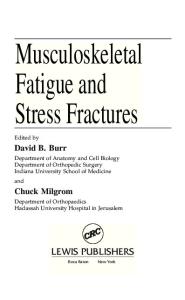
been no economic stimulus to make end result studies like the urge to evaluate the compensable accidents of industl')'". The diagnosis is complicated in infants by the lack of an a.ccurate history, and by the lack of cooperation during the examination. Even the roentgenogram is difficult to interpret because the ends of the long bones are composed largely of cartilage. The osseous centers appear at predictable ages which have been charted (Figs. 1 and 2). In older children the epiphyseal plates introduee translucent lines which fire sometimes difficult to distinguish from fractures. Fractures through the epiphyseal plates are difficult to rccognize if there has been a spontaneous reduction. Thurston Holland's (3) sign of a triangular chip of metaphysis attached to the epiphysis on the side lo which the epiphysis was previously displaced is reliable when present, particularly at the distal end of the radius (F;g. 139, p. 102). Most important in this cOllJlection and in general when taking roentgenograms of children is the rule: Always take both right and left parts in similar positions and each, of course, in two projections. Compare the roentgenograms of the injlll'ed extremity carefully with those of the uninjured. Faillll'es to observe this rule are responsible for most of the errors in diagnosis of children's fractures. A greenstick fractlll'e in a child may show only a wrinkle in the cortex, visible only when projected in pl'Ofile. The torus fmctmc of the distal end of the radius is an example (Fig. lOG, p.80).
At certain stages of a child's development minor twists and tumbles will cause fractures of the long bones that do not occur in adult-s. On the contrary, severe trl1.uma to the torso is required to produce spinal or pelvic fractlll'es which are common in adults with lesser injuries. The appe~ll"ancc of multiple fractures without adequate lrauma suggests at once the existence of a deficiency disease like osteogenesis imperfect-a. Isolated spontaneous fr3ctures in children are likely clue to benign bone cysts (Chapter XVII, Pathologic FractUl'cs). As compared lo t.he differcnces of opinion with regard to the treatment of f1'actures in adults thcre is relative unanimity among the few mell who have studied thc problem in childrcn. The pl'inciples of treatment are simple (142). Alignment is t,he chief rcquirement. The fl'actUl'e should not be grossly angulated 01' rotated. Rotational deformities are inexcusable and should be correct.ed at. an early stage of trcatment. Slight angulation is frequently compatible with a perfect end result. This means that many impacted fractUl'es in young children can be immobilized without reduc~ tion. On the contrary, angulated gl'censt,ick fractmcs neal' the center of long bones, particularly the forearm bones of oldcl' children, tUm;t be COlUpletely broken through and QccUl'ntcly aligned if permanent disability is to be avoided.
fNTHODUCTfON
3
Generall'ules may be formula.ted for 3ppraising a deformity and establishing a prognosis following the fracture of a growing bone. The degree of S1XmfalleOllS correclabilify of angulnr dcfOl'mities in long bOIle fractures of children is dependent UpOIl severn I factors: (A) l'hree local variablC$: (I) The nge of the child; (II) The distance of the fractllre from the end of the bone; (III) The amount of angulation. The younger the child and the nearer to the end of the bone, the more angulation one may accept. The older the child, [lnd the nearer the fractlll'e is to the middle of the bone, the more accurate the reduction must be. (B) The most complete spontnllcous correction of angulnr deformity occurs when the QJl(J1dation 1S £n the plane oj motion of Ct neighboring ginglymus joint. Just proximal to the hinge joints of the knee, elbow, and fingers, angulation with the apex lowal'd the flexor surface produces surprisingly little immediate disability. There is some limitation of flexion but usually this is not noticed by the patient. Hyperextension is insignificant, if present at all. }?unction is eventually normal unless the fracture occurs neal' the end of the growth period. Angulation in other directions is likely to pel'sist, at least in PUI't. Rot.a.tional deformities arc permanent. (C) Deformities oj ar ncar thejemoral neck arc not conected by molding. The proximal end of the femur is unique in its component struts and levers. In a growing child the results followlng It trochanteric or subtrochanteric fracture are complex. Apposition is of little significance since nonunion is unknown. Molding cares fOI' local irregularities of form. True to the preceding l'ule (A) of acceptability of maluuions in children, angular deformity near the proximal end of the shnft of the femur tends to straighten out with longitudinal growth. But t.he change .in the angle of the femorall1eek with reference to the slwft persists (Fig. 211, p. 165). If the fracturc produces a coxa vara, this deformity is permanent. Except flS altered by physiologic derotation, rotational malaligmnent is also pennanent. Similarly nn angulation osteotomy below the trochanter in a child will tend to straighten out with growth but the changed relationships of the neck ::\Ild the shaft persist. Coxa vara is ]Jermanently conected in a young child by subtrochanteric osteotomy but a similar ~lllgula,tiou osteotomy gradually loses its buttressing efTect in st.t1bilizing a dislocated hip (4). The effect of rotational osteotomy ill the subtrochanteric region is permanent. Apposition (the amount of end to end contact 01' engagemcnt of the ends of the fragments) and moderate shortening am of little significance in children. Long bones may be allowed to unitc \\ith ba,yonet (side to side) apposition in children as old ns t.en (female) or twelve (male) years with assurance that molding will produce a normal bone before growth is complete. Side to side apposition produces a rapid strong union. Al-
F\{ACTURES Ii\' CIfILDBEN
4
SHOULDER
HIP
~~
FIG. 1
FIGs. 1 and 2. Range of time of appcamnce of centers of ossification lOUt to 90th percentile. Figures followed by "m" = months; otherwise = yC.'lrs. Where two sets of figures am gil'cn for one center, upper hc.'\.vicr Iltllnbcr = males, lower lighter num· bel' ... females. All = visible at birth. Approximate (lgc of fusion in parentheses. (Reproduced with IXlI'mission of Dr'. n. R. Girdany and The Williams & 'Vilkins Co.)
INTRODUCTlO:\
5
VERTEBRAE OSSIFY
fROM
J
PRIMARY
SECONDIl.RY
TO
CENTERS
AND
CENTERS I
SECONDAR'f FOR
CENTERS -
ANNULIIR
AND ARCH
1-7yeors CENTERS CERVICAL LUMBAR
~ ..
FUSE:
16(25l
16(25}
ANT ERIO R_",AT"L"A S
CENTER APPEARS
~ A8
A 8-1(6)
.~
'='
~
SACRUM
SECONDARY CENTERS
,
PROCESSES
LOWER BODIES SACRAL FUSE AT 18; ALL FUSE BY
CENTERS
W
r ~~3~~;'~.,YJ! .l
VARY
PUBERTY
B-./\
TI===--
FUSE:3
DE'
8-25 FUSE
'C3 V
4-B
\j
~~~ \
A8
RIB
±
5-10
:~~:~
14
PRIMARY CENTERS AB, SECONDARY CENTERS
\;''''-,-1
APPEAR NEAR PUBERTY, FUSE 16-30 years. OCCASIONAL CENTERS
14 (25)
FUSE 4-8
Tubercle
lI 'h ond 12 'h RIBS HAVE NO EPIPHY-
I
~. I
Jr' '-') O~~S:GE
S =:;J 25=::;;.
INNOMINATE
16-18(25)
r6
'0
STERNUM
SES FOR
FUSE 3
COCCYX
FOR MAMMILLARY
\
FAIL
NEAR PUBEIlE MilV' APPEAR BY 1yoo's
AT 3 AT 6
I"I
(
THESE
MlNULII.R EPIPHYSES APPEAR
AXIS
I
MAY
-
ARCH CENTERS
16(25)
ANY OF
EPIP~YSES,
FUSE.
FUSE BODY
9
E~CEPT
PUBIS { Angle CresT
TUBERCLES
17(25)
FIG. 2
CLAVICLE
6
FRACTURES IN
CHLLDlU~N
though this position is usually not acceptable in adults, it is not only permissible but desirable in displaced fractures of the femur and humerus of younger children. The stimulus that follows:l. fracture results in accelerated longitudinal growth of the bone involved and sometimes of another distal to it. The result is similar to thaI, observed in bones with chronic osteomyelitis. The mechanism is not entirely clear and the results are not accurately predictable but the Rverage overgrowth of the individual bones following displaced fracLures may be used as an index of the desirable overlap during healing (Chaptel' VII). The end result will be better if the overgrowth is corrected in advance, because a bone that is too long is just as disabling as oue that is too short. There is no beneficent force which brings about a "compensatory" (5) shortening (Chapter VII, Injuries of the Femur). 'Vith the desiderata so simplified, definite principles of treatment can be outlined. In most cases, excellent l'e5ults are obtained by traction or closed reduction and a cast. When ovel'1apping is desirable, traction is the method of choice. Fractures neal' joints arc much more frequently treated successfully by manipulative reduction and plaster immobilization than in adults. There are definite and predictable exceptions to the success of conservative treatmenti. These exceptions include thr'ee relatively common fractures about the elbow and a few rare articular fractures. These will be enumerated ancl definite principles of treatment outlined under the val'ious ana,tomic headings. Open reductions of othel' fractUl'es are difficult to justify. For lack of knowledge of sound conservative principles 01' to suit the convenience of the surgeon, unnecessary operations may have been done for yeurs without serious complication. But even one unnecessary tragedy in a lifetime is reason enough to abandon such vicious opcrati0ns. A knowledge of prognos£s which IS valuable in adults is of the utmost importance in children who are going to continue to grow. It is this growth factor which makes fractures in childrcn so different from those in adults. Further growth is usually a help in the correction of the deformities of bones that have united in a crooked position or with shortening. All too frequently, growth is an inexorable force which produces deformity when the epiphyscal cartilagc has been damaged. These favorable and disastrous changes are predictable ~lnd should be understood by anyone who trcats fractures in children. In gencral, cp£physealjractllres are best treated by closed methods. All exception is at the proximal end of the femur. This subject is discussed with refcrence to fractures of the different bones particularly the distal elld of the tibia and radius. A violent longitudinal thrust producing a
INTRODUCTION
7
crushing injury to the epiphyseal plate will cause retardation of epiphyseal growth and serious deformity. Unless there has been damage to the growing cells of the epiphyseal plate at the time of injury, slight OVCI'growth is the rule, retarded growth an exception. Afte]' two weeks, considerable displacement is usually to be preferred to forceful closed reduction or an open reduction. The questions have often been asked, "How soon will such a deformity show up? When can we be relatively sure that there has been no serious injury to the growing cells of the epiphyseal plate?" These questions cannot be answered in a few words. It is not "how long", but "how muc;' growth" that determines the answer. The elongation of bones occurs unpredictably in spurts. If the fractured one and its fellow on the opposite side grow little or not at all in a given interval, the answer must be post· poned. :My advice would be as follows: When an epiphysis has been injured, take simultaneous roentgenograms of the alTecled bone and the one on the opposite side at six foot distance. The length should be accurately recorded. If, after three months 01' six months, the measurements of a roentgenogram similarly taken, show that there has been appreciable growth, and that lbe rate on t,hc fractured side is the same 01' a little faster than on the opposite side, then one can safely say that the epiphysis has not been damaged. If there has been no appreciable growth, then one just doesn't know. If the fractlll'ed bone has grown less, then one should give a guarded prognosis and repeat the roentgenograms at two month intervals. It doesn't mean anything to say "After six months", because bone~ may rest six months without growing appreciably. It doesn't mean anything to know that "The ehild has grown one inch in height" because occasionally the addition to height is entirely ill the tOl'SO while the extremities grow very little, if any. The technic of reduction and particularly that of fixation varies somewhat with the age of the child, but difTel's ~reatl.v from the technic in adults. Nonunion may be jgnored as a complication if opert operation is avoided. Fi.:xation is needed for a much shol'tcr lime than in adults; the duration being roughly proportional to the child's age. Immobilization should be efficient in ordcr to prevent deformit,y. Children are much more active than the older members of their families and their bl'oken bones must be held even more securely. In general, it is wise to immobilize one 01' more joints on either side of the fractlll'e until the callus is solid. Permallent stiffness of joints due to such immobilization is unknown in children. Skin tract,ion may be applied to straight fingers for three weeks without causing residual stiffness. Persist.ent limitation of joint motion
8
near a fracture is due Lo bOlly deformity andloJ' fibrosis of the soft parts resulting from the initial trauma 01' rough and repeated manipulations. Early mobilization is not. only unnecessary but is contraindicated. Let the soft p:nts heal. Reep the part at rcst until the bone fragments m'o solidly united. Physicallhel'opy, which is invaluable in the treatment of injured adults, is almost never necessary in the mnnagcment of uncomplicated children's fractures. Dasic pr'inciples of physiology and mechanics must be followed by the doctor in charge of I.he case. Reduction is gentle to avoid soft tissue damage. Casts, splints, bandages, and (,1'3ction arc applied with the part in a comfortable position. Thore must be no obstruction to circulation. Ice is applied locally and the part eleva,ted to minimize swelling. Heat and massage would be wasted and passive manipulation does more harm than good. At the propel' time active motion is supplied by the healthy child in unhmited quantities. Early passive motion which was strongly urged evcll as latc as Asbhmst's day (1) should bc strictly taboo. At the clbow where Lhere is frequcnlly a delay in the return of maLian, a "hands ofT" policy must be followed for many months. If the fraciure is j)ropel'ly J'educed, solidly healed, and thel'C is 110 complicating fil)l'osis of the joint capsule, normal motion will retut'll. The child knows instinctively better than his parents, physical therapist, or doctor what he may do ,,,ithout harm. Pain is his guide to restriction of activity. It is a good one. If left. to his own devices, he will recover in the shortest possible time. Any attempt to hastcn the roturn of motion by loading with pails of sand 01' manipulating, prolongs the restriction of molion indefinit.ely by perpetuating the prot.ective spasm. In children therc is no place for manipulation undor anesthesia. If extensive fibrosis prevents the retUl'n of motion, an operation js necessary to excise the fibl'osed tissue (6). Increased ext.ension may be obtained by a wedged cast or manipulation but usually the gain will be only temporary. The child's attempts at normal funct.ion plus "tincture of time" will produce the maximal permanent improvement that can bp obtained without the nid of smgery.
C II A I' T E H
I I
Injuries of tbe Shoulder Girdle
Ff1AC'I'ljIU~S OF 'l'H~; CL."VICLE
It should be no surpl'ise that. the clavicle is one of the most frequently fractured bones in the body, particularly during childhood. It serves as the only bony connection between the shoulder girdle and tho tnmk. Except for its articulation with Lhe stcrnum, the shoulder girdle floats in a. sea of muscles. Any medially directed blow on the shoulder is transmitted to the claYicle. This bone is likely to be the one to break when the force is applied to the outstretched hand, the elbow, 01' the shoulder. This means that the clavicle is subject to injury with almost any childhood accident,. ,,','actw'es at B£l'lh and Duriny JIIfaney
Compression of the shoulder girdle during deli\'ery will occasionally cause it fracture of the clavicle (2). If the force is great, the fracture may be displaced \\'ith overriding. A mother ma.y 1'011 on a newbol'l1 infant causing a similar fracLure. Sometimes an infant is (lJ'opped on the shoulder, 01' beaten by a mentally incompetent parent with sufficienL force to produce this fractul'e, The common symptom is pselldoparnlysis of the arm with obvious pain when the arm is moved on the ~llrccted side. If the fracture is complete, the pain will be considerable. A newbo1'll infnnt with a greenslick fracture may be mO\'ed more frccly, Rest in the supine posilion is all the tl'eatmCIl t necessn ry. The symptoms of a displaced fracLme may be relieved in the toddler by the addition of a figure of eight bandage of flannelette. Overriding of 5 mm. and nngu\n.tion of 10° mn.y be acceptc(1. :More deformity than this is not encollntel'ed, Healing is very rapid and the fracture may be ignored after ten dn,vs.
JO
FHACTURES IN CII LLDHEN
Frequently a. grccnstick fl':lCture is not discovered until massive callus has formed. The explanation of the child's previous fussiness is then evident. The mother should be :lssured thnt the bump will disappear entirely in a few months. In 1)8C1tdo]Jamlysis of Ihe arm, the physician is likely to suspect the humerus rather than the shoulder girdle because the child guards the arm so carefully, and the true diagnosis is revealed for the first Lime by the roentgenogram. Reduction is !'Urely necessary in a child under Sl:X ycors, even if the fracture is displaced (Fig. 20, p. 24). Grotesque I'Ocntgcnographic appearance is characteristic clue to the complex curve of the bone, and is compatible with an excellent cosmetic and functional result (Fig. 27, p. 25). In the rare case with gross oveniding, temporary lateral traction on the ann with eleyation of the bed on tlle alTccted side may be desirable (Fig. 12, p. 17). A child of three to six cannot be kept flat in bed. A figure-ofeight banclnge of stockinet, partially filled with sheet wadding (Fig. 3) may be tightened every mOl'lling by the mother. It will give comfort and is all the support necessary.
FmC[UTes of lhe CIa,viele in Ihe Child S£x to Twelve YeCt1's Old As the child grows aIdeI', games find sports account for falls on the elbow or shoulder with a direct medial thrust on the end of the clavicle. Greenstick fractUl'es are still frequent but overriding fractures are more common. The greenstick fracture is well treated by the cotLon figlll'e-of-eight dressing (Fig. 3). The infant should wear the support for three weeks, the older child, for four. The markedly displaced and overriding clavicular fracture of the older child should be reduced. Satisfactory anesthesia is obtained by injecting procaine into the hematoma. The child is seated in a chair and tied securely in place. A bolster is placed behind the small of the back. This position is ideal for the reduction (Fig. 4). To maintain the position, soft, all \\'001 felt is fitted as a figure-of-eight and over this plaster slabs and then turns of plaster llre added Lo complete the yoke popularized by Billington (7). While the plaster is still soft, it should be molded to form a, rope in either axilla and should be pulled away from the back of the neck. This is the most comfortable of the secLlre dressings fol' a fracture of the clavicle in u child (Fig. 6). 'J'he prO(jIWS£S is unirormly good. The "bump" of callus and the alarming "deformity" frequcntly seen in the roentgenogram will be obliterated by the molding incident to further growth (Fig. 28, p. 2.5). There is no justification for open reduction.
LXJUIUES OF TilE SHOULnEll OlllDLE FRACTURES OF TUE
11
8C,\PULA
Direct violence and particularly automobile injuries arc rcsponsible for most of the displaced fractmes of the scapula. Blows from hard objects and falls on the back of thc shoulder will produce lesser fra.ctures. The trea.tmcnt is conserva.tive even whell there is considemble displacement. Simple rest of the shoulder gil'dle "'itll a sling and a swath is sufficient. l"racturc of the scnpula is frequently combined with other injuries necessitating bed rest. In such a case, the fractured scapula may be ignored so long ns the patient is comfortable. Irregularities of contour disappear \\'iLh further gTo\\"Lh. If a bony spicule should persist, it may be removed at n Ia.tcr dat.e. It should be notcd that the entire blade of tbe scapula is removed without 101;.'; of function ill such conditions as scapula alta and deforming exostosis. Il\.JUHIES AT 'I'J-(g SHOULIH:ll JOIl\T
Injuries at the shoulder are c:1Used by falls on the outstretched hand Qt' elbow 01' sudden traet.ion on t.he arm. 80ft tissue injuries at birth vary from sprains to avulsion of t.he brachjal plexus. In children, dislocations arc extremely rarC as compared t.o their frequency in adolescents. Fractures fall into definite anatomic types according to ngc groups, 111.iury at birth from vigorous manipulation of the ann may cause an epiphyseal separftl,ion and/or a. dislocation which reduces spontaneously (2). Fractures below the epiphY1:ical plate arc likely to be through I,he middle of l-he shaft. Shoulder injlll'ies are extJ'cmel.v difficult to diagnose becnusc the bead of t.he humerus is still cartilaginous. The dysfunction associat.ed with a bone injury is often misint.erpreted as a brachial palsy. Fracture 01' disloca.tion should be slispected when there is marked s\\'clling of the shoulder wilh psetldop~1ralysis of the arm evcn with negative roenlgenogrflphic findings. One should always repeat the rocnt.genogram in a. week (8). By that time callus will be evident. (Figs. 7,8) if !'here is a. fracture. A dislocation cun be cletccted by careful comparison of thc two shoulders in bo01 the antcropostcrior and lateral projections. One of t.hese injuries is frequently combined with hrachinl bil't.h palsy. Tile coexistence of a nerve lesion should not blind one to I.he presence of a bone injury (9). In particular, onc should :l\'oid the mistake of abducting the arm Oil a splint ancl redislocat.in~ n shouldcr \\'hich had been l'educed spont:ll1eously. A splinted El'b's pal,,;y 1
l~
FIlACTUHES IN' CHILDHEN
head. The deformity resulting from this seriOllS injury is well shown in Figure 10. Although unsight,ly, tbe shortening of an upper extremit,y is of little significance. Function can be greatly improvcd by an osteotomy (Fig. JI). In all of these shouldel· injuries of the newbom, the treatment is easy compared to the difficulty of recognition of the lesion. Relative immobilization and rest of the part, is usually all that is necessary. Brief lateral traction is occasionally helpful as\in the treatment of similar injuries in the infant (Fig. ]2). The crib may be elevated all the side of the fracture for countcrtraction. A sling is ullsatisfactol>y in an infant. A collar and culf is betler treatment (Fig. 37, p. 30), and it may be combined with a swath. Rough handlillg of :1Il infant may cause the simpler injuries described in the preceding panlgl'aphs. The lesions to be differentiated are a true brachial birth palsy and/or a pseudoparalysis from congenital syphilis or scurvy. The latter two can usually be recognized ill a roentgenogmm. A true nerve lesion should be evident on testing with a pin. It is a common mjstake to treat IL brachial birth palsy with a. splint in the position of abduction 90°, rotation outward no o when there is an unrecognized complicating fl'acture or dislocation. This Cl'ror may cause permanent dislocation or limitation of motion. CII1:ldrenfT01n two to seven years sllstain injlll'les a.t the upper end of the humerus while at piny. Dislocations and epiphyseal iJljul'ies are rare. A lranst:Crsc subtubercuwl' .fmcltn·c is common and likely to be greenstick in the younger child. In the latter case, angulation of 10°_15° is compatible with a normal end result and t.he fracture need not be reduced. A collar and cuff is all the treatment necessary. Thc displaccd su.olllbcrru{ar .rraclu.n~ is normally aligned by the application of a hanging cast (Fig. 13). Bayonet apposition with 1 cm. of overt'iding is ideal posit.ion, Ten 1.0 twenty degrees of angulation are permissible. Although the initial appearance is shocking (Fig. 14), molding takes care of the l'oentgenographic picture (Figs. 15, ] 6). The shortness is out~ grown in a. few months. Occasionally following severe trauma such as sllstained in an aut.omobile accident) bed rest with lateral traction is desirable. In stich a case, care should be taken to ~1Void malrotation. The best plan is to flex the elbow and havc the forcarm pointing straight upward from the bed in neut.ral rot.at.ion (Fig. 12). In most instances, fl, hanging cast with Lhe elbow flexed to 80° should be substituted in a few days so that the child may be ambulatory. The cast may be changed for :\ collal' and cutr 01· a sling in three 01' four weeks. The rnt;ionale of the hanging cast is simple. The long head of the biceps crosses the shoulder joint. The biceps functions in flexion of the elbow and in supination. Immobilization of the elbow helps to place the shoulder
[:'iJURIES OF TIlE SHOULDEB. GIHDLE
11t rest. A padded bandage around the neck supports the wrist and lets the cust hang fmc (Fig, :13), so as to produce:lIl idenl traction effect. The child should sit up in a semireclining position in a chnir 01' bed for the first night or two. After that he may lie fiat in bed. A swath about the chest and cast may increase his comfort fOI" the first few days. in lhe cMhl oj eight La Jom"leen ycars subtuberculnl' fractures am less common and cp1:physcal. fracturcs of the proximal end of the humerus occur during games and SP01'tS (Fig. 17). Disloca.tions are uncommon until adolescence. The treatment is conservative. It is lhe greatest fallacy to think that accurate reducLion of an epiphyseal fracture at t.he proximal end of the humerus is important cllough to require opcn opemtion (10). Nothing could be farther from the truth. If :;:een prompUy, this fracture can be 1.lccUl"aiely reduced by closed methods. Usually simple Lradion and the applicntion of a hanging cast is all that is necessary (Fig . .13), Occasionnlly it will be found difficult to rcduce such a fmcturc unless t.he arm is placed in a pivotal position (11). Traction with the ann stl'aight overhead and the elbow noxed to DO° will bc found efTeetive for reduction (Fig. 18). If the fragments become displaced when the arm is returncd t.o the side, a light plaster spica should be applied holding the ann in the pi"olal position (Fig. 10) for four to fivc \'"ceks (12). The ....ounger the child with an epiphysenl fnlcture, the more displacement may be permitted" A tCll year old hoy with bHyonet npposition of an cpiph~"seal fmcture may be trcated consen'aLi,'cly with cver,'" assurance that the rcsult \,"ill be perfect. At twelve ,veal'S, 50 per cent apposition is satisfaclory but, there should be les.<; lhan 10° of angulation" It is easy to obt.ain this position by the method,;:; mentioned above. Open reduction is likely to damage the epiphyseal plat.e and cnuse shorLening as well ~lS some permanent stiffness of the shoulder (la). It is mentioned only to bc condemned. One of the commonest shoulder injuries is cHused by a solieiLous but ignorant nursenulid or pm'ent, of the young ehilcl. Tile toddlcr is helped across t.he street by holding olle hane!' Whell the opposite curb is reached a vigorous jerk pulls the child up to the sidcwalk (Fig. 21). If t.here is not full cooperation from the child, the shouldcr mn,v be OVCl'stl'ct.checl causing an acute sprnin. The snme mcchanism opemtei'i when the parent.s try to keep the child from falling by jerking on the ilrm. The injU\'y may be of considerable sevcrity" The child characteristically refuscs t.o use thc arm. The delt.oid may seem 10 he paralyzed (pseudoparalysis), and the condition has becn confused with poliomyclil.is, Pulled s)wuldcr is diagnosed on the history with a negat.ive roentgenogram, pain 011 att.cmpt.ed motion of the shoulder, but powcr in Lhe ann below thc shoulder. ""hen the force of lhe jerk is difrcrenlly t,ransmitt.ed, pulled elbow i,,; t.he rcsult (Chap leI' IV).
FfiACTURES IN CHILJ)HEN
FIG. 3. Frn.cturcs of the clavicle without. dispbcemcnt. mft)' be treated with n simple figure of eight bandage Illnde of stockinet, IlfI.dded inside with sheet. wlldding under the axillae. It is convenient to circle both shoulders :r.nd then pin the bandage loget.her in the back rather than to fonn a figure of eight. primsril.r. The bandage may be tightened each morning by the mother.
FIG. 'l. A. A displ:lced fmctul'C of thc clavicle in lin older child Clln well be reduced ullder local anesthetic with the patient sitting in a. chair. A rolled bath blnnket is placed at the smull of the back. A muslin bandage is passed across the thighs and Hed 8CCurel.r to the chiliI'. B. While the shoulders nre held upwnrd and bnck, n. Stril) of felt is formed to a figure of eightnnd stitched in plnce. Felt mny be enclosed in stockinet if the child is sensitive to wool.
Rolled both _. blankel
B
--
[5
JNJURll;;S OF THE SHOULDER GIRDf,E
FIG. 5. J..ong Sl)!ints of plaster of Paris :Ire seCll!'ed by tU1'IIS of plnsle!' b:md:lge. The layers :1l'C molded into a rope under the axillne. Firm pressure is made between the shoulder bhdCls so th:Lt the weight of the Ilrms is carried here.
I,
I'~
i
FIG. 6. The completed yoke is a comfortable, efficient dressing if properly applied. It should be \\'01'11 for foul' to six weeks.
16
FHACTUBES IN ClIILDllE!\'
FIG. i. A. H., 1l1:l1c, newborn, lOlli/Hi. Elliphysc:d separation [It the proximtl] end of the right humCl'll~
II"ns
110t
rccognized
until
dnrs aftcl' dcli"CI"y. Callus hud ucgun to form. Note the dispbccmcnt of the capital osseous
fourt()()ll
center'
Oil
the I'ight
:15
C01l11Xl,l"cd to
the left.
FiG. S. A. H., tll"clnJ days :lftcr Figmc 7. With tl":lctiOll (Fig. 12)
satisfactory position \l'as lIlaintaincd until mussi\'c c:lllllS formed. Support WilS no 10llger nCCC5Sill'.\".
FIG. O. A. H. FoUl' months after the delivery there is nlmost complete restoration of the osseous clemenls of the right shoulder. Note the precocious development all this side.
l~JUll U~S
010' TilE SHOULDER
FIG. lO. J. G., male, ag() 11, 0/2/36. Tlie ]Jl"oxiepipllysis W:lS completely sep:lmted at birth. The epiphysis \\"tlS rotated almost 1800 11lld healed in thi~ position wilh rmuled shortening of t.hc tll'l11, limited forwnrd flexion :lnd :t1xlucOon. This roent,.. genogram taken :1t the time of the eorrcctil'c osteotomy shows the deformity dendy. lll:ll
17
FIG. 11. J. G., G/17/53. Seventeen years lifter the preceding film sholl"s solid heltling :lnd molding of the osteotomy, wit.h greatly ir\lpro\"(~(1 l"lltlge in [01'11"111"(1 flexion lind :11)(!llctiOll.
~
"
GrlU)J.I~
...
,..-
\
!
FIG. 12. l~li.lnnelctte secured with Ace udhel"ent m:1Y be used for' !:Iteml tmction. To prevent rotation of the fmgmellts, the fore:mll should be slIspcndl-.:1 1.lCI'pcndieuiar to the bed.
18
....-.
FRACTlJRES IN CIHLDnEN
\ ,"i~
" .FIG. 13
FlO. 14
• FIG. 13. A h:U1ging elst Ina)" be suspended from the l\C<'k Il"ith :l pmld{!() lIluslin b:uldagc. The elbow should he f1c,'l:cd slightly lllore :lcutclr thnll :l. right
:lllg1c. The hC:l<1 of the hed must he cnlllkcd Ull for the first fel\' days. (Reproduced Idth pcrn)issioll of tile Jourrlill of the American i\hxlic:ll Association (·Ifi)I.
FIG. 14. T. K, !Unlc, nge 7. Epiph.\'sc:ll fracture of the [ll'Oximal end of the left hUlllerus. Treatment wilh a hanging ('[1st lind then :l sling.
11-
FIC. I';
FIG. I;). T. K., fOUf months ::lfter the preceding roelllgcllogmm. The child was :lmhuhtory while the fmgl\l('nls hC31cd ill 1){I~'ollet nplx>sition but S3ti&f:lctorr alignment.
INJUHIES OF TIm SJlQULDEB GIHDLE
FIG. J G, T, lC Three lind one·h:df years :tftcr Figure 15, thcre is nlmost complete elimination of the deformity, The humeri In'l'e the same length, (·linic:ll1y 1l01'nw1.
lew. ii. J. B., m:)le, H!;C to 4/3/53. Epiphysc:\l frHcture of the proximlll cnd of the hUlllcrus with characteristic dispillcemcnt. The fCduction could not bc maint:\incd with tho arlll lit tllC sidc ami open operation WM contemplntcd.
JD
FII.\c:J'UIlES IX CIIII,DREN
20
1
FI\'. IS
FIG. 19
FIG. IS. J. 8., 4/G/53, the $.'tme fl-:l.cture :LS 8110\\'11 in the preecding l'OCntgenogr:lI11S. Reduction W:lS ctlS-
i1r accomplished and lllaint{lincd by c1cnl.ting the arm Q\'cr the hc.'ld.
FlO. 19. J. B., 4{6/53. A light pbstcr SpiC1l 1\":lS applied with the arm in the pil'otul position. A(tel' foul' \mcks the
CHS~
was removed und
traction applied while the lIl'IlI wns W:I!I brought down to the side gmd\lally.
FIG. 20. J. B., 12/11/53. Anteroposterior dew of both shoulders eight months after the preceding rocntgenogr3m, showing nc:arl.r sym-
mctricnl nppe:u:mcc. Function \\'as entirely Ilonna\.
FIG. 20
CHAP'I'EB
III
Fract.ures of the Shaft of the Humerus
E'l'lOLOGY
As compared to the frequency of fmctlll'es t.hrough the middle of the humerus in :ldults, t,his fmctlll'e is rather l":1re in children. It may be a complication of delivery (2). It may OCCllI' with rough handling of infants dlll'ing dressing. In older children it is usually the result of direct trauma. The indirect force of a, fall on the outstret.ched hand 01' on the elbow ...causes a fracture elsewhcl·c. TREAT~l Er\T
Fracture of the shaft of the humerus in infancy is ideally treated by lateral traction (Fig. 12, p. 17) fot' a week. The elbow should be flexed with the forearm pointing straight upward from the bed in neutral rotation to assure the surgeon that a rotary displacement is not oecllrring. Traction with the forearm straight has allowed rotary displacement particularly in a squirming infant. When soft callus has fanned traction may be disconLinued and a collar and culT with a swath used insl'cad. Apposition is noL necessary. Fifteen degrees of angulaLion are permissible (Figs. 20-28, pp. 24, 25). If there is no displacement, 0/' if traction is not feasible, It collar and culT with a. swath may be used as primary troatment. The inf:lnt is less comfortable than wi th t.raction. Ambulatory children are best treated with hanging casts. Two em. of overriding is not an indication for f\ change of treatment. One em. of overgrowLh is common in a displaced sh:lft fracLure of Lhc humCl"LIS and some o\'erricling is desirable (Fig. 23, p. 23). The mtionale of I're:~tmcnt is thc samo as for fracture of the femur. Bayonet apposit.ion is llsually preferable, but a fmclure which is not displaced will not overgrow ilS much and should be left as it is. Healing is very rapid. In foUl' woeks all :-llpport. 21
22
FllACTURES IN CI-llLDHEN
may bc rcmovcd, Rcduct ion is required only in t he rare oblique fracture with the hone clld~ impaled in muscle. Traction under anesthesia is necessary to free tho fmgments. Traction is then continued or a hanging cast applied. A spica with Ihe firm abducted was common treatment before 1930, It is unneceSi>nril,\· cumbersome and tcnds to cause angulation with the npex medinlly. Thero is no just.ifioftlion for open I'cduction. Complications fire nlmost, unhenrd of in simple fraclUl'es of the sha.ft of the humerus in children. Nonunion is not encountered. Hadial lIcrve palsy which frequently accompanies this injury in ndults, OCClll'S \·cry rarely in children, Dr, Cave (14) cited ,I fl·enk case of radial nen·o palsy, A ten year old girl fell in the bat.hroom catching her arm betwcen the wall and a t.owel raek, A complete closed transverse fracture of Ihe midshaft of the humerus with minimal displacement was produced by direct trauma, Thcre was immediate loss of power in the radial distribution \\-ith hypesthesia to pin prick over the radial ~ensol'Y nrea. A cast for six weeks was followed by a cockup splint, Three and onc-half months aftor {,he injury I,herc was elect.rical but not fun(;lional evidence of neurolization. One year following the injul'Y all motions wero present. except flct.ive extension of the thumb. Twenty months after the injUl"}' , function was entirely normal. Fl'act,ure of the shaft of the humerus may be one of mult.iple injlll'ies which confine the patient to bed. Tl'adion is then the best treatment. If the hllmerus is left. ill the hOl'izontal position, t.he elba\\" should be flexed to insUl'C correct rotation. Occ~\sionally it is better to (lex t.he humeru,,; at lhe shoulder with skeletal traction through the ulna (Fig. 25, p. 24). Care should be t.aken t.hat the wire OJ' pin does not damage the epiphyseal plate of the olecranon (15). It is a. good plan to apply a cast from t.he hand 10 above the elbo\\' to protect the pin from contamina.tion.
,
FIG. 21. The "jerk" will hurt the child's elbow 01' s.houlder.
FHACTURES OF TILE: llu;\IEHUS
FIG. 22. \\". T., malo, ago L2, i /2/3G. A fl':lCturo of the middle tbird of the left humems W:lS COIllplic:lh..'(l b.l· multiple rib frllctures and colb!>sc of the left lung. With traction in bcd, good :llignmcnt \\'~lS m:linbilled.
FIG. 2.3. W. '1'., 9/2-l/3G. Solid union of the fnlctul'c wilh good uJiglllllent nnd 2 CIll. al'cniding thl'CC months after Figme 22.
FL(;. 2·1. W. T.. 8/23/38, tll"O ye:lrs :lftcr the ol'igillnl injury. The l"oentgenogl':l1H sho\\"s persistent chnnp;cs in the intcrnal nl'chitccture
but fUllction \\":15 entirel.'" normal. Thcre 11':18 I Cill. of sllol'tellillg.
FIG. 22
FlO. 23
23
24
FHACTUHES IN ClIlJ,DHEK
~j
lew. 26. H. \V., male, nge 3, 9/19/51. i\'lnrkedly c1ispbccd fractures or the left. cbvielo und tho proximal third or the lert humerus caused by tho imp:lct of :LIl ilUtomobile, The child I"ns first socn six days after the injury. A h:mging cast was applied and t.he deformit.iC1l permitted to remain.
FlO. 25. Trnction on the humerus by:l. Kirschner wire through the olccl':lIlon is sometimes used to :ldmntage. Care lIlust be t.nken to plnce the wire dist..,1 to the epiph}'se:lI plate of t.he olecranon. If the skeletal traction is to 00 used for more th:m II few days, the trnction bow should be incorpornted in a light pbster C.:'lst.
FRACTURES OF THE HUMERUS
l~[G. 27. H. \V., 12/8/52, fifteen months following the preceding roentgenogmm. The anguhr clefOl'lnity :tll(] shOl,tl!ning hare both hcen reduced,
FIG, 28. IT. \V., [2/8/52. Symmetrical shoulder' joints as wen as c1:wiclcs without deformity in spite of the gross displacement in Figure 26.
CHAI'TEH
IV
Injuries About the Elbow
SUPRACONDYl.AR (DIACONDYLAR, TnANSCONDYLAH) FHAC'J'UJU~S
(60 per cent oj elbow !mctures)1 EtioloGlj Attempts werc made Lo subdivide these fractures according to the level, in the days when "skiagraphs" wcre illdistinct shadows (65). The more dist,al ones were called dia-(thl'ough) condylar (Fig, 29) 01' transcondylar [lnd the term "supracondylar" was resenred fol' those slightly TnOl'e proximal. But in the lateral view, most fmct-lrcs look "supracondylar" while in the anteroposterior vicw, the same fractures have the "tra.nscondj'lnl''' appearance, The level through which the break commonly occurs does not vary more than a millimeter 01' two. The dilference, if any, is so slight, that they arc grouped together in this book, The exact levels of the fractures do not depend on the age, They occur from three to ten years, with greatest frcquency bctween five and eight, Epiphyseal separation of the entire distal end of the humerus was a common diagnosis at the turn of the eentmy (1). It, too, has vanished with improved radiologic technic, The supracondylm' fracture is ch:u'acteristically produced by a fall on the outstretched ann with the elbow in hyperextension, If the fracture is complete, the distal fragrn.ent is displaced posteriorly, usually with some upward riding (Fig. 30), The defor'mit)' may be tremendous so as to simulate a dislocation with which it is commonly confused. If the fracture is incomplete, there is little deformity, Angulation with the apex anteriorly may be sufficient t.o reduce flexion by 25° to 30° and justify reduction, Less than 1 pCI' cent of supmcondylnr fra.ctures am the reverse, or jlcx£on type, This injury is produced by a fall on the flexed elbow with I The percentages ill this cha.ptcr 3rc from cnd rcsult !;tuclies at thc l\'Iilwflukce Childrcn's Hospital.
rXJUHIES AHOUT TI!l~ ELUOW
resultant, anterior displacement of the distal fragment (Fig. 33). This flexion fracture should be recognized in order to reduce and immobilize it in sufficient extension to prevent a recurrence of the angular deformity (Fig. 34). It is an errol' to confuse lhe types and to treat in extension the usual posteriorly displaced distal fragment, wit.h angulation, apex anteriorly. This nlistake causes prolonged hyperextension and limited flexion. Treatment If a supracondylar fntcture is seen promptly, before swelling has appeareel, even though the displacement is eXlreme, an exccllent result is obtained by immediate closed reduction and immobilization in flexion (Fig. 35). If reduction is delayed until there is marked swelling, it is better to apply traction (Fig. 36) in slight flexion rather lhan to attempt a manipulative reduction. The manipulative method may succeed temporarily even with swelling, but there is little but tradition to recommend it (10). It is seldom possible lo Hex the elbow sufficienLly to maintain reduction without obstructing the circulation, and the po:;ition is promptly lost (17). The application of a circular cast is nn im·it.ation to Volkmann's ischemia.. \Vith three or foul' days of elevation, tnlCtion, and ice packs, the swelling will sub":lle and a manual reducl.ion may be completed under anesthesia. The inlroduction of hyaluronidase into fracture treatment has somewhat changed t.he prognosi::; in Rupmcondylur fractures. Three to five hundred T.R.. units may be injected into the henul,toma of a badly swollen elbow. The accelerated dispersion of extra. cellular fluid certainly hastens the restoration of the tissues to normal and probably reduces the ultimate fibrous reaction. Theoretically t.he likelihood of Volkmann's ischemia is mduced. The use of hyaluronidase may make it possible to reduce and maintain the position of supracond.vlar fractures primarily, even when considerable swelling is prcsent. Not infrequently, satisfactor.\· posilion is obtained by Dunlop traction without manipulation. It is particularly indicated in the unstable tmnscondylar fractures (18, 19). (Fig. 29). One may continue the traction for three weeks and then place the nrm in a sling There is usually no need to maintain the traction so long. After ten days when the fracture is reduced and the threat of vascular embarrassment has subsided, I prefer to make the little p~l.tient ambulatory by acutely flexing the elbow. This Inay be done conveniently with rectal pentothal. A collar and cutl'maintain the position (Fig. 37). The ability to reduce but not to hold the position of a supracondylar fracture has influenccd some men to use intcl'l1ftl fixation. While the re-
"
28
FHACTUH-E8 J:\' CJlILDHEN
suits nre some~imes good, permanent, limHation of motion is all too frequent. This method cannot be justified. V\'ith ten days of traction as above, there is enough callus formed La mnke the fracture 'Isticky". It wit! then stay reduced in the flexed position. If there is evor un indication for open reduction in the absence of compounding, it is extremely rare in a fresh casco A considel'3bly greater exposure is necessary than for the open reduction of a lateral condyle. Operations on supmcondy!ar fractures arc frequently followed by rest.ricted motion. Blind pinning with protruding pins is always undesirable ill children, whorsc urge to wiggle and scratch cannot be conl,rolled. The use of internal fixation because conservative management fnils on lhe first try is the way of fill impeluous sUI'geon. Patience and gentleness payoff in Lhe t,rea.tment of fracturcs in children, rind in sl..lpl'aconclyl:ll' fractures in pnrticular. Before the reduction of any elbow fractlll'e, c:1l"eful cxamination should establish the normal funclion of the motor nel'ves controlling the hand, and the condition of the radial pulse. If the pulsc is prescnt, it should not be obliter:'1,tcd by the manipulative reduclion. If absent before manipulation, it may be disl'cgarded if the capillary circulation remains good. An absent radial pulse is a c1nngcl' signal which cannot be ignored, but it is not of itself fin indication fol' open reduction. The mechanics of closed 'reduction are simplc (Fig. 35). ""hile countertmction is made in the axilla (not by grasping the skin of the ann!) straight traction is made on l,he supinated hnnd with one of the operator's bands. The other band grips the arm just flbove the elbow with the fingers over the biceps. The thumb lies against the distal end of the proximal fragment while hyperextension is madc to disengage the fragments. 1\1alrotation of the fragment is corrected by ,"nrying the degree of supination. Lateml di::;placement is corrected by molding the fragments bcfm'c correcting the postcrior displacement. Then the thumb slips down over the tip of thc olecranon forcing the hyperextendcd distal ft':l.grnent forward into posit.ion. Counterpres:'illt'c is supplied by the fingers. The elbow is flexed to mnintain the I'eduction anI.,' nftcl' complete reduction of the fmcttll'e. The position is checked at once by rocntgenograms. If it is not satisfactory, a second reduction may be tried. Traction in bed is preferable to the insult of sever:'!l manipulalions. F1·xat.iol/. 'in fle:r;ion ma.'" bc vUl'iow.:1,r achieved (27). The simplest nnd surest met.hod is to join :.1, collul' find culT by :.lll inelastic bnndage (Fig. 37). A light compression dressing about the elbo\\' retards swelling and inercaSCR the child's comfort. The addition of a plaster splint to hold t.he fore::mn in supination is advocated by some. The rotation of the forcarm is actually unimportant once reduction has been obtained and the elbow tlexed. Adhesi"e plaster dre~ing~ arc sccure, too secure. Unless npplied
1~.IUIlIES
ABOUT TilE ELBO\\'
29
with extreme carc, they are u, threat to the clrcubtion. The worst cases of Volkmann's ischemia have occurrcd with the usc of ndhesive plaster 01' circular casts (Fig. 38). Following reduction of the supracondylar fructure, the child should be hospitalized for twenty-four hours to insure frequent observation of the circulation of the hand by a competent nurse 01' house oflicer. n this is not. possible, the parcnts must understand the (bnger signals. Most significant as warnings of curly ischemia arc: pain, swelling, coldness, cyanosis Ot' pallor, and loss of ~lbility to move the fingers. Remcmber the triad: Pain, pallor, and paralysis (Fig. 39). The most important, and constnnt of these ~igns is pain. A \\'elll'educed fracture in a child should requirc no sedation ol,her than aspirin. Pain sevcre enough to require opiates should be a warning that thcre is some complication, oft.en too tight a bandage or too much flexion. V o/.kmann's I schem'ic ConlTCtcfuJ"c If there is evidcnce of circulatory embarra!%ment, the acuity of t.he
flexion is reduced immediately by 20° or 25°. If this change is not instantly succes."ful flnd I,hore is still a threat of ischemia, all bandages are removed from the elbow and forearm, angulation is reduced to 20°_30° straighter than a right angle, and ice bags applied. Satisfactory position of the fragments and elevation of t.he forea.rm arc maintained by traction. Usually this is applied to the skill of the forearm without constriction by the use of flannelet.te strips secured by Ace adherent., (Fig. 36) and held in place by a loosely applied elastic cotton bandage. A Kirschner wire through the olecranon (Fig. 25) (21) is prefencd by some. If there are already blebs about the elbow the wire may be inserted in the basal phalanx of tbe thumb. J~oss of positioll of the fragments is insignificant compared to the tragic disability following unrelieved ischemia (Fig. 38). Prompt blocking of the appropriate sympathetic ganglia is sometimes helpful. If the symptoms are well a.dva.nced or do not subside with conservative measures, no time should be lost before exploring the cubital fossa and volar aspect of the forearm. The tough fascia enclosing the flexor muscles of the forearm is slit permitting the explosive extrusion of edematous muscles and hematoma (22). An injured or markedly constricted brachial artery should be resected. This relieves vasospasm of the collateral ar~ terioles and the reflex involvement of the intimate vasculature of the muscles (23). Delay is disastrous. Within three or foul' hours, irreversible changes have taken place. The all-too-frequent claw hand usually means inadequate or delayed therapy.
30
FHACTunES IN CI11J.,!)HEN
Rclcnf£on
Tn the avcrnge C:lSC, lhe reduced supracondylar fracture should be immobilized in flexion fol' throe weeks. At 1,he end of that j;ime, the callus is strong enough to flllow the elbo\\" to be lo\\"cl'cd La a right angle in ,l sling. In another week 01' b\"o, all fixation may be discontinued and the child allowed to usc the h.-wei at, his own discrotion (24). It hns been adequately proved that morc rapid I'ctUl'll of normal function will OCClIl' if the child is unmolested. Currying ft pail of sand does mOrc harm than good (Fig. 40). There should be no I11nnipuln.tioll to increase motion eilher with or without anesthesia. Hcpeatcd stretching of contradures not, only distresses the child but perpetuates the spasm Hne! delays thc rcturn of normal function. Occasional1y thcro is fill indicntion to continue tradion long enough fol' union to occur Wig, 43A). Usually tbe position will bc satisfactory. Tf there is undesirable angular deformity at the end of three or' four weeks, the fracturc may still be manipulatcd under allesthe~i~t :111d satisfactory position obtained. Bayonet apposit.ion with tlorm:1l alignment is satisfactory and is not nn indication for manipul:1t,ion at thi~ stage (Fig. 43B). Sharp spicules disappear "!ld contours impro\'c with molding as in fractures ncar tbe ends of othcr long bones (Fig. 44) (25), Angular deformity corrects very littlc nt, thc distal end of the humerus.
Prognosis The prognosis in supracondylar fractures must bc guarded. If the trauma is not ullusually Se\'el'C, illld if the reduction is promptly and gently pcrformed, the rcsult should be a normal elbo\\' (1). Therc al'O several chances for disability. The mo:st, significant complication, Volkmann's ischemic cont.racture has been disCIlS:;ed, Exuberant callus will llsually disappcar sponlnncously if unmolested. Physif.:al therapy is not only superfluous but it actually delays recovcr,\' (Fig. 45). Myositis ossifictlns Wig. 40) OCCUI':': most frequently following excessive periosteal stripping, part,icularly :,ftcr dislocation::;. [t will usually disappcar if unmolested (Fig. 47). Opcmt.ive removal of the exccss bone in children is invariably follo\\"cd by I'CCUl'l'Cncc. ]Jersistcnt capsu1:l1' contracture may require excision of t.he :mlcr'ior capsule (6). If osteotom~' is necess:u'Y to correct extreme angular deformilY or to improve the arc of motion it should bc postponed for 8cveml years, Reversal of {he carryin(J angle (gunstock deformity) (Fig. 48) may be due to uncorrected medial displacement of the distal frngment (Fig;. 40), usually the result of rotation (27). It m:t.y follow ~~ perfect reduction and be due to accclcl'u.tcd gl'Owth of the lateml condyle; rarely, retarded growth of the medial.
IXJURIES ABOUT nlE ELBOW
P(/m/ysis.froll! nerve hljury is usunlly only temporary. The I'nc1ial nerve and occasionally Lhe median may be involved in the absence of ischemia. Early exploration is contraindic:'lted as spontancous recoycry is the nile. Grossly malunited supracondylar fractures may rcquire open reduction afLer six to eight weeks (Fig. 50). Moderate permanent limitation of motion is the rule c\'en if renson:)bly good position can be maintained by pin fixation (Figs. 51, 52). After eight to ten weeks it is usually beLter judgment to allow the fracture t.o go untreated and correct the deformity b.\· o:o:teotomy a ,\'ear 01' more ~Iftcr 1he injlll'Y.
FIG. 29. Ie K., male, :lge 3, 3/8/5'1-. A. A true l!":lllscondylar fr:ll:tul'e of the distal end of the IiUllle1'1ls with disphH:cment of the distal fmgment to the ulnnr side. B. Tile reduction obbined by traction :oS iIlustmted in Figurc 36. The latcral "iclI" \\":15 also good. After fifteen d:lYs a collar :lnd cufT \VC1'C applied. The hoy \\':15 <Eschtlrgcd from the hospit:l\ tll"O lIn.vs bter. At 110 time \\":lS !.lIe circulation in jeop:ll'c1y. Function nOrllllll.
31
32
rHACTURES [N CHILDREN
FlO. 30. G. L., male, age 9, 3/28/53. Supracondylar fracture with marked posterior unci proximal disphcc.mclJt of the distal fragment, imitating :1. dislocation on clinical cx:\mination.
,:,
" ,~'
I,
'
: FiG. 31. G. L.,
:~/29/53.
Anlero· posterior and laleml dews of the same fracture seen in the preceding figures on till! day following: all ensy reduction. Note the ohlique fracture line which makes the fracture lmns· condylnr in the anteroposterior view but definitely supracondylar in the lateral viCl\',
;
y ~
y'
"".
."
IN,Il.HUES AllOU r THE ELBOW
33
fIG. 32, G. L., 4/7/54, the same ense Olle year II£ter the fmeture. Healing hns been complete. The internal arehiLectUl'e is restored. The left hurnems is 2 mm. longer than the right. Motions and carrying angle are symmct.rica1.
,
FIG, 33. C. :\f., female. J<'lm.:ion type of supracondylar {l'actmc c;lllsed by a fall on the tip of the elbow, fOUl" weeks after the arm \\'as imrnobilil,cd in flexion. Some extension would have improved the posi~ion. The end result wns a l1orn1:11 eloo\r.
3<
FHACTURES 1:\ CIIILDHE:'\
FIG. 34
I
FIG. 3D
I~JUlUES
ABOUT THE
El~BOW
35
FIG. 34. A. The mcchrmislll of supraCQndybl' fl':lCturc, flexion type. ll. Immobilization in some degree of extension is desirable to bring the distal fragment into :l1jgnment with the proximal. Immobilization is lleeCS5.'lI'Y for only three or four \reeks.
FIG. 35. UedueHon of a supracondybr rr:lcture. A-. Dctermine the presence or absence of t.he radial pulse; cxamine for motor paralysis. A. Under genernl :mcsthesia, apply tmction with the elbow hypcrcxtell(lcd. Countertraction in the :lxilla; assistant's hands loosely on the ann. B. The surgeon's thumb forccs the distal fragment into position. Accurate reduction is obt:Jined br rotating the forcarm and molding the fr:lcturc. C. Only after the fmcture is completely reduced is the elbow flexed acutely (not forcibly). D. Reduction is maintained by the f1exe<1 position, wit.h nn elJieient. coll:u :llld cuff. IRel)roduecd with permission of the ,IOUrlllll of the Americall :Medie:l.l Association (45).J
fiG. 36. Dunlop's traction us modified by Allen and Gmmse (19) is used temporarily if there is exeessil'C swelling or threatened Yllscubr emb:llT:\SSment. It may be continue
, I
~
36
FBACTURES IN
CJlJl.DBJ~N
J'lG. 37. A cufT of soft material like wool felt is placed aoout the wrist nnd sccured with several turns of adhesive plaster. A similar reinforced soft coll:tr is pbced about the neck. These urc joined by :lll inelastic btwdagc with Lhe elbow in acute flexion. ft is well to apply a compression dressing to the elbow for :1 few days. If the child is restless a swath Inny be added. A plnstcr slab mily be used to nmintnin supin:l.tion of the forearm. This is usunlly superfluous if the fmcture has been tlccumtely reduced nnd tile elbo\\" kept flexed.
FIG. 3(). Doctor, trent the p:Jticnt, 1I0t tile pictul'e.
!;\,JURIES AIlQUT TilE 1':LIlQW
37
FIG, 38, Volkmann's ischcmic contractUl'C. Whcn this st~lgc is renchcd thcre has been cnt{\strophic d:lIllllgc to the elbow, forenrm, 3.nd h{\nd. The wflmings were p:Lin, p:lllor, and p..'1mlysis. Prolonged rehabilitation I\'ill produce only :I. ,"cry poor substitutc for :1 nonnal hand and
a=.
J'·IG. 40. c.'1rry the snnd yourself and let thc child get well.
38
FRACTURES IN CllILDREI\'
FiG. -11. W. H., mnle, ngc 10, JO/27/;].I. Supracondylar fr:\cturc of the left humerus with marked dis-placcmDnt, considerable soft tissue injury, and swelling. (Bcproduccd with permission of the AmcriCllll AC~ld{)my
of Ol'thop:l.cclic Surgeons,
Jllstnu:tionrtl Course Lcetmes (142).J
FlU. ·J2. \\'. H., 1O;2i /;'H. Accurate rnnnipulati\'c reduction of the precc
A
{1.!2).1
FlO. ·13. A. \V.ll., lL/23/34, the appearance of the preceding fmcture :lftCl" fOUl' wccks of traction. There 11':18 110l'mnl nlignment in both dews. Apposition was normal in the nnteroposterior \·iew; o.'lyollet :l.ppositioll in the bternl dell". B. Seven months later. The spike of bone had absorbed and there \\'118 only 10° limitation of flexion. Othcr clbow motions wcre normnl. [Reproduced with permission of the American Ae[\denl~· of Orthopaedic Surgeons, fllstruetion:ll Course Lectures (142).}
!xJUIUES ABOUT THE ELBOW
39
FlGo .J.I, W. H., IO/3/-HI, bter:d views of both elbows fiftecil years later. Slight defOl'mity persistso E'(~ cept for WO limitation of flexion on the left, function is normal :It\d s.,om_ I1lctric:ll. ~o subjective symptoms. [Reproduced with permission of the Amel'ican AC:tdemy of Orthop:lCdie Surgcons, Instructional Course Leetlll'CS
(142).]
FLO. 45. Strong arm methods will not stmighten the elbow.
40
FRACTURES IX CHll,DREN
FIG. ·16. R. T., m:llc, age 6, 7/19/40. :\Jyositis ossificans following (lisloc:ltion of the elbow. !'I'lotion was gl'c:1t1r rcstricted. [Reproduced with permission of the JOllrnn! of the American l\'Iedical Association (45).J
FIG. ·~7. R. T .. 7/17 (la, A ]'()(mtgcnogmm three yC:lrs ;Ifter" the preceding. Tile abllol"lllnl bone has almost dis.'lPPc:ll'cd. Elbo\\" motions :Irc IWI'Ill:l!. The tfc:lImcnt \\',IS masted.l· neglect. [Hcproduccd with pcrrnis.<;ioll of the .lournal of the A mcritan .Id edical Al;SOI·j,llioll (45).
FIG. 48. B. 11., male, age H:i", 12/14/42. Gunstoek dcfonuity of the right cloo\\' following [\ supracondrltlr fl"llctul'c fi\'e months previously. Angula-
tion \\'[\S due to medi:d disphcemont nnc! rota.tiOIl of the distal fr'agmcnt,
IXJUHIE$ AllOUT TilE ELllO\\'
41
FIG, 49. R. H" S/26/.J.4. An 3nterol>osterior roeutgenogr:ml of both elbows two yc:Jrs 3fter thc preceding l>hotogr:\l>h. There hns been no change in the deformity. Note the prcoocious epiphyseal dCl'elopment Oll the right. Sometimes cubitus \':lrtIS is caused by o\'cr~ growth of thc lateml epil)hysclil pl:1te, sometimcs by injury of the mcdi:ll. l,,'\teral :LIId medial angular deforrnitil'i:l do not correet 81>011tnncously.
FIG. GO. ,I, I L, lIlale, :I).;"e '\ 10/17/50. 1'11'0 \'ie\\'s of :t ,e:rossl,\" displaced :lIld :tngulatCl:l SUpm('U11~ ely!.')r fracture of the left Immel'lls six weeks :lfter the injury. ?\Iarketl deformity, :tlld limit:ltion of f1cxioll
iO", c:dern:\1 robtiOIl 3D". Open reduction tmd pin fix:ltion secured lIorm311losition of the rr:lgments.
42
FUAC'TUIlES
1~
CIHLDnEN
fIG: 51. ,I. fl., 1l/i/52, two yc-.lI'S after tile jJrf:(:cdjng rocntgcllOgrnnl. There is still 10" limit:t.tion of flexion and 25°, of extension.
FIG. 52. J. II., 11/7/52. :\orm.:ll alignment of the elbows in Figure 51,
lateral ,'iew. Rocntgenogmms i:Iken eighteen months bier showed illlpnll'ed nppe:mUlcc. Little change on clinical e.~(uninatioll. The left humerus is 15 mill. O\"crgrown as the result of the fmcture :m
INJUn lES ABOUT TILE ELBOW
'13
FRAC'I'U1lES Ol~ TUE 11<\'I'ER!\L CONDYLE OF THE HUJ\IEllUS
(18,5 per cent of elbow fTOctm'es)
Lateral condylar fract11res m'e produced by hyperextension plus angulation with the apex medially, often a combinfllion of direct and indirect trauma (Fig. 53). The fracture line extends from the lateml epicondyle obliquely downward and medinlly into the trochlea (Fig. 54A). The defonnity is not great nnd the injury i,o; often djsmissed as a sprain, particularly in the infant whose capilellal' osseous center has not yet appeared (Fig. 56), Careful palpation of bolh elbo\\"s may be Ihe only diagnostic procedure of any value. "'hen a. cOlldylnr fracture occurs in combination \\'ith a dislocation, there is likely 10 be prolonged or permanent limitation of motion. Roentgenogram:; of both elbows should be taken \\"ith symmetrical positioning. Even theu the diagnosis is difficult in young children because a large pOl,tion of the distal end of the humerus is still cartilaginous. The small oval os.':leous ccnter of the cnpitellum may be the only bone visua.lized in the condylar fragment but usually a. flake of meta.physeal bone is carried "'ith it. The fragment is lIlUch larger than would appear in the roentgenograms. Tts cephalnd corner is characteristically rolated latel'ally and do\\"nward in the sagittal ns \\"el1 as the frontal plane, thl'Ough an are of 90° or more by Lhe pun of the extenS01' muscles of the forearm. The fragment is trapped in Ille joint. so that the articular cartilage of the capitellum comes to lie against the raw surface of the metaphysis (Fig. 57). In this posit,ion union does not OCCur. In some cases with only slight initial displl:lcementi, Ihere may be sufficient continuit,y of the soft paris Lo hold Lhe frngment in posilion. Then open reduction is not necessary. The posilion may be improved by lateral compression of the fragment. Addilionall'oent.genograms should be taken in two days nnd again at a week because dc\a.~'ed displacement frequently occurs. 'Vhen the fracLurc is seen \\"ithin a fe\\' hours of the injury (Fig. 58), a perfect reduction may be obtained by m:l.l1ipulation, p~lI'Licl1]ad'y if the fragment is l:l.rge (Fig, 50). If Ihis is accomplished, the elbo'" shOllld be put up in acute Hexjoll like fl. supracondylm fracture. Rarely will the position be m:l.intained after the patient \\'ake~ lip. The fmgment is likely to be displaced by the pull of the extensor muscles of the fore:l.nn. Its position must be accuratelr checked by roentgenograms in two planes on .the second, fOlll'th and ninth days. If the position is RaLisfactorily maintained, one mn,y consider himself forlunale (Fig. GO). If there has been any dela,r in the l'edllclion or if closed reduction has biled, the patient should be trnnsfened promptly to an institution whel'C nil open reduction may be performed sa.lisfnctol'ily. If ideal conditions exist, it is usually wise to opcmtc prim:1l'ily on the markedly displaced
44
I~R.ACTURES
IN CHlLDREN
fracture rather than to subjecL the patient to the trauma of closed reduction which is likely to fail, or be followed by tat'ely slipping of the fragments. Delayed open reductions after the failure of conservative treatment arc less satisfactory (Fig. 61). The operation is morc difficult, lim..italion of motion persists longer, and loss of the carrying angle from il'ritat.ioll and over'growth of "he lateral condylar epiph.rseal plate is likely (Fig. 62). Signific~lI1t arrest 01' retardation of growth following open reduction has not been encountered. The limitatioll of motion which frequently follow::; all (ullueec::;sury) oJJen reduct.iull of ,L i:1upracondylar frac-
ture, docs not occur flftel' such an operation on a blend condyle which requires almost no shipping of soft parts.
'/'he Operation The opcn reduction of a lat.eral condylar fracture is best accomplished tlwough a Kochel' incision (Fig. 57). A lourniquet is not necessary. After the clotted blood and bone spicules ha.ve been removed from the raw surfaces with a clIl'et, the mobile fragment may be rotated and pulled into position with shurp hooks. An aCCU1'ate fit should be obtained. Two ebliquely placed shol'l; pins arc efficient ill holding thc fragmenli. The periostcum is sutured, but suture alone is ra1'ely enough fixation to prevent some displacement. The pins may be lcft slightly long so that they protl'ude under the skin. After closure of the wound, a cast is a.pplied from the knuckles Lo the axilla with the elbow in flexion of 90" and the forearm in neutral rotation. AL the end of t.hree weeks, the stitches are rcmoved through a window in the cn.st.1f they h:\\'o not already pushed 1iI1l'0ugh the skin, the pins arc exposed wiLh a pointed knife and pulled out. A week lat.el', the cast may be removed and replaced b.v a sling.
p.roynosis Long term (ten to fifteen YCHrs) followup of cases treated by prompt openreducl iUIl has sho\n1 the rcsults to be uniformly satisfactory. Cl'O\\'th anest 01' significant limitation of motion has not been observed. Delay makes t,he opemtion more difficult. An open operation as late as four months after the injury should be successful, but moderate deformity is t.he rule (]i'ib'"S. 65-(7). Nonunion is not incompatible with satisfactory fUllction (Figs. OS, 09). If lhe fnlcLurc is oldcl" than three months it is probably wise to allo\\" jt to go untreated (Fig. 56) until the patient is full gl'Own. If the fracture is unreduced, nonunion invariably OCCUI'S (Fig. 71). There is usually lilnitation of extension. With longitudinal growth on only the mcclin! side of the humerus, the cllrrying augle becomes exaggerated
LNJUIlIES ABOUT THE
~LBOW
(Fig. 70). The end result is a painful l weak l deformed elbow. The most significant disability is the delayed ulnar nerve palsy which nppears fifteen 01' twenty years after the injury (Fig. 72). The deformity is best cOlTceted 3S soon 3S growth is complete by osteotomy and excision of t.he loose fragment (ll'ig. 73). Pin fixation of the osteotomy is desirable. This operation will relievc the pain of the nonunion and concct the deformity (Fig. 74). Late ulnar nervc symptoms arc prevented. If Lhe symptoms flrc already prcscnt, the ulnar nerve may be transplanted to the front of the elbow at the time of the osteotomy. If ulnar palsy is thc prcdominant disability, anterior tnmsposition of the ncrve should be clone without the osteotomy (28).
FIG. 53. A. S., Illnle, age 11, S/S/51. Fracture o[ the lateml can· dyle combined with disloc:ltion of the ulnohumeral joint, when hc fcll oil' a POl'Il]1 miling hlllding on his outstret.ched ham!. Ashhurst (1) (:alled this fracturc "POsndllS" type becausc it, lI"as first described by the professor of sUl'gery by lllllt name at Buenos Aires (2G). But this Wf\S in the d:LyS whcn "skiograms" were \'nglle Sll:lilows. The dislocation \l'llS L'ccognizC(], out the fragmcnt was thought to comprise the entire distal cnd of the hUlllel'lls.
l FiG. 5·1. A. S. A. Closed rtldllction two hours :lftcr the injury under general nncsthcsiil. S/1O/51. A roont];cnogrrlln two dny8 nftcr rcduction showed :lCCUt':'Ite rIlnintcl\nnce o[ position. The fr:\eturc iine \I":IS between the :11'1"0\\'5. A !;'Hlging onst II"ns :lpplied with the elbo\\" in !)O" of flexioll. n. 10/24/51, ten wcek~ :'I[tCI" reduet.ion. Solid union of the lnteral condyle with good position in both vicll's. 70" limitation or extension, 10" limit:ltion of flexion on the right. WllS obt~lined
46
rRACTUHES IN ClIlLnllEN
1"10. 55. A. S" 1/13/51, tll'O yeMs a£tCl' tbe preceding roentgcno-
Premature closure of the epiphyseal plate hud caused 5" increase in the elll'l'ying angle. The lUlgle of gl"cntest extension on the right was 180" [Iud all the left was 190". Othel' motions were normo.lj usu:tlly they arc not. The result wns bCUCl' th:ln thnt obtained by Ash· gl'lllll.
btCl~l
hUl'st (l) ill his Cflse 28. The iujury \\,:1$ nPI):lrclltly idcntic:d.
I
FIG. 56. D. ]{., male, age 3. Unrecognized £1':lC
ture of the lateral condyle of tll"O years' dur:llion. There were IlO subjective symptoms. Slight defOt'lllity of the clbow. Tl'calment !lot indicated unlil the child is full grown. (CoUl"tcsy Dr. Paul J. Collopy.) [Rcproduclld with pCl1nission of the Amcric,'lll Acad~ emy of Orthopaedic Surgeons, fnstructiOIl:l1 COllJ1iC Lecturcs (35).1
IN.lUlllES ABOUT TilE ELUOW
FIG. 57. A. Di:lgmll1:\tic
I'Cpro-
scnt... tiotl of fmcturc of the lateral condyle. The cxlAlllsor Illuscles of the forclmll h:\\'e rotated the fractured surface A-B lnlcr:lll)' and do\\"nward
through till nrc of 00". The osseous centers of the c:lpil.cllulU :Illd the medinl epicondyle lire shown in white.
A-I. Open reduction is perfomloo
A
FIG. 58. J. n., male, age 'I, 7/1/33. Typical btcr:11 condylar fracture with 00" l'ot:\tioll of lim fragment in the frontal l)bne.
B
tll1l)ugll the Kocher Itttcrni incision. B. The roL'lted hle!":.l cond)'\ar frngmont has been c1e:med of blood clots :Iud pulled back into rJOSilion with Slll:lll hone hooks. The fmcturod surbee, A-B, h:!.! its nonn:ll position. The frngmcnt is fixed with two s1il311 pins which lire 3110wed to protrnde slightly under the skin. The aponeurosis of the extensor musclcs is sutured. [ReproctuC'cd with pennission of the Jourlllli of the Amcric:m .\Icdir;ll I\S$O('i:ltiolJ (45).1
48
lcRAC'l'URE.') 11\ CIlTLDBEN
J'IG. 59 ..J. B., 7/1/33. Immediate closed reduction with f:lir position which was well maintained in the subsequent. rocntgcllogmms.
L
The dist:!! fr:l.grncnt of slIch a fracture frequently displaces within the
first two or three days after reduction. lcrC
nccess:l.r.,..
FIG. 60. J. B., S/2/3i. The case shown in the preceding I'Oclltgenograms four years after reduction showing persistent. enlargement of the left bteral condyle but no CIll1.11gC in the carrying angle and 110 limit:lrion of elbow lIIotion. Note the trccocious development of the left padial head.
R L
J:-.',!UHIES ABOUT TilE El.llO\\'
49
FIG. tiL A. K. L., !\laIc, age 3}i". Displnccd right hterol condylnr fracture without milch rotation. Open reduction was dehycd two weeks hCC:HlSC of illness. B. Three weeks after :tccumtc open reduction. The c:tnying nngle wns normn\. [Beproduccd with permission of the American Academy of Orthopaedic Slirgoolls, Inst,rudiontLl Course Lcc~ hlll..'S (a5).]
1'10. 62. K. L., two and onc-llalf the preceding roentgenogram. Solid hCtl.ling has been accompanied by :lcceler:lted longitudinal growth, enhrgement of the l'ight latel':l! condyle, and il'l"eguhrity of contOUI'. The carrying angle \\'as diminished. Note the I»)"ecocious llla,tUl'ity of the radi:il hClld. There W:IS bony enhl'gcment of the l:ttel':~1 condyle on clinic:1l examination, [Beproduced with permission of the Americnn Academy of Orthopedic Surgeons, Tnstructionnl Course Lectures (35).] ye:~rs l\ftel'
50
FRACTURES 1:-.1 CILlLDRgN
FIG. 63. K. L., antcrol>ostcrior roentgenogram of both elbows fifteen yenl's following reduction. The bony lJromincncc of the right lateral condyle has dis.'l.ppcnred. The decreased carrying angle hus persisted. [Hcproduccd with permission of the American Academy of Orthopaedic Surgeons, Instructional COUl'SC Lee· tures (35).1
FIG. 04. K. L. Phowgraph fiftccn
yc~~rs
follow·
ing open reduction shol\'s persistent loss or the carrying angle. 'Elbow motions arc
1l01'Ilml.
•
IXJUIUES ABOUT TIlE ELBOW
51
•
FIO. Go. J. S" male, age lOH, 1/17/50. Three and onc-h:l.lf months following a fracture of the left lateral cOI1()ylo when the patient fell from :\ \\"[\gon. There
W:lS bolly
enlarge.
ment of the btcnd condyle of the humerus. Elbow motions were symmetrical.
FIG. 66.•1. S., 1/27/50. O!>Cll reduction was diflicult four lllOnl,hs after the injury. Cast applied. Sutures and pin removed four weeks kiter.
52
FR:\CTUHES IN CIlILDREN
PIG. 67. J. S. Anteroposterior aud latcral views of the left elbow ill Vigmc GG show solid union but irrcg\llarity of the bony contours.
glbow flexion limited 10°. Other motions normal. Clinical
CXfllIlina-
tion one yea\" later showed 12° incrc3se in the C;~lTrjng <'Ingle. i\fotions still the Slunc.
litG. 68. A. N., male, ugc 7, 8/12/37. Admit.ted to the Hospital fo!' Special Surgel'}' on 8/11/37 with
nn eight week old medial dislocation of the radius :Jnd ulna on the humel'US with a fracture of the lateral condyle of the humerus (see case A. S., Figure 53). An open reduction was performed all 8/14/37, two months :lftor the injury. Through ::L posterior incision the soft tissues werc completely stripped from the bones. It 11":18 neccss.'uy to free the proximal fOlllth of the ulna. The lateral eondybr fragment 11':15 removed, cle:\lled of eallus and replaced. It was hel(] in position with n beef bone peg. A plaster cast was al)plied
until 8/24/37. (Courtesy of Dr. T. FIG. GS
Cmnpbell Thompson.)
FIG. 69. A. N., 11/27/37. The elbow in j<'igure 6S fourtcen weeks poslopel·ati\'e. The capit-elhl' fmg~ mcnt f:.l.iled to unite, with gradual increase ill the cnl'l'j'ing :1llgle. Fiftccll rears latCl' function was excellent with extcnsion 150° nnd flexion 25°. He 1I':l.'S able to piny footu:11I and l:l.'Iscball and had no complnillt. Tr:msplanta.tion of the uln;lr nerve may still be necessary. (Collrtesy of Thompson.) FIG. 69
Dr.
T.
Cnmphell
I~JUHIE.,)
ABOUT TilE ELUOW
53
1"10.70. H.. S., male, age 2l, 4/I:j/J7. The right bteml condyle W:18 fl·:\Ctured but untreated at age 2. J~imit:\tion of elbow motion :lIltl pain nppe:lrcd gmdually. Hrl)(!sthcsi:i. of thc ulnar distribution W:\S disco\'ered Oil C.'(.1.mination. [Reproduced with permission of the American Ac;\demy of Orthopacdic Surgeons, Jnstruetional Course Lcctures (35).1 FlO. 71. R. S., 4/13/37. Antcrol>ostcrior roent.genogram of the right clbow shown in the Ilrccec:ling Ilhotogr.J.llh. There W:IS 3. nonunion of the I3.tcl':ll condylc y,;th upward riding. failure of longitudinal growth on the btcral side :md increase in thc carrying angle. The acute :mgubtion of thc bone h:ld bcl.'Ull to tl':lumatize lhe uln.u nen·c. (Reproduced wilh pennission of the Joumnl of the Americ,1.n ~1C!dic,1.1 Association (45).]
FIG. 72. (A)Schematicre]lrcsentatiou of nonunion of the right btcml humcral l.'Ondylc with increlsc in the e:lrrying nngle. COllll):lre with the normill gidc (0). Prolongcd trauma to the uln:lr ncn'c results in the c!lOlmrlcristic picture of dehyed ulnar nervc palsy. (Reproduced with pcmlission of the Journ.1.1 of the Ameriran 'lcdiC':l1 Association (45).J
54
FHACTUllES IN CliII.DBE:\
FlO. 73. R. S., lO/S/37..\ntcroposlerior ment· gcnogr:Hll six mouths :lftm' the deformity in Figure
i l was ('ol'l"cctcd by osteotoll\Y and the loose fr:lgment l'ClIIOI'e<,1. P:lill II':lS relim·cd. The hypesthesia o\"er lhe !lln:l!" distribution dis:lppcared without lrimsplnntation of the ulnar nerve. [Reproduced with pcrrllissioll of the Amcl'iC:lll AC:lfJcmy of Orlllop:\cdir: SUI'!!:cons, Instl"llction~d Course I,octures (aM.]
FlO. 74. R. S., 12/1/37, photogr:.lph ~ix mont.hs following the osh.'OtOlll.\'. The carr,l"ing Il1lg1e is now normal on the right. There is still slight limit.ation of flexion nn(J extension. lRcpmdllced with Jlermission of the Arncl'iean Academy or Orthopaedic Surgoons, Tnstruction:ll Course Lectures (35).J
rNJURIE$ A130UT Till:: ELBOW
55
FRACTUHES Ol~ THE ~bDlAL ErlcoNDYJ.,E
(8 per cent oj elbow fractures) )'fedial epiconclybr avulsion is the result of suelden valgus st.rain of the elbow with or without dislocation of the elbow joint, The fragment is an apophysis and not an epiphysis, and tnkes no part in longitudinal growth, If it is displaced 110 mom than a fe\\- millimeters (Fig. i5), the aponeurosis fo the flexor muscles i:'J not completely torn. Brief immobilization in flexion is all that is neccs:::ary. Bon." healing willl1sually lake place without deformit,y or disnbility.lI j-he displacement if; greater than fiye millimeters, one must decide between leRving the fragment displaced or opening the fl'acLure and pinning the frngment in its normal position. Attempts at closed reduction are futile. If the di:::placcmellt if; allowed to persist, nonunion is t:he rule. Usually therc is :::light deformity but no disability. In girls this may bc more de:::irable than a scar. Occasionally the displaced fmgment is reunited to the metaphpis by a hornlike bon," projection, \"ith marked displacement of the epicondyle there may be pl'olonged flexion deformity wit,h perrn:1nent wenkness of elba\\" flexion. If the injUl'y is diagnosed promplly and t.here is no eontrnindicntion to surget".", open replncemcnt is the procedure of choice in n male. There arc only two urgent indications for operation, First, if there are ~,vmpt.oms of ulnar nerve injury, the 11erve must be cxplored prornplly and the fr[lctul"C cnn be dealt with at the same time. Second,:\ lempol"lny disloeation of lhe elbQ'" joint not infrequently leavcs t.he bone fragment incnl'ceratcd in the elbow joint (Fig. 76). While it is occasion:llly possible to angulale the clbo\\' into valgus under anesthesia nncl pop the fmgmenL out again, there is the danger of causing or aggravaUng ulnar nelTe injury (20). After lwentyfour hours, such a manipulntion is futile ilnd open opcration is indicated (Fig, 77). \\"hen the surgery is performed within :l fe\\' (I:1Y8 of the injury (Fig, is), the fragment should be pinned back in position (Fig. 7\)). T]le ulnar nen'c is usually not exposed and n'la~' be left in the groo\-e unle:::s preoperative findings suggest cxplomtioll. The elba\\" :::houlcl be immobilized in plaster at a right :1.ngle for foUl' weeks postoperatively ElS described for Interal condylar fract\ln~s, It is well to remove the pins nnd stitches t.hrough a ",indo,\' a week earlier, If operation is clelnyed until two weeks after the injury, it is better Lo excise the bony fragment, and suture the aponeurosi;.: of the Jlexor muscles of the forearm as nell!' I,heir normal position :IS possible. The loss of the apophysis has no significant en-ect upon the future development of the elbo\\", Prompt open reduclion uniformly gives all excellent result, If operation is delayed flS 101lp; as four weeks, prolonged or permanent flexion
56
FHACTunF:S IN CJ-IILDHEN
deformity may occur..After that lnlcl'ntl, conscn'fL,tive treatment is usually better. Aftol' a dela)' as long as three months (30), even if the fragment hns caught in Lhe elbow joint, the prognosis is better with m.asterly neglect than wi th operation. :;\Iedial condylar fractures ;11'0 cxcl.'Cdingly rare jn children (Fjg. 81). If they cannot be reduced by manipulation, an operation should be performed promptly (31). Ashhurst (1) mfldc Lhis observation about a case of his in 1910. CO;o,I.\lIKUTED CONDn,..\lt FHAC'J'U1U~S
"1''' fractmcs and comminuted fractures of the lower end of the humerus, which arc common ill ndlllt..<; nrc vcry l':lt'c in children (Fig. 82). Open reductions arc frequently nccesstll'y in adults and they :wc notorj4 ousl)' difficult. In children, such operntions arc rarely justified. Permanent limitation of lnotion is the mle following ill-advised surgery. If the fracture is seen promptly it ma.y be reduced with smprising accuracy by closed manipuhttion. "'ith (nlclion in right angle flexion, the fragments are molded into good position and held t.here by the capsule which is kept taut by the ll'action of a, hanging cast (Fig. 13) for four to five weeks, Temporary limitatioll of motion following removnl of the plaster is treated by lllnstcrI.r neglect (Fig. 84). FIl,\C'I'UHIM:i OF 'l'ln; RADIAL NI;:CK
(4.5 per cent of elbow fractures) The longitudinal thrust t'hat produces a comprc:ssion fracture of the radial head in an adult causes a fracture of the radial neck in a young child (Fig. 86). Characteristically the buttolllike osseous center with a thin flnke of mdaphysis is broken olT transversely, displaced and angulated radially. Tf the fr3ctm'e is incomplete with minimal 3ngulatioll, no reduction is necessary. If the displacement, is partial so thnt thel'c is some apposition and Lhe nngulation is GO° 01' less, one may manipulate a fresh fracture by angulating the elba\\' into \'nt"us (Fig. 85) and pushing t.he radial head into improved posit.ion. Then 1he treatment should be conservative (Fig. 87). A c:tst is :lllplied with the elbow flexed to 90° or somewhnt more ncutcly. ViTith ,wgllialion less than 4:')°, Lhe end result will be surprisingly good. There is frequcnll.v some limil;t(ion of supination, but t.ne patient is rarely aware of this. For :l. period of ."evenll mont.hs all motions are greatly re~ strieted. The temptation is great to excise the radial head in ol'der to incrense the range of motion. While the immedia.te elTeet of such an operat.ion is favomblc, the late results arC tragic (Fig. 88). III the cases that have been follo"'ed for ~veral yenrs (32), there has been gross deformity
INJUIUES ABOUT TilE
Er~IlOW
57
with radial devin,fion of the hand, shortening of the forearm, increase in the carrying angle, und weakness. Never excise theradial hcod in a growing child. rr the radial head is fingulated nearly DO° and di.<;placecl so that its frfletUl'ed surface lies against the shaft (Fig. 89), closed reduction is rarely successful, and prompt open rcclur:tion is the treatment of choice. If the operation is performed ill Lhe fir,.;t fow days, the immrdiate ancllate results should be excellent (Fig. 90). f\ b:ld prognosi"i (:13) is not justified (Figs. 91,92). Great care should be Ilsed in h:ll1dling the cpiphysis. It should bc gently cleaned of clots and spicules, und pulled into position wit.h small hooks. When the fl'actUl'c alld Hw t'ndiohumeral dislocation are accUl'ately reduced and the elbow flexed, rota.tiotL of the forearm wilt not displace thc fmgment. The flexed position .o;hould be maintained \\'hile t,he wound is closed und plaster upplied. Tnternal fixation is unclesimble and usually U1l1leeessal'y (34). If the opemtive reduclion is delayed for a woek or 1,\\"0 or if the fractmo is comminuted, there will be cOllsidernble new bone formed and motion will be greatly restricted. As in C::lses of trauma.tic myositis ossif;cnlls, t,he treatment should be conservative. Sal,isfactory motion wilt gl'ndually return in most cases. Physiotherapy is contrflindicnted. After three weeks, gross deformity is preferable to an open reduction (35). FractUl'o of the radial ncck associated with dislocation of the ulnohumeral joint is likely to en\l~e pormanent limitn.tion of motion due to thc initial soft tissue injury (Figs. 93, 04). In such a caw, with persistent restriction of radiohumeral motion, 01' in the rare frac!,ure of the head of the radius (Fig. 95), opera.tive resection of Lhe head is indicated but should be postponed until the child is nlmost full grown. DISLOC,\'I'IOi'\S OF 'l"ll~~ ELBO\\"
(6 per cen!. of elbow injuries)
Dislocation of the elbow joint \\'ithout fl'nctul'c is more common in children than in adults. If seen promptly. it is usually reduced ",ith ease by simple traction with the elbow at right nngte flcxion. If adequate treatment is delayed a week or two, open rcductioll may bc IIccos.'3ury. Ancient dislocations (several months) arc best leI, alolle (Fip;. D6). The historic pseudol'eductioll of Roberli Jones (20) by manipulating under anesthesia ma.y be hclpful in restoring parlial flexion, if the elbow is extended. A three month old dii
58 suits child head head
FB.:\CTUHES
I~
CllILDllE:\
in permanent limitation of motion. Excision of the radial head in a. causes the deformity nnd disnbility tha.t were discussed uncleI' I'ildial fractUl'cs (Fig. SS) (30). Afler the child is ful1~' growll, the radial ma," be excised if there is an indication. :'fISCELL,-\NEOUS ]~I,BOIV [NJUHlt;:S
Olccntnon/ratlllrcs ocelli" rarely ns j:O
'!Olllcggia fractures that :1I'e three months old ,.,hould be allowed to go untreated. The adapt,alion of tl, child to this situation is remarkablc (Fig. 103). A normal range of motion lIsually develop;") in spite of the un..- igbtly hypcrnlobilily of the radial head (Fig. ]04). Thc latter can bc corrected by excii
l!\,Jcnll~:;
I\BOUT TIm ELBOW
59
At this age the radial hend is lIO greater in diameter than the shaff, (41), AILhough it is accuratel," described in the e:11'l)" Engli>:h (42) and French (43, GG) literatme in the 1:l.';L cent,u1')" (44) ,l!ld is well knO\\'n to pediaLrists, this lesion is !"arely menlioned in pnpel's on [mctmes and disloca.tions (45), lL j." ext,rcmely puzzling to the uninitiflted (fig. 105). The injury is cOlnl11onl,\' produced b," a jerk on Ihc child's uprnised nnn by the mother who is helping him 10 "tep lip OIl fl C\lrh~IOnc (Fig. 21, p. 22) 01' stl'cctcal' step (46). It. m:l.'" OCCUl' fllso during rough pb)" wit h oldcr children or when tbe arm is caught in Ow f:bts of a crib. The subluxation is extrcmely painful. The elbow is held sliffl,\' in ll10dernte flexion Hnd the forearm in midpl'onat·ioll. The child f'.creams ami rCf'.isl.<: all nttempls at help, The roentgenograms nre negaLive, The diagnostic fenLl.ll'c is limited supination find pain on ;1n.\" attempted motion. After an appropria.te wnrning, a deft. quick mlmipuhltion into supination produces an audible and palpable click :J.ssoci:1tecl with sharp pain. Motiolls then become nonl1:J.1 :J.nd the pain disappears, The elbow should be immobilized with collar and culT fol' a week to pre\'ent reClll'l'ence of t,he subluxat,ion which is not uncommon when support is omitted,
Ylo.75. F, K" 1l1:L1e, age 9. Avulsion of the medial epicondyle of lhe right humerus with mininwl dispbcement, The child I'llco\"Cl'ed ('olllpletel,\' without treatment, olher lh:m brief immobilization,
FHACTUH.ES IN CHTLDHEN
60
fIG. 7G. W. L., male, :tgc 10, 10/1-1/-18. fnjuryof the right elbow playing: footh~ll. Immedbtc pain :wel swelling; with lilllit:ttion of Illolion. Open reduction foul' d:tys Ill.Wr. The IIIcdi;11 Cl)icolldyle \\'35
rCllIovetl from the elbow joint and pinned in phcn
wilh LlI'o pieces of Kirscllllcl' wire. The ulnnl" lieI've wus lIot trautuatize
1
FIG. 77. W. L., 1l/12j-!S. Foul' weeks foUowing the operation at tltO time of removal of the pins. Subsequent complete recovery. (Courtesy Dt,. Bl'Uce Brewer.)
IXJlJltlES AIlOUT
TIII~
ELBOW
61
r PIG. 7S. W. i'll., Il1ll!C, :lge 11, 10/24/52. Injury of the left elbow with considerable displacement of the mcdinl cpicondrlc. Pain nnd swelling. This dcgrceof displ:u:cment was likely to cause Ilerm..mCllt dis:lbility.
Jig. 79. 'r. :\1., 10/28/52. Open reduction lmd pin fix:J.tioll of the frncturo in Figure is fOllr days after the injury.
G2
FBAC1'unES
I~
CJI[LnnEX
FIG. SO. "',:\f. Eight months followin!!: UIC OpClI reduction, function is ('ntirclr l1orJn:l1.
l:\'JCBII~S
ABOUT TilE ELBow
FIG. 81. D. P.. m,lle, i.ge l3. Fr:lctUl"cd medi:ll cond.de of the righ~ humerus when he fell striking his right clho\\" (lgainst [\ tree stump. The I;ond,dar fr;].gment was rotated 00°. Open reduction and fix:ltion with two Kirschncr Il"ires on Jul.\· IG. HI-tO. Cast :Hld wires remOI'ed on August 5, IO·W. Examin:ltion two ye:HS later Ranga of //lotion (It Ihe e1bQ/j) Hight Lefl Angle of greatest cxtclision. . .. 1650 lSO° Angle of greatest flexion 350 25° Pronation and supination were !'.\"!llllwtric:d. ILight medial condyle slightly prominent, Carrying angles s.\·mllletric:11. All epiphyses on the I'i~ht Il"erc closed while those on the left WCre still open (31),
FIC. 82. B. J., female, nge 11, .,/27/37. Comminuted condylnr fr:lc~ turc of the distnl end of the humerus.
64
FHACTUlms fN CHI LDREN
lelG. 83. B. J., S(29j3i. Prompt manipulation under 1lI1csthcsia and :lpplication of !I htlnging Cllst secured fair reduction. Hocntgenogram on removing t.he enst. Function mpidly returned to norma!.
Fie. 84. B. J" 9/30/38. J'omteen
months after the
~ccidentJ
futlction
normal except for lOa )imHa.lion of flexion. This persisted when tile W!lS
I)aticnt wns exmnincd t\\"cke years ateI'.
INJUIHI::::i ABOUT Tlill ELBOW
,I
I A
,
I ilV/I", _
'\
8
l~iO. 85. Reduction of a displaced radial neck fracture can sometimes be nccomplishcd under geneml anesthesia by angulating the elbolV into varus and rcJucing the fragment with firm digital pressure. With gross dislJlaccment, this method is not usually successful.
65
66
nlACTURES TN CHIf,DRCN
FIG. 86. C. N" {emilIe, age ,I, lO/19/53. The 1':ldi:d !leek was fractured, the cpipll)'se.'l.l fragment II':lS displaced n,di:ll1y nnd :lIlgubtcd iDQ by :1 fall on tile left lmnd. (Courtesy of 01'. P[\,u1.r. Collopy.)
FIG. 87. C. N" 10/10/53. CloslX! reduction of the frncture in Figure SG IIlIder genoml nneslhesia cut the deformity to 20° of :mgubtioll which 11':11'; acceptable. When c.~amined three months after the injury, all mal ions wel'c norrnnl except flexion II'hit;h was limited 15°, Six mouths bter molions Irere symmetrical. (Courtesy of Dr. P:ml J. Collopy.)
INJUHIE:S ABOUT THE ELBOW
FIG.8S. B. L., fellwle, age \.j. Hocntgellognll)\ of bolh forc:mllS nine years f(ll" n displ111"cd fracture uf the 1'1I1!ial neck. The mdius II"ns short ,md rlcliente. Thcrc \\'1IS mtlial dedation of the hand and increase in the carrying angle. These deformities lI"erc llSSQcialC(l with weakness and discornfort. :\101':11: Xc\'er rcmove the mdi:d head in fl growing child. [H,cproduecd with permission of the ",oum:ll of the Amcric:ln :'Ilcdical Association (52).1
:"ifter J"Clll0\"111 of Lhe left mdi,ll hend
67
68
FRAC'l'URES IN
CfHLDIH:~
}'IO. 89. C. let femnle, age 7, 8/13/40. Rocntgenogrom lwentyfour hours after the patient fell 011 the outstretched JcJt h:md. The rodial !leek was frncturod :llld the head displaced and :mgulatcd to the radi:ll side. The nrrow points to the mdinl 11C:ld in the ht.eral vicw.
FIG. 00, C. K., 0/27/40, six weeks after the pl'ccodillg l'oonlgCllogl'n.m. On 8/14/40 tho left mdinl hoad \I'll!! gently pulled into posilion with smnll shnrp hooks. No inl.emnl fixlltion \\":lS nc<:cssnry. A molded plaster splint was used to hold the elbow in €Ic..don. The position wns 1J)3intnincd with n collar and cuff.
INJUIUES ABOUT TILE ELBOW
69
FIG. !)J. C. K. Nine yc:u-s after the fracture, the elbows nrc radiolugically !;yllllucLrical except tlmt the left mdi:d ncck is slightly thick.
FIG. OZ. C. K. In the nntel"o!>asteriol" view the left elbow is llOl'llml. Clinically the motions nrc entirely symmetrical. Thc c:ll'l"ying angles are the same.
70
FR,\CTUHES
l~
CIIII.DREX
FiG. 93. R. 7.., lll:llc, age 12, 9 S 48. r'rncture of the neck of the radius with dislocation of the elbow ;md e"tcnsil'c 80ft tissue injury. Closed reduction of the (lisloc:\lioll. Open reduction of the radial he:\d 011 the Ilext day. {llcpl'Oduccd with t:lCrrniS!lion of the American AC:Ldcrny of Orthopedic Surgeons. [Ilst I'urj iOlllll Course tcrtures (35).]
FIG. !).J. It Z.. 2/23/49. Fi\"c months nftcr Figure 93 there is cxcessi,'c nell' bone fOTllwtion I\-jtll very little motion. Resection of the radial heM! fit HgC shtccn ended in complete flllk,dosis of t.he l':ldioulnal' joint. i\l:\rkcd restriction of flexion and e:dension. [Rcl'roduecd with pennissioll of the Amcrimn Acmlcmy of Orthopedic ~llrgeolli=, 'nslnlr~ tion-'ll Course T.A:!Chu'e!l (35).1
j
ri'l.JUI'tlI~
i\lIOUT TilE ELnoW
71
Fl(;. Hi). C. N., male, agc 13 Bocillgellogmm cleven llIonlllS after :l cnlsh injury of the right radial Ilead. AII.hough elbol\" motions II'cre J'cstrid,ed :15 [ollo\\"s: cxtension '10". llcxioll 15", Slipill:t.tion GO", and pronation 20", he II":IS a(h·isc(1 not to k~I'e the radial licild resected unti he 11':18 lIe;trly full grown.
FIG. 90. E. C., female, :Ige 19. The patient did not realize th~lt there WllS :l1Iything wrong wit.h her right elbo\\" until a rocntgenogram I\'~IS t~ken following :\ minor ~eci dent. The di:lgnosis of :Icute disloc~\ lion or the radi:ll head was not justified in I·iew o[ the small atJ'o[Jhi(~ hC:l.d which h:ld obl'iouslr been out,o[ position for man~' years. COllljlflriSOli of this plate with Olle or thc OPIXlsite cl1>ow would h:l\'c lliscloscd at oncc the abnormal :lpl'e~\f[lIlce of the right r:ldi1l1 he~d. :\[or~tl: Childhood dislocatiolls of the r~ldi.'ll he~lfl ncc<1 not be dis.'1bling. (Comtes,l' of Dr. T. Campbell Thompson.)
FHACTURES IN CHILOllEN
72
FIG. 97. W. G., 10/18/43, male, IIge 10, rccci\'e<1 multiple injuries
when he was struck by :t cnT. A rocntgcnogmrn revealed a fracture of the olecranon in satisfactory position. The left elbow WIIS splinted in extension. When
examined
six
weeks later the left elbow had llormal motion. CheckuJl rocntgcllogt':ltllS
four years !:tter showed no
trace of the original injury. Motions were symmetricaL
Fw. 98. 1". B., male, :tgc 22. Palclh cubiti, congcnibI rmomnly which should not be t1lnfuscd with :~ childhood fmcturc. This case II':\S discovered when trauma to t.he elbow produced a tmllSvcrsc frrICture of the projecting dist.al process of the abnormal bone. The lesion \\"[\5 not bihltCl'Il1 ::l
in this case.
I I
FIG. 09. C. C" female, :.lgc 10. Fracture of the 01eCl'llllon find radi:l.l ncck without gt'OSS displnccmcnt. No reduction was Ilccessary, Colbr llnd cuff were worn for three weeks. There were no complications. E:mmin:llion :\t one year showed the elbo\\" motions to be normal. The c:HTying angle was inerensed 5°,
IXJURlES AIlOUT TilE
~LI30W
FIG. 100. 1.. F., fClll:llc, :lge 9, 8/14/50. Photogrtll'h of :l grossl.., displ:ll.:cd :\Ionteggi:l fracture of the right. elbow (disloc:ltioll of the r:tdi:ll hc:\(l with fracture of the proxim:d third of the 1l111:~).
FIG. 101. L. F., S/l4/00. RoonLgenogmm of the preceding dispbccd Montcggi.'l fmclure.
,3
FRAGTUHES 11\ CJrlLDREN
FIG. t02. T,. F .. S/IG/50. Tllc fr:lctul'C in Figurc tOl :lfter closed I'cducLion fllld :tpplication or a hanging (":\~t. )io internal lix:ltion lI"as used. Completc rCCO\'cry fullowed, If there is:l t.endency to displ:H:clllent of t.hc rrnr-ture. :Ill intl':ulledull:u'.\" pin mn~' be used in the \llml as n, tClllpor:lr~' ll1C:1Sllrc.
L
R
FIG. 103. S. S., fern:llc, inf:mL G/18152. A
~[untcggi:l
fracture in:111 infant six \\'ccks
after t.he injury. TlI"o \'iews of tIle Idt fOI'C:II'1il shOll" grccnstick :Hlgulation or the ulna with
dispbcclllcnt or the mdi:d hend. The tll"O 1I01'l1l:lI.S on t.he right for comparison are absolutely cssential in sll<,h :l eiISC. Xo trcatnlcllt. (COUI'lcs.\· or DI'. A. C. Schmidt.)
IX,JeIlIES .\130L"T TilE ELBO\\"
75
J'IG, 10·1. F, L.. male, :lge G, [1/28/31. In this t.\·pic:d i\lonteggin frndure, the dislo. cated radial hCl1
3·11'"
FIG. \05. In II child from t\\'o to li\'o ye::!rs of n,ge, t.he circumference of the cartilaginous radial head is the SUllie 01' sm:lller th:m the neck. There is little to keep the annulnr ligament from sltblllxaUng. As shown ill the 1:l.tcr:t1 view, t.he lipmcnt slips between the capitellum :Hld the rMli,il helld II"hCll the eloow is h.\'l)Crextcndcd. By eight years, the radial head is rcbti\'c!y brgcr ,Hid "pulled" eJJ)Q\\' docs not occur, [Reproduced with permission of 1["gilJ :lnd Aitken, :Hld SUl'gery, Gynecology :lIIt! Obstetri('s (.\\ ).1
CIIAPTEH
V
Fractures of the Forearlll and Wrist
E'noLOay Ii'ract.ures of the forearm occur in order of frequency in the distal third (75 per cent), the middle third (18 per cent), and I,he proximal thil'd (7 per cent). At fhc dislol cnd, they are commonly produced by indirectfol'ce from a fall on the hypeJ'Cxlcnded outstretched hand. The force is the same as that which produces a Calles' fracture in an adult. In a child the roentgenographic appearance Yflrics with the age. In young children there is usually a both bone fracLure, frequenLly greenstick, 1 em. above the wrist (Fig. 106). From six to twelve, a both bone fracture is likely to be a little highCl', with displacement of the distal fragments dorsally (Fig. 107). At this ngc, the fmcture mflY involve thc distal epiphY.'5es with partial 01' complete displacement. Fradu-res of Ihc middle tlli1'd. may be produced by direct or indircct trauma. A direct injury may fracture the radius without the ulna, or the ulna alolle. Fracture of one or both bones js likely to be "greenstick" in children and anguhted with ihe apex volar, producing a characteristic defot'lnit.v (Fig. 108). The mechanics of the proximal fhi1'd fmct1l1'c8 mc complex, like Lhose of elba\\' joint injuries. THE:\"J':\IJ~N"l'
The trent,ment wll'ies wil;h the level of the fracture. Open operation is almost never justified unless the mdial head is also dislocated. G"ecnsliGkjracfurc8 of the distal rhi"d may be left unreduced if the angulation is not grent (Fig. 108). The younger the child and the neflrer the fracture is to the end of thc bone, the marc angulation one may leave unreduced. One ~hould not ordinarily tlccept more than 300 cven in an infant, and this would be excessive in:l child of six (Fig. 109). 'Vith gro\\·th and molding even grca,Lel" angulation will eventually disappear (Fig. 110). At six 70
FHACTlfRES OF FOnEAHi\1 A1\D WIlIST
77
or seven, a 30° angle will produce a crooked arm for several months. The pal'ents should be informed that the forearm will be bent at first, but will be perfectly straight; eventually. The closed reduction of a displaced fracturc of both boncs (If thc distal thh'd of the forearm may require considerable skill. Afier a delay of a few days it may be impossible. It should be accomplished in the fresh case as follows: The patient's hand is grasped in one of the smgeons, palm to palm, with the thumb o\'er the dorsum of the distal fragment. The ot.ber hand grasps the forearm ill symmetrical position with the thumb just tlbove the fracl.me. The forearm is then angulatcd as much as 90° wit.h the apex volanvard at the same time that strong traction is applied (Fig. lll). This is possible WitJl both thumbs as a fulcrum. The proximal thumb slips down lo make finn pl'essure on t.he distal fragment of the radius while ~he opera.tor's other hand straightens out the alignment. Usually both bones can be reduced simult,aneously. Checkup roentgenograms should be taken immediately in two planes. Do not. use a fluoroscopic exnmill3.bon. It leaves lIO permanent l'ecord-only n permanent elrect on the tisslles of the surgeon's Iwnds. If one bone is l'educed and the olher not, the reduced bone can be used as a level' to distract the unreduced one which is then slipped into position by simple mnnipulntion with the forearm stl·night. If two or three attempts at closed reduction fail, there may be muscle interposed (Fig. 112). This is not fin indication for open operation (Fig. 113). Fixed traction should be applied (Figs. 114, 115). The result will be ::t functiol1f111y normal forearm within six months (Figs. 116, 118) nnd normal bone architecture on roentgenographic examinaj,ion wilhin eighteen months in it child of about six years (Fig. 118), 11 !mclurc of lhe Tadius olollc or of both bones at the distal end should be reduced in full pronation (6G) unless the fragments sta,y reduced better with somewhat less pronation and immobilized in a cast ext.ending above the flexed elbow (Fig. 119). A circular plaster is betler thnn splints. The wrist should be in neutral flexion. M.olcling under the volar aspect of the radius to accentuate the normal curve, is more elTective than forced volar flexion of the ·wrist (52) in maintaining reduction. Pronation will relax the pronator quadratus and by twist.ing the rndius "around the ulna", overcome the strong tendenc,}" to rccurrence of the angulation with the apex volal'Warci. The cast should extend distally to Lhe knuckles. In a child there is no need to permit free flexion of the mctacnrpophalangeal joints. Fingers do not become stiff following five weeks of partial immobilization. The hand portion of the cast will be battered to pieces with loss of position if one is too careful about fl'eeing the child's hanel.
78
rHACTUHES IN CIIILI)IH:i\
Following reduction and the application of a cast, a. roentgenogram that shows satisfactory alignmen.L while the patient is asleep, must be repeated in a few days to prove that angulation lUIS not occul'red from muscle pull. Anotlier plato should be t.aken in L\\"o \\"eeks. It is bettcr to discover angulation in lime to correct it easily than to find it when t,he cast is removed (Figs. 120-122). Fragments that remain in malposition call be refractured at fh"c weeks but it is milch casicr at two (Fig. 123). Bayonet apposition in good :llignrncnt is not to be confused with angular deformity. The side to side con tad produces liD deformity or Iimib"l.Lion of motion lWei is acceptable. UC:lling is solid in less time than \\'ith end to end apposition, Slight enlargement of the wrist rapidly disappears, Length should nol, be a problem. If thore is appreciable overriding, it should be corrocted alld the position mainbined with Lruction. When there is no tendency t,o displacement, t.hore is no objection to midrola,tion. The wrist should be in neutral f1exioll (Fig. lL9), slight palmar flexioll rat.hol' than dorsiexl.ension as adviscd in frllct,lIrc!' of the hanel. Extreme volar flexion of the wl'ist is awkward, Occasionally it is permissible to cut Lhe cast and wedge it but care must be used to [l\'oid harmful prcssure, Prcssure pads or pressurc rods betwcen the bone:; arc not necessary in childrcll and should be avoided because of the danger of ischemic necrosis. Casl.:~ must be split and spread, or bivalved if there is any threat to the circulation, Too many mell treat rocntgenograms instead of children. I have been asked fl'equenlly how I satisfy parents who wish to sec t,he l'Oentgenogram. which looks like Figure 117. The formula is simple, I bring out a set of roentgenograms which 1 keep in readiness which look evcn worsc than the case undcr tl'eatmcnt. I show how the deformed bone returns rapidly to normal appearance, The gratitude of the parents at the end of trea.tmCllt, is worth thc trouble expended in explaining tho prognosis. IVlany of my colleagues who ha.ve taken the easy COlll'SC and replnccd the fragments in anatomic position b,\' open operation ha,\'c confessed Lo various complications, Nonunion is all loo frequent (5). Several cases of pseudarthrosis at the distal cnd of the radius requiring bone graft have been reported by pet'sonal communiC:ttions. Thcro 1ll'e sevcnll reports in the literatlll'e (47,48,49), Delayed union is common following opel'3.tion. Deformity due 1.0 uncqual growth is not Ullcommon (Figs. 124-12G). Infection cannot he ignored, The 8C3l' is object.ionable in 11 girl. Disastrous complications arc rarc but they nrc inexcusable when the rcsults with closed methods lue so good (5J). A reverse dis/cll, forearm fracture in which the f01'ce is applied \\·ith t.he wrist in volar flexion, is somewhat morc frequcnt in children than in aclult-s, among whom it [llmost c1is~lppearccl with the nutomobile cmlll.;.
FHACTUHES OF I'OHI':AIL\! A);J) WHIST
79
It ocelli's in childrcn who fnll on lllc back of the vol:ll' Hcxed hane!. '''hen the fractlll'e is di:.;placed, rcduction is Ilswllly Oil;;y. This fractillre should be put up in some degree of dOI':'>iextension. Fractures of Ihe middle tln'rd of 'h" fOl'cW'JIt should not be ~lIowcd to remain angulrttcd to any nppreciublc degree except in very young children. Reduction may be carried ouL l1S described for t.he distl1l third, usually with the fore~ll'll1 in midrotation. }\n angulation or l'Otation deformity here will persist for years (in oldercllildt'cll , pCrllll1llCllUy) and linlit pl'ona~ tionandjol' supination (G6). A !TUC est.imatc of the amount of angulation frequently neccssitates nccllrnte roentgenograms of both forcarms in symmetrical positions and scveml projections. The normal eun'es of the bones nre deceiving. Greenstick INc/.llres ali this level (Figs. J27-J29) ~lS recommended by l\lalgaigne (65) should he broken completely through by u, quick, deft bending force ovcr t.llC "llrgcon's knee (Fig. 130), (not "jcrking, uncontrolled movemcnt") (50). Anesthe"ia is not rcquired ill n. fresh case. After the frl1ctul'CS luwe becn completed with an nudible ~Ilap, the frl1gments willl'crnflin in good position while the forcarlll is su;;:pended by the fingcrs and a plaster case applied from the knucklcs to mid~nm with the elbow at u right anglc and thc fOl"Cmm ill Illidl'otalion. There i::; no tendency to displacement. Cross union has not occurred ill our series, and I ha ve ne\"cr heard of a vcrified case (G6). If a grccnstick fracture is not, broken through but merely bent to the straight pOf;ition as advocatcd by some (50), lhe deformity will usualt.·.. recur within the caf;t,. \\'cdginl-\" of the ca~t or the use of a pressure pad to prcvent the return of deformil-y (50) i::; an in\'itation to Volkmann's ischemia. The appearance of pain is n c1nnger signal here as it is at the elba\\". The position of a fracture of the middle third can be maint.ained by a simple cast (Fig. 131) wiLh neutral version in most cnses. ]..os:,; of fllignment is no indication for open reduction. Satisfactol'Y position C:ln be restored with skin tracliolll1pplicd to lhe fingers with rubbcr bands (Fig. 115) O\'er a banjo wire incorporatcd in the convention:ll arm cast wit,h the elba\\" at a. right, angle and the forearm in lllidrota.tiOtl (Fig. 114). There is no risk of stif1'ncs,,> when the fingel'~ of .'l child ~lre immobilized with traction but the dangel' associated with open reduction of this fracturc cannot be ignored. Apposition is not neccs~al'Y and 5 mm. of overriding will be equalized by accelcrated growth (Figs. 1.30-138). Nonunion does not occur, even in the ,lbsence of IrCl1tment. Slighl .-:hOl"teniJlg is insignificnnt in the forearm.
80
FIlAC'rrnES IX CIIILI)HEX
FIG. lUG, H. \r. t lIlalc, llgC 6, Greenstick (tOI'IIS) fmel\II'C of both bones ju~t :1001'0 the wrist. lmll\ol)ili~:LtilJll witll 11 short C:lst 01' :1 splint fol' two to three wccks rclic\'cs tile symptoms. COtlllllicatiolls "do not ocellI'.
FIG. 107. L. T., nulc, age II, 1/5/33. Bot.h oone frnclul'C Ch3r:.letcristie of the :ll,'1l, 3 to -1 CIII. :J.lxwe tolte wrist joint. Closed reductioll failed. The fr~lgmcl\t.s were inUllobilized in nonnal alignment with lxa-yonet apposition, dOI"S:ll dispbce-
mcnt of the distal fragments. flealing WM prompt without deformity. A folloll'up studr fil'c real'S latcl' Rhowcd normal motion, strength,
and
appearance. There was
radiologic c\·idcncc of fractuI"C.
no
FRACTURES OF FonEAIL'1 AXD WRIST
81
Fw. lOS. T. S., fClllnle, ;lgC 7. Grccnsti('k fr:u;lurc, distal end, both bones or the forc:lnll. A t:Ist W3S 31>!Jlicd. The Il.:llient did not return for five week:;.
FIG. lO!). T. S., (i1'C wccks nJlcl' thc preceding rocntgcnognLlll when the east WilS removed. A checkup roentgenogt:lm should have been ta.ken ill one week. The child had :I. 0 b..~1 cold. The angulation of +1 remained. The bowing with the con\'exity \'olar was [lImos! gone ill six weeks.
.....
- -.
--..--..-...
-
• • •
I"RACTUHES I~ CHILDREN
82
]~IC. 110. T. S., £ollo\\"UI) roenf... gcnogrmns three and ooc-hsU yC:ll'S :\rt.er the fmclUlll. The left. forearm :md wrist :U'e c1inic:tllr nnd radiologicalll' normal. MornL Angu13r deformities in Ihe JlI:mc of motion of 11 joint of a young child straighten out to a romark.1.blc degree.
FIG.
ill. The displaced
both
bone fmetuTc of the disbl end of the forctlrm (A) cun sometimes be
reduced by simple
tmction :Iud
IDnuipulatioll (B). [n the difJic111t
Cllse this is not possible. Theu it is well to combine traction with angubtioll apex vobr. One of the examiner's thumbs is used liS
fulcrum
fl8
:I
shown in (C). The frag-
ments ma,\' be nngulntcd ns much ns
00" if nCCCSSlll"J'. After the ends :I1"C engaged the thumb is sli]lped dowl1 O\'CI' the disl:ll fmgmcnt
the bones
:lTC
CD) :lnd
stmiglltcncd out..
FIL\('TUltES OF '1'111;
F()I:I~AH:-'I
.\:-JD WHIST
83
1"1(;. 112. P. L. female, :1gc 0. :\ closed hoth honc fractul'C of the r1i:o.till third of thc forC;L1'Ill:lt. eh:"lr:lc· lcrisfil' Icvcl. :'IIUH:lc tissuc 11:\.1 been ill1'lurlcd betwccn the !Jonc ends :15 seen in the Inteml \·icw. This disappe:ll'c<1 wilhin three months. ~olid union ,\relltTe<.I.
FIG. 113. R. J., m:tle, nge 5, 1/28/38, SUSlaincd the ehnract.cristi(O forca.nll fr~lct\ll'C fOI· tilis ;lgC wilen his sled slt'uck a parked eM. Following two unsuccessful :lttClllptS ;11. closed I'cduction undcr Illlestlll'"i;l, :Jignmcnt, \\':lS I1wintnincd IUllI o\'crridillg rcdlll'e,1 to fI minimulll by tr;lCtioll, :l]l]llied as illustrated in Figul'C 1].1. [Bepro(lureci with permission of tltc Journ:ll of tile AilleriC:lIl 1Icdical Asso('iatiOll (52).)
FRACTURES IN CHILDREN
8.
Rubber, band
,,
Applicator _/
Coat hanger
wire ' .......
"
(
,, ,, cast
~
,,
,l~==='" ,
Bias-cut
. ( Sfralght-cut III Flonnelet 1"10. II-I. Skin tr:lction m:lY be used on the fingers of ~l child (not ill l\ll adult) for throo weeks or morc without ill effect. By this mC!LIlS, the :llignmcnt of :IllY forc:l1'lH fracture 1l111y be m:lintflincd. Apposition is of no consequence. rUeprO
Flonnelet
.hJ1
FIlAt.'TUIlES Olo' TilE FOREAlli\1 AND WHIST
FIG. 115. Thc bnnjo traction was attached to long arm Mst wilh the elbow flexed. This was changed for a long lIl'm cast without traction, seventeen days after the [mcture.
:l.
'.
",..-'
,
~
I,
'I
,
So
~
lo'IG. lJ(j. R J. Except for 1;1ight s\\'elling the tlppenr:.\l1ce of the wrist was normal on relllovl\l of the second cast. Clinically normal nlignment is iml>orl1.nt in forc.'\rm fmcturcrmission of the Journal of the Amcri· C3n ;\Jcdic.'\1 Associ.'\tion (52).J
86
l'jU(;"ITllI:S IX ('IlILDREX
•
FIG. Iii. R. J., :V-Ij3S. Fh'c weeks following the f mcturc tlj('re W:lS ~ood :lligmllcllt, solid union, and minimal shortening. [Hcllro<)uccd with permission of the Journal of the Amcric:\ll )ICllil':11 ,·\s..."OCiatioll (52).1
FIG.
1I~.
H.•1.,
months arler
the
~/19/:t1:.:.
fmclure,
Eight
the
roentgellogr:lI11 ~hows :almost <'Omplclc rcstorutioll of the illtcnllll :lrehitcr'lun', Clinic:llly the forelrm was norm:.!. In the nJ<:n1b-enogr:ulls taken eighteen months after the injury, the len looked jllst like the rigllt. (Hcproduecd with permission of tlle Journal of tile Americ:Ul :\ledie:tl Associ.'llion (52),1
I<'RAC'l'UHES OF TIlE FOREARi\1 .'\.'\'O
wn 1ST
87
FIG. 119. Fmctun,';:< of the distnl t1lird of the fore:trm should be immobilized ill 1)1'01I:\tiollj slight pron:ltion for fr:u:tul'e'S of the mdius only, lmd somewhere hetll"cclI mit] :lI1d full pronation for both bOlles. In:1 child, the wrist nced not be in the position of fUll(·lion. Extreme palmal' flexion sel"l'eS 110 useful purpose either. Slight pnlm:n flexion is the ncutral l>osition. The {'ast should be molded under the \'obr slIrf:H'e or !III' rndills.
FIG, 120, F. Y" male, age 10, 12/29/51. This grccnstick fracture of the left. radius with :1 coillplete rrilCture of tlte ulna occurred when the ehild fell out of n double deckcr bc
•
88
FI~ACTUJtES
11\ CIIIJ,Dl\EK
FIG. 121. F. Y. This I'ocntgcnogrnm was taken through the Cilst just after the incomplete l'cduclion, before the patient resumed activity .
• • wI"""
FIG. 122. F. Y. Whcn the
C:15t
was rcmol'cd SC\'Cll weeks Inter the
deformity had recmrcd. Pl'Ollation was obliterated. Tn (~ boy of tell years, \\'ith the fmcture so l1e(~r the middle of the bOIlO, and angulated to this degree, the deformity \\':lS not acccpt:lbic.
FH,\CTURES OF TilE FOHE.\R:\I A:'\D WHIST
89
FIG. 123. F. Y., 3/25/53, one ye:t.r :lfter the preceding film. The aml was refractured 011 2/20/52 and put up in marked pronation. The alignment of the rndiu5 \\-35 restored. The uln:l was not refractured. At the end of :l rear, function was normal. :\Iornls: I. .-\ngublioll of greenstick fractul'CS of the forcnrfll is likely to recur unless the fracture is cOlllpleted. 2. Fractures of the mdius Ilcal" the distal end nre put up in I>ron:lliol1.
'FIG. 12-1. G. B., female, age 8, 10/25('-8. CornI>lcw closed fraclure of the distal end of the radius. Closed reduction attelllpted unsuccessfully on two ocensions. Open reduction on 11/2/28, one weck nfter the fracture. Discharged from tho liospit:tl in a plaster splint.
90
I"!lACTUHES IN CHILDREN
FIG. 125. G. B., Ll/30128. In the plaster splint Qile month :dler the frm.:tul'c. The position of the fl':lgmcnts i~ 5:\tisf:lCtor,\'.
I FIG. 126. G. R, 6/30/36, cig"ht }"cars following: the fr:l.cture. Note that open reduction h:ls "done something" to the left fore:lrrll. l"ol1owing closed reductions, deformity docs 1I0t occur (COtll!):l.rc R. J" Figure 113). There was bowiug: of botll mdius find ulnfl- with the eOllYcxity latcmllr. Appl'cci:lbte shOltenillg of the {orcMm bones. Sixty degrccs of limit-utiOll of l)ronatioll, to of supinntioll, Other Illotions were normal. Slight loss of the cUrI'ying angle. Pain in the wrist with hen vy work.
-
FH.\CTUUES OF '1'111:: fOREAIDI .\XD WRIST
91
Fw. 127. n.. K., lila Ie, :lge 3H', 6/21/·17. Typie~l greenstick fracture of both bones of the fOI'O:LI'I}). The true nngulatioll is mnsked by baby fat.
FIG. 12$. H. K., G/21/-17. Greenslick both bOllO rmcture of the middle third. 130t.!1 bones were bl'Oken through (Fig. 130) and 11. long cast was npplied with the foreal'lll in midrot:\tion. Thc soft tissue hinge that Ch:lrnlc)' describes (53) is dcsimble, but tl bony bridge will sen'c as :\ spring to rcl>roduce the deformity unless it is broken through.
FIG. 129. R K., 8/1/47, healed fmcturc shown in the prc\'ious roentgenogram six weeks 1:J.ler. Union is solid. The alignment is nonl\al. The ulna Ilormall.y bows with the colI\'c,,,,itr dOl'&\lly.
92
f FIG 130. An angulsled grccllstick fl'3cturc of both bones of the foreanu (1\) should not almost be almighleoed out, but should be broken through by a deft bending force m'er the surgoon's knee. (B). The defonnit}· should be slightly re\-ersod. The
bone is broken but the rlCrioslculn renl.'lins intact. Following this m31lC\l\'Cr, the forrol'm \\;11 renl.'lin stmight while susl>endcd by the fingers. Recurrence of the defomlitr docs not occur, liS it dl'.M..'S so of1cn if the fmcturc is not complctor.l.
Flc. 131. A long ann enst with the elbow fie-xed to 00" llnd the [oreaml in ncutrol version is routine for fractures of the middle third. It ill not llCCC!SS3.ry to le:we the knuckles free. ImmobiliZ3.tion of II child's tinger joints does not. cause stiffness unless there is 11 hand injury.
FB.\CTUlll·:S OF t·OHEAIUI AXJ) WHIST
Displaced fmctures of tile proxi/llol third arc reduced \\'ith the forearm in complete supinntion (Fig. 132) :H1d held in 3. plaster cast (Fig. )33). Overriding is /lot objcctionable but, nlignmont must be good (52). If there is ~1. tenclency to angulation, it must be corrodod by tnletioll applied to the fingers as suggested for fractures of the middle third. Bayonet apposilion is acceptable :,:incc there will be perfect restoration of the bony contours ill fi few months and roentgenograms will look norm31 within a year. runction will be nOl'm31. :V[lllpo.':litioll is an indication for mOl'e efficient conscrvnlive measures and not for open reduction. Fmclures of a s'/nolc wne in thcforcann at any level ofTer an interesting problem. Tf the fragments arc displaced nppreeiably 3n :lttempt should be made at reduction. The sound bone may be used as a lever ILnd considerable foree exerted in 1metion on the broken one (Pig. 134). }lronation is useful in maintnining tlie position of a fracture of the distnl end of the radius; .'
El)iphyscalfrllclllrcs 0/ the distal cnd of thcforcann are unique in children and usunlly OC(;UI" between the ages of six :lnd ten. The radius nlolle or both bones may be involved. Sometimes displacemcnt of the radial epiphysis is combined witll a greenstick fracture of the uln:l. Marked displacement, of the epiph.\·seal fmgmcnt is usually produed by direct l!'Uuma (Fig. 139). The diagnosis of a displaced epiphyseal fmcturc is readily made by the I'Ocntgcnogl"3m. OecasiOlwJly thcre is spontaneous reduet,ion. Then one should recognizc the diagnostic importance of 11 tiny chip of mctaphysis
JiR..\CTFHES I>: CIIII.DBEN
which lISlI:1lly l'em:1in;: att:lched t,o the dorsum or radial side of the epiphyse:ll fragment (3). It is fill indication t hat there had previously been dis~ placement. \\"hen :-:een promptly, the fracture is easily reduced (Fig. 1.40). As ill fractures through 1"110 distal end of the shaft, minor displacements Ilecd not be reduced. There ,rill be prompt, restoration of both function, and form on t he roentgenographic film. Slight overgrowth is common but retardation from epiphyseal injury is rare in this type of fl·acLlIl'c (Fig. l41) . The slightly displaced fracLure through the epiphy;:is of the radius (Fig. 142) produced indirectly by a fall with considerable force on the out,stl'etched hand m:I.. . . appear insignificant, but the associated cl'w:;hing injury to the growing: cell:; of the epiphyseal plate is likely to cnuse retarded epiphyseal growth. Angulfll' deformity appears promptly as the bone c10nga tes (Figs. J43, 144). The prognosis should take this pos;:ibility into consideration (Fig. J47). Prompt recognition is important, if Lreatment is to be efI1eicnt (Figs. 145, [46). Repeated osteoLomies are preferable Lo early closure of lhe radifll epiphysis to pmvcnt the retul'l1 of angular deformity. Not marc than ;j rnm. of elongation should be att.empted aot one operat.ion. The radial deviation of the hand secondary to epiphyseal fracture of the di!-;I,al end of the radius must be ditTerentiated from a true i\[adelung'!-; dcfonnit,v (Fig, 148), which is usually bilflter31, and asymptomatic until puberty. Epiphyseal fractures are treated like other fract.urcs of Lhe distal end of the forem·m, Open reduction is rarely necessary (Figs, 149-151). TlJe end result is inHuenced less b,'· :lccur;.lC~' of reduction t.han by the amount of initial In'Huna to the growing cells of the epiphysis, Forcible ot' repeated reductions ~lnd pnrlioularly open reduction of an ephiphyseal fracture which is more than two weeks old should be avoided because of the dangor of addilionnl tl'auma to the epiphyseal plate. After displacement of an epiphysis, realignment takes place subpel'iosteally and Lho shaft. soon follows the epiphYi
Injmies of the carpus :ll'e exceedingly r:ue until a child is almost full grown. Fresh disloc~ltiOIlSof the semilutltU" ~lre I'educed in the same manner as for :J.dults (54), Fractures of the carpal s{'ol'hoid :He usually noL displaced. They may be difficult lo recognize as in adult.s. Hoenlgenograms of both wrists should be taken with lhe fists clenched in Lhe dOt'sipalmat' projection, and in the oblique position. Hemy in a. week if tim first roentgenogram is negati\·e.
I'IlACTUIlES OF FOHE,\!B! AND \\'IlIST
Pbster immobilization including the thumb must be continued until there is rocntgenologic evidcncc of healing, This usually means cight \\'eeks, but it may take six months. Occasional carpal fractul'es will benefit, by open rcduction, l"ract.urc of the carpal scmilunnr is ral'ely encoulltcrcd as an isolated injUl'.r. Such a fracture with displacement of onc f!'figment could not be reduced by closed methods (Fig. 152). An associated crushing cpiphyseal fracture of thc distal cnd of the nldius which was caRily rcduced b.\" closed methods caused permnncnt shortening:, while the carpal semilumll' healed perfectly following open reduction (Figs. 153 to 155), Ligamentous injuries of the cnrpus require 1\\'0 \\"eeks of complete immobilization plus one week of splinting with Slll:>el'\"iscd gentlc motion at the times of dressing.
Etiolog!J A fracl.urc of the middle third of thc forearm, and occasionally at anothcr le\"cl in the upper extl'cmil;y of a child is sometimes t.he rcsult of a wringer 01' mangle injury. Tho fracture may occur at the time of the accidcnt 01' later as a pa,Lhologie fracture follo\\'ing delludation of t.he bone (Fig. 156). In either case the fmetmo is of scconch!.I'Y importance, The mcchanism of the soft tissue injury should bc thoroughly undcrstood if treatment is to be apPl"Opriute, The young child is fascinated hy 1he mO\'ing \\Tingcr of a ,'lemiautomniie \\"nshing machine and catchcs his fingers in thc turning rollcrs if not \\'flLched, Thc \\Tillger ovcrpO\\'CI',-; the ;-;crcaming child and draws purt or all of thc arm into the machine, At limes it may pull a small child off his feeL. If the \'ictim is fortunate, Romcone ,o:(ops the motOl' nile[ scts off the safel-.\· rclea!"c, If rc,o:clIc i:'< delayed, (he wringer meehnni!"m will churn the >:ofl p:nt!'lllt cithel' clbo\\' or axilla, damaging sc\'crcly fhe underlying muscles, ves.-:els and nerves at this point of injll1'Y. This 1"0\-el'O "crush injur.... " is a (rue surgical emcl'gency and Rhould be hnndlccl by someone who is familial' \\'ith l,he propel' treatmcnt. Neglecl; or impropcr tJ'eat~ ment at. this time rcsults in furthcr dnmage,
Examiootion The arm 11m}" nppenr completely 1I1lhal"lllCd ot' only f'lightly d;Jmagcd, A fc\\" superficial abrasiolls. lacemtions, ccch,\"lno>:es, 01' n moderate amount of edema Il\ny bc all that is noted. \\~hen "churning" hnsocclll'I"ccl, thero is usually a small ,)t'ea of "brush burn" in the tl11tecubital fossa 01' 2 Thi8 section is bn.sed ll[)on the pioneer w"rk of Dr. rrwin Schulz of l\'[il\\"[\ukee, Wi5' consin [lnd i8 written with his colbbomtion flild tIll' help of Dr. Fr:wkr.llon.
96
THACTUHES IN CHILDBEN
axilla. It is impossible to estimate the degree of dllmage at the first examination, since the most seriOllS element of the injury is the progressive tissue destruction that is hidden beneath the skin surface.
Pathologic Changes III addition to the Cl'llshing of underlying muscles, tenclons, nerves, and blood vessels, t,hcl'c is hemorrhage into fascial planes, extravasation of blood and tissue fluid. A sleeve of skin and subjacent tissue separates from the underlying fascia shearing oil the interven.ing and connect.ing vascular plexus. The arm may be compumd to a semidenuded musculoskeletal UndOI"structure incased in a pedicle graft sleeve. The pedicle graft sleeve has been stretched, twisted, and crushed. Salvage of this damaged slcevc is dependent upon limiting the "while bleeding" of tissue fluid extravasation and pooling of the hematoma and by gi \rinl;! turgor support to tissues to improve their circulation. Vascular spasm and ischemic necrosis begin soon and may progress to a disastrolls degree within I,\\'enl,y-foul' hours. These changes can be minimized or prcvented by proper treatment instituted immediately after the accident. Even arLor thirty-six hours, progl'cssive damage can be :1lTested. A poorly handled C<:"1se may go on to extensive necrosis of muscles, tendons, nerves, and bone (Fig. 15G) of the entire Ijmb. Infection mrty supervelle in the damaged tissue. This disaster means complct.e destmotion of tissue that might, otherwise have been snlvaged. Ischemic necrosis is the most damaging component of \\Tinger injury. It is preventable if proper treatment is begun early. Hot compresses and strong autisept.ics increase tissue ross and should be avoided. Proper treatment stops progressive loss of tis.'3ue and salvages structure!; which have been injured but firc still viable. 1'reatment The arm is examined and roentgenograms takeu. If there are fracturcs they are reduced find splinted \\'hen the pressure dressing is applied. The the injured extrcmity is cleansed thoroughly ,,,jth soap and water under aseptic precautions. Complicated cases arc treated ill the operating room with the surgeon in cap, mask, gown, and sterile gloves. Anesthesia is used if needed. Superficial and deep lacemtions are repail'cd but not closed tightly. Any large hematomas arc evacuated by aspimt.ioll. Sterile petrolatum strips are applied lengthwise (not circularly) on the abrasions and repaired lacerations. Sevcral layers of gauze are intcrposed bet"'ecn the fingers to avoid skin to skin contact. Kerlix roll bandage is wound about the extremity f!"Om the fingertips to well above the point of maximum injury. Mechanics' waste has been discu-rded because it is difficult to apply
FR:\CTUHl~S
OF Fon EAIL\1 M\D WH 1ST
c\'cnlr. A cott,on cbstic (not l"tIbberized) bandage gives further firm compression. The fingcrtips are left exposed so that the circulation may be carefully watched. This type of elastic pressure dressing gives support to the damaged vessels by preventing further edema and e","t,ravasa.tion. \Vhen a fracture is pl·esent" a light plaster cast is applied over the IJl'CSsure dre;::sing to maintain reduction. The forearm and arm nrc clevated and surrounded with icc pncks. Never apply heal. A suit.able antibiotic is given in the presence of abrasions and lncemtions.
Reslills Two end result studies have been made at the ::\filwaukee Childrcu's Hospital (55,56), the first of 4.5 patients in the period from 1935 to 1941 and the 8econd of J 14 cascs including the first series from HI35 to 195]. Many of the 45 patients (1935 to 1941) were treated late and by inadequate 01" harmful met,hods. The application of hot comprcsses produced the \mrst results. The other 69 cases (1042 to 1951) showed markedly improved results. This was a reflection of local publicity regarding the seriousness of wringer injury find its proper treatment. The patients in the second group were brought to the hospital cal'licr; the late ones recelyed better medical care. Skin grafts were necessary in fewer cases (57). The periods of hospitalization were markedly reduced.
FIlACTURES IN ell II~DREX
98
FIG. 132. Fractures of the radius or both hOllcs ill the !ll'oxinml third should be re
by the supinator muscle. B. The dislal frogment is influenced by two !lron:itors which must be stretched out by pronating the fOre..Ulll to nlign the dist:!.l fl'3gmcnt with the proximlli.
,
,
A
j
,
I
FIG.
133. A long fOl"C:ll'lll I':lst
aho\lld he applied ill sUI>innlion for fmetures of the proxilll:\1 tllird. H there is all.\' :mgubtion or cXecl!Si\·c o\"crriding. n b:mjo IIUl\chmcnt with skin Il'tlCtion on the fingers 1Il3r be Ildclcd (Fig. 115).
B
FRACTUItES OF THE FOHEAIBI AND WHIST
FIG. 134. Vulgus straltl of the elbow is llseful ill distr:lttillJ; the l'adi:11 fr:lgments when only the r:tdius is broken. Tmction is k~s illlpOI'tant than the ]m'er:lge fiction at the wrist (A) by using th{) ulml :IS a fukl'lJll\. 13. The distnl frngrnent is mnni(llllntcd into posilion with the thumh.
99
100
FHACTUHES.lN CHILDHEX
FiG. 135. L. B., llde, nge (i, 1/4/3G. Befr:lcture of both bones at the site of a grccnstick fracture of both bones UlI'CC :mcl ono-half months previously.
Good
:llig"llment
but no apposition
Il"af,;
obtained in
n simple C:1St. [BcproduL1xl with permission of the Jounml of the American i\Iedical Asso('intion (52).1
FIG. L3D, L. B., 7/22/3/J, six months :\flcr the preceding 1iI1ll. Union had occurred Idth good alignment. There was still slight limilnlion of pl'ollation. This disappeared wit,hill three months. [Reproduced with permission of the .JoUl'll:l1 of the Amcrie:Hl :\Iedical Associ:ltion (52).]
FHACTUIlES OF THE FQHEAR\[
.\~I)
WHIST
101
J'lC. 13i. L. B., 11/8/40, fl\·c ycnrs ~lftcr the origitwl injulT. There II'IIS no mdiologie evidenec of t,he fl"!H'turc. [Reproduccd with permission of the .JolIl'lwl of the American :\IOOic:ll Association (52).)
PIG. l38. L. B., 11/8/40, hter:Ll viell' of the preceding c:lse. The fOI·e:lrJlIS were syrl11lletl'icn1. Clinielllly they cannot be distinguished. [Repl·oduced with permission of the Jour/lill of lhe Amel'il\[edicill Associlltioll (52).1
FH.\CTIJHE:-i (:\ (;\\[1.11111>::'\
102
10'10. 139. H. E.; male, :Ige LO,
8/18/39. Epiph)'SC31 frncturc of the distal end of both bones of the right forelrm e:lU5t'd bY:l fall from:l. straw 8t1\ek. Xote the chip of metaJ)h)"Sis :lltached to the radi:ll Cpil>hyse:II fragment (nrrow). Il:eduction w::as ens)· under gemmll :mesthesCl. A long c:lst was !IPI>licd for three
weeks.
Fu.:. \·10. B. E., {l/llj:39. Roelllof thc preccllinj::" cpiphyS(':l1 fmctul'o on reilloval of fhe
:;CIIOj;I':llll
C:lst. E~arnim\tioll ten \\"f'cks latCI' showed llol"lnrd fUllc[.ioll.
FRACTUHE:::; OF TilE FOHE,\IBI AND WIlIST
<03
FIG, 1·11. R E., 1O/2S/-H. A five .'·car followllp showed normal function with 5 mill. of shortening of the right r,ldil1S
li'IG. 142. D. B., nUl,le, age 7, 3/19/37. :\ fillilllal displncement of the left dist:tl melial epiphysis :lssochted with n greenstick fracture of the ulna. II':IS caused by :~ f:lll on the left hnnd. Quite properly a enst was applied without fUl'tlICl" reduction.
Il>l
FHACTUrtES [)/
C[lJr.DBE~
FIG. l.J.3. D. B., 8/7/42. The injury must. hllYC been sc,"ere, with considerable dtllll:lgC to the growing cells of the left distlll rodi:11 epiphysis bee-luse this defonllitr 31lp
1"10. 144. D. B., 8/7/42. An anteroposterior roentgenogram or both wrists showing marked rctmdation of growth fit the left distal mdi:ll epiphysis.
FHACTUHES ,)F TIlE FOHI~Am.(·AND \VIlI~T
105
FlO. 1-15. D. n., 8/3/43. One yenl' nHer :ltTestof growth of the dist:ll ulnar epiphysis nnd osteotomy with inscrtion of :l hone block in the rndius. The operation on tlle radius stimubt.cd furUler growth :It both ends or the OOne.
FIG. l--lG. D. n., i IlL/;j(}. A good cosmetic result. Shortening of II. fore:ml1 is not of great consequence. The deformity could llot have been prel'ented but could hal'e been corrected c:\I"Jier with less snCl·ificc or length.
FRACTURES IN CFIlLDBEN
106
Humerus' Proximal Olqb..
.
a,GondA,,"
Gre
·
Dislal Diqbl} . . n,GondA Green.
-}-
Radius.
·.80 " 'J' -
Proximal {
. 19 'J'
.20 ,..
·-
'"
Oiqb,.
••.
S,Goi'd A . Green
,,25 •
•
Dist.al
~j\,.,A· Greer; .
-
Femur. Proximol Digby . . . B,GandA.
Green,
Dist.al Digby . . 5,Gand A
Green.
-
.75'"
. 75
~
.,,"}.--f-o-@ ~roR"
. 210 ~ .. 30~
.. sgo} .. 70 ~ .. 70 %
BJ •
.80 'J'
Proximal Dig"" . .
Fibula,
8,GohcI A
Tibia,
Proximal
Green . .
Proximal
Dig"" ..
a.G ond A
{
Green.
Oist.al
'7---{
Digby . . B,Gand A al'ceo.
Digby . . e,Gand A Green . . .
Distal Diql>y . .
l\Go"d A
Green . .
. 43 \t'i .45 SliS . 44 ~
Percent.C1qes represent. the contf'ibution of each epiph';lsis
w
total growth of each })one
... -Ble-qord, Gill and Abbot.t...
K.HelpJc, M.D. '53,
FIG. 147. Epiphyseal contributions to longitudinal bone growth. III making a prognosis as to growth stimuhtion or ret:t.rdaUon resulting from epiphyseal injury, one must consider the relative normal !':llc of groWtll of thc epiphysis involved. The averages al'C Il"ell shown in the above C~lrloon. (Courlcsy of Dr. Clwdcs Frantz.)
,.
20
~ ~
~
-~
FHACTURES OF TI·rg FORE:ABl\! AND::WRIST
107
FlO. 1·18. A. P. L., femille, age 8. This II'lIS not :l. fracture of the diet:d r:ldinl epiphysis on both sides. It 11":15 i\lndclung's deformity, idiopnthic shortening nnd bowing of the radius. (Courtesy of Dr. W. W. Hurst.)
FIG. 148. B. P. L. tatem! \'iell"s of the wrists of the preceding clIse to show the unusual I'obr de\'i:t~ tion of the distal end of the radius with increase in the norlll:ll mdinl articular :lngle. (Courtcsy or Dr. \Y. W. Hurst.)
FHACTunES IN
lOS
CilILDHI~N
FIG. }-I{). 8. L., lIIalc, 5/23/53. Complete dislocation of the Jist:!.l Cl)iphyses of the left. forc:lrm with fm.ctUt'c of the ulm).!' styloid when a
tmclol' o\"crturncd with the boy's arm caught in the steering wheel. (Courtesy of Dr. Hobert Iiolise.)
FrG.
150. E. L., 5/20/53.
Hcduetion of the radiaJ
A.
epj[lh)'si~
wus easily accomplished. The uln:\!' epiphysis remained caught in the distnl I"adinl :utiellhtion. It could be dislodged only witll fln open reduction. B shows accurate rcpbccmcnt of the ulnar epiphysis with pin fixation. (Courtesy of Dr. Robert HOllse.)
FR:\CTIJIlI~";
tOO
OF TilE FOIlEAHM AXI) WHI:-lT
FIG. 151. E. L., 5/28/54 (at. 6' distanCil), one yenr after thc prcced· ing roentgenograms. There h38 been slight ret:llllfltion of growth of the left. radius (closed reduction) but considerably less growth of the left uhm. (OI>C1l rOOuetion or the el>iI>h~ cis). Right l':ldius Lcft r:l(lius
2'Z.7 cm. 21.8 cm . .!) Clil.
I{i~ht ulw\. IAt Ull\:1
. .. 25.3 crn. 2-1.0 Clll.
l.3 em. FUllction II":LB good. (C'out'lcsy of Dr. Hol>CI't HOllSC.)
FIG. 152. G. 13., m:llc, :t.ge 12, 7/23/4J,. A cr\l.'lhiug l~'pc of epiph:rscal fracture of the right. distal mdi..'l.1 elliphysis with :l. fnlcture dislOC:ltion of thc scmilt1ll3r. Closed m:l.nil}uI3tioll :l.cellratel~· reduced t.he mdi.'l.\ Cpil}hysis but. 1\'38 illcfTce· tU:l.l in restoring thc scmiluntlt.
FIG. L53. C. B., 7/26/43, the wrist. shown in Figure 152. On 7!2-l/·l3 the semilunar W:IS reduced by open oj>cmtioll through :l \'olar incision :lIld 3. enst tlpplied. No intcnml fix.'l.lioll W:l.S neccss:J.ry.
110
FIlACTlJHES l:-l (:IIII.D]{EN
FiG.
15·1. G. B.,
4/30/47. A
811011'5 Ill:lintcnnncc of good position
of the semilunal" lrith rctHl'dation of growth of the radius. 5/28/47. B. The distill ulnar epiphysis hnd been curetted, packed \rilh bone chips lInd pinned.
FlO. 155. G.
n.,
1/Ll/50. 3.6 em
shortening of the right )':ldius \\":18 not a great hnndir.:\]l. Function W:lS nOl'mnl.
FHACTUBES OF THE FnIH:AH1\1 AXD WHIST
FIG. 15G. D. It, m:lle, :lge 5. 1/29/3$. Unullited p:lthologic rmctllrc of bolh bones of the left furCal"lll six mont.hs followillg n \\"ringel' injury with slough of the skill. The frllctllre OCC~lrred fort.y d:l~·s before this l"OelltgellognUll. Rescetioll of the r:ldius :\lul debridement of the ulna shortened the fore:\nn. Thel'e was 8ponl:l1lcolts he:llinJ.( of thc I'ndius in eight wccks.
rIG. 157. D. B., 4/30/50, twel\"(~ ye:ll"S :\fter the preceding roentgenogr:un. At eight months the left ulna \\":IS still lmUIl itcd. Aftcl' six .\·e:\rs, both 1)OIIcs were solidly united. /\ t twcll"C ~·C:ll'g, llronution wns limit<.'{l 10°, uln:\l" ndductioll limited ·10°, \"o1:lr flexion limilcd 10°. Dtllel' wrist motions wcre 1l0rlll:l1. Grip nOI·mal. The left radius WHS 2.9 em, short; the left uln:l, ·1.2 elll. :IS ('olllp:ll"cd 10 the bOllf.ll of the ril,;;hl side in :1 six foot pbte,
111
CIIAI'TEII
\',
Injuries of the Hand'
The child's most frequent injury is to hi::; hand. Wide recuperative and cOl'l'ect.ivc healing power::; aid the surgeon but do not free him from the obligation of intelligent m:\Ilagement. One W110 treats hand fmcl,ures must be pl'epared to lrcat all injured structures concomitantly.l\Iuch of the child's fut.ure social activit.\, and economic position is dependent on the prc:<;crvalion of good hand function. S'pl'ains (t/ld dislocal'ions arc relatively common among the closed injudcs to thc hane!' Ft"tlctlll'cS arc more often opcn than closed, nnd accompanied by serious soft tissuc injuries. 1\[ost statistical analyses of children's fractures have a low hand incidcnce-7 pel' cent to 10 pel' cent. This undoubtedly results from the omission of the most frequent hnnd fra.cture of all-the distnl phalanx in lhe crushed finger tip, Treatment of this injury at the office 01' alit-patient level excludes it from tnbulation in a hospitnl series. :\Jost closed injuries OCCUI" with f:111s. playground or athletic accidents. Open injuries corne from crushes in dOOl·8, nnd injuries by toys. Care of open and closed types is so difTerellt that t,hey \\·ill be considered separately. Cr,OSIW FHACTUHES ANI> D1SLOCNI'IO!'\S
Thc treatment of Ihc c1osedfraclul'c and dislocation has for its goals pre\"(lnLion of fmther displncement and added damage to soft parts, reduction to positions of normal bony architectural relations, and retention of the corrcct alignment during henling (ul). Though carly reduction is es.':ientifll, no manipulation should be done until the exact dingnosis is established 3 This Ch:lptCl" was contributed by Dr'. William H. Frackeltoll, Assist.'lnt Professor of Surgery, i\'l:trqllette Uni"Cl"sity, School of ;lledicine. 112
TN,J{jHIES OF TilE HAND
1I3
nne! skilled nttcntion call be supplied. l\1eflnwhile, splinting of Um hand nnd forenrm in position of funclion (Fig. 158) with a, sterile compression dl'cRsing sntisfies the fir~t-aid requircments (Fig. 159). Dio!lllosis. Inspect. for swelling, ecchymo:'
114
FflACTCHr~;:;
IX CHIl,DHEX
FlO. 158. "Position of Function" wrist dorsicxten
one. There is considerable leeway WhOIl lhe angular deformity has the apex in t.he direction of f1exio/l, that is with the fragments h,\'pcrcxtendcd at the t.ime of healing (Figs. 100, WI). Somewhat, less correction takes place whon a phalanx heals ill :Hl atlgubtcd position with t.he apex danially (Cl13pter I). In obtaining ai' maintaining I'eduction of oblique, comminuted, or joint fractures, traction may be helpful. This is adequately and pnl.ctically obtained by applying g:HIZC strips held 1,0 the digit with collodion. This simple method is sufficiently secure for the application of traction necessary fOI" phalangeal and metacarpal fracture control. A specific technic is necessary to avoid con!'trictioll. OJ10 inch wide gauze bandage is used. A lcngth slightly OVCI' twice that of the finger is cut. With the finger in extension, the skin of 1.ho lateral sides is painted with collodion. The gauze is applied in loop fashion, the froe ends of the gauze at the la.teral sides of the base of the fingcl', and the loop extended beyond the tip of the finger. Addit.ional collodion is pninled on in successive layer's :.lS it drys. Since there is a strip of exposed skill both dorsally and volady, circumferential constriction is avoided. The collodion is allowed to dry \Yell befol'e actual traction is applied to the loop (Fig. 162). Pulp traction through a transfixion wire is unnecessary and too fl'e~ quently complicated by soft tissue damage from infecLion 01' impaired circulation. Tractioll by means of JI hole in the finger nail is unsa.tisfactory and damaging to the nail. \""oven constricting devices such as "Japanese finger traps" Me unthinkable. Adhesive tape applied in spiral fashion is likewise dangerous. Since the amount of 1mction necessary rarely exceeds the capabilities of the collodion strip method, skeletal traction is seldom necessary. A thin Kirschner wire Ol' metal traction pin may be placed through the bone transversely at the distal end of a fnwturcd balle, or in the terminal phalanx of the finger. The hand and injured finger or fingers should be supported in a, position of funclion on :1 volar cUI'ved metal splint integru,ted into a. hancl-fore:ll"tn cast.l\illlitiple digit injuries may be better supported upon a hand~forearm 'Iposition of fUllction" splint (Fig. 159).
115
TNJURIES OF' THE HAND
shape
A
Surgical pad
,,, / ,, ,
,
: Mound of gouze
B <,, , ,,, ,, ~
,,, ,
t
,
","_~ =:~~==-=~~~g;,_~_~~~_::c:~_~,_:",~_~.: • ~
,,
_ _ _ ----e::
_
------~
/
Yucca shaped while damp
"
•
Sugica/ pad 8 mound of gauze
~ ..
I ..
i-_~ __
. ~
"
" . • '\
Y\l(;~:l
f!0uze
wrapped
around arm board soaked with wuter is
FIG. 159. Hand-rorC:ll'lU splint 'Hld dAl'CSSllll;. d' 'r g,uze gil'es proper pnhnnr arch. A . shape Il - moun 0 • , G b t t easily cut and ell 0 ploper . ,', t 'd '",,'olnction 'mel compression. C. auze
ndthcsplll al511 .... . r 'I ' surgical pad over lIS gauze:t" ' .. btU 1 nd before applying the extenor !nyer 0 is placed between the fingers; h.erhx.\ Oll Ie 111 ,
gnuze btLndage.
FltACT(JIlES 1:;"1 CIIILDHEN
116
FiG. tGO. B. P., female, age S, 7J30/40. Two wocks before this rocntgcnogm111 \\"ilS taken the patient sustained n frnct.llI'c of the b:\$:ll phnlanx of the middle fingol' when she wns slruck by a flying handle. The finger hnd been splinted in Aexion with the present deformity. Union had occurred :l1ld the deformity W:15 :\l1owed to remain. Symmetric:d rocnt-genogl'ams were tllkell but. the left olle Iras omitted to &'\ve space,
]'IG. 161. 13. P., 3/11/53. BocntgCllograms thirteen j"C:ll"S following the fr:tclure in Figure 100 shows
complete restor:ltiOIl of the fractured bone. The r:lllgc of motiOIl of the adjacent joints was clinically normal and symmctJ·ica.] with that of the other hrmd. -:'Iraral: Angulation of I> fracture in the ]llnne of motion of It hinge joint dislIPPc:l.rs to :l remarknblc degrcc, particularly
if the apex is toward the flexor side (Ch!lptcr I).
Exceptions to Reduction in the Position of Function l'hc three "B's" are reduced in e1ilTel'ent fnshion although by closed methods. BcnncU's fracture of t,he base of the thumb melacal'paJ through the joint and with dislocation of the "hook", is not seen ill young children. 'Vhen it occurs in adolescents, skeletal f-ixn.tion through the head 01' shaft of the proximal phalanx of the thumb is the treatment. Boxer's fracture of the !leck of the metacarpal, is reduced by right nngle Aexion of the proximal phalnllx 011 the metaenrpal. The head of the metacarpal is stabilir.ed in position hy its joint ligaments. The usual method of npplying n, posterior plaster slab to hand and forearm continued
rXJUHIES OF TilE IIAND
Rubber bond ,
117
A Collodion strip applied to sides of finger
,~{
"Q~"",
B
\
\
,
.
Sofety pm
------
...~
;;
FlO. lG2. Traction for l'c{luction~retclltion of digital metacarpal fractures. A. Traction is obtained by sccuring; gnuze strips to thc btcml :ISj)cctS of thc finger by 1l:linting the finger witll a fell' hyel's of collodion. 13. The commercial aluminum splint can be bent to sli:\pe or a strip of metal fnsliioned to a similar design. C. A hand-forearm cast is applied and flllolI"ed to set. The metal splint. is t.hell incorpomtculI'ith additional turns of plaster. i\loleskin 01' gauze separates the fingcr from dircct cont:Lct with tllc metal splint. When the plaster find the collodion fire sufficiently ell',\', corroc traction car be applied by a rubber blmel.
on O\'CI' the proximal phalanx ill its right ~lI1glc pO:$ilion may at limes be inclTecliive fol' continued fixahon. It is Buitable in older children to pass a hJrschncl' wire through the head of lhe melacarpal on into Lbe shaft of the metacal'prd to emerge dorsally Imd slightly obliquely to the long axi" of the shaft and on out through t.he skin proximally. Excess Kirschner wire is cut olT distally. The proximal end of the Kirschner wire emcrging on the dorsum of the hand is t.hen pulled further proximally until t hc distal end of the Kirschner wire is withdrawn beyond the metacarpal joint surface and entirely within Lhe head of the metacarpal. The protruding end is cut olT, bent, and buried
118
uncleI' the skill. A metal strip splint padded with moleskin unci bent at right angles can be applied on t.he c1ol'F;um of the hand and beyond its I'ight angle bene! to the dorsum of the proximal phalanx. Such a splint may be removed int.ermiLtcntly during the third to fifth week of the healing time for skin care and general c1ennsing. Boseball Iracl/lTc, or mallet, finger, i" rupture of the extensor tendon ancl/or h:1Ctlll'C of t,he dorsum of the base of the distal phnlanx. This injury is fairly frequent in childt'cnj often in an open wound. It is reduced and mnintained wit,h the distal joint in hyperextension and the mjdjoint of the digit in flexion, thereb:.. securing Lho greatest relaxation to the extensor tendon nppuraLus at the distal joint. IL is splinted in t.his position rather thnn by opel'fltive fixMion, even if it is an open injmy, A plasler of Paris splint is often Ildvocated but is difficult t.o apply [\nd maintain in children. SaJisfactory fixation is obt::tined bY:1 narrow metal gutter splint padded wit.h moleskin and placed vohnly, The splint is so bent as to allow for hyperextension at the distal joint, I'ight angle flexion at the midjoint, slight flexion nt the proximal joint, and with sufficient Spljl1t length 1.0 extend pl'Oximally within the palm. It is held in place by st,rips of adehsive npplied nbout the pnlm, I.he proximal phalnllx, the mjdphalanx, and over a. small felt pad pl:lced on the dorSUIll. of the distal joint. Weekly or biweekly adjustment:.<: ;1I'e necess[\I'Y during the succeeding five to six week herding period.
Open Reduction
A closed fractmc of the lw.nd of it ehild is operated UpOll only when manipulation fflils 1.0 produce a satisfactory result. A well equipped opel'ating room; good light.ing; adequate assistance ancl instruments; complete anesthesia, usually gcneral; and a bloodless field obtained by the use of fI, pneumatic cuff [tbout the upper arm; careful skin preparation find draping flrc essential. Of closed fr[lctllrc;-; :H1d dislocations requiring open reduct;ion, the most common is dislocation of the metacarpophalangeal joint of the thumb. We have discussed the reasons why closed reduction is likely to fail. If hyperextension of the proximal phalanx and gentle rocking to free the metacarpal head hos been inelTective, a rudiolateral incision is mflde over the proximal joint. The skin :md subjacent tissue are elevflted. The digitul nelTe is \'isualized and protected. The volar c;1psule rent is onlurged longitudinally to expose the pl'otrudillg metacarpal head. The encircling tendinous portions of the flexol' pollicis brevis are withdrawn from each side of the met.acflrpal head :lnd reduction then obtained by manipulation under direct vision. The joint is fixed in moderate flexion for three weeks and then motion i1l ~ll1o\\·ed. Some limit.nlion in range is to be expected. The
[1'\.JUHIES OF
Tim
IIA1'\O
119
longer the period before reduct ion, Ihe gt'catcl' will bc Ihe ultimatc I·cst.rict.ion of mot.ion. llfefacal']Jop!lofal/(jcol dislocations of the fingers develop a somcwhat. similar obst~lcle to closed reduction, but les.., frequcntly. The proximal joint, of the index or lit.t.le finger cnn be appI'oached from t.he radial or ulnnr latcral side respectively. For the middle und ring finger a dorsal inci.<:ion spliUing the eXICllf:;OI' tendon and dot'.'e:1dily maint.ain COrrect alignment. of a. phahmgeal head fracture, pinning of Ihe head on the sh:1ft with an ohlique fine wire may be required, M:linlenance of reduction is secured by immobiliza.tion of the bony illjury area. It. is secured with npparatus 01' bandaging that pre\'ents motion at site of injury, This nppamtlls or bandaging should not interfcrc with c.lreulat.ion b,\' constriction 01' pressure. Except as previously discussed for the three HB" fmcl.ures, po.<:ition of function is indicated during retention. Normal concavity of the palm preserves the 3rch of the hane!. The wrist is in 30° dorsiexten.o:ion, the met.aearpophalangeal and distal interphalangeal joint.~ in 45° of flcxion, and the middle interpliabngeal joint in almost 00° of flexion. For multiple digit injury a position of function met.al splint of the },lasoll-AlJen type is ideal although not commercially avuilable in a child's size, An unpadded splint made from plaster strips is highly satisfactory, FOI' multiple digit injury, t.he plaster splint can extend distally and volady sufficient to form the fiexion support for the digit.s. Unless t.he thumb is involved it is aclv:.mtflgeously left free of plaster even to its base For one 01' two digits the plaster need not extend f:lIther t.h:1n the dist2.1 palmar crease. The involved digits can be supported on a. volar curved melal splint int.cgrated into the plaster. Freeing the uninvolved digits for movement is practical though not so essential in children ns in adults. The tendency for stiffening of uninvolved but splinted fingcrs is les.'5 in the child than in Lhe adult. If traction wlth I'he colloction gauze loop is to be uscd, a rubber band from the loop is fastened wit.h a safety pin into the plaster cast. at the navicular volar wrist area (Fig 162). Small children have an amazing ~Ibility to remove dressings and splints. The only satisfactory counter move is to fix the entire limb from finger tips to the upper al1n in a complete cnst, or a plaster splint for purt. of the circumference of the :UID. The IEl.tter is applied over light padding with
FBACTUHES IN ClITLOHEN
120
A
B
4"
o~
11
.,-~
of strip rolled up lacu!
)
!~ o
I " 'j I
~
"~,I
~I.. ~
(~
C
A~"'''\\~':'
,~.
1
lJ
~[
ifi'l \ :' ...-
\~ Hand ond arm irlserled info cuI ond ~ porfion rolled down over
nond
FlO. IG3. Fixn~ion sling for hand injury cascs. A. A 3 n 01' 'lH (7 cm.~l0 em.) width of stockinet is cut to propel' length and notched as shown. B. The short third is rolled bnck. C. The llotched opening :md the roll of stockinet arc passed over the hand and splint dressing to nn upper arm position. The short third is then unrolled to cover the fore:mll and hnnd. D. The remaining length is passed about tllC shoulder lind neck and slipped over the hund. The end is sccured with safety pins with the hnnd :It UHl proper height. The snrety pins :I1'C covered with adhesive ~1.pc.
the elbow at right angles .find maintained in place with a spiral gauze bandage. In add.ition, a. halte]' type of stockinet sling (Fig. 163) will add comfort as well as prevent the destruction or dislodging of the cast through the child's banging, dunking, or other activities. The use of Kil'schner wil'es at time of reduction has been mentioned. Occasionally they may also be used transversely for metacarpal fractures, especially oblique ones. Even here position of function splinting and gentle collodion strip gauze tnlction will ordilHlt'ily suffice, Wiring or plating of hand fl'actures is unnecessary in children.
121
INJUH1ES OF THE IlAND
_ _ "4 "Y -.
2
34
i
Average
union time
"
be. 10·1. The level of n phabnfl1lctul'e affects the llcaling time. At thc base of the phalanx, t.he hone is more vascular ~nd c:mcellolls. ] fealing is morc rapid. At midsklft, the amount of cortical bone is grc:ltcst and vascularity prop()rtion~tely less; healing time here is slowest. At the he:ld of tlw pht\bnx, cancellous bone is not us :lIml1(bnt ~IS ut the buse but is in ~rcllt,cr proportion fJlUn in rnidshaft. The figures gi\'en rn~lY be decreased for childrcn down to one-half for toddlers. [Hcdruwll from Dr. Eric :'.Iobcl·g (58).1 gc~l
In the early post injury stagc, elevatioll is desirable. It is mandatory in the severely contused limb. When the patient is in bcd, the upper ann is horizontal nnd the forearm at right nngles and vert.ical to it. The arm is suspended by a. loop of bandage pnssed along either side of tbe forearm and undel'llen.th the elbow. Tn the ambuln.tol'Y p:1tient the hultcr stockinet sling will prevent dependency. Immobilization should be continuous until healing of soft tissues and finn bony ullion have been established. Ligamentous and capsular injuries accompanying dislocations require two weeks of complete immobiJir.atioll. Dlll'ing the third week the hand may be removed from the splint for bathing, but should be splinted between the dressings. The healing of fractures of the phalanges and metacarpals requires immobilization for three to five "'eeks in children. Distal phalangeal fractures willlolerate activity somewhat eadieI'. Ph:l1angeal fractures vnry in healing lime with the level at which the fractme occurs. There is hard, slow healing, cortical bone in t.he mid shaft position. Ncar the head 01' bnse of a. phalanx, there is more cancellous vflscular bone (Fig. 164). In the mid portion of t he shaft healing is slower by one to two weeks. l\'1ail1tcnallce of reduction mllst be t,imed nccol'dingly. em'paljractures rarely occur in children except with direct crushing iujuries (Chapter V, }'I'aetures of the Forearm and Wrist). After immobilization is discontinued, the progrnm is dilTcrent for the child than for the adult. Formal occupational 01" physical thenl.py treatments arc unnecessary. The restoration of function will occur through the child's own hand activity as he pla.ys with his toys or cngngcs in ordinary games. Indeed, attempts at passive stretching mny prolong and increase
122
FHACTUHES IX CIlI1.DHI·:?\
disability of sprain.,; ,HId joint involvements. A ;.;prained joint, with or without ligamentous te,1I", may require two to throe months of use before maximum mobility find subsidence 01" thickening has come about. It will recover best without pas.o;ive m:ll1ipulnt,ions. 01'1,:', 'FH,\CTUHES A.'iD DISLOCATIO.:\S
Open IWlld injLlrie~ with fracturcs :tnd dislocations require comp0",itc care of all damagcd ti~::>lles (60). Such "'oumls may ill\'okc damage to skin by blll'lling 01" :1\'1l1f;ntiRfies thc~e rcquircments, E\'cn the hemorrh::lgo from n se\'cl'cd t'ndiol ;1rtory can bc controllcd by the gentle comprcs,"-ion, The ,"tet'ile dresRing protects thc open wound from contaminatioll by intcrested bystandcrs or dirty instruments, The splint ancl dres~ing immobilizc the hand nnd prcvent added tcaring, sepamtion, or piercing of damngcd tissue, Preliminary to dcf-iniLi\'c t!'Catmcnt of opcn hand injuries, t.he "how when, and where" history should be oblained, The manner of injury"how"-\\"i11 give nn index as to the type of wound and the severity of the crush factor present, An estimate of forthcoming edema and fibrosis, and the consequent limitntions for operative manipulation is thus gained. If the time interval is not loo groat-"\\'!len"-(four to :o;ix haUl'S) an open contaminated wound can be cOl\\'erled to a clean one. Primary elosllt'C is then desirable. The placo-"\\'here"-the accidcnt occu!'red will gi\'c ful'the!' index as to cont~,mil1ation-did it happcn ill thc manured farlll field, or in the relatively clean area. of tho household kitchen? Inquiry as to first aid treatmenl; gives fUl'\hor information as to possiblc added contamination and trauma, DiaallOsis prior 10 the child's enlmllce into an opera.ting room is limited to functional testing rather than cxamination of the wound ibelf. Ctlll the patient flex the distn] joint of the finger ngainst resistance? If lIoL, likely the profundus tendon is cut. Can the patient perceiyc the touch of a cott.on applicatol' to the digit surface? If not, perhaps the digital nerve to that urea has been severcd or scveroly contused. Can the patient oxtend the last two joint.s of his fingers, spread his fingers :lpart, und bl"ing them together?.If not, likely the motor nlnar ncrvc has been damaged. Can the
li'l.JUlln:s OF TilE
J[:\~I)
123
patient bring the thumb out, n.-:; in t,be po.o;jt,ion of rca-ching for a glass? If not, there is probably d:llllflge lo the median motor nCtTe. All of lhese tesls can bc cUlTied out, without rctllO\'ing the gauze co\·cring thc wouncl. If the child is too young for coopcration or undcl'st:lllding, fUllclionnl tcst~ ing is of little I\"orth. Inspect,ion:ls to defol'mit;y and the locntion of the wound will hrrve to ."'CITe as a guidc 10 probablc damage. Boentgenogr:1tns arc taken without disturbing the o\·crlying dressing. Under opcrating room condition:"', with masked nose :lnd mouth, an ade~ Ciuate \I'olltld eX:1tlliuHtion cun be mndc by sterile handling. Defi nit£ve Ireatmenl. The pa tien t is t rca tcd syst em:1 tiea Ily for pflin, shock, and hemorrhage and :"'lIr\"cyed for athol' injuries which m:ly require concomiUtnt 01' priori I,,," cnl·e. Antibiotics arc gi\'cll prophylactically. Tet:ullIs toxoid booster (tel-anus nntiloxin in lhe :lhf'ence of prior active immunizal"ion) is adminislcred (ChapLer XVI). For opcmtil'e trca/meni, the following arc c~.<::ential::l well equipped op~ crnting room, good lighting, fldoqunlc assist:ltlCe nnd instruments, complete nnesthesia (:llmost invariably gencra!), [Inc! a, bloodless field secured by strlnll pneumatic cutr to the uppel' arm. The area. about the wound is gently and thoroughly cleansed \\'ith white soap or bland detergent and water. Saline irrigation of the wound along with inslTutmmlall'emoval of devitalized tif<sue and foreign hodies completes Ihe mechnnical cleatlsing. Whcn the crush or contamination fflctor is significant, 01' the wound is mllny homs old, repair and closure arc deferred. Nonetheless, mechanical cleansing is carried out gently, but thoroughly. The highest gO:ll of immediate definitive care of h:\I1d injury is healing withoLlt infection. Fl'icd~ rich (08) said ill essence, most wounds cont[!.minated wit,h cet'tain IX1tho~ genic bacteria, but cleansed by mechanical flushing within six hours \I·in heal without infection. 1\lo::,t wounds contaminated f'imilarly but lIoL so cleansed until aftel' six hours will develop infection. If this six hou!' time intl'!'val is not exceeded, severed stt'uctUl"e.<: ma.\" Lc rcpflil'ed and the wound closed pt·imnrily. Whcn ~uJ'fncc I'issuc has becn lost but clOSUI'C is otherwise indicated, a 8kin ~raft fflcilitaLes closure without (,en."ion and is nature's "best b:lndage". A voluminous gentle compression dl'essing is flpplied and the hand and forearm splinted with metal or pl:1ster in optimum position of function. Post-operative elcvntion and re~t of t,he hand pro~ mote healing. Noninterference with the initial dressing (six to len dnys) permits un· disturbed hen ling. Early evidence of infection is c1evaled temperature, odor, and pain in that sequencc. Gangrene is indicated by pain, ado!', and temperature ("Top" and "Pot"). Active motion is start.cd when thc stnlCtures flrc sufficiently healed; usually ten days for skin and subjacent tissues, two to three weeks fol' muscle, fascia, and capsule, and three weeks
J 2-1
FRAC'l'UItES I:\' CIHLDHEN
more for tenclons anc!ncrves. Supervised pass.ive motion may be instituted earlier in certain instances. The schedule for bone and joi.nt healing bas been listed previously. During operat.ioll, joint dislocation::; fire reduced. Bone fragments in the open wound are restored tlS nearly as possiblc to normal position. They nre almost invariably saved when any periostcum with blood supply remains attached. Even detached pieces may be put back into position except jn highly eontarnin:.lted wounds. Loose bodies that could cause joint irrita.tion are removed. In open fractures, reduction without foreign material fixation is best. Occasionally it may be necessary to stabilize the fmctmcs with a suture of stainless steel wire or a. stainless steel pin. Maintenance of reduction of fractures or dislocations can usually be obtained by appropriate splintillg in position of function. If traction is required and contusion of the skin of the fingers makes collodjon strip application tlIl\\"ise, then a transverse pin ma.j' be inserted in tbe distal phalnnx. Tendons W'e seldom repa-ired when :1 fracture cxists in the same wound area (63). Only fille nonabsorbable suture such as 00000, or 0000 silk or nylon, or fine stainless steel wire (No. 35 or 3G, .0056 01' .005 inch; .013 or .0125 cm.) are used. Crushed, severed ends of tendons nrc m..inimally debrided to maintain length. Maintenance of approximation depends not on strcngth of suture, but on relaxation by splint.ing. Tendons are relatively avascular and particularly susccptible to infection and indurated healing. Additional operative trauma may be disnstrous. Suture by intratendinous crisscross suture using a double ann twelve inch strand is a suitable technic (Fig. ]65). Suturc is passed transversely through the tendon %" (9 mm.) or so from the proximl:lt cut encl. T'he needles pass alternately transversely and somewhat obliquely to emerge at the end of the tendon. They are similarly inserted :llld crisscrossed in the distal end of thc tendon emerging ultimately on the same side at an :lrca neal' to each other and again about %" (D mm.) away from the severed end. Each end of the strand is pullcd individually alld nlternately to tighten and "accol'dionate" the tendon into finn approximation before tying the suture. The suture is placed transversely in a. single plane. By this technic there is mjnimal aseptic necrosis from the presencc of the suture and no circumferential strangul:ttion. In the narrow channel of the tendon sheath within the finger, adherence of the injured flexor tcndon is all too common. "No man's land" lies between tJle distal crease of the palm and t.he midflexioll crease of the finger. Primary tendon suture seldom sllcceeds here .wen whcn the sublimjs tendon is removed; it invariably fails when both tendons are united withjn tbe shenth. It is invariably unsuccessful when a fracture coexists :1t the 01'
-,
\ 0-
INJURIES OF Tl-U; HAND
-.C· .•.
.
,, ,,
"Pull on sutures must be straight
FIG. 165. Suture of tendons with silk technic. [Hedrawn from Dt·. Sterling Bunnell (59).] A· to D. With a thread :Illd t\\·o needles thc sutures arc placed tmversirlg the tendon wit.h eaeh needle from two to four times and emerging through the end. E. All slack is dmwn out. I? and G. The suture is continued similad.r Ill> the other tcndon.13ot.h ends arc brought out at the 8.'1!l\e spot. In I>!acing the last strand in the second tendon end the needle lllust not spenr the other tllread or the threa.ds wil1not slip. By keeping lIle needles on sep:lmte sides of the wndon this is avoided, or !.>cUer, both needles ma.y be thrust through the tendon simult.'1neously. H. :loud 1. To prevent the tendon ends from scpara.ting under strain the slack is removed from tile second tendon. To do this one suture is pulled at a time as the tendon is sho\·ed :llong it to snug against the other tendon end ..1. and K. There is but one knot; when tied it sinks inlo the tendon :Ind at:t I>bce where it receivcs the least str.'l.in as knots nre the weakest p.'l.rts of a tendon suture.
12G
FIlACTUl\ES IN ClilLDnEN
same level as Ihe tendon injury. The cn!lus about the fracture is accompnnied by indurnLion and fixation or the lendon. G00d results ::lccrue from secondary tendon repair nftel' 1he induration of injury has subsided (three t.o five weeks). It is far better to gain primary wound healing in the hand and accomplish secondary flexor tendon repair than to jeopardize eventual function through complicated indurated healing. Flexor tendons distill and proximal Lo "no man's land" giyc good result.s when repaired within foul' to eight hours following a lueerated wound without n crllsh betor. Crush is thus :lIlother contrnindication to tendon repair undor almost all.\' circumstance. Only when the contused aren. is so sIHnll thnt it can be excised, is repair feasiblc. Then one deals wit.h an "incised", not a "crush" wound. Extensor tendons nrc :lnatomicnlly difTerent from flexors . .Although they are relatively avascular and lenel to become adherent at levels of bone injl1l'y, they nrc not loc:lted in tubular sheaths. Simple mattress tendon sutUl'C is used. The results from extensor tendon repair arc usually good. Even after the so-called "safc time intcrval" for tendon repairs hM passed, it is wise to unitc :"cvel'cd ncrve cnds. When crush is not a factor, primary definitive neurol'l'haphy cnn be done, 000000 eye silk sutUl'C with an atrllumat,ic needle is uscd fol' the interrupted sutures placed in the ncurilcmma sheath. In cruslled 01' grossly contaminated wounds, only tempOl'aI'y inilialullion should be sought. Secondal'y repair (ncllroplasty with lysis of nerve, removal of neuroma nnd neul'ol'rhaphy) is done laler, following subsidence of indul'aLion. Open wounds with avulsion of tissue and exposure of bone 01' joint, nrc not suitable for .o:plit thickness skin grafting. It is bett.er 1'0 s\\'il1g a pedicle of skin and subjacent tissue over to the wouml from a neighboring digit 01' implant the wound into a subpectoral arca of the trunk. Tue bed from which the pedicle was advnnced is covered with a split thickness skin graft, Discussion of the crushed jin{}l3rt# merits special consideration because of its frequency (64), In a child, and especinlly the infant, one might hesitate to consider hospitalization and operatiollunder gCllel'a1 Rnestllesia. for so small fln injury, The following plan alTers an adequate substitute. If the tip is crushed but tbe bone is not denuded by avulsioll of the soft parts, general anesthes.ia. is unnecessary, One may use a barbiturate sedative by mouth, or for the infant by rectum; codeine by mouth or hypodermically. The fingertip is gently but thoroughly irrigated with sterile water und saline solution. Even lissue of questionable vitality is wOl'th preserving. Approximation of the tissue is obtained by a simple collodion dressing. Narrow strips of fine mesh gauze moistened in collodion and brought over the tip of the finger in crisscross fttshion will sel've (Fig. 166).
INJURIES
Ol~
TI-lE HAND
127
Overlapping collodion strips
FlO. 16G. Trcatmcnt of cl'Ushed _----7,7;;:---~""""-- fingcz' tip lI'itllout suturc. A. A .... I ':5f!i\, child's crushed and lacerated finger is thoroughly cleallsed, n. nnd C. Reduction und closure obtnined by I):linting the finger with collodion nnd npplying !~ .. (6 mrn.) strips of fine mesh gauze.
c
A gently compressing dressing and splint are necessary. Tetanus immunization should be given (Chapter XVI, Open Fractures). Fractures of lhe d1'slal phalanx require no additional treatment.. If the nail has been partially avulsed, it is best to trim it off proximally, to such a level that it does not overlap the epinychium. This preserves the distal end of the nail for its splinting and protective effect and yet aUows m.:udation of hemorrhage outward from j.he pulp. This treatment of the nail overcomes dislocation of the soft tissue, allows reduction and alignment of comminuted portions of the distal pha.\anx, and serves better to counteract subsequent nail deformity. 'l'rawnalic mnpulat·ion. When an open fr:lcture includes the loss of a part of a hanel, portions of digits that are partially attached but of ques~ tionable vitality should not be amputated. The regenerative powers of the child are remarkable. The time required for healing such a digit is not im~ portant in a child. In the adult such a prolonged treatment period would be accompanied by post injury fibrosis in the other fingers. This is llot true in the child. When the amputation has been entirely completed by the injury, treat· ment is aimed at coverage. Further bone should not be rongeured a.way merely to provide a local pedicle skin flap. Instead, if local skin is insuffi· cient, a. free split thickness skin graft can be used (Fig. 167). Often it is all that is necessary fol' permanent re(~overy. If not, it will preserve the digit end for later reconstruction. Whr,.n all conditions al'e suitable to consider the wound as converted f!'Om contaminated to clean, a denuded bony end may be covered ,\~th a CI'OSS fulgcr pedicle skin graft from an
128
FHACTUIlES IN
CJ11LDHJ~N
"" _c---~:.~_.B
c
;;
'.'
.ce::::;Z:::::. :c. ,J -_ .. .
"
J'
FIG. 167. Free skin graft for partial amputation of linger tip. A. The length of the :ltilputatcd finger tip should be presen-cd; the lXlIlc should not be cut back merely to obtain soft tissue closure. B. A thin "split-thickness" skin gr.1ft taken from a donor hip or forca.l'I11 al"o:\ is placed o\'er the delluded lip and held with [I. few fine "t.'lcking" sutures. C. A small compression pHd overlying the skin graft is held in place with collodion or adhesive strips :lpplicd in crossed fashion as shown .
~
Overlapping collodion or adhesive strips adjacent digit, :l pedicle skin graft from the thenar emjnence, or from a subpecLoral pedicle. A thumb should be saved uncleI' any circumstance, for cven a stilT one is very useful. A denuded thumb should be covered with a pedicle graft. A completely denuded finger, however, seldom regains satisfactory function with :U1 encircling pedicle skin graft. Partial preserva.tioll of length, especinlly in a girl, may be indicated in order that 1\ prosthesis may be applied to the slump later. The story that the "finger wns completely cut ofT and sewed back on as good as ne\\'" must generally be relegated to the category of old wives' tales. 'Vhen the tip of a finger has been severed and that tip has not been crushed appreciably, it is feasible to use it:. as a composite graft, lightly stitch it in place. Treat the wound expectantly. The need for preservation of partially amputec1 pArts 01' digits has already been emphasized. Survival ::md usefulness may be gained; failure does not constitute nil added loss. It is worthwhile to emphasize that the highest goal to be obtained jn hand injury is primar)' healing ·without, infection. An analysis of the wound, the appreciation of the potentialities of repair and limitation of treatment within these potentialities will best serve the patient. The child's spontaneous hand usage is all that is needed ill t.he way of therapy to restore form and function upon the compleLion of healing. Reconstructive surgery on the tissues surviving iJljUl"}' has wide scope. Even one sUl'viving digit is more v~lluable than the most perfected mechanical hand. A single digit 01" a metacarpal stump can grasp or pinch ngainst a prosthetic post attnched to a glove 01' a forearm cufL When the hand is totally lost, t1, prosthesis and early training are worthwhile.
CII,\I'TEU
VII
Injuries of Lhe Femur
" Etiology AbouL 70 per cent of all fractures of the shaft of the femul' in children occur in the middle third, 18 per cent in the proximal third, and the remaining 12 pel' cent at the distal end. The distribution varies with the age . ..\. transverse fracture of the middle third of n child's femur (Fig. 168) is usually produced by direct trauma; a long spiral (Fig. 169), by torsion. Comminuted fractures are less common than in adults. Greenstick fractures are less frequent at this level than in the distal third. To the simple ones that occur in childhood games arc added the complex injuries of the modern machine age. Birth fractures of the femur are llsually trnnsverse of the middle third. Rarely nrc they complicated by a. palsy of the lumbosacral plexus analogous to, but obviously much more difficult to produce than a similar lesion in the bmchial plexus. This exceedingly rare nerve lesion may oecU!· without bone injury. In making the diagnosis, one should not overlook an epiphyseal fracture. Treatment
Closed fractures of the shaft may be t.l'Cated by conservative measures with a routinely satisfactory outcome. An incomplete fmcture without defonnity is inunobilized in a. cnst for six to eight weeks depending upon the nge of the child. In treating t.he displaced fracture, only a few principles need be observed. Rotntion and angulation of the distnl fragment must be avoided. Both can be eliminated by t.1'3cLion wit.h the legs held in symmetrienl positions. In Lhe ~'oung ehild I.bis is curried out with a double overhend system (69). It is better not to tie the ropes 10 a rigid
'29
130
FRACTURES IN CJ-JJLDHEj\
bar but to usc separate weights find pulleys (Fig. 170). An efficient 1'0stl'ajnt is imperative (70). A child over five or six years is usually better controlled by single or double Russell traction with n, weight of three to six pounds (Fig. 171). Pulleys must be kept oiled. A..Il efficient foot piece will prevent rotation and keep the fool; n t a right angle. Tile knee sling must be soft and so placed as to avoid pressure on Ole peroneal nerve. A steady pull on the fractured leg will lIsually maintain correct alignment (71). Rotation is controlled by the fool. picce ",vith the occasional addition of adhesive traction with a Internl pull. If there is angulation, it may be corrected by application of traction to the other leg. In fractures of Lhe junction of the middle and upper thirds there is frequently adduction of the proxi.mal fm,gment. This is overcome not by increasing the pull on Lhe broken leg but by diminishing it and pulling: harder with traction on t.he opposite side (Fig. 172). This lilts the pelvis distally on the opposite side, thus adducting the distal fragment on the affected side. If there is excessive adduction of the distal fragment, the legs can be widely separated to correct alignment. In spite of careful observation, the fmctme of an unusually acLive child may develop early consolidation \\"ith angulation. The fracture may be I'ebroken easily under sedation or gene!,;11 anesthesia (Figs. 173-177). The callus will have a plastic consistency which will aid in maintaining correct alignment with further traction. 11 Intclionframc for newbol'1ls and infants m~\y be mounted on a board with a foam rubber pad (Fig. 178) so that the child may be cared for at home. Rubber bands replace the pulley tmction. Nursing care is easy if the buttocks arc held just oil" the pad. The child remains comfort.able during transportation to the office 01' hospital for careful inspection at frequent intervals and [01' roentgenologic examinations. R.otation is con~ trolled by keeping the feet in symmetrical positions. Twenty degrees of angulation apex posterior (loss of the anterior how), and one ccntimetcr of overriding are permissible. There is less ch:1nce of complication and a higher percentage of good results \,ith traction than with otber forms of conservati\re treatment. Union is more prompt than with a, cast ::lnd there is less likelihood of angulation. The disadvantage is the need for a hospital stay of three to six weeks depending upon the age of the child. In very young children, the fracture will be solid in us short a time as two weeks but traction should not be removed until the tenderness of the callus has disappeared. There are dangers associated with skin l1'Oclion in children which must be recognized (72). Tight bandages may cause circulatory embarrassment. If the,· f11'p. not released immediately, they will soon cause sloughs.
TilE SHAFT
Ol~
TilE FEi\lUR
J3l
worse, ischemic necrosis (Fig. 179). This is particularly true of overhead traction in infants. The well leg is more often injured in this manner than the broken one. Pain in the leg, swelling, cyanosis, or pallo!' of t,he toes require immediatc removal of the bandages and inspection of the skin. Never disregard l;be complaint of pain. The pain from 11 fl'aclure should not be sufficient to requirc opiatcs in ;"\, child. Traction may be applied with ordinary adhesiye pl:lstel'. Even better, is the use of reinforced flannelette strips glued on with Ace adherent. Allow the liquid to become almost dry before the strips are laid on smoothly, and held in place wiLb a loosely applied elastic cotton bandage, Apply traction at once instead of waiting, as with adhesive plaster. Tincture of benzoin is lcss efficicnt in a. child because it soon loses its hold, Disp1flccment of the traction strips must be detected promptly. If lhe child kicks vigorously the circular turns of bnndage "'ill slip up around the heel and cause pressure sores. Frcquent inspection and rewrapping \\'hen necessnry will eliminate this complication. Skeletal traction is rarely nccesslll')' in a child because alignment can be mnintained by skin traction and complele rcslol'ntioll of longt,h is not desimble in a displaced fracture (67 1 73 1 75). When gross shol'lening is not detected promptly, skeletal traction may be indicntcd. A I
132
FR:\CTUHES IN CHILOREN
callus is soft. The fragments are likely to angulatc. When the callus is no longer tender, traction may be discontinued safely. Then a cast or a caliper is superfluous. !(cep the child in bed at home for an additional week and then permit partial weight be:lring with crutches. As the child gains confidence he will automatically transfer weight from the crutches to the foot on the affected side. He will not overdo unless mischievous boys steal his crutches. Ll the absence of complications there is no reason to gild the lily with physical therapy. For the t,ransportation of a child with a fresh fracture of Lhe femur, {hed tra-Ction mny be npplicd with a Thomas splint. For an infant, the usual overhead may be mounted on flo board (Fig. 178). Hoke traction is ideal (Fig. 180). It provides a constant pull on the affected leg by means of a ratchct with the opposite leg for countertraction. It is comfortable and may be left on for the duration of treatment. Complications arc less likely than with ordinary cast fixation and healing is prompt. In the usual displaced shaft fracture of the femur, end to end apposition is not only UllneceS.':ial·y but undesirable. In a large series of cases, the aver~ age overgrowth of fractured femurs was 1 em. Then 1 Clll. of overriding is the ideal position for union (77, 78, 79). Side to side apposition gives a more rapid and stronger union than end to end contact. With accelerated growth on the fractured side, the final leg lengths should be symmetrical. A fractured femur that is reduced so that its length is equal to the one on the opposite side, will likely be too long at the end of treatment. A long leg is just 313 undesirable as a short one. If the fracture is !Jot displaced, the periosteum will not be stdpped. Growth 'I-ill be accelerated little, if uny. Then the existing normal length is desirable. It should be obvious thut all fractures of the femur must be followed for a year or marc, until one knows that the mte of gro\\-th is equal all the two sides, und that the legs al'e of about the same length. The exact degree of acceleration of growth is unpredictable (67). As a general rule, the amount of overgrowth is proportional to the initial displacement of the bone ends and the amount of callus formed. The surest way to get excessive oYergrowth is to remanipulate a fracture after callus is formed so as to produce a huge exuberant lllass (Fig. 176). One inch or lllore of overgrowth has been observed repeatedly after such manipulation. In refracture of a malunited femur, traction should be adjusted to leave 1 inch of shortening (Figs. 181-]83). Inequality of leg length a. yelw or more following a femom.! fmcture is a permanent deformity. There is no beneficent force that intercedes to produce
THE SHAFT OF TilE YE.'I1Un
133
bone will be accelerated, so Lllat epiphyseal closure takes place a few months pl·ematul'ely. But during that few months before final closure, very little longitudinal growth occurs. At best the unaffected femur will gain in length only a few millimeters at the end of the growth period. If the cliO'erence in length is a sjgnificant deformity the long femur should be stapled (80, 81) in time to equalize the length. Occasionally in a young child of short parents, the opposite femur may be stimulated (82, 83,84,85) so as t,o gain desirable height. "Marked overriding" or "gl'OSS deformity" nre usually given as indications for open reduction. There is no excuse for malposition if skin traction is used. In the case of a neglected fracture with such a deformity, refracture with skin or RkeletaJ traction will suffice to obtain a normal extremity. In such a case intramedullary fl.. . . ation is contraindicated. End to end apposition, plus the stimulation of refracture plus the irritation of the foreign body produce mnrkcd overgrowth. It is interesting to note that in Cole's records (86) abnormal bowing of the shaft "\\'as seen only in the operated cases". The plea that open reduction js necessary to remove interposed muscles is groundless in childl'elL Most strands of muscle will pull out of Lhe way with traction. Callus will form in and around fibers of muscle which remain interposed (Fig. 112). Defects in the callus that are visible in the roentgenogram as transluseellt areas are soon completely obliterated nnd solid union results. It is true that undcr ideal circumstances I he immediate hazard from open reduction is small, but it is ever present. Plates anel screws are particularly bad in children. They nm prone to break \\'ith the inevitable constant movement, even in a plaster east. The operation restores the exact length of the bone, which immediately starts to overgl'Ow. The callus is scanty and weak, however, and must be protected for a long period of time. The plate must be removed later. This means an additional operation and furthel' stimulation of gl'Owih. The intramedullary nail is less likely to break in children thnn the bone plate. The cases that have been reported to me have shown overgrowth of the femur of variable amounts from one to tlnee centimeters. The stimulation from the nail alone is probnbly not more than one to two centimeters. Further elongation would be dependent upon massive callus formation or low grade infection. Removal of a nail probably produces no further overgrowth. The operalion is unnecessary, however, and as such must be condemned. It introduces the hazard of fin unnecessary anesthetic, unnecessary exposure of the bone ends, nnd lraumn. to Lhe entire m:lITOW cavity of the femur. There is no reason for doing it. "'ith tmction, in a growing
FRACTunES IN CIIILDHI::N
child, there is solid healing within four to eight weeks. Nonunions do not occur except following opcn reductions. In an adult the healing time i::. much longer. Nonunions and delayeclunions are common and there is n. real reason fol' using the intramedullary nail. .''l.fler open reductions in ehildrcn by competent surgeons we have seen a "ariety of serious complications: nonunion (Figs. 184, 185), osteomyelitis (Fig. 186), multiple refractures (li'ig. 187), gross defol'mitiy (Fig. 188), and in one early case, death from scpt,icemia (Fig. 189). This chamber of horrors mel'iLs the serious consideration of anyone who contemplates open reduction on I,he shaft of the femur in a child (87). One postoperativc osteomyelitis in a lifet.ime is enough to cure tl. surgeon of a casual attitude toward open reduction. In the Milwaukee Children's Hospital there has been no scrious complication with the usc of tl·action.
FIG. JG8. K. L., lll~lc, ~gc 4. Transvcrse fr:tetul'C of thc middle third. A. On admission. n. Aftcr three weeks of tractioll. Bn.\·onetnppositioll wilh 1 em. o\'el'l'iding isdcsimble. The ~ngubtion with the apex postel"ior1~' could have been prevcnttXl by the support of 11 firm pilJo\\". C. The sarnc femur two years laler. :'Ifolding lifts t:lkcn eare or the bony dcformity. Roentgcnograms at six foot distance eight years llftm' the fracturc show tllll.t the left femur measUl'cd i mm. longer than lhe unbrokcn riglit. [Rcproduced with permission of the Southern Mcdical Journal (Si).1
INJUJUES OF THE
FE~lUR
FIG. 109, IT. \V., IlInle, nge 4. Spir:,j fl':1ClUrC of the middle third ufter foul' weeks of traction. Union is solid with o\'erridillg und some :\IlguJation. Better position than this CUll tlsllully be obtuincd. In eight months the length II"ns restored and :\Jignment corrected.
- .... l,lt:::::l1
LL ,
FIG. 170. Bymnt originally :\ppJied l'erUcal traction to only the fructurcd femur. It is better to pull on both legs, Tmetioll m:\y be udjusted better if sepnr:lte weights and pulley5 nrc used. Some form of rcstrnint. is impet:ltive.
135
136
FRACTURES iN CIIILDIlEN
--
,J'
•
--
\
II
\,
, \
\
-
I
-
. •
,.-.0::
I
FIG. 171. Russell lmction is idenl for the trenlmcnt of fmcture of the femur in older children. If the IlOSitioll is diflicult to mnint.'l.in the other leg should be treated simii:J.rly. If the child is coopcl':l.ti\'e, unilateral traction mlLY be used. A box is then placed at the foot of the bed for counterpt'CSSure on the welt foot ns nil alternative to elevation of the foot end of the bed.
U,JURIES OF THE
A
B
Incorrect
Correct
jl' f \
Ii i
¥ I
1:l7
FIG. H2. When the proxilll.nl fragment is ad· dueled (A), do not increasc the pull; nl>I,I}· tmction to the other leg (B), tilt the pclvill do\\·nwtLrd on the other side nlld relense the ndductor SI>:ISfll.
I
. ·'1
FE~IUIl.
lj I Y
FlO. 173. G. K, mille, age G, 12/1/33. li'mctul'e of the right femur in all automobile accident. [Reproduced with permission of the Amcric;\n Academy of Orthop.'wdic Surgeons, Instructioll:ll Coul'$(! Lectures (142).1
138
FRACTURES IN
CI-lILlllll~N
1'10.174. G. K, 12//5/33, two weeks after the j}l'cdous rocntgenogram. Salisfactorr apposition but witli ol'crpull. Beginning callus formation. [Reproduced with pCJnlissioll of the American :\c:\dcrny 01 Orthopncdic .Surgeons, Instruttional COlll'SC Lcdur'CS (142).]
lelG. 175. G. K., 1/1J/3"'-. At six weeks the Russell tmction was l'cmo\"cd. The fmgmcllts angulntcd :\JHl rcfl'actul'cd. With l em. of ovcdap, union would 11l~\'c heen solid.
INjUn IE.r:i OF THE FK'IUB
139
FIG. 176. G. K., 1/30/34. Three weeks :l.fter the rcapplie:\tion of Husscll It':l.ction, thc c:lllus WAS lIlatum (not tender). There is now end to end :ll>position with lIl:lssi\'c c:lllus. O\'ergro\\"th WtlS inc\'it:lblc in such it casc. [llcprodueed with permission of the Americ:lll Academy of Orthopaedic Surgeons, lustl'Uclionill Course tccturcs (142).]
Fla. Iii. G. K., 11/8/-10, seven yC:lt'S after figure 17G. One inch (2.5 cm.) overp;rowlh of the right feruur has persisted. A Phemisler cpiphysiodesis W:lS performed :It the dis!.'ll end of the right femur with 8.'\tisr:letory cqualizi\tion or leg length. (Rcl>rodut'C<1 with permission of the Americ:1Il Acndemy or Orthopnedic SurgcolI.~, Instructional Course Leeture!l (1-12).1
R
FHACTUI\ES IN CIl[LDnJ~N
'40
FIG. 178. Bryant traction may be mounted on a board. A foam rubber pad rllld ;1 restraint about the torso are lidded. Rubber bands suspend thll fecI,. The child is casily cared foJ' :lnd Cfln be transported.
FIG. li9. N. S., fcm:tle, age 5. Ischemic necrosis is a catastrophic compliC:ltion of 13ryant's tmctioll. Double overhead traction for a fractured right femur brought on an acute ischemia in tIle left calf :IS is frequently the case. With p:Lin, pallor, :\nd pamlrsis of the toes, b1\I\
INJURIES OF THE
FE~1 Un.
14)
FIG. 180. Traction in :lbduetioll with well Jeg COllnlcrtraet.ion by lllC:InS of :1 Hoke casl. is ideal for the tl~lllSPOl·L.'l.lioll of children with fmcturcs of the femur who arc too old to be suspended on II bO:ll'd. Constant tmction is maint..\illed by the mtcheL at the end of the bars. Sometimes the alTcctcd leg is jJ:l.l'tiall.r incl\l{h..'
FlO. lSI. R. L., m:lle, age 8, 9/26/16. Roentgenogram fh'e wccks ;\fter a fracture of the right femur. Alignment is perfect but ovelTiding is ex· eessi\'e.
142
FHACTURES IN CHILDREN
lelG. 182. H. r.. The union shown in Figure 181 easily broken up under anesthesia. With skeletal traction, tile ol·crriding II"f1S reduced to 1 em. Thirtccn wccks lIftcr the fmcture, union was ngnin solid. II":IS
FIG. 183. it. L., '1/15/47, six months nfter the fracture. The !)Osition and the length were s:ttis· £:tctory then but therc hnd been II tremendous stimulation to longitudinal growth. Two :rears btcr the right femur WflS (j mll1. too IOllg and the right tibia InlS 7 mm. too long. In It case of this type, overriding or 2 elll. should lwve boon permitted to remflin. Witll stnpling of the right felllUl', the inequality I\·US eorredoo.
l~JUIllES
OF THE
rE~lUIl
143
FlO. 18'1. V. S., femnle, nge 21. The end result of a closed frneture of the middle of the right femur treated by open reduction and plating nt the age of six. The resulting nonunion was unsuccessfully tren.tc
FiG. 18.'>. V. S. The catastrophic cnd result of 3n unnecessary open reduction and phting. The girl 1I'0uld have been better with nn nmputntioll. [Reproduced with permission of the Southern 1"ledic.'11 Journal (87).J
144
nlACTURES IN Cl:ill.J[)llli::-l'
c FIG. lSD, R.l\'L, male, lIgo 8. A. 7I2l/29. Closed fl'actul'cof the left femur would have given a perfect cnd result if :tllowcd to remain in this position. n. 7/29/29. End to end npposition wftS the g01l1 of the S\lq~con who perforllled :In opon operation and applied a Lane pliltc. The plate and s(:rcws were rcmo\"cd ten weeks I:lter lind in :1l1other month Il sequestrectomy was performed be<:llusc of infcdion. C. 0/22/31. SflllCeri1.lItiOIl was 1\C(lCS5..'Lr.r two years after the fmcture. Five roars tlftel' the frnctul'C tile qu:tch'iceps mcclmnism was lengthened. There was pcrmnllcnt disnbility. [Reprodnccd with pm"mission of the Southern i'I'!cdical "ourmll (87).]
IXJl'nn:s OF '1'1-11-: FE:\lUR
FIG. Ilsi, H. J" 1II,.1e, :tge 10, 10/31/29, The rigllt femur WItS fr:lcturcd five months before this rocntgcnogr:ull. Aftcr two weeks of tmelLon perfect cnd to cnd :11>Position WllS not Obl:liucd. An open reduction and :l. ,'cry poor ]llnling job were done, The surgeon's judgmcnt wns :Ipp:lrentlr on a p:n with his technical :lbi1itr. In:l (':t81, union oCl'ul'rcd" Befraeture on remo\"111 of the l':tllt. The 11(\1'<111":11'0 11":18 rOmOVl'l'! :1Il(] :1, S('{'OU(] l":lllt, applied" Tllcrc II':IS !l refracture one ,I"C:l!" after removing it, Two YC:lI"S ht.el' there was :1 third refractnre llccl'SSitnting a C:lst for four months" The pnticnt worC:L br:lcc fol' three ~"e:Lrs until the femur 11":15 ;t()li(! enough to permit unsllpporllXl wcight re.lring. The Icrt femur \\"IIS 51\\.'111, :Itropllic :'Ind 4. 1ll1II, short. The sc:lr of two oper:ltiolls W:IS :'Idherent to Ioone. YC:lrs of Ilccdlcss in\"lIlidisllI were cnused hy:'lll unneees..'\Ijo· oper:llion.
FIG, ISS. J.. Po, fem:'lle, :lge II, IOmt.. . 7. The left femur of t.hig clevcn ~"C:lr old girl was broken two ye:u'$ IIredously. The unllccess.:.lr.\" insct'lion of thc tIUCC!" h:tr
14G
FHAC'TL'HES IN ClIll.DHEN
I~JG. Ib9. The boy is fine, doctor. Ko h:lrd\\':lro is lleedOO!
THE SJI.\JlT OF TIlE FEI\lUB
147
IKJuHn:s AT THE .PnOXL\lAI. EI\l) O~' '1']11': }'J';.\lUlt
A thrust in the long !lxi."i of t.he thigh particularly with the femu!' in a distorted position will ocr(lRiOJ'lally produce a fracture of the neck of the femur, 01" n traumntic cpiphyseal separation (Figs. 100-103). The !'Ccord should sho\\' elendy the difTerentiat.ion from a pathologic epiph....seal displncement (adolescent coxa v:nn) which is discussed later. TrllC fmcLlu'es at the proximal end of the fomtll' nrc so nne that 110 one has a great experience with them. The.... arc ll."unlly indilTcl'enlly t,rented with bad results (Fig!", HJ4 to U)7) , The published reports (88, SD, flO, !H) show that t.he gn'at hazard is a\';lsculal" necrosis wit.h l'csulUlJIt deformity and disability during adult life. Some mon havc obtaincd good results Idt.h clo:,ed reduction and n, en."I,. This trCtltmcnl. is more likely to succeed in epiphy~cal frflctures (Fig. HH) t.han in those through the middle of the neck. If necessary, for DN:umte !'cduetion and efficient immobilizatioll, prompt internal Fixation is juslificd in this diHicult fracturo, The u.<:ual Smith-Petersen n~lil is 1.00 large for use in the neck of a fomur of a ....oung child. One-eighth inch (3 mm.) adjuslable nails nrc entirely !":ltisf:lclor..... Injur.... of lhe epiph.\·seal platc is unlikely from tr~1/1:,fixiol1 with two or throe sue]} pins, [lnd longitudinal growth should continue. Prematurc closure is assllred, ho\\"c"or, and tlie cai<e should be followed until growth is complete, Slipped femoral epiphZJI$is (adolescent coxa vara) is ;'~ disorder of both sexes, much morc common in the male, H usually appear:) fit puberty, but may occur as early flS ten. 1\1ost of the patients nrc of thc flabby fat type with ret.arded scxllnl development tllld obvious gtandutnl' dysfunction. An occasion::!! one if.! ~l lean, phys.ically ilctivc, !'iexually precocious adolescent,. The e~lrly s.... mptoms of pflin in the knee or hip, and limp may exist for many months before the disability is 1'ufficient to bring Ihe palient to the eloctor. Theil, too oflen, the cxamination is limited to the kllee because the pain is referred th~l'e. It. is impcrative tha,t physicians who treat fractures in child!'cn and adolc.scents become fflmiliar with the symptoms, findings, ~1ncl trea.tmcnt of this tmtity. The patient walks with a limp on the affected ::;ide with the foot ex~ ternally rotated, the hip adducted, He has [\, positive Tl'cndclenburg sign on this side. Phy~ical eXllmination at t.his time shows restrict.ion of all motions of the affected hip, but particularly of abduction and rotation inward. If posterior slipping hM progressed to fl considerablc degrce, extension may be abnormally increased, while flcxion is limited. The roentgenogram in thc anteroposterior projection will frequently show \\'iclening of the tmnsluscenL zone of the epiphyseal plate due to dis· placemcnt of the caput, but morc often this zone is obscured by supel'~ imposition. Slipping of the epiphysis is usually, but not a1"':lys, nssociatecl
148
FRACTURES IN CHILDREN
with some coxa vara (Fig. 198). The lateral view of the neck in the Lanen· stein position (Fig. 201) is llsually more revealing than tIle anteroposterior view (Fig. 199), pU.I'Licularly in the early case (Fig. 202). The diagnosis should be made when the symptoms first appear. The treatment is conscl'vntive and/or operative. Conservative management includes preliminary bed rest with traction in the longitudinal axis of the limb as 'veil as in internal rotation in combination with medical management of the hypoglandulal'ism, if present. In the case without complete separation of the caput, muscle spasm will disappear rapidly and a considerable degree of actual cOITcction of the deformity may be obtained. The hip must be protected for a prolonged period. If the caput has been suddenly completely sepamted, reduction is easily accomplished by the same method of traction. IJl either case, the hip is vulnerable until the epiphysis has fused. Aggravation of pre-existing symptoms or an acute separation may occur when activity is resumed. For this reason, one is justified in the primary operative treatment of a slipped femoral epiphysis if an experienced orthopaedic smgeon and a first class operating theater are available. The operative illdicatjons change with (1) the chronicity of the disease (duration and amount of slipping), and (2) the recentness of the complete separation, when it occurs. The following table gives these indications in outline form. Conservative treatment (92) is used temporarily in adolescent coxa vara (slipped femol'll] epiphysis), or because operative treatment is not available. OpcraLivc LrealmenL of various stages of the disease 1. Incomplete separaLion of capuL and metaphysis. A. :Minimal slip of cnpuL 011 metaphysis (less Lllan 1 em.) (93) (I) lJnfused epiphyseal plall,>--fixation wiLh nails. (2) Fused epiphyseal plale--Ilo treatment necessary. B. l\'Ioderale slip of caput on met.aphysis (about J em.) (1) Unfused epiphyseal plate--fixation with nails and simultaneous oste-
otomy (4, 7-1) (Fig. 202). (2) Fuscd cpiphyscal plate-subtrochantcric osteotomy without nailing.
C. Extrcme slip of eapllt posterior on metaphysis (over I em.) (I) Unfused-open operation with separation at epiphyseal plate, reduction, find nailing; or wedge osteotomy of thc neck, reduction, and nailing. (2) l'used. (n) Good motion-wedge osteotomy of lhe neck, reduction, and nailing. (b) Only 30° tolal range of hip motion. Unilateral-with pain on motion-arthrodesis. Bilateral-arlhroplasty, displacement ostc-
otomy, prosthesis or other palliativc operation Oll one 01' both hips selecl{.'{j according: to age, se~, ,lnd occupation. In male, an mthrodesis on olle side is oft.en desirable. If. With suelden complcle separalioll of caput. and met.aph.\'sis. A. Early in disease, minimal ncek ehanges, (1) Fresh separat.ion-dosed l"l..'{luct.ioll and nailing. (2) FOil\" wceks old-wcdgc resection, opell reduct.ion find nniling. B. Late in the disease (deformit.y of the ncck csl:lhlished) (t) Fresh separat.ion-wedge resectiOn of the neck, open redllction and nailing (Fig. 200). (2) Foul' weeks old-sn-me trentment a.s 13 (I). (3) Ancien!" (a) Good rnotion----8nrnc treatrnent as B (I), (b) Only 30" t.otnl range of hip malion, Unilateral--arlhl'odcsis. 13il:ltcniJ-arthl'oplnsty, displaccmcnt. osteotomy, 01' prosthesis, or other pn-llialive opcral,ion on one Ot' both hips. Tn a ml1le, fill arthrodesis on one side is often desirn.ble. In maintaining the position of the caput on the metaphysis of the femur, three 78"" (3 mm.) adjustable nails are llsually preferable to a single trifin nail. The Ia.tter is designed to traverse the neck of ll1l atrophic adult femur. Here it is an efficient unit. ·When two dense sl\t'f,lees of bone are to be held from slipping it is less dTeetive than t.Jwee widely dispersed nails. It is particularly clumsy when there is considerable displacement, The slender antis may be crossed within the neck so that they obtain :1 good hold on the tbicker portion of the caput. A single tl'ifin nail must necessarily aim at the cephalad and anterior portion of the epiphysis, which is vcry thin with vcry little margin of safety. Often it must be driven through the posterior cortex of the neck in ordel' to reach its goal. There is less trauma associated with drilling in three small caliber nails thnn with driving of a composite nail through the exceedingly hard metaphyseal zone. This means less likelihood of injury to the blood supply, and less danger of subsequent avascular necrosis. Pinning alone is the method of choice even when the caput has slipped as much as 1 em. When the deformity is sufficient to justify osteotomy in the stage of continued slipping, then pinning may be combined with subtrochanteric osteotomy to obtnin more abduct.ion, internal rotation, and flexion of tbe distal fragment (Fig. 202). The model"fite slips with fused epiphyseal plates which have become painfl'ce with residual deformity are best corrected by a subtrochanteric osteotomy (4). Coxa vara may be fully corrected. The operation is contra· indicated only when the posterior displacement of the head is exceedingly great. The opera.tion is rarely followed by avasculnr necrosis and is mueh
150
FRACTURES fK CJIILDBEN
safer than a direct:. attack upon the /leck of the femur. It oflen eliminates the limp find establishes a very satisfactory range of motion. Wedge osteotomy of the neck of Lhe femU!' at the metaphyseal zone with tbe base cephalad nnd [Interior, is justified in the absence of complete separation, in the aeLive stage ancl even after healing has begull, if the deformity is extreme. No other proccdul'C is of value. The operation is a
difficult one and mllst be performed with a. minimum of trauma. It should not be attempted except uncleI' the direction of one who has performed the operation many times. \Vhile thol'o is a. definite risk of avascular necrosis of the femoral head, Lho incidence is evon greater with cast fixation atone, or with attempted forced manipulation and cast fixation. The latter combination is rarely successful even temporarily, and almost invariably eventuates in a stilT hip. 'Nith minimal trauma 01' none at all, the capital epiphysis may separate completely ofT the neck in either the carly 01' late stage of the disease. If seen promptly in the early stage there is no doubt that gentle closed reduction followed by blind nailing gives an excellent result in most cases. If the separation occurs suddenly in a latc stage of the disease with existing marked deformity of the neck and eburnation of the exposed portion of the metaphysis, then manipulative reduction replaces the capital epiphysis only in the position of deformity where it was before the finlll slip and not in its normal position. The manipulation has served only to endanger further the blood sllpply of the capital epiphysis. In such a case, primary wedge resection of the cephalad portion of tho neck, gentle curettement of the epiphyseal surfllce of the head, and accurate rcplacement in the normal position, is the operation of choice. This is not as hazardous as a wedge osteotomy in the ancient case which has not completely separated. In such a case, all of the trauma of the osteotomy is transmitted to the head. "Vhen the head has already separated spontaneously, and an anvil retractor (94) is slipped under the neck, the osteotomy can be performed without any trauma being transmitted to the head. Once the neck is cut away, the head may be replaced, still without trauma, by inward rotation and abduction of the shaft. In the hands of an expert, this operation yields a high percentage of good results. In the treatment of slipped femoral epiphysis, the high incidence of bilaterality (20 per CCllt to 40 per cent) must be recognized. In some clinics, the appeara-nce of the disorder in one hip is considered an indication to nail both hips (95). A less radical approach is to examine both hips clinically find by roentgenograms at the time of the first visit. If the second hip is normal, the parents are warned that it may become similarly involvedat any time. Very often the child is brought in within a year to have the second hip nailed. Prompt treatment is needed to prevent the com~
THE
SIlAl~T
01" THE I'Jo:)\IUR
15J
plcte slip which greatly complicates the problem. The parents must understand t.his fully. Congenital coxa varo., sometimes wrongly designn1.ed cOll{fcnilal.fracture of the neck of the femur is a developmental 1ll1Omaly of one or both hips characterized by shortening, limited abduction, find a roentgenogram showing a varus position of t.he neck of t,}lC femur. In the infant or young child the middle portion of the !leek may be entirely cfll'Lilflginous so that the appearance is suggestive of a fmcture (Fig. 203). Bone grafling has been done t.o "secure union". It is supcl'fhlOu:'l. Subtrochantcric osteotomy with blade pla.tc fixation in the young child (Fig. 204), leads to prompl. ossifica.tion of the cartilage zolle and conection of j,he limp. If the varus is extreme, it ma.y be corrected in two or even three sLageti to avoiu sacrificing length. 'l'rmt11/(/l'ic dislocation of lhe hi1) is excecdingly rare in young childrcn (Fig. 205). Reports in the literature are mostly of isolated cases (97, 9S, 99). In adolescents, it is less likely to be complicated by a. fracture of the acetabulum than in adult..s. The diagnosis of the postel·jor dislocation (70 per cent) can be made from the position of the extremity in intel'llnlrotation and fte)"ion (Fig. 207), ns contmst.ed to the shortening and external rot.ation of fracture of the neck of the fenlUl' (Fig. 208). A roellt.genogrum is necessary for the record. Reduction of the posterior dislocation is nccomplished under genera! anesthesia with the least trauma by flexillg l.be thigh Lo a right angle and lifting l.he flexed knee toward the ceiling, in adduction. The thigh is abducted and extended only after the femoral head slips into place. The same llUl.IleUVer will convert an anterior dislocation to a posterior one and then reduce it. The historic violent reduction methods by appal'fI,tus or levering against thc "Y" ligfllnent were developed in the absence of anesthesia. They are exceedingly traumatic. Open reduction is unnecessary unless treatment has been delayed for two weeks. After two 01' three months an angulation osteotomy (4) will give a better ultimate result thun an open reduction followed by fibrous ankylosis. Arthrodesis in good position is an a.cceptable outcome in l,he male, but is not bettel' than the osteotomy if the length of the legs is equalized. Avascular nccrosis is it less common sequel than in adults but when it occurs j,he end result is even more devastating. For this reason a dislocated hip should be protected from fuJI weight bearing for three months. If there is no evidence of aVllsculnr necl'Osis by thal. time, it is unlikely, but delayed osteochondral challges much like those of coxa plana do occur (LOO). Troc!wnlC'ricfmclw'cs are nIso pl'one to develop avascular necrosis of the caput. A hip spica with wide abduction for eight weeks will usually im-
152
FIlACTURES
rx
CH[[,DHEN
mobilize the fragments adequately. The sl/ucervicalfractllrc is one that is likely to heal in the varus position, and it should be operated upon in most cases (Fig. 195). A child size blade plflte is used to restore and maintflin the normal angle. It is true that 11ngulal' defol'mities belo\\" the lesser trochanter straighten out with growth, but when 1.l'ochantcl'ic and subtrochanteric fracturcs heal with u. coxa V~lra deform..ity (Figs. 209, 210), the loss of the neck angle is permanent in spite of molding, as discussed in Chapter I (Fig. 211). The short proximal fragment of a subtrochanteric fractme is pulled into flexion by the psoas (Fig. 212) while the hip extensors act more strongly on the distal fragment. The tempkttion to do an open reduction in a child should be resisted. In young children and somctimes in older ones, accurate reduction may be obtainecl by acutely flexing both hips with overhead traction. If Russell traction is used in an older child, an additional sling ma,y be placed under t,he knee of tlle afTected side to produce more acute angulation at the hip ancl knee (Fig. 214), 'Vhen the distal fragment has been brought in line with the flexed proximal fragment, the position enn be maintained by continued traction. Bayonet apposition is permissible and even desirable in the displaced fracture. Slight angulation is acceptable in a young child (Fig. 213). One must be careful to avoid excessive angulation with the apex laterally wbich produces permanent coxa "ara (Fig. 210). In a high fracture of Ow shafl, the short proximal fragment may be abducted from loss of adductor pull. The alignment is corrected by increasing the traction on the affected extremity in t,hc abducted position. In the low fracture of the upper thircl, as ill one of the middle third, an adducted proximal fragment is aligned by lessening the puil on the aITected leg anel adding more traction to the opposite leg so as to abduct OlC pelvis to the side of the fracture (Fig. 172). The need for close supervision of traction cannot be overemphasized. Failures al'e due to the way the method is applied r:1..ther than to any fault with the method itself. Van Eden (101) observed that the traction treatment of fractures of the femur in children was better in small hospita.ls where the attending surgeon pel'sonally supervised the entire treatment than in the large hospitals where treatment was left to the house officers. This should not be true if the residents are trained in the principles of fracture management. Avltlsionfracture of the (Ircatcr trochanter may occur as an isolated injury or may be associated with dislocation of the hip (Fig. 215). The simple avulsion heals promptly with the leg immobilized in abduction. Avulsion of the lesser trochQ1ltcr is the result of muscular violence. It heals with displacement but little or no disability.
THE SllAFT OF THE FE:\IUll.
153
F'UAC'l'URES OF THE: Drs'!'",.. END OF TilE FEMUR
Forced hypcrextension of the kncc may producc nn epiphyseal separation of the distal end of the femur (Fig. 216) with gross displncement; the wagon ·wheel injury of the horse and buggy days. If the injury is seen promptly, accurate closed reduction lIsually offers 110 problem uncleI' general anesthesia. Once tho fmctme is reduced there is no tcndency to displacement (102). A long cast \dth the knee in slight flexion will immobilize the fmelul'C until union is solid in three to foul' weeks. If reduction is delayed, it may be very difficult. Occasionally this fmelul'C becomes it surgical emergency because the metaphyseal fragment protrudes against the poplitenl vcssels (103).]f thel'C is circulatory embarrassment :md the fractul'C cannot be easily reduced by closed methods, prompt open operation is indicated (104). Legs hflvc been lost t.hrough delay. An epiphyseal separation should be accur:lte1y rcduced in most cases, but with modcrate displacement and good fllignment it will heal without loss of function. In a chjld undcr ten, ,mgulation of 10 0 to 20 0 with the ~pex posterior will cause no significant bnck knee nnd only temporary loss of fleC\1on. Angulation with the apex nnterior will cause prolonged flexion. Knock knee and bow leg deformities diminish only slightly with growth. Supracondylar fracturcs of the femur just abovc the epiphyse:ll plate arc rare. They may be treated in traction with the knee and hip Hexed at right angles (Fig. 214). In young children when t.here is difficulty holding; the fragment it is occasionally cxpedient to immobilize tbe knee in acute flexion. The fracture is analogous to the supracondylar fracture of the elbow and may be s.imilarly treatcd. The same care must be taken to watch the circulation. The "1'" fractures of the fe/lloral, condyles so frequently encountered in adults are exceedingly ral'e in chjldl'en. They may be treated with traction below the knee. A Kirschner wire through the tibinl crest may be lIsed if necessary. The soft part'i serve to hold the fnlgments in fail' npposit,ion. Considerable displacement of fI, supracondylal' fracture is permissible. Bayonet apposition is acceptable jn fI young child, but alignment should be reasOJlably good. Obliquc fracturcs through the distal femoral epiphysis are usually the result of direct violence..<\..nat.omie rcduction is not necessflry. Thc management may be difficult enough to justify a Kit'scllner wirc through the crest of the tibia. A long leg cast with the knee flexed should stabilize the wirc. Open opel'ation will increase the likelihood of epiphyseal damage. The prognosis is good in most disttll femoral epiphyseal displacements of adolescents. Premature closure of the epiphyseal plate llsually occurs,
154
FHACTUTlES IN CI/IJ,DREK
but there is rarely more t.han a few millimeters difference in leg lcngUl. Fractures produced by great yiolencc with injury of the growing cells of the epiphyseal plate, \dlCthcl' the..." flrc incomplete, transverse, or oblique, arc likely to be complicat.ed by rct:l1'c1cd growth with both linear and angular deformity (Figs. 217, 218). Prompt recognition of tbecpiphyseal damage and npPl'Opriatc treatment arc imperative if permanent disability is to be avoided. ]{epeated osteotomies arc necessary in it young child to corl'Cct the angulation and at the same time accelerate growth of the damngcd bone. Sometimes a 'wedge or II napkin-l'ing" of bone from the bank I11fty be insetted, but. one must, be careful not to extend the host bone too much. Skin sloughs, and marc :;erious complications have oc· curred.Internal fixation is desirable in t.he oldcr child. Growth of the opposite fcmur should be retarded promptly by stapling, but not before eight years (Fig. 219). An ~lngular deformity in a short bone should not be correctod by stapling. In j,lie treatment of defoml.ities from epiphyseal injury, it is impcrati\'c that one understand the rates of growth (Fig, 147, p. lOG) of the various epiphyse~ which have been computed from actuarinl studies (105), It i.'; also necessary to know the time of clOSllre of the epiphyses (Figs.l and 2, pp. 4, 5). Variations may be anticipated by obtaining an accw'ate gro\,·tll history of the progenitors. Treatment of fractures of t.he femu]' in children in tbe past has been lacking in appreciation of the late sequelae. ]~ach pal..iont must be fol. lowed Ullt.il one can be sure t.Imt l..hol'o \,·ill be no inequality of leg length, The surgeon who treats such deformities must keep as his goal, the ideal appearance and function of the extremities when the children are through growmg.
l~la. 190. I. K., maJc, agc 4, 5/2L/29. A fall from a fil'e escape caused this traumatic sepumtion of the proximal epiphysis of the right femur. [Reproduccd with pct'll1iss;on of Southcrn :\'ledicid JomnaJ (87).1
INJUIUES OF THE FEi\lUR
155
FIG. 191. r. IL, 8/1/29. The fmcture wus reduced under :lI1esthesift, and immobilized in plaster for eight wccks. Kote the t'ehtive increnscd density of tIle right ealJit,11 cpiphysis :)t ten weeks. Unfortunntely thc boy was not pl'el'ented from weight beal·ing. [Reproduced \lith permission of the Southern :'Iledienl Journal (87).]
FIG. 1!J2.
r.
K., 12/11/30, SCI'en
months :lftCI' the fl':tdure. Anlscul.'ll·
necl'Osis of the capit:d epiphysis had ah'ead.,· :lppeanxl. \Veight bcaring \\':15 continued. IHcprodnccd with pCI·lIlis.~ion of lhe Sonthern :'Iredical .foul'1lal (87).1
rIG. 19:>. 1. K., 5/'1./35, six ye:ns after the fr:lCturc. RC\'llScubrizntion h:ld occurred but with deformity which would have been prel'cnted had weight bearing bccn eliminated. In the next scI'en yC:HS the rants (lcformityof the right hip incrc:lsecl. [Reproduced with permission of the Southern l\ledic:ll Journnl (87).J
156
FH.\(,'TUBE:; IX
('IIILonE~
Flo. l!}·t R. S. P., m:tlc, :tgc 5, 1/7/51. A subcen,jc:l1 frncture of the left femur c.'\usOO by :t 10bogg:1Il llccident.
1"10. 195. R S. P., 1/30/51. After traction in bed for two weeks the :tniero-posterior rocnlgenogr:ttn did not look too bad but the Intern.\ view showed gross disl)hccmcnt. Further 1'C(luction WllS not attempted. The boy w:ts :tHowed to wl)lk with crutches.
lXJl,;HIE.., OF TilE
FE~lrR
FIG_ 196_ R. S. P., 8/15/51. Scl"flll months following the fmcture in Figure l!H, thCl'C were COX:l. '-:11':1, el)iphYl'iolysis, :l.nd :I\":l.scubr necrosis or the c:l.pit:ll epiphysis_ Adduction, f1exioll, cxlcl'llal rot:ltion dcfol'lnit.,- were extreme rmd the limp \ras l:\rolcsqlle.
157
ISS
FH,\CTURES IN CHILDREN
FIG. 19i. R. S. P., 10 2-1/52. ji'oUrlCCIl months following :m angubtioll, robtion osteotomy with internal fixation. FUllction was grmtl.r improved. The h:lrdll':lte W:lS rcmo\'ed :l.t this time. The right distn,1 femorn.1 epiphysis \\'M st.'\I)lcd to cqu:llizc leg length. :\Iotioll of the left hip remained oonsidcrnb1r restricted but the boy walked with scared)" a limp.
FIG. lOS. It II., m:llc, :lI;C 14, 1/28/·12. Sudden sepamtion of the right c:lpital epiphysis after six months of Iimpil\~. Kota the chronic changes in the metaphysis. This 11':\5 a ]J:\thologic, not an :lcute traumatic fmcture.
iNJURIES OF TIfE
15()
FE~lUH
PIG. 1Of). H. II., 1/23/42. A latcml \'iew of the hip shown in l"igure If)S. The deposition of bone on t.he posterior mnrgin of l.he metaphysis was clearly sllo\\·n. There was an neute complete sepnr~ltion of a femontl epiphysis engrnfted on n chronie slipping.
VI(;. 200. R. n., 1/12/42. Alllcl'o posteriol' :tl1d lateral dell"S of the s,'l.llle hip sho\1"1l in Figure 1f)9 following wedge resection of the met:\physis, open l'c
FlG. 201. A lateral vic\\" of the I'rOxim:11 end of the femul' is besl obtained by the Laucustcin position wilh .150 flexion and 45° nbductioll :IS shown. The lube is cenlered directly o\"er tIle capital epiphysis. The femoral llcad :Itld neck Me 5110\\"11 in profile. Sec Fib'!.lre 202.
[60
FllAC'l'URES IN CHILD HEN
FIG. 202. A. S" male, age 13, 12/12/·12. Limp of a year's duration W:\S cxpbined by moderate slipping of the capital epiphysis without ncute scpurat.ioll. There Iras coxa varn. in the anteroposterior view :1.Od posterior displacement of the cllpital epiphysis in the lateral view. Internal rotation :llld abduction were limited. The roentgenogram was taken eight weeks following a subtrochanteric osteotomy with intern:\.l fixation. The coxa yara was corrected by tlllguintioll, apex medially and the rotatioll, by inward torsion of the dist-nl fragment. Further slipping was pre\'ented by the simultanCQus insertion of two adjustable nails. [Reproduced with permission of the Jotll"llnl or Bone and Joint Surgcry (74).]
FIG. 203. R. le., rcrnalc, :lgC 5. Congenital eoxa vara of the left hip, sometimes improperly called "congcnit:ll fractUl'e of the Deck of the femur". Tho abnQrmality w3s3S5ociatcd with limp. Abduction limited 40°, internal rotat.ioll limited 25°; cxtcl'llal rotation 50° in e:xccss or normal. Early correction of t.his dcformit.y WllS desirable.
INJUHms OF THE FEJ\IUH.
161
FIG. 204. R. K, the same case as Figure 203, se\'en weeks after an osteotomy to coneet the angulation :l.nd rotation. Internal fixation II':IS seCllre and a cast unnecessary. A bone graft lI'ould have been superfluous. The cartilage islands which ga\'e the :lppC:Lrllnce of a fra.eture readily dis:ll)' pcared \\"hen the weight bearing thrust \\,:IS corrected. The blade pbte and screws were removed in six months.
FIG. 205. E. \V., [emide, :lge 21 months, 3/1/52. Roent.genogram immedbtelr :Irtci' :111 automobile ba,eked over her. Ko injury other than supcrllcial abrasions alit:! postCl'ior disloe:\tion of the left hip. Closed reduction under gcncr:tl :If\CSU'CSia. Pbster illlmobili7.11.tion for fl\'e \\·eeks. She started weight IXl:Iring fOllrteen weeks after the injury. The child lilllpc<] occasiorl:lll.\' during the lil"St ,\"C:II', but reeo\"crcd ('omplctcly. (COlll'tcS.\" of Dr. F. H Forclll:Ill.)
162
FRACTURES IN CHILDREN
FIG. 206. E. W., tJ)19/54, roentgenogram following figure 205, two years after the closed reduction. The child was clinically well and had a bilaterally negative Trendelenburg sign. (Courtesy of Dr. F. H. Foreman.)
FIG. 207. The right leg is in the characteristic position assumed with a posterior dislocation of the hip; shortening with internal rotation and flexion Compare with Fijl;ure 208.
INJ U1UES OF THE FEMUR
Fracture of femur
FiG. 208. The clml".'I.eterislie position of the left leg (A) following a fracture of the neck of the femur with shortening, cxtcrn:ll rot.atioll and with th(' troch:lnter riding high, (D) with fr:LCturo of th(' shaft of the femur, th£! Ie:; eXlCTlHllly rotated but the relations of the hip rellmin normal.
163
164
FIlAOTUIlES IN CIIILDREN
FlG. 209. J. H., Ill:l.!c, llge 26 months, 2/7/53. Comminuted tro· chnntcric and subtroth:~ntcric fractures of the left femur in an automobile collision. Trc:J.tmcnt with Bryant's trnction. (Courtesy of Dr. R. S. Rilling.)
FlO. 210. J. H'I 5/15/5'1. Healing of the preceding frncture in coxa yam with 15 mm. shortening. This is a dis:l.o1ing deformity that will require correction by osteotomy. Such :1 fracture is Ocst treated by wide alxluctioll. This may be obtained by a Ctlst or tmction or both (Fig. 180). Courtesy of Dr. R. S. Rilling.)
165
INJURiES OF THE F'Ei\Wlt
c 1yr.
Fresh
Fresh
N
o
3 mos.
10 yrs.
,,
, , ,!z I
I
y 3 mos.
1,
I
5
yr~.
FIG. 2L1. Subtrochalltcl'ic rrflcturcs in rt child should be normally aligned. A, ba.yonet 3ppositioll is S.'ltisfactory, B. l\'[olding takcs care of the locnl deformity. Slight flexion of the proximal fragment is permissible, 1\'Todol":lte overriding is equalized by accelerated growth. In one ycnr, function should be normal. 11. Angulation into coxa "tua is not permissible, N. Coxa var:\. remains as :1 permallcnt deformity, There is no change in the neck angle :IS a result of growth. O. The loc..'ll irl'cgularity disappears but the coxa "am re~ Ill:lins. X. Converscly, :\. subt,rochanterie ostootomy to correct cox:\. ,,3m is permanently elTccti,·e. y, The tlllgle betwccn the neck [lnd the long axis remains constant. Z. The subtroehanteric a.llgular protrusion gradually diS:Lppe:l.rs. If the operation was performed to stabilize a dislocated hip, then its elleetivencss gradually diminishcs and the angulation osteotomy must be repcated.
)66
FRACTURES IN CHILDB.EN
FIG 2[2. H. T., malo, :Igc i, 1/2G/3'1. With tmctioll this subtl'och:mtcric fracture healed with u:l)'onet apposition. fn the lateral vicw (all the left) there W:"IS flexion of the proximal fr":tgment. This could h:lVC been flc:..:ing the hip.
FIG. 213. 1-1. T. Six years ufter' the preceding ligurc the locnl deformity had 1)Cl)1l obliterated i;y molding. Alignlllent was now normal. Leg length was the same and motions of the Iii!) were 110rI11111.
eliminated
by
INJURIES OF TliE 1;'EMUn.
16i
\
I
o
PIG. 214. Russell traction is modified by adding all additional strand to the knee sling flnd pulling IIpwMd. A. Care must be t-akell lo :woi
l68
FIlACTURES IN ClllLDnEX
FIG. 215. D. II., nmlc, llge 8. In :l.Il automobile :accident the left hip W:lS dislocated and the greater :md lesser trodl1llltcrs avulsed. Heduclion of the dislocation restored the lroch:lIItcrs to a l"ctl50nnblr
good position. 1'hey united without ddormity but the (,llpikl1 epiphysis wont on to aV:lscul:u' necrosis. Conl.rust with Figufes 205 and 2OG.
FIG. 2IG. J. S" m:llc, age 15 Complete SCI)llrtllion of the <]is-
tal femonl! CIJiphysis \\"1IS reduced promptly under genoml :\llcsthcsia without difficulty. There were no complic;atiolls. The distal femoral cpiph,\"sis on the right side closed [\, few months before the one 011 the left. There \\":\5 3 mill. of
shortening. The length of the bones before the accident is not.
known. (Courlcsyo( Dr. Bmce Brewer.)
IXJUIUES OF THE i-"E:\I(;R
J \\ I
(
169
FIG. 217. l~. K., female, age 9, 9/17/H. Scnm rc:us after a horse kicked her right knee injuring thc distal remornl cpiph~'si5, shortening and angular dCrOl"nlity weN! disnbling :15 well liS unsightly.
FIG. 218. F. K., the case shown in .Fihrurc 217. The centrol portion or the right disl.:ll femorol el'iphrsenl plate had been destroyed. Growth hfld been more retarded btcr3l1y than mcdi;t1lr.
170
FBACTUIlES IN
CHILDBI~N
FlO. 219. F. K Supr:l.cond.dar osteotomy on the right. with insertion of :1 brge block of bone from the bank corN!Cted the nngtllnlion. With t.his W:lS combined stapling of lhe mcdi:ll side of the dist.'ll fClIloml epiphysis. U8\lully multi]l[C osteotomies arc prefemblc. but the parents were rcluti\"cly tall. The left distnl fcmornl epiphysis wns stnplcd to retard growth of that bone.
FIG. 220. F. K., IL/IZI5!. A stl':l.ight leg has been Il'I.'linlained on the right. The right femur has p:utilllly caught up to the lert. At the completion of growth the total dilTerence ill length wns 2.8 em. It was not nccessnrr to remove any of the smples.
C II ,\ I'T E It V I I ,
Fractures About thc Knee
FI1ACTUHES 01~ TT:rE PATgLI,A
Transverse frcwtU1'es of lhe patella. are caused by [l, direct blow, by the indirect force of sudden violent flexion of Lhe knee, or by a combinat.ion of the two. They occur much less frequently than in adults, because fill of the tissues arc more resilient. Following a previous injury, stiffness about the knee mny predispose to such a fmature with a long te:lr of the extensor mech:lllism on either side (Fig. 221). As in the adult, fill open reduction is necessary to repnil' the extensor mechanism. The bone fragments may be held in the normal position by n. circular turn of wire about the patella (Fig. 222). The capsulc is rcpaired with nmttrc&~ sutures which are placed before the redueLion and lied nfterwnrd. If secure fixation is not obtaiJlcd, the pa.tellar fragments will separate and form an elongated patella. R.emember when interprcLing roentgenograms that the peripheral port.ion of the patella is still cartilaginous. Partite patellae may be confused with fractures of this bone in adolescents. The abnormality is usually present bilaterally. Comminuted fmctures are ram and excision of the patella is unnecessary. In a child the intact patella may be avulsed from the distal half of its normal fibrous covering in cOlmection with a transverse rupture of the quadriceps mechanism. In such a case, t.he roentgenognlill shows an upward riding patella and only a few flakes of bone adherent Lo the patellar ligament (Fig. 223). Replacement of the patella into it~ former location is accomplished like placing an egg in an egg cup (Fig. 224). Suture of the soft parts is sufficient. Plaster immobilization ill extension for four weeks is desirable. Postoperative extellsion contractu res do not occur in children, even if the immobilization is twice flS long. Avulsion of lhe anterior portion oj the pro:t'imal tibial e1>i1>h.ysis is a rare injury of adolescence (106). Before the epiphysis has united at age seven171
172
FRACTURES IX CHILDREN
teen to nineteen, the tonguelike anterior projection, ot" a large!' portion may be torn loose and displaced upward by a violent extension of the knee. If the displacement is more than lor 2 mm., open reduction and suture or fixation with pills will be required to maintain the position of the loose fragment that has been carried upward by the patellar ligament. Following tlris injury in growing children, the prognosis should be guarded. Back knee due to premature closure of the anterior partial! of the epiphysis is likely. Fortunately thjs injury occurs in older boys and the chance of gross deformity is slight. Osgood-Schlaller's osteochondrosis of the lib1:al tuberosity (96) must be distinguished from a fracturc. It occurs chnracteristically in boys thirteen 01" fourteen yeal"s old who have become active in violent sporlis. A tender bump appears just below the patellar ligament. The symptoms are usually unilateral although symmetrical la tel'll I roentgenograms of the knees frequently s110w a bilateral irregularity of ossification of the tonguelike portion of the epiphysis. The diagnosis is mnde on the clinical findings. The roentgenogram is important in estnblishing the prognosis and plannjng tl·catment. "Then the symptoms are of only a few month's duration find the roentgenogram is indicative of a simple inflammatory reaction of tbe epiphysis, the prognosis is good with plaster immobilization for six to eight weeks. The tenderness and bump will both disappear permanently. Drilling is superfluous and likely to lead to immedinte epiphyseal closure, which is not desirable. The case of long duration is likely to be confused with a· fracture. A separate osseous center forms anteriorly. If trcatment is delayed too long the SCpal"ation becomes permanent find the bony enhlrgement is perpetuated by an ossicle which lies in a, nest of granulation and/or fibrous tissue. The bump and tbe tender "J1OIlunion" make kneeling painful or impossible (Fig. 225). When the symptoms of Osgood-Schlatter's disease are as.'sociated with the fOl'nlation of this definite ossicle, much time Ifill be saved by excising not only the ossicle but the bed. The patellar ligament is split longitudinally and the two halves elevated to either side by sharp dissection without disturbing the peripheral attadunent of the ligament. The entire prominence is removed with a large gouge. The cut does not go deep enough to endanger the epiphyseal plate. The halves of the patellar ligament arc sutured together and the knee immobilized in plaster for three weeks. llrompt and permanent relief of the symptoms is a.fforded. AVULSlO:-< OF THg 'i'WIAI" SPINE
Torsion strain combined with hyperextension of the knee of
11
child
FHACTUHES ABOUT THE J(NEI::
[73
will avulse the attachment of the anterior cruciate ligament more frequently than in an adult. There is hemarthrosis and if the bone fragment is displaced, the knee cannot be fully extended. The symptoms nrc similar to those of a locked semilunar cartilage. The tl"lle nature of the injury should be suspected, because semilunar cUI·tilagc tears are exceedingly rare in children. A roentgenogram will show a thin flake of bone raised from the tibial plateau (E'ig. 226). The diHercntial diagnosis between this condition and osteochondrosis dissccans or chip fracLurc of the articular surface should not be difficult. Avulsion of the anterior cl'tlciate usually occurs in children between six and twclvc years, while the other two occur after that age. If the injUl'y is recognir.od promptly, nrtbrotomy through all anterior incision will permit nccurale rcplncement of the fragment. It is held in position by :t loop of wire through the nnterior cl"llciat.c ligament at its attachment, oVOr thc fragment, and down through drill holes in the front of the proximal tibiol epiphysis. The trick is to drill two holes that converge slightly to the medial and lateral mnrgins of the defect. A separate loop of wirc is passcd upward through ench drill hole. An end of the first wire is caught, with cach loop and pulled down through thc corresponding hole. The ends of the wire fire twisted together or tied just distal to the tibial tuberosity (Fig. 227). Great care must be tfiken not to damage the epiphyseal cartilage which should lie entirely distal and posterior to the holes. After six or eight weeks, the wire is easily removed. The end result should be perfect. If the epiphyseal cartilage is damaged, a back knee will result. If the fractme has been neglected for two or three weeks and/or the fragment is not grossly displaced (Fig. 228), a better result will be obtained with conservative treatment. A cast should be applied with the knee in 10° Icss than complcte extension for three or four weeks. Roentgenograms will show a bump on the tibinl plateau, but function is surprisingly good immediately and eventually it will be entirely normal (Fig. 229). CHIP FHAC'l'Um:s OJ~ 'THE FEMORAL CONDYLE
Athletic injuries of adolescents, particularly from football and basketball occasionally combine nexion of the knee lI'il:h a. direct blow so as to chip off a piece of bone nnd cartilage from the femoral condyle, rarely the patella. Symptoms of internal derangement of the knee appear immediately. Thcre is hemarthrosis nnc! grcat disability. Usually the knee CflLlnot be fully cxtended; occasionally it cannot be flexed. Intermittent locking may dcvelop. Carefully posit.ioned roentgenogra.ms will disclose a paperthin (Jake of bOlle (Fig. 230).
174
FBAC'I'UHES IN CIIlLDHEK
The diagnosis is sometimes diffICult. The exact origin of the bone is rarely seen on a roentgenogram (Fig. 231), and sometimes the defect cannot be seen at operation without a lflrge exploratory incision, which is not justi fied. Prompt arthrotomy should be performed fol' removal of the loose piece. The roentgenographic shadow is no indication of its size, A thin layer of osseous granules is attached to a full thickness picce of articular cartilage. Spontaneous repair of the defect is usually complete. Unless removal of the free body has been delayed (Fig. 231), the pl'ognosis is good for normal function evoll if the injuI'y borders on a, weight bearing surface. A differential dingnosis between this chip fmcture and a true osteochondrosis di,ssecans is necessary fot' insurance ot' medicolegal reasons. The fracture comes on without prodromes and is the result of a sudden violent injury. Disability is immediate and complete. Osteochondrosis dissecans will usually grumble for several weeks. In this early stage, roentgenograms will disclose t,he picce of bone and cartilage still in situ (F.ig. 232), surrounded by a zone of rarefaclion on the lateral aspect of the medial condyle of the femur. Only if the loose fragment has become displaced as a free body, is opel'aLion indicated. In the early stnges, the sequestrated body will l'evascularize if not molested. A plaster C3st for six to eight weeks hos permitted complete healing in most cases. Conserva· tive treatment should not be abandoned unless the fragment is actually displaced. AU too frequently on operation has been pCI{ormed only to find the cartibge surface .intact, Do not remove the roentgenologically demarcated body in a child unless there is a break in the cartilage. Drilling is superfluous in view of the excellent results that are obtained with simple conservative measures. It is unwise in the late case when partial separation has already occulTed. Operation should then include removal of the dead piece of bone and cartiloge and smoothing of the edges of the crater. Healing of the defect with fI fibro-cartilaginous scar gives a. satisfactory end result, but not as perfect flS the one obtnined by conservative measures when tl1e case is !;Cen cadier. 'l'ears of thc selllil.wwr carl'ila(Jcs arc rare before puberty. Before that age symptoms suggestive of this diagnosis will usually disappear with rest in a cylinder cast for lhree weeks. They may be caused by a partially torn tibial colb.teral ligament. or a contused cartilage 01' fat pad with persistent flexion deformity. In the adolescent, operation may be required. A tom cartilage 01' a discoid cortilage should be removed. FR,\C'rUHE:S
INTO AI)JACENT Epl1'HYSES
Incomplete fract.ures of tho uppel' tibial or lower femoral epiphyses are frequently referred to as knee injuries. Those of the lower femu!' have
FRACTUlU:S ABOUT THE KNEE
175
been discussed with fractures of that bone. i\'rol'e frequent in young childl'cn are the fractures into the proximal tibial epiphysis. A metaphyseal fracture without displacement may seem insigltificant at first, but later may prove to be a serious injury of the growing cells of the epiphyseal plate resulting in valgus, varus, flexion, 01' back knee deformit,y. The prog~ nosis must be guarded (Figs, 1,2, pp. 4, 5 and Fig. 147, p. lOG). It must be remembered that angular deformity following fracture of the proximal end of the j,ibia may be the I'esult of sliimulation of one~half of the epiphyseal plate rather than retardat.ion of the growth of the other. R.oentgenographic measurement, by six foot plates, scnnograms, 01' hy other accurate means should determine thc length of tho bone immediately after healing of the fracture. This I'ecord is invalunble in determining whether the subsequent deformity is due to overgrowth or retardation. The most frequent cause of growth arrest is the ~lighl'\'y displaced epiphyseal fracture with a cl1lsh injury of the mdnphyseal plate due to a forceful longitudinal thrust, rather than the widely displaced epiphyseal fracture due to laterally directed trauma, In the latter case, Lhe growing celts of the cpiphysis are all carried with Lhe epiphyseal plate. They cont.inue to grow with temporary acceleration so that the shnft soon lines up "iLh the displaced epiphysis. In the crush type of injury some of the cells are destroyed and growth is retarded thereafLel'. 1'ib/:a, vam (107) is a relativcly common lesion of idiopathic origin. The infant.ile type is an cxaggeraLion of the physiologic development. It ma.y be unilateral (Fig. 233) or bilateral. The diffcrential diagnosis from the preceding post traumatic lesion is casily made by roentgenograms. If a fracture occurs in a bone which was prcviously bowed, the diagnosis should bc mude at the time of the fracture. Frequently the original deformity may be COI'l'ected during the treatment of the fradure, The tendency to recurrence must be noted. Checkup rocnLgenognuns should establish the accuracy of the nlignment at the end of thc healillg pcriod, If a varus (bow leg) develops later as a result of cpiphyscal damagc, the surgeon will then have proof that the reduction was nccuratc. Tibia valga (Fig. 234), jClIwra varu, and fcmo1'(/ volga (l07) :1rc l'l'Il'e but have been observed as idiopathic lesions. Such deformities are usually clue to injury or disease, Befol'e any angulal· or rotational deformity ill the young child is ascribed to trauma or disease, the physician should be familiar with the normal variations of the developing child (l08, 109, liD, Ill). The new· born baby has flexed knees and hips. The knecs are rotated externally while the feet are rotated inwul'd. Thcre is lateral bowing of both femora and tibiae. These "physiologic" deformities are normally overcol'rected during the first two years of life. A thme year old child has a knock knec-
176
'~'H.AC'rum:s
IN
CHILDREN'
back knee stance with the feet pointing straight forward. A pre-adolescent girl should be appreciably knock kneed if the tibiae me to be parallel after the enlm'gement of the pelvis JU1S occurred at pubel'ty. If t.hese "deformities" are corrected dul'ing the course of fracture treatment they will go on to overcorrection as the child develops. DlSWCATJONS OF TilE Km:E
Dislocation of one or both knees is occasionally observed :J.t birth. The condition may be aggl'u,vatecl by the delively, but is usually the result of a developmental anomaly with hyperextension and markedly restricted flexion. Treatment should be started promptly. With maintenance of the flexed position, it is sometimes possible to correct the deformity by can· servative means. In the severo case, operation is indicated (126). Traumatic d-islocation of the knee is ext.remely rare in a child. It may be produced by great violence such as tbe impact of an automobile. Closed reduction and the upplication of a. plasler cast will give a. good result more frequently than ill an adult. Forceful medial or lateral angulation of the knee in the adolescent may cnuse t.he complete avulsion of either end of the tibial or fibular collateralligamen t. Novocaine injection of the tencler ligament and roentgenograms taken with the knee in varus and valgus strain may be of great benefit in establishing the diagnosis. The knee joint will open widely uncleI' strain if the ligament is ruptured. The results with six to eight weeks of immobilization are satisfactory fot' ordinary activities but may preclude the prompt return to vigorous athletics. The question as to the management of such lesions will arise in the football injuries of high school boys. The operative I'eplacernent of the completely avulsed ligament should be considered if t.he case is diagnosed early. Recovery is more prompt and more complete. If the l'upture is incomplet.e, or :lfter a delay of a. week, immobilization \\'i-~b a cyHndel' cast in extension and bow. leg (or knock knee) strain, is the lr~.!!.lmCJ:it of choice.
FItACTUIU-:S ABOUT TilE KNEE
177
FlO. 221. D. J., male age 6, 7 /51. A 1)f'C\,jous e.xtensi\·e lacera.tion of the qutu.lreicps meehlUlislR limited f1c.xion and predislXlSCd to the l.rnnsverse fracture of the left p..'\telb 011 sudden forceful flexion. This injury is rore in the child's norm:tl kncc.
m
FlO.
m.
D. J., 8/13/51. TIle
!l3me Crise :\5 ill Figure 221, following
repair of the extensor lIIcchnnism aDd spproxim:'l.tion of the Iml"cs of the patelb with st.ainless steel wire. Kote that most of the p:t.telb is still cartibginous. It looks as though the wire loop were too brge. Recover)' was complete \\'ith slight enlargement of the left ptltella.
FlO. 223. H. :1.., mnle, nge 13. Sudden flexion of the left knee during n footb:\11 game tore tho extensor mechanism tmnsversely. Tho I)tl.tclltl. was pulled upward nnd avulsed from the p.'ltcllnr lig.'llncnt. The nrrow l)(lints to the tillY chips of bone left on the ligsmcnt, indiC31ing the fonner position of the patella.
178
HtACTUHl~S
iN CHlJ.. DHI~1\"
lelG. 224. H. Z., t\\'cnty~scvell months following the IHClleding roentgenogram. There is complete henling nfter replacement of the patella ill the distal ligamentous pocket :lIld suture of U1C extensor mccklllism. No internal fixation of the bone was necessnl'Y. 1'1.11 extension cast was worn for six weeks.
FtG.225. J. 1l., lIlale, age Hi, with a history of painful lump on the fWllt of the right knee (Os. good-Schhttcr's clisc:lsc) of two years' dumtioll. 3.
No Uc!cquilte treatment. Note the SCrX\I'ittc ossicle which had forllled in front of the pntcllar tubercle. This was n. dcvclopmcnhl anomnlr and not :t fracture. Scpamtioll had I)rogl'csscd too hI' for the lesion to heal by conscl'I':ltive mcnsul'C$. Excision of the entire urca. through :~ longitudinal incision in the patellar ligament resulted in :l. complete cure. Excision of ouly the ossicle docs not l'Clic\'c the
s)'mptoms. Note the corlical cyst (non-ossifying fibroma) in the pl'Oximal cnd of the tibia. This is of no signifi-
cance (sec Chapter XVII, P,lthologitl Fractures).
rllACTURES ABOU'r TIrE KNEE
li9
FIG. 226. E. H., male, U/3/41. The portion of the tibial plateau to which the anterior crucia.le lig:unent was attuehed was completely avulscd by the ligament. Symptoms of intel'llul derangement were completely disabling. The knee could not be e_~tc!1dcd.
FIG. 227. E. n., 4/9/42, fil'e months following the preceding roentgenogram. The :wulsed fmgment wns replaced the day aftcr the injury flnd maintflined in position by n loop of stainless steel wire. Rceo\'cI')' of function was complete. The wire was flIIsily I·crnoved.
FIG. 228. S. V., female, age S, JI/7/52. Avulsion of the anterior tibial Cl"llcbtc lltlnchtnent similar to thnt in tlte pTCceding case, but with less dispbccment of the fragment. Aspiration of the knee joint :wd immobiliz:ltion of the knee with a long cylinder Mst relieved the symptoms. (Court-csy of Dr. M. L. Stmus.)
I:'HACTUHES IN CIIILDRE:N
180
FIG. 229. S. V'l 11/27/53. A ycal" following the preceding roentgenogram shows complete hea.ling with slight prominence of the [ll'ulsed fmgrncnt. runction I\"as normal. (Courtesy of Dr. I'd. L. Stnllls.)
FiG. 230. W. K., male, llgc 15U, 1/23/39. The :mol\' point.:; to :l chip of bone :Uld cfIltib.gc which was dislodged from the bteral fcmoml condyle when the pntient jumped
aud twisted his knee in
fl
h.'1sketball
game.
FIG. 231. W. K. A t.:l.llgcntial roentgenogram of the preceding case showed the defect from which :t chip of bone and cnrtilnge wtlS broken. Acute symptoms of inlcrn:11 derangement were produced. R.cmonll of the free body was debred for six months, and there was some residual disability wilh fibri]]:l_ lion of articubl' cnrtil:lgc.
FHACTUHES ABOUT THE
l\KEI~
181
r FIG. 23'2. J. T., fClll31e, ~ge 10, 6/1/';·1. Iliblcr:.ll osteochondrosis disscc:llls in :111 elrlr st:lge. This was not. :I frncture. There had been 110 free body formation. Symptoma. tic II'ClltmCllt. :tne! immobilization of the knee with :1 cast for four to six II'ccks WHS all of the treatment lhnt II'ns
FlO. 233. Typical bil:lter:ll inf:llIt.ilc type of tibb vnr:l. The angular nHUS dcfol'lniLr Wl1S con'CCtcd by osteotomy ncar the l»)"oximu] clld of the tibia.
Normal nligmncnt \\'ns f11t.aincd. This t.ypo is not
:IS
1)I'one to recurrence us the adolescent. (Comtcs)' of Dr. Leo Mayer.)
llC1'CSSI rr.
182
FnACTunES IN CHIT..DREN
FIG. 234. lV[, II., rn:llc, age 3, 10/28/53. Typical bilateral tibia valga. This deformity ami infantile tibia \'am are developmental cxnggerations of "physiologic" deformities. Ther lire not produced by injuries of the epiphysea.l plrrtes. 'rhe deformity is rarely t1nil:l.tcml. (Courtesy of Dr. I. S. j\'[cReynolds.)
CIIAPTEH
IX
Injuries of the Leg and Ankle
THE SHA~"I'S OF '1'IlE '1'1131:\ AND FIBULA
Fractures of the shaft. of the tibia v[ll'~. in ehnmctcr with the age of the child. In infants and children up to six years, t.orsion of the foot produces a spiral ft'adure of the tibia with no break of t.he f:ibuln. There is frequently little or no displacement. This type of injury results from catching thc foot in the crib nnd t.wisting in an clTort to get loose. The older child falls wit,h the foot enught and produces the Sflllle injury (Fig. 235). In children from five to tOil years, direct. traum~l frequently produce::>:l :::implc transvcrse fract.ure of both bones with or without displacement (Fig. 236). Among adolescents. f<ports f
184
FB,\CTUHES IN CI-IfLJ)BEX
slightly flexed (Fig. 240). Traction of five to Lell pounds may then be placed on the cast and good alignment and satisfactory length maintained. As in the case of the femur, overriding of 1. em. is considered a favorable position. Overgrowth in the leg Occurs less rapidly but as consistently as it does in the femur (Fig. 257 and Fig. 294, p. 245). All closed leg fractures should be treated conservatively in children. After the completion of growth, the complicated spiral ski 01' footba!! fracture with it butterfly fragment ancl marked displacement, may require open reduction if nn ideal end result is to be obtai.ned. Closed reduction with overriding of the fragments, eventual slight shortening, and slight bony deformity arc to be pl'eferred to an attempted open operation by a tyro. Unless the injured youth is La have definitive surgery within an hour or two, before the swelling has become a problem, the leg should be immobilized in a. splint or cast with a pressure dressing, ice bags, and elevation. It is unwise to attempt the open reduction of a tibial fracture at the height of the swelling it day after the injury occurs. It is better to wait several days until the reaction has subsided. Hyaluronidase may reduce this time. It should be emphasized thnt until growth is complete. open l'eduction hus ltO place in the treatment of the fractured tibia. Even then, closed I'eduction nnd simple plaster fixation is usunlly the method of choice. ]i'RI\CTUlll~S JUS'!' ABQVIi; 'rUE ANKLE
The typica.l ankle fractures of adults do not occur in children. Chamc· teristic injuries occur for the various age groups. In the jnfant there is a tramwerse greenstick frac!'w'e Just proximal to the cpiphysis due t.o direct trauma (Fig. 236). Thc tibia alone, or both bones may be involved. There is usually minimal angulation, and immobilization with a long cast for three weeks is all that is necessary. In the Cllild from three to twelve years, un ankle fracture often involves the epiphysis (Figs. 241, 244). As at the knee and wrist, the direct lateral force is less likely to produce growth arrest than the longitudinal thrust. The distal tibial epiphysis is the most prone of any in the body to tbe type of injury that causes damage to the epiphyseal plate and subsequent angular deformity and/ol' shOl'tening (Figs. 242, 247). Closed reduction of epiphyseal fraclures at the ankle is usually easy if the case is seen promplly (Fig. 245). Open operation is not justified even with considerable residunl displacement. A long cast with the knee flexed should be applied for four weeks in a young child. As puberty is ap· proached the time should be extended to six weeks. It is imperative that the parents be informed that epiphyseal damage is not unlikely. Careful examination at intervals of three and then six months with accurate roentgenographic measurement of the individual bones is necessa ..y for
IN,WlllES OF TilE LEG 1\:\'1)
AXl\LI~
185
the first year aI' marc until one can be sure that growth is progressing satisfactorily. Jncl'ense in length of the legs may be determined by direct measurement or by subtrncLing the ~itting from the standing height. If thore is no growth, an opinion as to pmgl'ess must be deferred, even if considerable time has elflpsed. It is important to recognize an incipient deformity cady so that the appropriate conective mC;Jsures can be instituted promptly before gross deformit,y has pmduced secondary changes (Fig. 243). A knowledge of the growth expectancy is imporlnnt (Figs. 1, 2, pp. 4, 5 and 147 p.106). When t,here is no epiphysenl damage, overgrowth of 1 cm. is the rule in a displaced fracture. This is equally true whether the fracture be in the middle of the bone 01' neur the end. Epiphyseal fractures with considemble displaccment Wigs. 248, 249) should be lreated with mast.erly neglect after a delay of two weeks. Closed reduclion is usually impossible, and open operation is not justiFIed. As previously discussed (Chapter VII) the gro\\'ing cells nceompany the epiphysis, and with molding, the angulation diminishes (l<'igs. 250, 251). As the child approaches puberty, morc accurate reductions are necessnl'y if unsightly deformity is t.o be avoided. !l{alleolar fr!lctures occur occasionally .in older childrcn with a version or rotational strain (Fig. 252). There is uswl11y no displacement. On the latent I side, the epiphysis is likely to be involved. Here a fracture without displacement may go unrecognized until roentgenograms ten days later show subperiosteal callus about the epiphyseal plate. l\laltcolar fractures heal better than they do in adults. A ShOlt unpadded cast witl a walking heel is usually the best treatment. Internal fixat,ion is not necessary. Pseudarthrosis is not encountered in children. Sl'RAINS
The feet of a normal child are p1'OIlaled until thC:lgcof six years or more so that his ankles nre extremely stable. Sprain is very rarc. Foot strain is common. Overactivity and insufficient rcst frequcnlly cnuse ligamentous strain of the inner sides of the foot and ankle. A child with such strain often stands on the outer border of one or both feet with the toes curled under. He limps. He has a grcat urge to remove his shoes. Adhesive strapping with the heels invertcd is a therapeutic test flnd good treatment. An inside heel wedge with an elevation of ~6" is good for the pronated foot of the older child. It should nevcr be used all fin irlfant who still walks on a wide base to mnintain balance. 'Vedging the heels of the toddler 'vill mnke his feet more unstable and prolong his awkwnrdness. Rigid nl'ch supports have no place in the treatment of the feet of small children. i\l[uch of the thunder would be stolen from the shoe qu:tcks who prey on over solicitous mothers if all doctors would broadcast the fact that small children are normally flat footed.
FllACTURES IN CHILDREN
ISo
FlO. 235. J. NI., male, age 9. Spirul fracture of the distal third of the right tibia due to toJosion. The fibul:\ wus not broken (other plates showed the proximnl end).
FIG. 236. R. D., mule, :tgc G. Typic:l! greenstick fmctul'C both bones of the distul third of the leg;. Immobiliz:-.lion witlLout reducl.iOll for fll'c to six weeks in a long (':lst
with knee bent.
Case. R IJ. u...~\.\:""A t"
fIG. 2:l7. J. S., mule, agc 4. Hocntgellograpllic appcarance on rcmor:ll of the cast SC\'CIl wecks aftcr t.he fmetmc.
\.':>. f'I~-,'
CDoe":.;.,,, "4 o.."'AIo'o\oX,o""
FiG. 238. J. S., SCI'Ctl ycars after the preceding rocntgenogmm. Func· lion 11':"15 complclely nOI'l11:11.
1S7
188
FRACTUlU:S IX CIlIJ...DREN
O",,,,,,\,,:{,,,,, t,,~,,!~(",d
FlO. 239. J. S., blcmJ vicw to accompnny Figure 238. The legs were rocntgenologically s.rmmctricn\ :ll~ though the site of tlte fmcture could still ue identified. MornL Bayonet npposition is acceptable. Moderate nngulntioll of the middle of the tilli:t with the npex posteriorly is 110t procludi,"c of permanent dcfol'nlity.
FlO. 2·lO. i\ lost fracturel:i of the leg in dlildren a rc well treated with n long leI; cast. The foot should be at rig:lltanglcs nll(] in ncutr:ll ,·crsion. The knee should be flexed between 00" :md 130".
IN.JOB IES OF
TIlI~
LEG
A~D
AN I,Ll::
189
fIG. 2·11. C. :11., fcmale, uge G, 10/19/46. l;'mcturc of the distal tibial epiphysis with greenstick fracture of the liuula 11enled satisfactorily without. reduction. [Reproduced with permission of lhe American Academy of Orthop:'lcdic Surgeons, InstructiouaJ Course Lectures (35)./ (Courtesy, Dr. James J. Cnllahan.)
FIG. 2·12. C. :U., 5/25;'19, t.wo )'c;lrs :md SC\'cn months after Fil;llre 2'1l. 'l'llCL'e had been p.artial ,Inest of cpiphyscal growth of the distal end of t.he tibb. on thc medial side. This had resulted in disabling VllrtlS of thc anklc :uJ(1 shortening of the right tibia. [Reproduced with permission of the Amcrican Academy of Orthopaedic Surgeons, Instrnctional Course Lectures (35).]
190
FH,\CTlJHES 1:'1: CIIIL.DHEN
FIG. 2·13. C. ~r., 2/25/52. Three months after the second 05tookllny to correct the :mgulal' deformity. A wedge shaped segment of cylindrical bone from Lho bnnk was inserted. There W:J,S complete correction of the deformity !tnd good acceptance of the graft. This osteotomy will be penna-nent becnuse there was epiphyseal closure. Inequality of leg length was cqualiztXl by st{lpling the Jll'Oximal epiphysis of the opposite tibia.. (Reproduced with permission of thc Amcrican Academy of Ol,thopacdic Surgeons, Tnstl'uction:\l CO\ll'!lC T..cctures (35),]
FIG. 2-14. E. .E., mnlc, age S, 10/9/34. Dist.'l.l tibial epiphysea.l sepamlion with oblique fmcture of the middle of the fibula when the leg \l'llS crushed by :11l :llltomobilc. The vicll' on the left is an oblique :mteropost€l'ior dew, the right a htCl'a1. [RclHoduced with permission of the American Academy of Ol"thopaedic Surgeons, Instruetiollitl COlli'&} J...cclures (142).1
IKJUIUES
Qlo'
TilE
I.l~G
f\NO I\Nl\LE
191
FIG. 245. K E., 10/15/3'•. The proceding fmcturc niter C3S,y closed reduction and imlllobiliz:ltion in :I long leg C:lst.
FIG. 21(;. E. E., ll/I[j/~fi. '1'11"0 :thcr the fr;u·turc there II":L8 shortening" of I~· (12 nUll.) due to injllly of the Ilist:d tibi:d cpiphyscnl plate wit·ll ~lig"ht '·Iml.'! of tllC anklc. {Rcprodm'l)1l witli permission of the Amcrielm Academy of Orthopaedic Surgoons, Instructional C'oUl'se J.,oo.. tures <(42).1
.'·C:II'S
192
FRACTUlU:;S IN CIULDlillN
FIG. 247 E. E., 2/28/39, five years following the fracture shown in Figure 24't Epiphyse."L! growth was ~lmost complete. There h~d been premature closure of the Icft dist~l tibial epiphysis. The varus deformity II'~S minimal but there was 2.5 cm. shortening. Today we would nnticip:l.te the result a.t the end of the first two yetus nnd st:l.ple the opposite pl"Oxima.1 tibial epiphysis. IReproduce<1 with permission of the Ameri~ C:l.11 Academy of Orthopaedic Surgeons, Instructional Course Lectures (1"2).1
FIG. 248. n. P., female, age to. Anteroposterior viell"s of both ankles three wcek!> nfter n fmeture on the right. There was;) mms. of shorte])~ ing. l\Ioderate swelling. 1\0 tre:ttment. [Reproduced with permission of the AmcriC:lll Academy of Orthopaedic Surgeons, Instrue~jonal Course Lectures (35).)
INJunmS OF THE LEG AND ANl\LE
FIG. 249. B. P. Latcral views of both :llIkles of the preceding C:lSC sholl' the deformity which 1I"11S masked by swelling, The p:\tient walked with crutches. Because of the likelihood of epiphyseal dam:l.gc with :\ltempted reduction, the dcformity II":lS accepted find a II"fllking: C:lst applied.IRcproduced with pcrmission of the ;\ mcriCilll Academy of Ortho]JflCdic Surgeons, Instruction:ll Course J_cctmcs (35).]
rIG. 250. B. p, Antcropostcrior vic\\' of both 10'';8 two ye:l.I's [lnd foul' months following the preceding rocntgenogr:\m, The :lI1gul:ll" deformity W[lS still visible but diminished and ffitlsked bidy well by the soft tissues. The right leg was noll' slightly longer than the left. [Reproduced with permission of the AmeriC:l.n Academy of Orthop:ledic SUl'geon$, Instructional Comsc Lectures (35).J
FRACTUHES IN CI-llLDllE:\'
194
FIG. 251. B. P. Improyc
plate. [Reproduced with permission of the American Academy of Orthopaedic SUI'gCOIlS, Instmctional Course LllCtut'CS (35).1
n..,
FIG. 252. L. male, age 12. A longitudinal fmcturc t.hrougJI the medial side of the epiphyseal 1'1:110 alld lllct.'lphysis wns immobilized in plaster without reduction. The fmcturu hc:ilcd promptly and there wns no growth disturbnllcc.
CllAl'TEBX
Injuries of the Foot"
The child's foot is so flexible and resilient that a force applied to it is usually tl'allsmitted higher up Lo produce lhe fmcturc there. Almost all fractures of the foot are due to direct trauma. A fall from a great height will cause a fracture of the cllicaneus and/or the wlus (Fig. 253) as in the
For "f!:lHoot" see the last two ptll'tlgrnphs, pl'cccding: chapter. 1!l5
196
FRACTURES B\ CIITLDREN
heal satisfactorily without serious residuals from avascular necrosis of the trochlea. Reasonably good position is necessary. The occasIOnal displaced fracture of the talus with complete rotation of one fragment in an older child may require surgery to replace the fragment. Flak6fro;;turcs of the articular surface of the trochlea (Fig. 258) appear suddenly after a violent turist of the allkJe of an older child (112). Th~y may be difficult to demonstrate by roentgenogram unless the ankle is sharply plantarOexed. If displaced, they will not unite and must be re· moved like the chip fractures in the knee. The prognosis is good for DOl·· mal function The rare fraclute oj the calcaneus in a child should be treated conscl'vatively. Amazing degrees of restoration of form and fUIlction have been observed. Mutilating operations are contraindicated. Avuscular necrosis occurs, but creeping substitution is rapid (Fig. 255). The subtu1ar post traumatic arthrosis which is so disabling in adult.s does not occur. The bone should be protected from weight bearing until it is revascularized. Crush fractures 9/ the naviclllar are almost unknown. Followmg an injury, the osteochondral lesion of this bone known as Koehler's disease (Fig. 259) (and a sirnihr lesion of the cunei forms) should not be confused with a fracture. The extreme flattening, fragmentation, and increased density of tbe osseous center are characteristic. Koehler's disease is seli limited. Symptomatic treatment with a cast is desirable for three to four weeks until the tenderness und swelling have disappeared. After several months the roentgenogram will appear almost normaL SLight permanent thinning of the navicular is compatible with perfect function. There is no likelihood of permanent deformity as there is with coxa plana (LeggCalve-Perthes' disease). [i'reiberg's disease (127) (I{oehler's disease fI, 2) is a similar osteochondral lesion of a metntarsal head, usunlly the second or third) in a girl of about thirteen (Fig. 260). The entity is much more prevalent since the advent of lonfers which causo pressure on the ends of the toes. Pain frequently brings the patient in for roentgenologic examination. There may be an osteochondrosis dissecflns which is sometimes misdiagnosed as a fracture. In contrast t.o Koehler's 'II 1, the prognosis is not good. Unless the disorder is recognized very early deformity results Weight bearing should be relieved by a cast in the sevem case, or by approprifbte modification 'with anterior pad and Jones' bar of a shoe that is long enough. Pm'maneut limitation of motioll is likely and the joint may become very painful in later lifc even requiring resection of the basal phalanx. The prevalence of this disease should constitute a plea for long enough shoes fOl' growing girls.
IN.lUlUES OJ? THE mOT
FIG. 253. L. K, male, age ~. Crushing injury of both legs :lnd the left talus :lIId calcaneus in n fn11 of fi"e stories. After shock lind been eontl'olled, the legs were immobilized in plaster using a l\:irsehncr wire through the proximal end of the lert tibia. There were :lIsa fractures of thc left femur, both ischia.l tuberosities, the s,'lcrUnl, :\11(] several dOrsal vertcbr:\e. [Reproduced with permission of the American Academy of Ortllopaedic Surgeons, Instructionnl Course Lectures (35).J
197
[98
FI{ACTUIlES IN
CmLDIU~X
FlO. 2,y1. h K., fil"c lnonLhs after the preceding I'oentgcnog:rmn. Healing had taken placc but tllerc was ([vascular necrosis of the trochlen of the hilus and the tuberosity of the calcanells. [Reproduced with pcnnission of the Amm'ic:lnAcadcmyofOI'thopaedic Surgeons,
Instructional COIlrSC T,cctures (35).J
FiG. 255. L. K., seventeen montllS following Iigure 2iYL Creeping substitution had ocCllrl'ed in the areas of 1'lHlSCuiar necrosis. Function 11':18 good nlthollgh the!'e was limitation of motion of the left nnklc nml subtalur joints. [Reproduced with permission of the American Academy of Orthopaedic Surgeons, Tnstl'llction:\l Course Lectures (35).J
Jl'\.JUH
[l~S
OF THE FOOT
100
FIG. 250. L. K., :I pbtc si:-t weeks after Figure 253. There WilS ·1 mill. shortcning on the left. Xotc t)IC channel of the IGl'sdlllCI' wire ill the proximal end of t he left tibia.
FIG. 257. L. 1\., 5/7/52. Two ,1'eMS following the preceding roentgenogram there h:ld bccn :1 gain ill the len:;th of the left tibi:l of 7 mill. due to :1Ceeleri\loo growth. The left tibia lI"as nOli" :l mill. too long. Angular deformity was still I'isible hut not di&'\uling.
200
FRACTURES IN CHILDREN
FlO. 258. M. Eo, female, age 13. A flake fmcture of the trochlcu of the astragalus was Cfisily recognized five months after illjury. In the original film the fracture \\'115 missed although it II"llS visible on careful sCI"ulin)'. The 1I1Ikie WM treated liS :l. sprllin. Complete I'cco\'ery aftel' removal of the loose fragment.
FIG 259. B. B., femnle age 4. Koehler's disease of the tnrsal rea.vicu13r; not 1\ fmcture. There was incren-sed density, Battening, and fmgmenl'ltion of the osseous center nssocinLed with swelling fLnd pfLin. The symptoms were relieved by :l walking plaster cast. The form of the llIwieulnr \\'[18 gradually restored. The disc.'1se is self-limited. Significant dcformity does not occur e\'en without treatment.
,
INJURIES OF TilE FOOT
FlO. 200. P. W., female, age l4. This IVIlS flat a fractmc of thc head of the second metatarS:ll. Freiberg's disease (Koehler's disease #2) is :1I\ ostcoehol1dnll lesion silllihr to coxa plana. There is the same tendency to deformity if the foot is not protected nt an c:l1'ly stage. Short shoes prcdispose to this lesion.
201
CHAI'TEH
XI
Injuries of the Rihs and Sternum
Like the fibulae, the ribs in children nre so elastic that they Me rarely broken except by a crushing injury. Thc genel'al condition of the child is then the first concern and the fractures may be disregarded. Rib fractures ,n children due to a blow al·e likely to be greenstick. A swath made of an elastic bandage is comforting. A strip of muslin over one shoulder will keep it from slipping down (Fig. 2Gl). A "Sam BrO\l"ll belt" may be used. Adhesive produces more discomfort than the fracture in most cases. Fractures of the sternum are of rare occurrence in older children. If the fracture is displaced, there is no trouble with closed reduction if treatment is prompt. The patient is placed in hyperextension with the arms abovc the head. Undcr local 01' general anesthesia thc fnl.cture is reduced by head and foot traction and digital pressure. In nn old fracture, closed reduction is impossible. There is no indication for open reduction. Function is normal. IHolding COl'rect-s nn~· deformity.
I);'J DRIES OF THE nlllS AND STERNmI
Muslin sling shoutde/
/ /over
,
,
Colfon etaslle --
I~_
J
I r~
_.1
" I
~ _~.
,-'''
,_
Sofely pins
(boekBfronfj
t'~:
''EJ /
FiG. 261. Fractures of thc ribs in children may bc made morc oomfort.lblc by Il. cotton clastic slVath. It is kcpt from slipping down by n muslin band over the shoulder.
203
C II A I'T E II
X I I
Inj llries of the Pelvis
Crush fractures oj IIw pelvis occur I'Hrcly in children of any age because of the elasticity of this bOlly ring with its interposed cartilage buffers. Ulliess they arc complicated by injury of the urethra or other pelvic contents, they arc of litUc significance. They heal very rapidly. :Moderate degrees of malposition arc of no significnllce as LJley disappear with moldlng. In older girls, restoration of the pelvic outlet is necessary. This should not require surgery. If the fragments nre separated, they may be pulled together by a. hammock (Fib. 262A). 1\lore commonly, there is compression of the pelvis (113). This may be corrected by the application of two long leg casts with a fulcrum at the knees and a. tum buckle or Spanish windlass to pull the feet together (Fig. 262B). Either type of force Illay be combined with traction on OllC leg if there is upwal"d riding of one-half of the pelvis (Fig. 262A). Boys between twelve and nineteen, rarely girls of equal development, ma.y have a tra-u1nalic sep(l1'(ltion oj the a.pophysis of the ischium. The crescent of bone is pulled ~lWa.y by a. violent muscle effort such as a jump 01" a sudden start in running. 'Vhcn there is wide separaLion, union does not take place spontaneollsly 1111d considerable disabiliLy results. Thel'e is weakness und pain with activity. \Vhen displacement is minimal, the apophysis will unite (114). Molding will corl'Cct the deformity if the adolescent grows for several years. Some thickening of the ischium persists when the injury occurs nearer the closure of the apophysis at the twenty-fifth year. Open operation and intel'llal fixation have failed because of delay in some cases. The fragment has pulled loose again (115). An attempt at reduction and secure fixation of tbe grossly displaced fragment would be justified if the diagnosis were made early. Excision rnay be considered in the late case with disabling symptoms, but thore is a marked tendency to ~O·l
205
INJURIES OF THE PELVIS
the reformation of new bone at Lhia location. The scar superficial to this bone is tender when the patient sits. Just before the £.scMopubic hiatus closes in children frem four to eight years of age there is frequently n, consirlerable exaggeration of the normal fusiform enlargement of tbe syncbondrosis suggesting callus (Fig. 263). This osteocbondrallesion (116) may cause a. limp and limitation of abduction. It is not a fracture. A few day's rest isaU the treatment that is necessary. The enlnrgement disnppeal's with time. Os acetabula mny appear as a permanent separa;te ossicle at the rim of the acetabulum. The osseous center here may temporarily present the common picture of an osteochondral inflammatory lesion. Irregularity of ossification may give the appearance of a fracture (Fig, 264). The lesion is rarely symptoma.tic. If there is hip pain it will disappear spontaneously.
-
- f~. . ~. I 4
,
,~
'\
c_· '.-
~
,
'.
~'.
!'
J
%
;~ "\~ 1,
1
,
/
.;;,:z
.
l
BlOCk'" /
;
.. "" , ..~:;: ,
..~
'
~.
:':'
J
Jl
I
~
'\
.
FIG. 2G2. A. l~ractures of the pelvis with Sel)aralion of lhe rragments mar be eOIJ~ trolled by a peh'ic sling suspended by weights and pulleys Oil either side. 1f there is upward riding of the lup and iliulIl on olle side, lhe sling mn.r oe combined with traction on the affected side as showll by the fIlTOW. B. If lhe fmgments :Ire o\'erl:lpped by compression, lateral separn.tiOll of the felllora is indieuled. This may be obtained br applying long casts nppropriatcl}' padded with felt ill the groins, Illedial sides of the knees, and lateral sides of the ~lfikles. A block is placed between the knees :'Ind the feet :ll'e approximated with a Spanish windl:lSS. Wllen tIle desire
206
FRACTUIlES IN CIlfLDRE:N
FiC. 263. J. D. Osteochondrosis of the ischial pubic hhtus is a lesion produl:ing; cnbt'gcment and irregulaxity of density. Symptoms of Limp, llsually without p.. .,illi limited abduction of the affected hip with other motions normal, arc relieved promptly by bed rest. The roentgenographic changes may oeem' in the absence of symptoms.
FiC. 264. L. D. IlTcgubl'it.\, in the dcn:loplllcnt of the rim of the :lcct~lbulum m:l)' simubtc:1 fI-:lCtUl'C in the roentgenogram. Symptoms of hip dis:lbility arc rare. The centel' 11811:111,\' unitlls I:llcr, but may form an os ncct.a.buh. This 11":1$ not a fracture hut :L bibtcnll ostcochondl':llicsion associ:llcd ill this cuse with IIbnorrnal dcn;loprncnt of t.he cnpit:L1 epiphysis.
CHAI'TEIl
XIII
Injuries of the Spine
THE CEIW1C,\ L
Spua:
Except in cases of cxtrcme Yiolence, jTuct1/TCS alld dislocations oj the cervical spine arc rnrc. They occlir in older children us diving accidents; rarely from the wl1iplash transportation injuries which arc so common in adults. They nrc less likely to cnuse t.ranseclioll of the cord tban are similar injuries of adults, but their trcatment is otherwise essentially the same (117, 118). Following injury, nn nsymmetricnl attitude may lead one to misdiagnose a congenitnl anomaly as n fracture. Roent.genograms should be carefully sL1.ldicd and compared wit,h those of a child of simjlur age. Apophyses are often confusing. Klippel-F'cil syndmme, failure of differentiation of the cervicnl vertebrae must be recognized as ~l congenital deformity. Subluxation of a pail' of facets of the cen'icnl spine is relatively common; complete 1.tnilalcl'al dislocation, more rare (Fig. 207). During a game a child's head may be jerked to one side and rotnted. It remains in that position (Fig. 266) due to a. fnilure of olle pair of facets to return to the normal position. A subluxation is difficult to demollstl'ate 011 a roentgenogram but a complete luxation may be identified (l;'ig. 267). Witll head traction the pnin and SIXlsm will disappenl' and the ill\'olved facets will reseat themselves (Fig. 268). A few days in bed with a pull of a fclt harness (Fig. 26SB) is usually the treatment of choice. The conunercinl Sayre head pieces are less comfortable. When neck motions arc normally free and painless, a collar made of stockinet filled with coarse coLton is applied as illustnl.t.ed (Fig. 269). It is more comfortable ~lS well as morc efficient than the adjustable neck braces sold by instrument honses and brace makers. It lifts the head with n definite traction elTect. III older children a ::\lill.erVll jacket (Fig. 265) may be preferable. 207
208
FIlACTURES IN CHILDREN
All acute subluxation may be reduced by manipulation without anesthesia. While traction is applied and the child is completely relaxed in the position of dcfonnity the head is given a sudden deft twist in the direction of increased deformity. This unlocks the facets .mel permits them to seat again. The head should then be similarly manipulated in the opposite direction. The InfHlipulation must be paiuless. Motions wilt then be entirely normal while traction is maintained. It is well to continue to support the neck for sever[ll dayr- with n, cotton collar (Fig. 209). Traction is not necessary wben the subluxation is seen promptly and completely reduced. If there is any delay, traction in bed is the only tl'catment to consider. Spontaneous dislocation of the alias and axis may occur as a complication of upper respiratory disease (119, 120), A careful history should distinguish this entity from. the preceding one. Bed rest with traction is indicated, nevor manipulation, A plaster collar may be desirable for a few weeks after the patient is ambulatol'Y. THE
Dons:\ L
SPINE
Small children may be t.ied in knots without fracturing the dorsal spine. After the age of eight yeul's, a jack knife injUl'Y in football, a blow from a falling object, a fall from a height, 01· a transportation accident will com· press a vertebra (Fig, 270), but the fracturc is rarely unstable, Usually the compression is not great and it is unnecessary to reduce the deformity, Until healing is complete the child should be placed in a hyperextension cast to prevent furthel' collapse, Active treatment without support, which is so desirable in most spine fractures of adults, is not applicable in rela. tively irresponsible children. After a few weeks the roentgenogram will show some irregularity of contour and bone density due to osteochondral changes of the secondary osseous centers, The vertebral body will slowly become homogeneous and there may evcn be partial restoration of form (Fig. 271) if extension has been well maintained by a cas~ or n. brace. No disability results even though there is slight pennancnt wedging, In tbe absence of extreme violence, :l. fracture of the dorsal spille is likely due to a. pre-existing lesion. Eosinophilic granuloma is one cause for collapse (Chaptel' XVII), There are occasional cysts of the vertebral bodies ·which disappear following the fracture. If a pathologic fracture produces lateral wedging of a vertebra (Fig. 272) the resulting local ang;u· lar deformity should be held overcorrected for many months to be sure that the deformity does not progress during a mpid gl'owth SpUl't, Reversal of the unequal pressure on the compressed vertebra. permits equalization of the deformity by overgrowth of the involved and adjacent vertebrae according to Hueter (121) and VolkmalUl (122) and demonstrated repeatedly by Risser.
I>lJURIES OF TIlE SI'I>lE
209
One must not mistake for a fracturc nn idiopathic oslcochondrallesion of the primary (Fig. 273), or secondary (Fig. 274) osscous centers of the spine. The first, knowll as Calve's disease, (128) and orginally believed to be an idiopathic osteochondrosis like coxa plana, is a lesion of the primary osseous ccnter of a vcrtebra. The body becomes dcnse, very flat, and broader than l.he contiguous ones (Fig. 273). Tho adjacent intervertebral spaces are incl'e:1scd mther thall diminished. With bed rest, the height of the \'ertebra is almost restored, and the densit,y returns to normal. It is recognized no\\", with bettcr l'Oentgcnogrnms, tlHl.t several lesions may cause this symptom complex. JlI some, lhe loss of height may be occasioned by a pathologic fracturc ::IS in eosinophilic granuloma. (Chapter XVII, Pathologic Fractures). SchC1./Crmann (140) W:15 the first to describe the irl'cgular development of the sccond(ay osseo-tls centers of the spine causing juvenile l'Ound back. The etiology is unknown. The deformity is probably associated ,,·ith metabolic dysfunction. It is stl'Ongly hereditary. Increase ill the deformity ma.y be minimized by prompt eHicientsplinting (Fig. 275).llapidly progressive Curves nre best treated by correction in :l ~[il\\'aHkee bmce and prompt fusion. Therc is an immediate cosmetic impro\·ornent \\'it,h definite roentgenogmphic evidence of correction of the kyphos. If fusion is solid, the deformity docs not rclapse, although the grafted area. will clongate with further growth of the spine. THE LUMBAR SPINE
In the lumbar 1"(~(lion whore the disorder is less frequent, the difTel'cntial diagnosis is more difficult (Fig. 276). The isolated irregularity of develop~ ment of a single epiphysis (Fig. 277) is often confused with a fracture. R.emember that fractures of the spine llrc almost unknown in childrcn belo\\" the fIrst lumbar segment. One mllst not 0\·crtre3t an adolescent or an adult as a result of such a mistaken diagnosis. Not only are there likely to be far reaching mcdicolegal complications, but gravc and wholly unnecessary ncurosis may develop from nn otherwise trivial injury. Fmchtres oj the Imm:na.c or pedicles are exceedingly rarc, but must be looked fot' in cases of extrcmc violence to the lorso in which there is an clement of torsion (Fig. 278). AIUlOUgh the fl'nctul'e ma,y be unstable, reduction is not necessary. With rest, complete recovcry is the rulc, and in children there is no indica-lion fot' spinal fusion. The support of a cast or a, brace and later a corsct, may be indicntcd. In tlle older child, tho diffcrcntial dia.gnosis between fracture oj Q trans~ versc process and an wt1tniled opophys£s mllst be dctcrmined by c:lrcful scrutiny of sevcralroentgenographic films. Roentgenograms after several \\'ecks lllay be nccessary to be ccrtain of the diagnosis. A fracture will heal in the intcrval; the anomaly will remn in unchanged.
210
l'HACTURES
[N
CHILDREN
ConGenital anomalies in the low lumbar region are likely to be mistaken fol' fractures. SpondyloNsthes£s with an isthmus defect and some forward slipping of the superior vertelmlC is usually diagnosed for the first tim.e when back pain appears spontaneously at age thirty; or younger, when the low baek is subjected to unusual strnin such as a tour of miljtary service. Occasiollnlly a spondylolisthesis will become symptomatic in childhood. Such a case was followed from nine years until twelve (Fig. 279). Disabling symptoms were not relieved by the prolonged use of a surgical corset. Complete relief of symptoms WfiS obLained by fusion of the fourth and fifth segments to the sacl'um (Fig. 280). III a ten year followup the young man reporlcd p:wticipation in basketball and all ordinary sports without disability. Spondylolys£s, the isthm.us defect \\'ithout tbe slipping, is diagnosed in the oblique roentgenogram, occasionally better in the lateral. It must not be confused with fracture of the isthmus which is sometimes caused by great violence. Later l'Ocnlgcnograms will show healing of a traumatic bone lesion.
FIG. 265. Minerva jacket to be applied ill the more serious injurics of the cerviC:J.l :lnd dorsal spine. It is useful in children who will squinu out of nlmost nuy neek bmce that is known.
B
[~JURIES 0).'
,
\
FIG. W7 . .:\UL A.S 13r52.Sublux/1lion of the beet betwoon the first and second cerl"iCtlI "crtebrl'le on the righl. 11 10/27/52. HCtld trtLctioll (Fig. 2GS) promptly reduced the subluxnlion. She wore :1 coHon co1l:1I" (Fig. 2(j!)) for t\\"o \\·ccks. 1\inclI"ccks after the injUl')" the symptoms lind OOIllI)!etely diS:1ppenred.
THE SPIXE
2Il
FIG. 266. \1. :\1., rcnmle, age 6. A wry neck produced bY:1 jerk on the head with painful subtuX!ltioll of t.he facet between one find two on the right (Fig. 2(7).
212
I'HACTURES IN CHILDREN
A
~ _ ~i~,. ~~~~
I _
FIG. 268. A. Head trnctiOIl with elevation of lhe head end of the bed is the safest method of treAting !leek injuries in children. B. A sling IMide of felt and stockinet is better tolerated than the commerci:\l nrticle. C. A restraint is necessary for II10St children.
INJURIES 01- THE SPINE
d~
(11>«r' ,i)vl ,If A
I/
A
~\
FIG. 269. A modificfl.tion 01 the Schanz coU:lr is well :ld:lpted for usc in children. Three inch (l7 em.) stockinet six feet (2 m.) long is stuffed with CO:lfSC cotton. The cotton is ru-st rolled lengthwise into n. sausage :lnd then pulled into the rolled up stockinet. Some empty stockinet is left at euch end. The support is applied loosely under the chin :lnd occiput (A). The second tum is \)clow t,he first :md somewhat tighter (ll). The third turn is made snug enough to m.ake the child uncomfortable at first.. The first end is incorpomled in the \mod· tLge and the loose end is pinned fast.. This dressing may \)c tightened by the mother at frequent intervals. It affords n. support to the hrod with 3 distraction effect on the neck.
213
214
FRACTURES IN CHTLDHEN
fiG. 270. T. 0., m:de, age 11, 11/4/-17. A compression fr:lctUfC of
the twelfth thoracic \utcbru was susblillCd when tl1i8 patient wns "jack-knifed" while playing footh.'\ll with larger oars. Immediate !l!l.ill ill the back. TJlcrc was compression of the sUllerior table of the twelfth tllOntcic \"Cl'tcbf:~ with an unterior break in the cortex. A hyperextension cast WllS applied for eight \\'ceks follOlled hy :l hYPCI"C:l:tCllsioll brace for a ycar.
FIG. 27 J. T. 0., Llj28j52. Ossification W;lS verr incgular for sCI'oml
years in the compressed Lhoracic vertebra sholl'lI in Fig\l['c 270. After five yoal'S it Imd 1Jccn parti:llly reformed. There wore no symptoms referable to the back. Clinical recovery \\':1S complete c.~ecJlt for slight' giubus at UIC site of the fmcture.
215
IX.IUBIES OF Tim SPIXE
FIG. 'Ii2, K. \'., fem:lle, :'gc 6, ":Iin in the b:.~k :lS$OCi:lted \\ ith compression of 1110 nillth thor-.lcic \"Crlelm\ illlilletli:ltely following :1 fall Oil le\'l'1 ground. The dlild landed on hcr U:lck with her spine fle....ed. There m:IY IIII\-e Ill!Cll btcml fle:l:ion liS well. Paill pensisted until she W:lS illunobiliwd in :1 C:ISI.. For II yc::u she wore :1 .\Iil* \\":l1lkco hml'c wilh a btf't~ll ~lmp 011 Ille IcJl. Xote lite <''01l1pl~ion of lhe ninth tllol':leic \'cl"ldmt 011 the l"iJ.\lit ,~ide. The fnltlmc HI'" peu'cd to Ita\"(' OcclIrn.... l :tl tile ~it('(>f II
pl'(·('.I:i;;ting (',1";1. illl'oldng tile right. ~idc of lile bod,\' lind rigltt pcdi('lc,
:!7~t
F. .\1. This is olLe of till' t\\O C:ISCS h,\' (':Ike (128). Collapse of:1 n:>r!ehr:' o( ;1 .\·Ol/ng ('hill! \\ illl hro:lliening, incrc:I5eC1 den~ity ;Ind somelime;< fr:lgmen!:ltion has sinec hc.'t"n illcntified \\itll C:,ln!;',;; n:llIIe. Other ea~ h:l\'c J,een t·itetl ill thc lilemture with sulJ6C(IUent rcstor:llion o( 111 most norm:ll hei.L:ht and (oml. II. is 1I0W rt'CC>l(lliZCI:1 thllt sc\'cr:11 \·t'rlehml lesions of knowll elioIO'.(\· 1ll:IY t·:tll.-(' Ihi,~ unui'llal ~ll\ence. (Courlesy of Dr. Elliott G, Br:l('kf'll.) \Repl1lllucl'd wilh I)('rllli,;,~i(ln of lltc .Imll'll:tln( Bone un,1 .Joint ~ut'J::er,\' (12").1 Fw,
plllJli~hcd
FIG. :!7·1. G. :\1., frm:,lc, :l~C ],;, All o~IC."J('I[(n[(Il"ld lesion (,[ OIl) 1'('('Ol1dlll'," OSSft'II,~ ('('lIters of tile dOI'~ltI spill(' Il!:c..;(will!l'll with r:1g;g;t'll dc\'c1oplllellt or Ihe "{'I'tehl'al rpipllySt'S :lIld :IllICrilll' II'l'llgin;.; of the itl\'oh'et! I'crh·hnl{' i.;; :t,..;:o('illted with HII' n:ltll(' S,'hClICnmlllll (1-10). If lIle pl'oc<.."iS is lilOlitr.1 10 (Jill' (II' two \'crtehr:le it lila}' he ("(JtlfuSClI I\"itll fmdurc. Pain tII:l.'" he ("Qllsidemble while the Jiselsc is :\('1 i\·{'. The Il'enn:\ncnt rounding of lhc h:lek i,; ohjcdionahle 1.l:lrtieubrly ill girl!!, Thc lIi~lse is self-limited but 1ll:1}' he Itl'1':ltl.'" improl"€!(1 hy tll:\in!:lining pro!)('r posture during tlle
or
aclh'c I'l:lltC.
216
l?lG. 2i:i. The ),lilll'aukoo brace has been modificd to trelt \·crtebr.ll epiphysitis (Figs. 2i4 and 276) in its Ileti\"e st.:lgc. It is used fllso following spine ru.';ion ill tile sen!t'e e:U;e&. It is equt\lly alb".... able to use in higlt dOI"S.'l1 rrneture:s in children. [II the e:ISC in Fib'llre 2i2 the bmee W:l.'; uSCfI with :~ left lateral str:~p to rorrect the latcml elm'btion of the spine.
INJURIES OF THE SPiNE
217
FIG. 27IJ. I. S., fem:llc, :,go 12. Locnli1.ed cpipllYliitis in the lumbar rcgion is more likely to he confused with fmctme. Pain was prescnt in the :~cth'c stage and the lumbar rUI"\'e 11':1$ re\"ersed.
FIG. 277, Del'elolllllent:ll :molllal,\' of a IUlllb:\r vertebra. This is not a fr:.cture. This lesion is SOIlletimes the sequel to the \'ertebrttl epiph.\'sitis SCCn in figure 276.
FIG. 278. :\1. K., femalc. age 5H. The l:unin:l on the left side of the second JUlllb:u' \"ertehl~~ \\as frae· tmed when this thild \\"IIS struck ill the back by a C:lr. Tile :ultcrior dis-I,hlccd :lJ"tieuhr beet is cleilrly \'isible in the oblique \"lew (riJl:ht). The 1:.. lin:! of lumbar one is tilted. The child \I'M trcated on II Bradford frml)c :'Ind llll'ldc a eomclctc recovery c.linic:llly. There were no neurologic symptoms.
2lS
.-HACTunES IN CllILOREN
FIG. 27\). A. E., nUlle, age 11, 10/1/45. Spondylolisthesis caused
dis.'lbling symptoms in spite of fill clliciClll surgic:ll corset. The defect in the pars inlra·articularus which is I·isiblc in the Iatcr.ll \·icw (sometimes better shOll-n in all oblique dew) should not be confused with :l fr:l.cture. It. is usll.'\lly dc\"e!opmcnllli in origin. There is gmde one forward slipping of the fifth lumoor ycrtebrJ. on the sacrum. The fourth and fifth lumoor yertebr:l.c were fused to the sacrum.
FlO. 280. A. E. Roentgenographic [lpl>car:lIlcc OIlO year after the opcmtion in the
C:l5e 8110\\'11 in Figure 279. The graft firm!)' united the fourth :lnd fifth lumb..'ll' \'erlcbrne to the S.'lCnllll. The symptoms were coml>letelr relim"cd.
ClIAI''l'I':H
XIV
Facial Bone Fractures and Dislocatious'
ETIOLOGY
Facial bone fractures in childrcn arc uncommon. Dislocations are limited to thc temporomandibular joint and are even more rare. The potentialities for disfigurement and dysfunction arc so great that immediate diagnosis and definitive treatment are necessary. Even with (food treatment, disturbances in growth may be disfiguring in later yc:ws (123). The parcnts should be a.cquainted with this fact. GENERAL CONsm811ATIONS
Open wounds \\'ith copious bleeding offer sufficient stimulus l.o the parent to bring the child to the physician for attention. Closed fractures may be promptly obscurcd by thc rapidly appearing edema. l1nd swelling. If the parents I'ationalizc that "cverything is going to be ullright and Johnny just had a. bad bump", treatment may be postponed) or even omitted until too 1ale for an optimum result. In closed injlll'ies. lhe surgeon must suspect a fracture under an nrca of contusion and s\\"clling. Diagnosis by gentle palp~ll;jon must supplement mere inspection. Speculum examination is indicated in all nasal contusions. Palpation :1S well as observation of occlusion is necessary in injuries ",bout the jaws. Examination of cxtr[l· ocular function must sllpplement palpation of thc bony orbit in cheek IllJunes. The most common injuries of the face are to the nose, mnndiblc, maxilla, and malar bones in that oreler. They are most frcquently caused by direct impact of the windshield 01' t.onneau of a car, a. hmtled object, 01' a ~ This ch:'lptcr II":\S contributed by Dr. WilIi::un H. Frackcllon, Assistant of Surgery, Marquette Univcrsit:r, School of ?-.'lcdicinC'.
2Hl
Profcs~or
220
1~IlACTURES
1:';; CI rr LJ)llEN
blow from the fist. A child's fall injures ft nasal bone marc frequently than the mandible, ll1axilla or lrlfllar bone, Roentgenogrnms are esseutial for qunntitative as well as thc qualitative analysis of fncial injuries. They may not rcveal some nose injuries, but :H'C essential in m:1ndiblc, maxilla and malar bone fractures. Tcmporomandibular joint dislocfllions can hc adequately dingnosed by palpation and testing dent:'!l occlusion, but :-:hould be vcrificd by rocntgenograms for I,he record and for an est,imide of progress, Treatment of closed fractures and dislocations can wcll bc considered in conjunction with open injnries .<.;incc manipulation and fixation proceed nlong ~imi1ar mcLhods, Laceratiolls OJ' (tvllflfiol!8 rcquirc prompt atlentioll and usually this is obtained. The repair ~hould be exacting in ordcr to prcvent deformity. The rich vascularity nlloll"s prima!'y clo,<.;urc of soft tissue wounds of the face eighteen 01' twcnty-folll' hOllrs after injury, much later than those of the extremities (Chnptet' XVI). This leewa,v should not be used as an excuse for undue delay. It docs ~el'\'e to pro\,icle sufficient time for roentgenograms, accurate diagnosis of fraclurcs, trealiment of shock, find evaluation of oLhcr injuries requiring simultaneous carc, It is unnecCSStH)' to postpone trcatment of injur,v of the face because of coexisting cerebnl1 damage. Even in children mueh can be done uncleI' local nncsthesi:t if gencml anesthesia is tempol':"U'ily eontl'i1indicnted. With open facc injuries, it is best t.o trent underlying frflctures simultaneously rathcr than to follow Lhe frequcnt pr:lctice of rcpniring laccrations :md defctTing treatment of fractures for sevcrnl days. If fmcturcs are treated early, reduction is easier, less tl'Uurnatic, :md le!':s likely to C:.luse growth disturbance, In addition, an open wound may give ready access to an underlying fmcture nnd permit direct m:Hlipulntioll without nccessory tIlcision. INJUIUES OF THe NOSE
Palpation fot' nbnol'lnal movement nnd for crepitus is important. Intranasal inspection will revcnllacel'aLion of the mucosa, dislocation of nasal cartilage 01' sepl,um, and Ihe presence of hematoma requiring evacuation. Trentment is usually uncomplicalied except for severe comminution involving the nasal bones as well as the septum, In the older child a preliminary barbitunlie sedalion and 5 pCI' cent cocnine cotton intranasal packs supplcmented with novocaine innltl'tllion provide satisfactory ancsthesia. In the younger child general anesthesia. is needed for complete inspection as well as treatment, Reduction is casily carried out by the usc of n blunt instrument or a l'llbber sheathed probe in thc nose, :lllcl fing:cr m:lnipulation extemally_
FACIAL BO:\E FRACTURES AKD DrSLOC;\TIONS
221
Elevation alld correctiOll of lateral deviation will bring about realignment. The less comminuted fmctures are re!ati\"ely stable. The severely commjnut.ed fractures require t.he supportive splinting of fl light illtmnasal packing of fine mesh vaseline gauze in each nostril and up into the nose. This separates thc :scplnl from the latcral wnll. A sec lion of small rubber tube laid in the floor of each nostril serves, nol, so much to allow breathing, as to prevenl, discomfort dlll'ing t.he uci, of s\\"aJlowing with obstructed nostrils. The tubes :1l'C held from slipping back into the nasal cavity by an extcrnal suture pa~sed through them and across the midline (Fig. 281). The suture in lurn is held in place ",ith :t small piece of adhesive. The nose is protected e;,,:!'cl"llally und stflbilized furthcl' by a· lightly pfldded mctal splint. Light ",heet aluminum or coppel' is best, but a splint can be cut from a tin can and bent into propcr shape (Fig. 281). It is held on the nose with a ccnl,ral strip of adhesive fastcned to the forehead. Adhesive strapping is upplied trnnsvcrscly across the chceks t.o keep the splint in contnct wit.h the nose und to prevent btcnd displacement. The intn1lHlsnJ pack.ing 1113,y be l'cmoved in two days if internal support is not needed. When intel'llal support is requircd because of scvere comm..inution of the septum, the pncking may remain for as long as a week, but it is betler to replace it beforc thnt t.ime. The cxLcrn[l! splint should bc maintained fol' ten to fourteen d:1YS Lo allow fibrous :lIld bony union and also to prcvent inadvertent trautnn and displacement of the fractured nose. Even minor injUl'ies to the nosc nrc likely to result in deformity. Well reduccd llasnl fractures mny devclop overgrown bony ridges requiring rhinoplastic correction in later yen!'s. The operation for improvemcnt of the shape of external nose and septum is best postponcd until the age of fifteen or sixteen. Occasionally severe septal thickening 01' displacement mfly require earlier attention (124). :MANOlllur,AR FRACTURES
Mandibuhl1' jmctmcs (125) are dingnosed by distul'lxHlcCS in occlusion. Light pnlpation will rcveal the il'l'egularil,y 01' tenderness of a fracture. Ecchymoses uncler the mucosa. will point to thc fracture site. Early treahnent is dirccted t.o maintenance of nn nil'\vay. The child should be transported in the prone position with the face clown and head lowered l'nt,hel' than supine. Ranc1agcs are frequently unneccssary except foJ' lacerations. The commonly applied Barton bandage is actually harmful in that it forces the lowcr jnw backward and upwfl.rd. A barrel typc hitch (Fig. 282) is better and rna.y be ut.ilized even fol' definitive retention III some cases.
222
1~IlACTUHES
IK CHILDHEN
Left and right lateral roentgenograms show the vertical ramus of the mandible, the angle, and the body. Posteroanterior projections show the complete mandible. Specialized views or laminographic and stereoscopic films may be needed to visualize conectly the temporomandibular joint, the condyle, and coronoid process. Uncomplicated fractures of the body of the mandible are ndequa,tely treated by manipulation, alignment in good occlusion, application of a bnrrel type hitch, nnd overlying plaster strips. More sevel'e fractures and those with marked displacement require reduction under anesthesia, Fixation by interdental loop wires at molar, canine, and incisor positions is suitable in the older child (Fig. 283). When only deciduous teeth are present, banels 01' wires nrc ineffective. Acrylic splints can be used. Extl'aol'nl fixntion by pins is inrtdvisable fol' fear of dama.ging unerupted tooth follicles. A fracture at the angle of the jaw may require asmnll infra-angula!' incision and direct wiring tlwough small bone driH holes even in children. The wil'e need not be removed. At the symphysis a similar method is feasible 01' a transfixion Kirschner wire may be placed in the towCi' portion of the symphysis without damage to tooth follicles. Accompanying condylar dislocations are cOl'rectcd by bringing the teeth into occlusion. When necessary a thickness of rubber can be placed between the upper and lower teeth to act as a fulcl'Um during part of the period of gradual reduction and splinting. Subcondylcn' fractures are treated similarly by reducing thcm and bringing the teeth into occlusion for fixation. The potentialities of growth disturbance are great since the greatcst elongation of the mandible occurs in this area. Following reduction and during retention of the jaw in occlusion, the young child can be fed with a rubber tube attached to a syringe. An ordinary diet run through a blender can be managed by the older ehild thl'Ough available tooth spaces. It is unnecessary to extract a tooth. A simple mouth ,,'ash should be used after each feeding. An antibiotic should be given prophylflctically in open fractures. -Much of the foregoing applies to fractures of the maxilla (125). "rhen the alveolar process alone is involved, the loose teeth are supportcd on an arch bar stabilized to the adjacent teeth. In small children, acrylic splints may be neccssary. Extellsive maxillary fractures require specialized fixation. iVL\ LAn FRACTURES
Malar fractu1'cs may be limited to thc jlygomatic arch. A palpable depression may exist and there may be accompanying difficulty in jaw ac-
FACIAl, II01'\E FHACTUln:s AND DISLOCATrOi\S
223
lion due to coronoid impingement 01' invokement of lemporal and masseter muscle. If the injury is limited to this urell, c1evation of the fracture can be carried out. The bone may be simply elevnted by fl, hook introduced subcutaneously. :Morc severely displaced arch fmctures require a tempor.al incision and eleval ion of t,lie arch by a. probe passed beneath the temporal fascia. After reduction the fmclure is stnble unless it is severely comminuted. INI"IlAOHBI'UI, FHAC'I'UHES
When a maIm fracture is confU1cd to the orbital rim, the diagnosis is made by palp:'llion. Abnormal extmocular movement should be noted. The cooperative older child :should be tested for diplopia. Fracture in this area frequently involves the infraorbital foramcn and infraorbital nerve and leads to numbness of the cheek, nose, and lip. If the nerve is .~evel'ed, paresthesia and hypesthesin may be permanent. Treatment of in!nwl'b1:lal/1'((clw'e with tl,:sl)lacemcnl is suitably carricd through an infraorbital incision, direct exposure of the bone, and elevation of the fracture by a hook. If the infraorbital floor is severely comminuted the fllltrum is entered through a buccal incision. The antrulll is cleared of blood and tom mucous membrane and packed sufficiently firmly with vaseline gauze strips to elcvate the orbital (1001' and correct any ocular displacement. Instead of leading the pack out through the buccal incision within thc junction of the gum and cheek, a nasal aperturc can be made ",it.h a. blunt hemostat and the pack led out through the nose. The buccal intraoral incision can then be closed with lcss opportunity for an oral-antral fistula to form. The pack rcmains in plnce for two wccks and it is then withdrawn in stages.
224
FRACTURES TN Cf:lILDREN
A Tin can
~"~~2.:;:rt~ ; -i~n)
",---'--~';
,
!, ~
_-f-Tape
D
Tape ,
"
Metal splint
\
'Eye pad
II
Rubber tubing sutured together ond secured by tape FIG. 2$1. Splinting of:1ll injured now. A. and B. A light nluminlllll nasal splint is best. A can l\lUY be cut with tin snips according to puttern sllOwn, tiS un effective sllbstitute. C. The splint is bent to the shnpe of the !lose_ D. AftCI' reduct.io!l and suture of the injured parts, the posit.ion is mainklined by intra.nas.'\.l petrolatum gauze packing. Within the gauze pncking but along the floor of the nose 2}2 ineh (6 em.) rubber tubes will provide an nir challllCl. The skin of the nose is painted with tincturc of benzoin, o\-crbid with transverse strips of adhesive, and the metal splint applied. If therc :Ire externul1a.ccrn.tions, a thin fine mesh gauze padding (eyc pad) is used instead of adhesive hlpc. In childrcn especially, it is necessary to prevcnt the displaccment of the intnHl:lsal gauze packing :mc1 rubber tubes by an external stitch through th(J g:IUZC and rubber from side to side as showl1.
FACl,\L 130XE FllACTUnES A:\'D DISLOCATiONS "~
..
'-"" . .-., .."..",.•.." ",
.,'<"~::.\-:
-,:'
~k .i:.';':.':
A Right
B Wrong FIG. 282. Emcrgcncy hClld bllndllgc fOI' frnctmcd jnw. A. B:ll'!'cl t.ypc hitch supports \\'ithout rclrllcting the lowcl' jaw. B. B:nton t.ype hitch inl·OI'l"Cctl.\" forccs the IOll"er jill\' l:lOstel'iorly nncl thus cnuscs tonguc obstruction of the airway.
225
FRACTURES IN CHiLDREN
226
c
D
FIG. 283. i\'lethod of :~pplying interdental fixation. A. A 6-ineh (15 em.) leugt]l of w28 or #20 wire (.OI2 em. or .OW cm.) is gi,·cll a single twist about [I. rod of %2 inch (.23 em.) diameter. B. Both ends of the wire nrc passed between the selected pair of teeth at the gum line. C. One end of the wire is brought :Lbout the crown of each tooth nnd one end is passed ulldem!m.th the twisted loop before it is approximated to the other. The twisted loop is then given additionnl tums to mnkc the fixntion snug. D. After the pnired l\"ires nrc in plnee, small rubber bands (mny be cut from a rubber tube) flrc plncerl over the twisted loops as shown.
CHAl'TEIt
XV
Skull FracLure and Craniocerebral Injnries'
ETIOLOGY
Injuries to the brains of inf!mts during uelivery are less common than was previously believed. There is often some underlying abnormality predisposing to cerebral injury during delivery. The actual cerebral defect usually exists prior to birth. The designation, "birth injured" is unfortunate :md should be replaced by "cerebral palsy". The incidence of skull fractures and craniocerebral injuries in children has increased in direct proportion to the gain in Lhe number of vehicles and the speed at which they travel. The number of children injured in autos, by cal's while at play or while crossing the street has grmnl to an alarming le\"el. Falls from porches and windo\\'s hnve added t.o the toll. The spreading distribution and incrcasing number of these cases calls for understanding of the basic principles of treatment, of head injuries by the general physician or surgeon. This requires knowledge of the fundamentals of anatomy and physiology of Lhe nervous system. GENEBAL CONSillEHATIO:--'S
The skull of the child must be considered in three stages of development. The first phase is that of the infant skull with open suture lines. In the second, lhe suture lines arc joined but ossification is incomplete. In the third, the skull is completely ossified like a. rigid box. The transmission of force through the sJ.....ull to the brain varies with the phases of development. A blo\\' on the inelastic closed box skull transmits the force in nil directions through the semicolloiclal brnin and its fluid envelope. The presence of the forllmen magnum, minor foramina, nerves and blood ves5 This section \\":lS prep:lrccl by Dr. D:l\"id Clc\'eblld, Professor of Neurologic Surgery, j\[Ill'quet.te Univcrsity, 8('ho01 of l\fcdicine.
227
228
FHACTURES IX CIIILDBEN
Falx c,erebri
,
~~~~~~~~ su~~!png sinus
In! long
Great cerebral
Straight
Sigmoid sinus"
, Cerebellar fossa
FIG. 28--1. :l\lidsagittnl sctltion of
~hc
hC:l.d
sels, dura, falx, nnd tcntorium alters the mechanism only slightly (Fig. 284). The skull with closed suture lines, but incomplete ossification is much more clastic than the rigid box. It will absorb sonle of the bioI\" and lessen the force transmitted to the broin. The efTeet of tl'fluma to the infant skull with patent sutures is more local with minimal transmission through the brain, because of the great elasticity of the unfused portion of the skull. The lines of strcs.'S of the skull and I.he mechnnism of skull fracture are well rccognized. With impact, there is an area of inbending immediately beneath and ul'ound the point· of the blow. SUl'roundlng this is an area of outbending which has a tearing ajXll't efTect. If the force is nc1equate and the vclocity high, the area of inbending faih and a depressed fracture results. If t.he area of inbcndillg docs not fail, the tearing apart effect may result in ,l linear fracture which radial"es from the point of impact. The type of fracture depends upon the speed and energy of the blow. The high velocity injury tends to cause perforation and depression at the point of impact. A blow of low velocity and great force causes it stellate fracture with depl'ession of an extensive area.
SKULL FHACTUHES A.\'D CHA;,\JOCEHEBHA], IN.WHlES
229
The ebstic skull of the infant with meager ossification nbsorbs much of thc blow by t.he give of the skull plate as a unit. A blunt object striking this membranous typc of skull with low velocity will MUse a "derby hat" defonnity (Fig. 201); a dent; in the elastic skull without teming apart or frncture. The elastic inbending from a low velocity blow may dissipate the force flnd permit relatively little cerebral injury. High velocity force results in severe local cerebral injmy clue to inell'ective skull protection. The presence of fl frllcl.urc in the skull is of minor importance, unless it crosses a sinus so as to admit air or infection into the cranial cavity, or tenrs a blood vessel or nerve. In contrast 10 fractures elsewhere in the body, there is no muscle pull to add deformity. The prime consideration is not that of the fracture, but rather of the brain damagc. \Vhen sudden force is applied to the ::ikull, thcrc i:.: llll immediate incrcase in intracranial prcssure which can be mea.surcd. At the same time there is a sudden movement of the brnin, which is most marked at the midline 01' falx. This is fo11o\\"ed immediately by a, rebound stretch. As a result of the brain movement and increased tension, there may be severe molecular and chemical nlterations, smnll blood vessel rupture, and petechial hemorrhage throughout the brain. The rebound stl·etch may tear the ~urfacc yessels and cause subdural or extradurnl hemorrhage. When the skull is moved by sudden force, there is n :.:Iight lag in the mO\'cmellt of the bra.in, due to its inertia in its fluid ell\·clope. CO/wersel)', when movement of the skull stops suddcnly, the brain moves further. lAlcerations and hemorrhages may occm in the frontal and tempol'nl lobes f!'Om the trauma. of the ridges of the floor of the skull. The cerebrum consists of two hemispheres which flre separated by a tough, sickle shaped falx which extends from the nnlerior tip to the tentorium behind (Fig. 284). The ielltol'ium diyicles Lhe cerebral hemispheres from the cerebellum which lies behind ancl belo\\'. The brain stem nlJ1s through an opening in the tentorium to the pons which is surrounded above and laterally by the cerebellar hemispheres. Sevcre distUl'bances frequently OCCUI' at this opening in the wntoriull1. Massive increase in prCSSUl'e above the tentorium may force the unca.l portions of the temporal lobe to herniate through the incisura and compress the br:'lin stem to such a. dcgree thut its function is llbolishcd. Decerebrate rigidity may result from such herniation. 'i'/wej)ects of cerebral inju1'Y depend to a great extent upon alterations of the arterial, venous, and cerebral spinal fluid circulation. The cerebrospinal fluid is produced Inrgcly by the choroid plo:\..1Is lining the ventricles. It circulates from the Interfll ventricles through the foramen of ::\folU'o into the third ventricle, nnd then by WflY of the aqueduct of Sylvius to the fourLh \'entl'icle in the posterior fossn. It escapcs into the subarachnoid
FR..\CTlON IN CUILDREN
230
Sensory cortex
,,
\
Sight
Motor speech
,,
I
Hearing FlO. 285. Lateral view of the hemispheres with the motor [trcas named (l29)
spaces by way of the two lateral foramina of Luschka, nnd into the cisterna Inagna through the medial foramen of Magendie. It leaves the cisterna magna between the inferior portions of the cerebellum, to enter the subarachnoid space around the cord and brain. The fluid is absorbed into the venous sinuses through the ul'chnoidal villi and granulations to complete circulation. The flow is aided by the pumping effect of the arterial pulsations of the brnin. Increase in the volume of the brain may compress the subarachnoid spaces and impair eircula,tion and absorption of the cerebl'Ospinal fluid. Damming back the fluid further increases the intraventricular pressul'C. Detailed cC1·ebrallocalization is beyond the scope of this book, but gross area localjzation is important (130). The left hernisphere is the dominant one in a right handed individua.1; the right hemispherE.:, ill a left handed perSon. The frontal lobes lie in front of the central sulci of Rolando. They are primarily mot.or. The parts immediately anterior to the central sulcus control voluntary muscular activity on the opposite side of the body (Fig. 285). The frontal rind medial por-Lions of the frontnllobe are concerned with such functions as psychic activity, cerebration, attention and judgment. Injury to 01' destruction of motor portions, results in contralateral hemiplegia whereas irritation will produce Jacksonian convulsions. Speech is initiated in the portion of the frontal lobe above the temporal lobe in
SKULL FrlACTUHES AND CltANrOCEHEBIlAL INJURIES
2;:\1
the dominant hemisphere, and injury to this area will result in motor aphasia. The parietal lobes which lie behind the frontal lobes and ahead of the occipital lobes are concerned with sensor}' reception for thc contralatcral half of the body. The anterior inferior parietnllobe in the dominant hemisphere controls visual or sensory speech. The occipital poles of the brain contnin the visual centel's (Fig. 285). Dnmnge to this portion of the brain results in visual field loss Lo the opposite side. The temporal lobes in the dominant hemisphere fire related to auditory speech, hearing, smell, and associated taste. A lesion deep in the tempol'l1llobe will involve the optic radiations with loss of vision or visual hallucinations to the opposite side. The medial ancl basal st.mcturcs of the brain, including the hypothalamus, thalamus, and mesencephalon, are concerned with vital fuuetions such as blood prcssure, respiration, metnboli"m, and temperature regulation. CLINlCA L 8\':\1 PTQ:\I$
Craniocerebl'fll injuries in childrcn vary in scverity with thc stage of skull development and the force. Infants fire frequenUy injured by falls from a. bath table, high chair, bed, 01' the nttendant's nrms. There may be n period of unconsciousness lasting from a. few seconds to minutes or hours. There may be a lucid interval followed by dclnyed stupor or unconsciousness, frequently indicating subdural or extradural hemorrhage. The durntion of unconsciousness vnries with thc severity of the injury. Syrnptoms of pupillary change; motor partdy.o:is; changing blood pressure, pulse and respiration arc indicati\'c of SCVCl'C cerebral injury 01' hemorrhage. A ris"cllg, temperature follows bl'nin stem find hypothalamus injury. Rapidly increasing neurologic signs 01' symptoms should arouse suspicion of arterial hemorrhage. The depth and duration of unconsciousness lHe of prognostic "alue. Unconsciousness for d3ys or weeks usually means scrious cerebral damage and pcrm~lllent symptoms, but injury 10 thc tips of the frontal or temporal lobes may cause prolonged unconsciousness with ultimnte complete recove]·)'. Disorientation, restlessness and agitatcd tossing about arc associated with laccration and hemorrhage of the inferior surfaces of the fmutal and temporullobes. Deep surgical shock, com:l, and pnralysis are indicntive of cerebral lacel'fllioll and hemorrhage. Extensor rigidity in which the head is thrown backward and the arms nnd legs rigidly extended with the hflllds flexed at the \\Tist [lnd the feet plantar flexed may takc place in severe cerebral bcerution and hemonhage .....il,h probable compression of the unenl portions of thc temporal lobe on the br:lin stcm at. the tcntorial opening. The dC1Jlh of unconsciousness is of immediate prognostic importance.
232
FIlAC'lTHES T.\" CIIII,DHE.\"
Failure to arouse from the st.imulatioll of orbital prcssure, deep muscle prcssurc or sphincter dilation i." a grnve sign. Hcsponse to such stimuli by defensive or purposeful movements, or the return of restless movements arc morc hopeful SigHS. [nawsino TeSp'iNI{ory 1'Ole is nll indication of se\'crc cerebral damage \yith n pOOl' prognosis. This is frequently nssociated with :1 rising temperature and increasing pulsc rflle. The rapid increase in pulse and respiratory nltes is ,<:;uggestive of intracerebral hcmol'rhnge and mesoncephalic failurc. Sudden slowing of rcspirntions or pl'Olonged periods of npllea result from mesencephalic compression at Lhe incisure of 111'.xlullary compression at the foramen magnum rflthcr lhnn from h:::morl'hngc within these structlll'CS.
An ahnmingly low pulse rale alld 7'i,~ill(j blood 7mJSsw'c me suggcsti\'c of an extr'ndul'al or subdural blood clot., A slo\\" pulse rnte, forty 1.0 sixty per minute, may be physiologic for I,he pntient but should be con~idcred with other symptoms, It WfIITant,s c1o'ie obscrvntion for changing or in· crea,.ing Ilemologic ~igns. A rapid pulse following a slol\" pulse is a poor prognostic ,.igll and indicates medullnry f:liiure. A sudden slowing of the pulse oecurs in mass le~iolls, :"lIch as extrndural or ,,:,ubdural hemalomns with ...udden compl'cssion clTccls, Observntion of the size :md renctio]] of the pupils is ,"elY important. for di:lgnosis and prognosis. Dilated, fixed or small contnlcted pupils arc \'er~' unfU\"ornhle filldings. A ullilat.crally dibted pupil occurs on t.he Hide of subdural 01' extradura.l helllonhage, but rnay also occm as a result of injlll'y to tho optic nel'\"C ill fI fracture of the orbit 01' sella turcica. A normnl pupil which becomes dilated nnd fixed suggests extradural and subdma! hemorrhage on the side of the dilnted pupil, rarely on {'he opposit.e ~ide, P:lpilledemn. hemonhag:eti or ('hoked disc:-i ~'eldom OCCUl' carly in :111 injul',\' unless thcre is a very bngc hematoma. Ocular paralysis usually occurs with gross hemorrh:1ge or ,<:;O\'eI'O damage on the side of the pal'alysis, There nl:lY be bleeding 01' fluid dl"flinagc from the Ilose, mouth, or cars in bnsal skull fracture, Peripheral facial pam lysis may OCCllr in fracturelS of the petrous bone through t.ho acoustic meatus and may be associated with hearing loss and bleeding; or draining ear on t.he ,<;amc ,;:;idc, Central fnoinl pnralysis in associntion with the pnral.n:j:-; of the arm or leg on the same ~ide occurS in mass lesions on the opposite side of the brnin. It is often diflicult Lo detec!' a parliol or comp(cle 7Jara1-ysis in a deeply unconscious patienL, 1-10\\'0\'01', in (\ prdiclIt who can be aroused by cxternnl stimuli, failure of movcment of aile or morc extremities may be significant of cerebral injul'." 01' hemorrhage. These changes flrc easily detected whell the p<1lient becomcs restless :llld moves abouL. An extremity \I'hich does not mO\'e ;;hould be examined for edc1ence of fracture before
SKULl, FIUCTUlU:S AXD CR,\Xl0CEllElln.\L
1~,jUH'ES
2;)0
it is moved by the examiner to detect paralysi,,:;. .A paralyzed :l,.rm will fall flail when it is lifted and held above the f,lce or ehest whereas a nonparalyr.ed ann will fall more slowly with greater tone rind a sLiding effect. A leg that is pat'alyzed will flop to the side when it is (]exed at the lUIee and hip and allowed to rest in that position. A non-paralyzed leg will rotate outward but at tbe same time it will slide to the extended position with definite muscle tone. COlU!lIlsions following cerebral tnmma may be significant of intracerebral or cxtraeerebra I hernorrhage, if there is no history of previous convulsions. Focal or Jacksonian convulsiolls 1\'hieh start in one extremity or muscle must make one think of ~1 localized hematoma or ,ill irritative lesioll which requires opcr::ltion. i\[E:J)ICAL THt~A'nlE!'\'1'
Common sense is the best prescription for medicallll:lllagemcnt. Injur.\· of the brain causes inhibit.ed or depressed body function. Metabolic necds of thc bod," should be met and natural body functions promoted. Adcquate circulation is neccss:lI'y for oxygenation of the brain, Unless he is in deep coma, an injured child finds fl, position which allow~ him Lhe greatcst comfort and fl'eedom of body function. Nursing c:ll'e mu~t, pl'Ovidc cleanlincss and comfort for the patient even whell he is semicomfltose. The (l'ca{lIIent 0.1 !$hocl~ bv lowering the hc"lcl, giving; fluids, f1lld stimulants, impro,'cs extracel'ebt'nl cil'cuJ::d:ion. The application of bandages 01' posiLion of the head should llot be :ll101\"Od to obstruct j,he throat 01' compress the carotid nrtel'ies. A free nir way must be mainhlincd to allow adeQuaLe oxygen exch:lllgc in the lungs. Secretions in j,he throat, trachea or lungs, which interferc shouicl be removcd by a~piration, postural dn)inage or tracheotomy, as indicated. When oxygen is nece&;Dr,", it js best administered in an oxygen tCllt. i\Irrny of the post lr:lI,llnalic cerebral t'Csicluals can be avoided by prompt correction of oxygen !leed. 'Restraints should be used only when absolutely tlccesl:iflry because even a. semicomatose patient will fight, them and ill so doing raise his blood pressure, increase his oxygen need :lnd counteract Lhc benefit of other trea.tment. The recognition and treatment a/he?' 'inj/wics including skeletal and visceral are importnnt in the treatment of craniocerebral injuries, Often the splinting of nn injurcd extremity, empLying of a distended bladder, removal of a foreign body from a \I'oUlld, or the npplication of a proper dressing will quiet a restless paticnt. Thc (:Iolhillg mrlY be irritating and harmfully stimulating nnd should be rcmo\'cd. The beel clothes and dillpel'S should be clean, dr.\', llnd free of \\Tinkles. Persistent restlessness is an indicat ion of intracerebral 01' subarachnoidal bleeding. During the first Lwcnt~'-foltr hOllrs, mild barbiturate sedaLiycs
0'
234
lqtACTUHES IN
CIlILDru~N
mrLy be necessary to calm the patient. Pain in tbe craniocerebral injury is best relieved by aspirin. The usc of narcotics cannot be too highly condemned in that they add depression to an already depressed brain. If pain is due to a fracLured bone, injured muscle or viscus, and is not relieved by simple analgesics, stronger medication is necessary. Codeine in small closes is the safest drug. It should be limited to those cuses in which it is urgently needed l:md in which cerebral depression is minimal. Spinal puncture is 110t nccessary as a diagnostic measure; the clinical signs are in themselves diagnostic. Lumbar puncture may temporarily relieve intracerebral pressure, but its elTect is temporary and there is clanger of promoting more bleedil1g. Spinal puncture in the lirst twelve or twenty-four hours iu the presence of a large intra or extracerebral hematoma may procluce a pressure cone at the incisura. Lumbar punctures may be a ,'aluable adjunct in the management of craniocerebral injuries and skull fractures, but must be used cautiously. Blood in the spinal fluid acts us a meningeal irritnnt with resulting )'estless· ness and mild neck rigidity. Removal of bloody fiuicl is sometimes necessary, but should be postponed for twenty-four hoUl's, if expedient. The intravenous injection of strongly hyperlom:c solutions is no longer routine. While such solutions have been shown to reduce intracrania.l pressure and brain volume, the after effect is to increase both of these above the original level. Hypertonic solutions clo not decompress the brain in the presence of punctuate hemorrhages. Food and fluid intake should meet physiologic requirements. Dehydra.. tion and starvation are not only contmindicatecl, but are definitely harmful. Fluids can be administel'ed by mouth, intravenously or subcutaneously, to maintain normal urine concentmtion and moisture of the mucous membranes. If the pa.tient cannot t:1ke food by moutb, it can be given to him in liquid form through a, smull nasal cat.hetel'. ACTIVI'JT
It is now genemlly conceded that the pa.tient should be allowed to change position following an injul·)'. He may roll from side to side and sit upright. Change of posture helps to maintain vasomotor stability and prevents many of the sequelae of vasomotor instability. ",Valkil1g or jarring should be a.voided for at least two weeks in the more serious cases. Such precaution is then unnecessary.:\S tissue healing is adequa.te to withstand ordinary activities. SURGTCAL TRli:A'I'~IENT
l'.ifany cases of craniocerebral injur)' rcqujl'e some surgical treatment varying from local scalp repair to major craniotomy (Fig. 286). Each case
SKULL FRACTUHES AND CHANlOCEHEBlIAL INJURIES
FIG. 28G, :\[ultiplc lincal' skull fractu!'es arc not of necessity serious, hut the patient must be \\':ltchcd for ('crclw:i1 S.I'I11ptOlllS.
is illdi\'idll3! and dogmatic rules cannot be given 10 guide treatment,. One of the most Ileglecled surgical needs in craniocerebral injuries in both children and adults is the maintenance of;lJJ adequote oir W(llj. If the exchange of air 1:.0 the lungs is hindered by obstrUdion that canllot bc relieved otherwise, a lrncheolomy should be performed early and rnnintained until the need is completely p:1St. Adequote oXlj(/elwlion of all injlll'cd brain is essential and JUny be the f:lClol· which will prevent dirc post traumatic sequelae. Scalp u:oullds constitute by far thc largest number of illjuries requiring surgical tl'Catmcnt. No scalp wound, regardless of how trivial its appen\'[\I\CO, should be neglected. The hail' is clipped and the scalp sha.ved widely. The wound is thoroughly clenllscd with surgical soap and rcpnired uodel' local nneslhesia, if posRible. Hemostasis is best obtaincd b.," digital prcssure wit.h the tips of the fingers on each :;ide of the wound. The wound edges ::Ire spread with the fingers and the area inspected and palpated for ft'Uctlll'e 01" contamination. The contused devitalized ;;calp is UlOl'oughly debrided and closed in Iaycrs (Fig. 287). If there is a fracture without depression 01· diSplacement the wound is closcd as above described. Major operations should be cHl'ricd out wiLh spccialiy,ed neurosurgical
235
FHACTUBES IN CHH.DHEN
236
$kin __
,, \
,
\
r
-,
", ,
," :~" , \ ", ,\ , ,
B
,,
-.
'-,
4
,
--
FIG. 287. Scalp laceration Ilnd rcpair. A. Outline of the "S" flap. B. Careful debridement of the contused ski! edges. C. The skull is inspected for :1 fracture. A dirty wound has been converted to a clean onc. D. Tight closure in bycrs Idthout dr:\inage in the fresh case. [Modified from Bancroft and Pilcher (120).1
equipmcnt and technic. No major surgicnl procedurcs could be carriee! out without an electro-sUl'gicnl unit. suction apparatus, Cushing clips, bone wax, cranial burs, rongeurs, retmctors, [lne! methods for hemostasis. The principles of 11'Cafme1l.{ oj open depressed fmc-lures of the skull (Fig. 289) are those of open fractures elsewhere in the body (Chapter XVI). A painstaking debridement andl'cpair should be carried out only by one skilled jn such surgery. Prior to the days of chemotherap); and antibiotics, the surgical repair of :"L compound depressed skull fracture was an emergency procedure that had to be finished within the first twelve hours. Prompt repair is st.ill 3d"isabJe, but surgical wisdom now demands tha.t the patient be brought to the best possible condition before surgical l'('~ pair is carried out, even though twenty-foul' haUl'S may elapse. The fresh scalp wound is made into a clean wound by removal of contaminated nnd contused tissue (Fig 287). Dept'Csscd bone fragments that can be lifted out without difficult.y are removed (Fig. 288). If there is the slightest question of contamination they [Ire diSCflnled. One should never
SKULL FRACTURES AND CRANIOCEREnnAL lNJURIES
8
/~.~>
•
A
"",,'
I
..
'
Incision
FIC. 288. Surgical tl'eatmcnt of a depressed skull fracture. A. Skin flap with the base caudad to cxpose the fracture. B. Diagralllatie rcprescnt:\tion of the depression, the bone fragment-s and brain damagc. C. Removal of thc deprc8sed fragments. D. Removal of necrotic tissue, forcign bodies, and blood by suction.
237
238
FIG. 289. 1\[. fl" male, age 7, 9/15/53. r~ltenll \"icll' of 1m open comminuted depressed fl'ont:l] skull fracture.
elevate the depressed fragment,s blindly. Whcn these fragments cannot be cnsily removed, a bur hole is placed at tho edge of the depression, the depressed urea thoroughly encircled with a rongeur', and the frngments lifted out cnrefully. In this way, t.om's in the dura and brain call readily be seen ancl properly handled. When the durn has been penetra.ted, the bone fragments are gently exiracted and the dlll'ul rent is opened more widely to allow thorough illspectiotl of the brain. Bleeding vessels arc coagulaLed and the contused brain is debrided by suction and irrigation. The dura is closed wutOI' tight and the scalp wound slitured in anatomic layers. Clean bone may be rcplaced in the cranial defect (Fig. 290). Closed depressed fractures should be elevated. The depression is always greater t,hnn appe~lJ'S in roentgenograms. The timing depends upon the pat.ient's condition. Depresscd fraetut'Cs seldom require immediate surgical correction. They can bc trcat.ed as elective procedures and wait fot' skilled neurosurgical management. Deroy hal or rubber blll1 skull deprcssion sometimes occurs in a newborn from forceps prc;;:surc or intl'llutcl'ine impingement against a bony
SKULL FHACTURES AXD ('II AX JOCEI1EBHAI.J INJURIES
FIG. 200. l\L n., 10/0/5.1. AnLC1'Qpostcriol" viC\\' aftcr dehridement, cb'ntion or thc fmgmcllts to thc normal contout·.
prominen(.'e. The indentation rarely causes neul'ologic dist.urbance and usually rcduccs itself spontaneously wit.hin ten da.\·s. Gentle pressure around the borders of the depl"cssocl [lrea will oftcn mold the depression back to normal contoul'. If lhe depl'c!';sion per.<.:ists longer thnn ten days, sUI'gical cOl'rection may be Ilcces,.::l1'Y. The safest method is to work through a bUt" opening nt the edge of the depressioll. A curved elevator is inserted between the depressed bone :lnd (he dum :lud the depressed arca. gently elevated (Fig. 291). Hemorrhage rcsulting from trauma to the head mn.\' be cxtradural, subdural, subarachnoid or intraccrebral. Exlradul"o! 1.Jlcccll:ng usually occurs
230
240
FBACTUHES IN ClllLOTWN
B
r~ '-/
Skull , Duro, ", ~ --~=--~
. ~~
~~"~.
D
FIG. 2!H. Elcnltioll of the "dcrby hut." depl'ession in a ncwbol'l1 inbnt. A. The depres-. siao with t.hc o\"cJ'1ying skin incision. B. 'l't'cphinc hole at the edge of the dept·ession. C. Insertion of tIle curved elevator hetll'ccn the skull :Uld the durn. D. Elevation of the skull into
1l0l'l11ft!
position.
in the middle fossa of the skull following a Lear of t.he middle meningeal artery by [\, fracture of the temporal bone. This arterial hemorrhage is viscious in its persistence. UnlcEs it is recognized early and opcrntcd UpOIl prompt,l)' it results in l'apiclloss of consciousness. contralateral pnralysis, and death. Oue mllst remove an ac!cquH,te anlQunt of bone to ullow aspiration of the hematoma. and coagulation, clipping 01' ligation of the offending vessel. Subdural. bleeditlg is usually the resulli of a. teal' of a vein running from the cortex Lo n, venous sinus. Although the bleeding may be profuse, the pressure of t,he brain fmd hematoma against the bleeding vessel may stop t.he blceding temporarily 01' pennallcntly (Fig. 292). A large subdural hematoma produces symptoms promptly. Days, weeks or months may
241
SKULL FBI\CTUBES 1\0:D cn,\XIQCEBfo:lIBAL lX,/UnTES
--Hemafama
B
FIG. 202. Di:lg:r:llwltic I'cpl'CSCnlatioll of ;1. Stl!xlllmi IICII\:ltOlllll. A. Compl'cs.~ion of the hemisphere with displacement of thc "clllriclcs 10 the opposite side. B. Crlllliotomy for rCll1ov:ll of the hcmlltoml\. [BIXlr:I\\'n from IhncI'Qft :llld Pill·her (129).1
pass before furthcr bleeding klkcs place or before 1he ellcflpsulalcd hematoma draws in fluid by osmolie pressure to enlarge it into n compressing, paralyzing or irritating lesion. Subdul'IlI hygroma is tl collect.ion of cercbl'ospiual fluid that escaped through it teal' of the :mlchnoid into the subdural space. The fluid becomes xanthochromic wilh nn incl'o[l!o;c in protein because of the stasis or associated bloeding. The symptoms of chronic subdural hygroma or hemorrhage fIl'e restlessness, irl'itabilit.y, vomiting, wcigl1t loss, convulsions, and occasional weakness or p:H"nlysis. Headaches arc nlmost invariable (lnd arc due to blood ves.<:el or ,.:;;inus Lrilction by the blood clot or hygroma. The diagnosis of subduml hematoma 01' hygroma mny he made by subdural puncture in the inbnt with patent: fontl'lnels. The collection of blood 01.' fluid should be removed surgically, hmYever, l':.llhcl' thnn by repented aspiration. If the fontanels nrc closcd and subdural hemorrhage is suspected cercbral nrtcl'iogm.ms, pneumocneephalogl'nm 01.' trephine exploration moy be necess:ll'y for dingnosis. "When the diagnosis has been established, all osteoplast.ic enllliotomy is necessary as ill an ndult fol' removal of thc fluid (Fig. 29213).
]lnACTlJ/I.l~S
IN Cllrr,DHEN
Subarachnoid hemorrhage clue to injury to a cortical vessel may remain.
in the subarachnoid space or extelld into one of tllC spinal Auid cisterns. Ir-
ritation from the blood or its components may simulate meningitis. Intracerebral hcmoT1'}wgc is usually diffuse, and rarely localized enough to permit surgical rcmoval. J-lemolThage into the brain stem produces grave distmbl1nce.'3 of vital functions. Subtemporal decompression operations are not to be lIsed indiscriminately for the relief of post traumatic incl'Oased intl'acranial pressure and edema" Even in the hands of the most skillful neurologic surgeon, this procedure adds tr:uuna, to an already injured brain. Herniation of the brain takes place :It the site of the decompression and produces a ringlike swelling and edema at the point of herniation with additional brain damage. Cranioplasty. Defects in the skull following loss of bone in open or open depressed sk:ull fmctmes seldom need covering in infants or children. Since sutmcs 01' fixed attachments to the skull may pl'event its normal growth, the material used to cover a skull defect ill a. child must not be attached to the sJ..."Ull. When a defect is potentially dangerous enough to reqUl'e prolection l a large plastic or tantalum plate to overlay thc defect on all sides, can be fL"ed satisfactorily to the soft tissue to prevent is dis~ placement..
CI-I'\I''I'EIl
XVI
Open (Compound) Fraclures
GENERAL PHINCIPLE;S
)'Iodern transportation produces 0POIl fracLures in children which are similar to those of adulls. The Lrcatment is the satHo with rcg~lrd to thorough cleansing and immediate debridement of thc wound. One who treats Lhe fractur'c must cvaluate the soft tissue injury and be prepared to deal with tendon and ncrve laccration. Fol' discussion of the subject see Chapter VI, Injuries of the Hand. Internal fixation is frequently neecssUl'y in the immobilization of adult bones that must be accurately reduced und securely helel. The rules far children are dilTerent. Thel'O is rarely need for intcl'I1ul fixation. Usually one can debride the wound and apply a ~implc cast. Skeletal traction with a Kirschnor wire incorporft.ted in a cast distal ta the wound may be used if there is considel'a.ble comminution 01' loss of bone substance (Fig. 293). In children the ability to bridge bono defects is indirectly proportional to the agc, and nonunion is rllre. Some shortening is 110t only permissible but desirable. The stimulus of the henlil1g frac\.ure with thc addit.ion of the inflammatory reaction about the healing soft part wound, will cause considerable avergl'Owth (Fig. 294). Accelerated growth is likcly to continue longer if there is local infect.ion. Internal fixation is justifiable in childrclI only \\'he1l it would be mechanically necessary wcrc it, a closed fracture like one of the lateral humeral condyle. When used, pillS should be rcmoved. A plate with screws is gcnerally bad. It is likely to brcak. If left in a child it js either inert and is completely buried in the bone so that it is very difficult to remove later, or there is considerable I'eact.ion with pl'Olollgcd overgrowth. Thc bone under a plate may be weakened enough to give rise to a pathologic fracture. 2-13
244
FHACTUHES [:'\ CIIILDHEl'\
l"IG. 293. G. G., male, age 7. An open fmeture of both bones of the right leI:; was associated with extensive soft tissue damage. Immcdi:ltC c:lI'cful debridement. and partin! c:losure. A Kirschner wire through the heel WM incorporated in n plaster cast. The roontgenogram was taken six dfl.ys aftcr the injury. The foot wflsclerllted in bed Oil traction. Note the bayonet apposition and the shortening of about 15 mm.
I An open fmctl1l'o is tl. true surgical emergency. Minutes :)1'0 pl'ecious. The frad-ul'cd bone should be splinted :'it the site of the accident and the child taken a.t, once to fI hospital whore definitive treatment can be carried out. Protruding bone ends should not be pulled back into the wound. Innocent looking pUllcturc wounds ha.ve bcen responsible for death from gas gangl"Clle. The compound fract.ure incident to the child's play is usually contaminated with dirt which harbors the spores of tetanus and gas gangrene. The coutamination of the wounds of highway accidents needs no comment..
OPEN
l~'n.ACTun ES
FIG. 2£1·1. G. G. Anteropostcrior \'iew of hoth legs olle 'yC:lI' Ilftcr the prcooding rOClltgenog;rmn. :\[olding hud nhnost c1illtinnted the 10c:ll defol'lnity which II"ns not e\"iclcnt cliuienlly. Acecler;ltecl growth hnd m:ulc up nll hut,'i nun. of the shortening on the right. Note the lines of gl"Owth nt the ends of the tibiae Il"hich c1cnrl.\· demonstrate the ilceeler:lbon of groll'th. The gain all occurred in the first five lIlonth~. Xo further cqu:tli~ntion W;lS nllticip:'Ited.
2·15
246
FHACTUHES [X CI-IILDBEN EMGRGENCY TnEA'l'J\ll~N'I'
A roentgenogram should be taken through the splint and the child transferred immediately to the operating room. Preparation is carried out with the technical precaution observed in n, major operation. A protruding bone end is covered with fl sponge Ilnd the sUl'rounding ,lren is cleansed with a, detergent 01' a bland soap and copious amounts of sterile water 01' saline. If hail' is present the part is shaved. With sterile glove technic the wound itself is then cleansed. If the bone end is dirty, it is removed \\'iLh a I'Ongclll'. No antiseptic is H.sed. Most of the unfortunate complicatiolls of compound fractures arc due to delay :md dirt. The surgeon trims awn)' contused \\'ounc\ margins and devitalized soft tissue. Compound wounds of children require the excision of less tissue than those of adults because the recuperative power is greater, but the prompt debridement of a crushed wound is impol'laut. Aftel> changing gloves, the sllI'geon rcduces the fracture by traetjon and a minimum of mflnipulation. The wound may be enlarged for inspection of the bone ends. It should be thoroughly cleansed with detergents and many rinsings of saline after the reduction. Exact timing of the procedure is ridiculous but the wound toilet will consume not lcss than one-half houl' in rt complicated casco n therc is only the t.ip of a small splinter of bone protruding through :1, minute clean wound which was obviously produced from the inside out by the spicule itself, it is sufficient to thoroughly cleanse and/ol' remove the protruding tip and pull it back into position. There is very little likelihood of contamination of such a wound, but in all caf'es. antibiotics should be started immediillcly. After these pl'clilninal'ies, the fraetme may be treated flS a closed one. A boosler shot of toxoid, or /etanus anlio/xin (not gas bacillus) must be gi\·en. The dosage depends on the local conditions and not. the age of the child. In administering antitoxins, one cannot do better than follow the advice of the Committee for the Study of Immunization as Prophylaxis for Tetanus and Gas Gangrcne (131). "As regards immunization ·with tetanus antitoxin: For wounds seen within twenty-four hours after injury the usual dose of rmlitoxin shall be 1,500 uniL'S. For a large wound or known gross contamination, the prophylactic dose of antitoxin shall be 3,000 units. "For wounds, seen later than twenty-four hours after injury the indicated dosage of antitoxin should be doubled for el1ch day of elapsed time up to a tolal of 10,000 units. "For wounds not suscepLible to adequate surgictll dcbridement. containing residual devitalized tissue or remaining unhealed there isan obligation to prolon~ the period of protection wilh I1ntitoxin. The preferred PI'O-
OPEN J"IUC'rUIU:S
247
cedme is Lo l'Cpeat the administration of J ,500 units of antitoxin every seven days until the wound is clean or healed. An altenmtivel.y acceptable method is to administer n larger dose (up to 100,000 units) initially with the expectation that the larger dose will provide a longer period (up to three weeks) of passive proteet,ion. The use of tetanus antiotoxin should always be preceded by appropriflte find meticulously performed tcsts for sensitiviL)' to horse prot.ein. The tetanus antitoxin must be diluted 1 :10 in saline solution for inti'flcutaneous or ophthalmic testing. Only a minute amount of the dilu ted malerial should be put into the eye or the skin. The skin wheal should be about the size of the head of a Plll lest false positives OCClll' \\'hen larger quantities are injected. When both eye ancl skin tests are positive, the danger of anaphylaxis is greater than the hazard of tetanus. The facts about serum reactions m3y be summarized as follows: (1) Negative tests ]'ender immedjate serum reactions unlikely, but fatal anaphylactic reactions have occmrecl aftcr negative skin and eye tests. (2) Negative tests have no pCl'tenency as regards vulnerability to delayed serum reactions occurring up to len days after administration of antitoxin. (3) The incidence and severity of reactions [Irc g]'eatest with intrathecal and intra.vcnous serum, and least with intramuscular senlm. (4) Positivc skin tests, a prcvious dose of antitoxin or known scnsitivity to horse pmteins render serum therapy dangerous. It has not been proven that descnsitization programs for administration of serum are capable of providing antitoxin immunity. (5) In the event equine antitoxin is contraindicated, bovine antitoxin should be used after appropriate skin tests are shown to be negative. (6) Adequate prophylaxis for serum rcactions may be defined as: (a) administration of 0.3 ml. of 1:1000 adrcnaljne in an oily base immediately prior to the injection of antitoxin; (b) antihistamine drugs for ten days. (7) Adequate treatn1ent for an established serum re~ action is provided with ACTI-!." If toxoid is used the "basic, 01' initial, vaccinat.ion should be achieved by the use of slowly absorbed alum precipitated toxoid. Cornbinat~on of alum tetanus toxoid with either diphtheria toxoid, pertussis vaccine or typhoid vaccine is not only satisfactory but appears to be desirable. "Rouline booster doses arc best given as alum pmcipita,ted toxoid. Wound boostcr doses of toxoid are best given as the more rapidly absorbed fluid toxoid. "After the lapse of more than four years after the last booster dose of toxoid there is often a delayed or inadequate rcsponse to toxoid beyond the foUl' to five day period usually requimd for the recall of nntitoxic antibodies. " Tetanus antitoxin and toxoid should be used at the same time in "individuals not previously vaccinated with toxoid and receiving Letanus
U8
FHACTURES JX CJ[[L1)I1E;.I
antitoxin, active immunization with toxoid should be started concomitantly because the scnsiltizing effect of the pl'Ophyladic serum renders subsequent antitoxin therapy hazardous and uncertain. Toxoid should be 1 Ill!. in quantity, given in a separate syringe and at, a separate site. "In inc1ividunls pl'cviou."l.\' vaccinat.ed \\"ith toxoid, although the mat~ tel' is controversial, the coincident usc of toxoid and antitoxin may be considered whenover: (1) A massively contaminated wound and delayed surgical care create the hazard of likely onset of tetanus prior to the lapse of a further fout' La five days required for response to toxoid. (2) The lapse of more than four years from the time of the !::1st booster dose produces a situation wherein a small but definite group of previously vaccinated patients require six 01' more days for response 1.0 toxoid." In the prophylaxis of gas gangrene, antitoxin is ineffective and is not recommended. "Gns gangrene toxoids show experimental promise but await clinical evaluation. The most reliable prophylaxis of gas gangrene is early and adequate I,'oulld surgery (debridement) "'ith the wound being left open. Several cbys later, when the wound is clean, it ma.y be closed by delayed suLlIl'e." The question of cfos!tTC of conlaminated wowlds is Olle thaL requires considerable judgment. It is wiser to leave fifty wounds open than 1.0 close one with the inclusion of foreign material which causes a serious infection. There are so many factors operating that dogmatic rules cannot be laid down, If the compound wound is fresh, relatively clean, and incised mther than crushed, it may well be closed. \J.,Then there is a dela-y of several hours, a large wound with Cl'llshing of the soft parts should be packed open with vaseline. At most, the ends may be closed for a short distance, Between these extremes various courses may be followed. The loose closure of many fresh, open, but clean fmcturcs is permissible in children. Open fractures of the fcmur with minimal sofL tissue damage may be placed in traction like closed fmctures. If thcro is extensive damage, it is better to use a cast combined with skeletal traction applied to the crest of the tibia for moro complete immobilization, ·When soft part healing is delayed, it is a. good plan to continue the plaster cast even after the bones have united. In children there is little danger of joint stilIness from prolonged immobilization. Constant movement ncar a joint will perpetuate a residual spot of granulation for weeks. PnOGNOSIS
The ]Jl'oanosis for open fractures in children dilTers from that in adults in several ways. The growth factor contributes greatly to the end result (Fig. 294). A bone which is short may bc stimulated to accelemted growth and be longer than its fellow at the I.erminaLion of the growth period. The
OPEN FRACTUHES
249
degree of bone disruption is:l rough index of the amount of overgrowth thnt will occur. The nearne~s to the epiphyseal plate is of little, if any, significance. If the fracture or a. subsequent infection ill\"olves the epiphyseal plate thcm may be shortening and/or angul:ll' defDl'mity. LATJ,; TRE:ATC\r£NT
SeqlLCsfrlt11l formal£on is less frequent in children th<111 in adults because of the more mpid revasculul'izalion of devitnlized bone. It is wise to wait for several months before deciding to remove a piecc of dend bonc (Fig. 296, p. 256). Frequently the wound will herd and the dead bone nbsorb. When thore is persistent drninage about a sequestrum, the latter should be removed if t.he sUl'l'otll1djng new bone is strong enough to support the cxtrcmit.v. The closed plaster technic with 1he application of it vaseline c1l'fiin is desirnblc in such cniSCS. Prim:'!!"y c1osU1'e is sometimes feasible if one can be sure of antibiotjc covemge. \\Then there is a, large area of (fTanulalion tisslLe, prompt healing may be obtnilled by the use of split thickness graft taken from the s:1Ine extremity 01' from the opposite thigh. Healing is more r:lpid than in naults, however, and surprisingly large defects will heal spontnneously.
C II ,\ I' T E It
X V I I
Pathologic Fractures
ETIOLOGY
Pa.thologic fractmcs are those which OCCUI' with minimal trauma because a bone is weakened by some abnormal condition. The deficiency of the bone may be due to local changes or may be a part of a generalized skeletal disen::;e. "Spontaneous frnctures" of apparently normal bones in children are limited to fatigue fractures (mareh fractmcs) (Fig. 295). They are not actunlly "spontaneous", but [\I'e caused by some unusual activity, repcatcd so many times that the bone fatigues and breaks. True pathologic fractures occur in children from different causes than in adults. Paget's disen-sc, senile atrophy, and Charcot's disease, which weaken so mnny bones ill adults, are not found in children. :I\'letastatic malignancy is an unusual cause of fracture. Excessive radiation therapy causes epiphyseal damage with subsequent deformity in the growing child but rarely causcs bone necrosis and fractlll'e. Tn children, on the other hand we encounter congenital fractures, osteogenesis imperfecta, and slipped fcmOl'fll epiphyses (Chapter VII, Injmies of the Femur) which do not occur in adults. FnAC'l'URI~S DUE '1'0 LOCAL CAUSES
Local atrophy of bone may be due to pl'Olonged postoperative. immobilization. It is rare in childrcn cxcept in combination with nn inAammatol'Y process such as tuberculosis or osteomyelitis (Fig. 29G); or a neurologic deficit flS found in chronic poliomyelitis or spina, bifida. Tn such cases, the prolonged immobilization of :L bone which is already atrophic leads to extremc and sometimcs disastrous bone atrophy with frequent fractures without actual trauma (Fig. 297). Premature closlll'e of the epiphyses (132) is part of the same process. Pathologic fractures associated 250
PATHOLOGIC FRA(,wfUllES
251
with the atrophic bones of poliomyelitis heal readily, Since there is very little soH tissuc injury, pain is slight nnd lhe fracture may go unrecognized. Fractures associnted with sensory as well as motor paralysis (spina bifida) may be entirely painless, They arc well treated with traction. Exuberant callus appears in unbelievable nmOUllls (Fig. 208). Bem'gn cystic bone lesions conlribute the greatest number of pfithologic fractures in chilcll"en (Fig. 2GfJA). BOllO c.'"sLs of the proximal ends of the humet·us, femur, and tibia, less frequently elsewhere ill long balles, are usually unrecognized unLil lhe fracture occurs. There is very little pain when the extremity is at rest. Tho etiology is obscure. Simple bone cysts are usunlly classified as neoplasms, but they may be only evidence of injury and repair, The diagnosis Ciln u,<:ually be made from lhe !'oelltgenogram. It is confirmed by microscopic sludy of the membrflnc lining the cavity. The treatment of the fracture con~ists of irnmobilizalioll \\'ith jraction Qt' a enst. Fmcture of the proximal end of the humerus is well Illnnaged with n hanging cnst. J:fenling is prompt. This fact lws led to some confusion in the literature, While the fracture heals, the cysts rarel,v disnppear completely and permnllontly except ill the lldolesccllt. As :1. ntle the cyst reforms and a succession of fraclurcs results, After the fracture he:ds, the accepted Ol'lhopaedic treatment is eradication of the cyst and packing of the ea\"it~' with bone chips, ReCtllTCLlCe :lftcr this treatment is not infrequcnt evcll if excision is cnrried wcll beyond the ends of the cyst (Fig, 29GB). It makes lillie diflercllce whet.her :llltogenolls bone or homogenous bone from the bank be used for packing. It is doubt.ful whether the result,,, :1I'e bettet" following cautery of the cavity with phenol fiushed out by alcohol. Bcginning recurrence of the eyst may be efTectively treated by radialion. This is safe if the lesion is neur the centcr of a long bonc. Frequently the lesion is ncar the epiphyseal plute anclmdiation cnrries a considerable hazard of growth retardation, Roentgen therapy should noL be given before the diagllOsis is established by biops,\', The study of cases of bone cyst during the entire growLh period has shown that lhe cysts clisappeflt" spontaneously at puberty. This is true whether the cyst has been operated upon or not. Proof of Lhis fact may be obtained indirectly from a review of cases in any large orthopaedic clinic, One rarely, if ever, obtains n history of multiple fractures through the proximal end of the humerus due to bone cyst in a child, with persistence or the rrflcture in the adult paLient, P:1thologic fractlll'es due to simil:1!' bone cysts in adults start dlll'ing adult life and are not carry O\'ers from children's bone cysts. With the fact in mind that bone cyst,s henl spontaneousl.y at puberty,
252
FRACTUHES 1:\ CllrLDlll':;';
It IS logical to treat these pathologic fractures conservatively in older children \\'ho may never have a. second fractUl'e. This is particularly true of girls. Even though they have a second fra.cture, the result may be more pleasing than a SCllr. Young boys would be greatly incapacitatcd by mult,iple fractures and should have Illl operation to eradicate the cyst and pack wit.h bone chips. The cUI'ettement should be carried well beyond the end of the cyst in both directions. Care must be exercised to avoid injuring an epiphyseal pbte. Cautel'ir.ation is not necessUlT Packing \\'ith bone chips hastens restoraLion of the sLrength of the bone. Homogenous bone is useful for t.his purpose. Fibrous dysplasia of bone is sometimes similar in appeanHlce and is not differenLiated from simple cyst in most text books. Monostotic fibrous dysplasia usually appears in the roentgenogmm as a fusiform enlarge,nent of the bone with irregularity of density (Fig. 300). Thero ma.... be several small translucent arcas 01' there may be one large one. In the latter case, the differential diagnosis from bone cyst is sometimes difficult. Usually the outlines of the translucent nrea, are il'l'egular and ill defined. There is a lncy appearance of the margins. The bone surrounding the cyst,ie lesion is usually more abnormal than in bone cyst. Fracture through the fibrous lesion is frequently unknown to the patient. An unexplained limp 01' weakness of an 3rlll may bring him in for examination. An incompleLe fracture ill the roentgenogram will then call attention to a lesion. If untreated the fracture frequently heals in deformity and [·efra.eLme occurs \\'hen acti\'ity is resumed. The differential diagnosis must be mnde following a biopsy and microscopic examination. Complete eradication of the cystic arcas and packing with bone chips usually leads to prompt healing. Deformity can be corrected itt a la.tel' operation after the diagnosis is established, frequently in combination with the excis.ion of
P..\TIIOI,O(;IG Ffl,\GTUHES
253
factorily but. the lc:':ion usually progresses locflll.\'. Completc \'c::;ection and replacemcnt with a bone graft is uSlI[llly nece1'if':[Iry [lnd should be done before the lesion hecOlnes loo Inrge. There is no objection to performing this resection at the time of the fracture. In thi:; it diO'ers from bOlle cysts. in which it is expedient to operate fOI" el"adic:l1ion of the cyst. when the fractlll'c is hcaled. Multiple hC'/IIan{Jiollw!3 oj bone olTer a problem which is still insolublc (Fig. 303), The.\· 111[1,\" occur in many bOlles simultaneously (Fig. 304), One cannot l'esect all of the bOllcs. The fractures must be tl'e~lted itS they occur, mueh like those; of osteogenesis imperfect.:1. Aneurysmal bone cysts, a nuc causc of fracture, occur in children flnd young adults, lIsu:llly under twenl'Y. Originally called 'giant cell tumors' they are eccentric swellings of the bone with a thin shell, coarse trabeculations of the base of the cyst and (inc trabeculations in the lesion itself. The spaces are full of blood ra.ther than clear fluid. Curettage llSU[llly effects a complet.e cure. The prognosis is good and radiatioll therapy is usually not indicated (133, 134). Congcnital fracturc oj thc tibia (localized neurofibromatosis). A ehild may be born with :t pseud;lrthl'osis of the tibia as a result. of localized neurofibromatOi;is. A similar fracture may occur during infancy usually at the site of an angular deformity of the bone with the apex anteriorly (Fig. 305). The circumference of 1he bone is usually diminished at the site of the angulation wit.h oblitemtion of the medulla so that the bone is charaeteristicnlly small and sclerotic. Smulll,ranslucent ureas mayor may not be presen1" If thc bone is Hot already broken through, bu!. ungulated with the apex anteriorly, an ill-advised osteot,omy or osleoclflsis chanlCt.eristically ter~ minates in a nonunion. The prognosis in a. spontaneous fracture is the same, i\,[assive bone grafts when they remain successful until ;lfter puberty give a better result than amputation. Rcfracture with persistent pseudart,hrosis is common in childhood (135). Amputation is to be preferred to permanent pseuc!:lr1,hrosis. New hope is ofTel'Cd with the usc of Sofield's methocl of serial section of the utTeeted bone wit.h intl"amedultar fixation (Fig. 300) (138). Fatigue fractures (mm'ch fractures) have been discussed, as they occur in the metatarsals in children (Chapter X, Injuries of the Foot). As in adults, almost any long bone might be involved, but the tibia is next in frequency (Fig. 295). Fatigue fracture of this bone appears following sustained exertion in older boys. The cause is not entirely clear. The fracture may be due to mpeated bending of the bone which finally fatigues and breaks much as a metal rod will bl'e[lk witb similul' trea.tment. It may be that fatigue leads to
254
FRACTUHES [l\ Cl-l J LDItE:l\
vascular spasm and local necrosis of bOlle which predisposes to fracture under excessive strai.n. The diflgnosis should be suspected when there is a. sudden onset of tenderness and swelling over both the dorsal and voltw surfaces of the distal end of tbe second OJ' third metatarsal. .P.ain, swelling, and tenderness may be present for a week before there is lllly roentgenologic evidence of either' fracture or callus. The canus may appear before the fracture line. If a roentgcnogl'aln is taken in ten days, the fracture will usually be evident. Later, massive callus is somewhat suggestive of a sarcoma. Legs have been sacrificed by amputation because fl fatigue fracture was wrougly diagnosed as a malignant tumor. l"nAC'I'UHES DUE TO GEKEBALIZED AI3NOIUll\I~I'I'JES
Generalized atrophy of the bOlIes of a child may be idiopathie (136). Severe rickets which is now mrcly seen in enlightened eommunities will cause a similar predisposition to fmcLUl"C. It is analagous to osteomalacia in adults. Osteogenesis imperfecl(£ is a generalized hypoplasia of the skeleton associated with multiple spontaneous fractures (Fig. 307). The fragility of of the bones is due to a defect in development rather t,han the inability of the patient to assimilate calcium and phosphorus, as shown by the normal metabolism of these element.s, Key (137) recognizes tllree types: (1) Osteogenesis impel'fecta nssocialed with fmctures at 01' soon after birth without actual tr:llllna. (2) Idiopathic osteopsathyrosis in which the fractures do not occur until aftel' the patient begins to walk. These cases ha.ve blue sclerae and the booes are slender and brittle, (3) Hereditary hypoplasia of the mesenchyme is like the second except that it is transmitted as a dominant hereditary factor and the affected individuals have a tendency to develop deafness in early adult life, True osteogenesis imperfeeta is frequently fatal during Lhe first few years of life. If the child sUl'vives there is n. tendency to improvement and the bony abnormality may clear up at puberty leaving in its wake. multiple deformities, 'Moderate bowing of the femms and tibias can be corrected by osteoclasis with imm.obilization by traction 01' a cast. The difficulty is that the contl'flcted soft parts prevent cOl'l'ection of extreme deformities by this means. Open operation is then necessary. Children with I,his disease stand shock very poorly and fatalit.ies have been report.eel. The chief drawback to the corl'ection of deformity is that the child wilt refracture the same bone agaill pl'omptly on being allowcd even restricted activity. A most ingenious and effective method of overcoming this difficulty has recently been deYelopeel by Dr. Harold Sofield (Figs. 308, 309).
255
PATHOLOGrc FHA(:TUHCS
Complete subperiosteal exposure of the bone and rei:iection from metaphysis to metaphysis permits the segmental section of the bone like thick slices of sausage. These slices R·m aJl strung on an intramedullary nail; the central Olles rotated so as to reverse the deformity. They are replaced with the intramedullary nail engnged in either metnphysis. Healing is prompt with elimination of deformity (Fig. 309). The intramedullary nail is left in situ to prevent displacement if further fracture occurs. The incidence of fracture is groatly diminished by the internal support. In some cases the rod has been replaced when the bOlle grows too long for it (138). Eosinophi/:ic granuloma and other disorders of the reticuloendothelial system must be differentiated from osteomyelitis ot' bone tumors. Pathologic fracture may be the first indication that the lesion is present (Fig. 310). Usually there is pain, swelling, and systemic involvement including a low fever and rapid sedimentation rate. Involvement of a single vertehl'a may be the cause of some cases of Calve's disease (Chapter XIII, Injuries of the Spine). Infantile scurvy is chamcterized by several types of pathologic frac· tures. The brittle metnphyses mity split 01' be compt'essed to a cup shape (139). In the severe cases, epiphyseal displacement at the ends of long bones occms (Fig. 311). All types of pathologic fmctures heal rapidly under appropriate medical treatment with adequate om-nge juice. The shaft of the bone aligns itself with the new position, of the epiphysis and deformity is almost unknown. The patient's postme in bed usually corrects the alignment. Gross angular dist.ortions should be corrected.
FIG. 295. i\farch Fracture R. C., male, age 14. ::\hrch fracture of the proximal end of the tibia :lppcaring in the early days of basketlJall scason. Swelling and tendcrncss ovcr t.ho site of the fracture. The l"Ocntgcnogr:ull \ras takcn about t,cn d:~ys after the onset of symptoms. Complete rceo,'cry with rcst. (Courtesy of Dr. John Pink.)
25G
FIl.\CTjjHES IN CIIILDHEN
FiG. 2f)fJ. Chronic Osteomyelitis
n. Z., male. P:lthologic fmeturc of the dis~l end of the femur through the site of :1 hematogcnous ostCQlllyclitis. JmmobiJizution in a plaster cast with hc~ding, Thc sC
FIG. 297. Spont{UlCOUS Fmctul'c J. F., fellltllc, age 12, 7/27/53. Extreme hone :l~roph.y due to ncurotrophic disturua1H:C associated with spina billdu, plus postoperntil'e immobilization follo\\"ing :l hil> oper':ltion. "Spontaneous" fraetlll'es ~hrough the metapllyses of the feillm and tibia occurred whilc the p:l~icnt w:llkcd on crut.chcs. The dense shadows were i\'OI'Y, used for stimulation of gl'Owth.
PATHOLOGIC FllACTURES
257
FIG. 298. Neurotrophic Cnllus J. F., 4/6/54. The cnse illustrated in li'igure 297 shows healing of the previous metaphysenl fractures. With minirnfll tmumn. the patient fractured the femur completely at, n. higher level. There was no pain. While on tmction the IXl.ticnt moved freely ill bed producing the tremendous callus seell in the roentgenogram. Healing was very pl'Ompt as it frequently is in bones which arc subject to n neurotrophic atrophy.
-. FIG. 299. Bone Cyst A. w.e., 4/23/47, m:l1e, age lO. l'athologic fracture of the right humerus through n. bone cyst. With n hanging cast, the fracturc healed but the cyst did not di.s.1.ppear. The cyst II'fiS completely er:ulicaled and packed with bone chips from the iliulll on 8/25/47. The difignosis was confirmed micl'Oscopically. The cyst reformed and was curctted ngain Oil 4/2/48 find packed with chips fl'Om the bank. B. 11/10/18. Note the growth of the humerlls pl'Oxililal to t.he cyst. Rocntgen thcrapy to the area but cystic arcas remained until the child re:lched maturity, when they disappeared. This sequence of el'ents is typical.
258
FRACTURES I),' CHILDREN
le1G. 300. Monostlltic Fibrous Dysphsi[l. 1\1. fl., mule, agc 14. Incomplete fra.cture of the proximal cnd of the right femur through a monostotic fibrous dysplasia. Five yen!'s previously there had been a similar fracture which healed completely but with the neck of the femur in vurus. Intermittent limp since, :Iggravatecl recently without the patient knowing about the present fracture. A biopsy was performed, the cystic lesion curctted, :md [l:lcked with bone chips.
FIG. 301. :\L fl., 5/11/53. After the lesion shown in Figure 300 had healed, all osteotomy II'ns pcrformed to correct the coxa v:'II"a. At the same time 11 small recurrent cystic III·CU ,,"liS curetted. Intcrnal fixation with a blade plate. The hardware was I'emoved on 2/12/53. This roentgenogram lI"as taken three months after the last operation. No limp. No complaint. Hip motions symmetdcal. There \\'as 1 em. of shortening of the right leg.
PATHOLOGIC r"RACl'URES
259
FIG. 302. Solitary J lemangioma :'lIale. nge S. Solitary hemangioma of the humerus with spontancous fracture. There Iras diffuse blooding at the time of the biopsy. L:ltcr the braehi:ll :ntcry W:IS elamped, the tumor resected and replilCed with a graft from the bank. Satisfactory hOlding with !;ood function. (Courtesy of Dr. l-f, C. Schumm.)
FIG. 303. :'Ilulliplc IlemangiollH\ A. E., lllale, il!;C ii, ii/13/ii'1. Patllologic fmelure of the right humerus thlUUgh a hcm:ulgiomil-. There were multiple lesions and h:ld been inllUllIerable fmctlll'OS. Ther healed l'oildi1r, this one with a hanging cast.
260
FHACTUHES IN CHILDREN
FIG. 3(}.1. }[ultiple Hemangioma A. and B. A roentgenogram of the C-'lSC in Figure 303 showing the multiplicity of lesions including ribs, elnvicle, sC:lj)ul:le, :IS well :IS humerii.
1',\TIIOLOGIC FRACTunES
261
FIG. :}O.J. C()Il:'::(lnil~ll Pselld:lrlllrosis .1. B., m:t1c, age 7, IO/G/53. Congcnit:d fraclul'C or the tihi:l foliowinJ.: :1II lllliillCf'cssful attcmpt at bone graft. Tllc ~lcf()l'lllit..\' waS Cll:ll'fletcl'istic. rcoul'tcs.)' of Dr. l!:Jrold Sofield (13S).]
FIG. 3OU. J le;t!l..'(1 Congcllit:,! Pseud:lrthrosis
.J. H., i /2/5·1. The s.'Ulle leg:ls in Figure 305 after
.,
rescction of the pseudarthrosis and rearmngernent or the llLet:lpll,rsis t.o hring hctllthy bone to the 10ll'er thin!. The defect. \\':lS filled with bank bone. Almost complete healing. Alignment II'US lH:lint:lined by an intra.rncdullur.\· rod. This method offers 1LI0ra hope of pcrm:mcnt. (:lIrc th:m somc of those previously tried. [Courtesy of Dr. Harold Sofield (13S).]
''2:62
FIlM..: TUBES IN CIIILD1U;N
FIG. 307. Osteogenesis Imperfect:.
.T. N., male, nge 8, 11 /16/33. Fmetul'e through the left forearm of 1\ case of osteogencsis imperfeeta. The dcFOI·mitil'.~ hlHI increased in sevcrity until thc child died.
FIG. 30B. Osteogenesis Imperfect:!' E. K., fC11lnlc, :lge B, 5/25151. Osteogenesis impet'feeta of the se\"(ll"(l type lI'ith multiple fractul'es, exlreme deformit), :md marked diffuse bone atrophy. [Courtesy of Dr. H(II"Old Sofield (L38).1
PATHOLOGIC rRACTURF.S
FIG. 300. E. K., 8/26/53. The s.'Ilne :Iz·m :15 in Figure 308 following resection of the shaft fllld "rOl.lding". The diaphysis was clll into segmcnts, 0 m:lIly of them rotated 180 :md the bone straighlcncd oul. He.ding WllS nCllrly complcl('. ICoul·tllsy of Dr. 11:1rold Soficld (138).]
263
264
FItA(..'TUIlF.8 IN CHII,OImN
FIG. 310. Eosinophilic Granuloma !\[. S., femiLle, age 14, 2/21/49.
Eosinophilic grnnuloma. with :l. path~ logic fmcture of the proximal third of the left humerus. The pnticnl. had had lesions of the left femur, left ilium, and second lumbar vertebra. The left humerus \\'lIS the ollly bone to fmcture.
FIG. 311. Inf::lntile Scun'Y S. N" 8j21P..s. Healing scurvy in all infnnt. Pathologic fmcture of the disl:ll mct;'l,ph)'sis of the left felnur with displacement of the epiphysis. The subpcriostcnl lie\\' bOllo outlined the hellllltom:~, which Inter calcified. The shnft rcnligncd itself with the
epiplt~'Sis tlnd en~ntll:ll1y
wns str:l.ight.
it.
RefCl"ellCeS J. ASlillUHSr, A. P. C.: An Anatomical and Surgical Study of Fract..llrcs of
II. CODM.\;\, E. A.: The Shoulder, p. -11. Boston, T. Todd Co., 1934. 12. 130lJlll>ILLON, ,J. F.: Fracture-sepamLion of Lhe proximal epiphysis of the humerus. J. Bone & Joint Surg. 328: 35, 1950. 13. AtitiTI;\, L. .1.: Fractures of the morphological neck of the humerus in children. Canael. i\fed. Assn. J. 40: 5·IG, lD3\). 14. C..WE, E. F.: Personal communication. 15. B~:B(;lm~'ELl)T, K: Traumatic epiphyseal separation of the long bones of the extremities (I:;eitl'iige zur Kenntnis der traumatischen Epiphyscnlosungell an den langen Hohrenknochen del' Extremitiiten: cine klinische-rontgenologische Studie). Acta Chil·. Scandinav. (Stipp. 28) 73: 1.10, 1933. IG. WA'l'So:-,-JO:-"'ES, n.: Vractlll'es and .Toint Injuries, 3rd cd., vol. 2, p. 485. Baltimore, The Willinms & Wilkins Co., 10·13. J7. A'M'EXllOHOUGll, C. G.: Hemodelling of the humerus after suprncondylar fmctures in childhood. J. BOlle & ,Joint Surg. 858: 386, ID53. 18. l)U~l.OI', J.: Transcondylar fractures of the humerus in childhood .r. Bone & ,Joint Surg. 21: 59, \D39. 19. ALl,EX, P. D., ,\NI) GH.\~ISE, A. E.: 'l'ranscondylilt· fractlll'cs of the humerus tl'catcd by Dunlop tmction: rcport of 21 cases from Children's Surgical Service, Bellevue Hospital, New York City. Am ..1. Slll'g. 67: 217, 1f1-l5. 20. JONES, n.: On injurics to the elba\\"joint. CHn. J. £5:17, \D04. 21. CARI,I, C.: Wire traction for supra·
the Lower End of the Humerus. Philadelphia, Lea & Febigcl', 1910. 2. 'l'JlUESDELL, E. D.: Birth I"racturcs llnd Epiphyseal Dislocations. :\ ow York, Paul 13. Hocbcl', Inc., H1l7. 3. HOLl,,\ND, C. T.: Uadiographical nole on injuries to distal epiphyses of radius and ulna. Proc. Roy. Soc. ~Icd.
·1.
22: 695, [929.
W. P.: Proximal oSleotomies of the femur. Am. Acad. Orthop. SUfg. Instructional Course Lectures 9: 1, 1952. Ann Arbor, ,r. W. Ed\mrds, Inc., 1952. 5. ODEIA.., R. T., AND LE\"DlG, S. M.: The conservative treatment of fractures in children. Surg., Gyncc. & Obst. 92: 69, 1951. 6. WILSO;,\, P. D.: Capsllicclomy for the relief of flexion contractures of the elbow following fract.ure..r. l30ne & Joint Surg. 26: 71, 19.t-1. 7. BILI,I:>"CTON, H.. W.: A ncw (pl:lster yoke) dressing for fracture of the clavicle. South. ?\'Ied. J. fJ,: GG7, BLOU;'\1',
1931. 8. SNEO,"COIl, S. '1'., ANO WIIA30N, H. 13.: Some obstetrical injuries to the long bones. .1. Bone & .Joint. Surg. 31A: 378, l.949. 9. !'\'lIClIEI" L.: Obstetrical dislocation of thc uppcr humeral epiphysis (Le decollement obst6t.I·ical de l'cpiphysc sllpericlll'e de l'humel'lls). Rev. d' orthop. 2.t,.: 201, 1937. Abstr. Sllrg., Gyncc. & Obst. 65: 2-13, 1D37. 10. AI1'KEN, A. P.: End rcsults of fl'flctul'es of the proximal humeral epiphysis. J. Bone & JointSurg. 18: 103G, 1036. 2C,5
2GG
FRACTURES IN CHILDHEN
condylar fracture of the elbow in children (Trazione col filo nelle [ratture sovracondiloidee di gomito del bambino). ChiI'. d. org. d. movimento 18: 3l1, 1033. 22. BLOU1\"T, W. P.: Volkmann's ischemic contracture. Surg., Gyncc. & Obst. 90: 24·1, 1950. 23. HARMAN, .I. W.: The significance of local vascular phenomcna in the production of ischemic necrosis in skelekll muscles. Am .. r. Path. 24:
3·1.
35.
36.
G25, 19-18.
24. 25.
20.
27.
28.
29.
30.
31.
32.
33.
:\.: Fmctmcs about the elbow. J. A. M. A. 89: J5GS, 1027. SI~VElt, .T. W., AND GALI,UPE, If. Q.: Fractul"c of the elbo\\'. J. A. M. A. 98: 1737, 1932. CllUTHO, P.: Frnctum.'l de la Extremidad Inferior del HUll1cl'o en los Ninos. Tesis. Buenos Aircs, J. Peuser, 1004. DUNN, I\.: Supracondylar fracture of the elbow. Brit. "i\'[e
37.
38.
39.
'10.
41.
'12.
'13.
-14.
45.
long bones in children. Am. J. SUi·g. 51: 736, 1941. ](81', J. A.: Treatment of fractures of the head and neck of t.he radius. J. A. :M. A. 96: 101, 1931. BLOUJ'\'I', \V. P.: unusual fraetlll'es in children. Am. Acad. OrUlOp. Burg. Instructional Course Lectures 11: 57,19&.1. Ann Arbor, J. W. Edwurds, 1nc., 105-1. SCBUI,iI, J.: Fract.urcs of the elbow in children. Wisc. 1\100. J. 36: 913, 1037. ZEITLrK, A.: The traumatic origin of accessory bones at the elbo\\". .I. Bone &; .Joint Surg. 17: 933, 1935. BOYD, H. n.: Surgical exposure of the ulna. and proximal third of the radius t,hrough one incision. Surg., Gynec. & Obst. 71: SU, 19·JO. EV,\N8, E. 1\L: Pronation injuries of fOI'cal'ln with special reference t.o onteriol" Monteggia. fracture. J. Bone & Joint Burg. 31B: 578, 19-19. BoYl), H. B.: Fmclures about the elbow in children. Surg., Gynec. & Obst. 89: 775, I g-19. ·MAGILL, T-l. K., AND A.lTK~:N, A. P.: Pulled elbow. Surg., GYllec. &. Obst. 98: 753, 1954. HUTClIINSON, J., JR.: On cerlain obscUl"e sprains of t.he elbow occurring in young children. Ann. Stu·g. 2: 91,1885. BOUHGUE'I': So-called incomplete luxa· Lion of t he proximal end of the radius (l'vJemoirc sur les luxalions dites incomplMes de I'exlrcmite sllperielll"e du radius). Hev. med.chir. Iii: 287, 1854. V",,",, AnSIMLE. W. W.: On subluxation of t,he head of the radius in children with a resume of one hundred consecutive cases. Ann. Surg. 9: 'JOI, 1889. Br-OUl\'T, W. P., SCHULZ, J., . . ".'0
267
BEFERENCES
·16.
47.
-18.
-In.
50.
CASSIDY, It. Jl.: Fracture.'; of the elbow in children. J. A. 1\1. A. 146: 6!Hl, 1951. OltF.E"',.I. '1'., AN"D GAY, F. 11.: Tmumatic subluxation of the radial head in young children ..J. Bone & Joint Surg. 36;\: 655, 105-1. I3011LEIl, L.: Treatment of complicated fractures of thc forearm (l3ehandlung scllll'cl'ct' Vonlcl'annbriiche). Arch. f. klin. ChiI'. 162: 713, 1930. LEU\'IWF, .1., AND GOD"\lm, II.: Bone pegs in fractures of the forearm in children (L'encheviJlemellt osseux dans les fractures de I'avant:. bras chcz I'cnfant). J. de ChiI', 48: 792, 1030. BOSWOUl'H, 13. i\I.: Fractures of both bones of the forearm in childr·en. Surg., Gynec. & Obst. 72: ti67, 19·11. WATSON-JONES, ft.: Frncturcs nnd Joint Injuries, 3rd cd., vol.~, p. 514. Baltimore, The Williams & Wilkins Co., 10-13.
J. C.:
57. HAIU)lN, C. A., ,\NIl BOIJIXSON, D. \Y.: Coverage problems in lhe treatmcnt of wringer injuries. ./. Bone & Joint Surg. SGA: 202, 195·1. 58. MOIH:tlG, E.: Usc of traction treatment for fl'actmes of phalanges and metacarpals. Ada chir. scandinav. 99: 3-11, 1950. 50. BUNNEI,L, S.: Surgery of the Hand, 2nd ed. Philadelphia, .r. B. Lippincott Co., 1!l-18. 60. l:HACKELTON, \Y. II.: Salvaging the Injured l-lnnd. :Milwuukee, Ne\\"berry Pub!., Tnc., in press. GI. A lIU?rican Socicly for Surgery of the Hand: The Care of Hand Injuries. Bulletins, Am. Soc. Surg. Hand, 1!l..j9. 62. FI,YNN, J. K: Problems with trauma to lhe haneL .T. Bone & Joint Sllrg. 35A: 132, 1053. 63. 1\'fAS01\',1\L L.: Pl'irnary and secondary tendon Sllture: discussion of significance of technique in tendon surgery. Surg., Gynee. & Obst. 70: 392,
l"ractures of the distal pnl't of the forearm in children: cOlTection of deformity hy growth. Minn. 1\fed. S5: 7.j·I, 1952. BI,OUN'I', \V. P., SCIl.\EFEH, .\. A., .\N1> JOIINSON, ,J. H.: Fmcl,ures of the fOrearm in children ..J. A. :'If. A. 120: Ill, [0·1:2. CII,\ RNI,EY, J.: The Closed Treatment of Common Fractures, p, :). B:\ltimorc, The Williams & Wilkins Co., 1050. KEY,.1. A., ,um Co"m'EI,L, H. K: The !vfanagement of Fnlctllrcs, Dislocations and Sprains, ·Illl cc1., p. 706. St. Louis, C. V. Mosby Co., IO,IG. SCIlUf,r., L: Wringer injury. Surg. 20: 301,10-1(i. TL\Us~I.\N"', P. 17. ,IND E\'I"lH~T'l', 1-1. U.: Wringer 111Jur.v. Rlll'g. 28: 71, 1950.
G-1. CBlI"';I,,\IIl, G. F., CHOWLEY, R. T., ,\1\'0 j\1ILLEH, H : j\,ranagemellt of severe trauma to distal digital extrcmities. Am. J. Surg. 85: 322, ]953. 65. i\'[AI.GAIGNE, J. F.:.-\ Treatise on Fractmes. Translated fmln the T·'rench by J. H. Packard. Philadelphia, .J. B. .Lippincott Co., 183D. 6G. D£s'['O'l', Eo: Pronation and supination of t.he fOl'emtn in tnl.umatic lesions (Pronation et supination de l'uvantbrns dans les lesions trnurnatiqucs). Pl"csse~fed. 2/: ·11,1013. 67. L~,VA1\'[)EI\, G.: Treatment of fructures of the shaft of the fernul' (Uebel' die 13ehandlung von 13riichen des Obel'schenkelschaft,es). Acta chiI'. Scandinav. (SI1Pp. 12) Gil: 5, '92!l. 68. FlUlmlllcu, P. L.: The aseptic prepUl'a-
;31. GIIH;/lSON", R.
5?
53.
54.
55. 5G.
0.,
:\NO
rVI"'~,
lO-lQ.
268
FRACTunES IN OIU LDREN
tion of fresh wounds (Die ascptischc Versorgullg frischer Wunden). Arch. f. klin. Chir. 57: 288, 1898. G!J. BHY.... NT, 1'.: The Pmctice of Surgery, vol. 2, p. ':105, London, J. &-: A. Churchill, ISiG. 70. COXWELl., J L K: Acute f)"ftutures of the shaft of the femlll' III children. J.llonc & Joint.Smg. lJ: 593, 1929. 71. HUSSI::LL, H.
n,:
FrtlCtlll'cs
of
S.
A.,
AN)) i'l!AI'lo:'mY,
82.
the
femur: It c1inic31 study. ]3rit.. 1. Surg 11: ':191, 191·!. 72. TIIOW:iON,
81.
83.
L ..J.:
"olkmann's ischemic cont.racture: l'clationsbip to fm.etmo of i he femur. .T. Bone &-: Joint Sllt'g. 3813: a36,
84.
19,) I.
73.
74.
75.
76.
77.
78.
7l).
80.
A. P. C" ANI) ~[;:WELI, W. .-t.: COllscl'vativc trclltrncnL of fracttll'Cs of the femur. Ann. Surg. 48: 7-l0, 1908. I3l..oul\T, W. P.: Blude-plate internal fixntion fol' high feilloral osLeotomies. J. Bonc &: Joint Surg. 25: 319, 19·13. :MAG:-i'USOX, P. L~.: Fractures, 3rd cd., p. 268. Philadelphia, J. B. Lippincott Co., 1930. EIIALT, W.: Traction and bed rest for fractures of the ferntit' and tibia in childl'cn (I!:xtcnsion lind l,agcrllng bie kindlichCll Untel'schcnkcl- und Obel'schenkelbri.ichen). Chirllrg. 21: 386, 1050. D.WlD, V. C.: Shortening nnd compcnsatory ovcrgrowth following fractures of thc femur in children. Arch. Surg. 9: 438, 102,1. BI::>GARO, J. D., AXD i\L\lln~N;';o:-", L.: Fractures in childl·cn. SlIrg., Oynec. & Obst. 65: 4G·I, 1937. ArrKEN, A. P.: Ovel'growl.h of the femoral shaft follo\\~ng fmc!,ul'e III children. Am. J. Surg. ,'J9: 1'17, I!)·LO. PHEmsTEll, D. B.: Operative [lITest,;f longitudinal gl·o\\·th of bones in !'he ASllllUns'l',
85.
SQ.
87.
88.
80.
90.
treatment of deformities. J. BOlle & .foint Surg. 15: 1, 1933. 111.0UN1·, \V. P., M'W CLAllKE, C. n.: Control of bone growth by epiphyseal stapling. J. BOlle &: .Joint SUl'g 3111: 46../., JIH9. HELn;lucll, 1-1.: Production of lllcreased new bone formation (Uebel' kunstlichc Vermchrung del' Knochcnncubildullg). Arch. f. klin. Chir. 36: 873, 1887. F~~BGU80"', A. 13.: Surgical stimulalion of bone growt.h by a ncw procedure. J. A. 1\'1. :\. 100: :lG, 1!):3:3. PI':,\SE, C. N' Local stimulation of growth of long bOlles. J. Bone & .Joint Surge. 3,'JA: I, 1052. Br,ou1\''l', W. P., AND %EI£II, F· Control of bone Icngth. J. A. j\l. A. 1.'18: 451,1%2. Cor,E, W. H.: Compensatory lengthenlllg of thc fcmur III childrcn after fmct.urc. Arm. Burg. 82: GOO, 1!l25. 13l.ou1'\'I', \V. P., SCU1\EFEIl, A. A., AND Fox, G. W.: Fractures of the femur in children. Sou1h. 1\Je
G.,
AND
I~EZ,\i\lA,
I.. G.:
Fracture of neck of the femur III childrcn. J. Bonc &:. Joint. Surg. 33A: 387, 10.51. 91. TNcruM, A. J., ANI) BACllYNSKI, B.: Fractures of the hip ill childrcn. J. Bone & Joint Surg. 85A: 867, 1953. 92. GHEEN, W. '1'.: Slipping of uppel' femoral epiphysis; diagnost.ic and thera.-
HEFE:llENCES
pClIt.ic consideralions. Arch. Surg. 50: 10, \9·15. 03. JOPLIN, n. .1.: Slipped capilnl femoral epiphysis. Am. Acnd. Ort.hop. Surg. Instrlletional Course Lectures 7: 210, 1950. Ann Mbol', .J. W. Edwards, fne., 1950. 04. BwuN'r, W. P.: The anvil-retractor. J. Bone & .Joint Smg. 25: :l08, H}.j3. O;j. J..\;\lIH;Wr, C. X.: Pcrsonal communiCU4 tion. 9G. 08000", R. IL J""i",,, of the tibiul t.ubercle occllning dmillg adolescence. Boston 1\[ed. & Surg .. 1. 148: 114,1903. !J7. CHOYCE, C. C.: Tr'aurnntic dislocation of lhe hip in childhood and relalion of lr:)uma to psclldocoxalgin. Brit. J. Surg. 12: 52, W2-1. DS. PI.. ATT, II.: Traumatic dislOf'ation of the hip-joint in It child. Lancet 1: 80, IOIG. Df.!. CL'\Il.KE, H. 0.: Traumatic disloc:ttioll of the bip-joint. in a child. Hrit. .I. Surg. 16: 600,1020. 100. EI,W,LlE, H. C.: Pseudocoxalgia following traumatic dislocn.tioll of lhe hip in a boy aged fOllr years. ,I. Ort.hop, Sll1'g. 1: 100, 1010. 101. V,u''o' EnKN, P. II.: Hcsulls in the treatment of fmclAlres of the shnft. of the femur (BclHI.ndlullgscrfolge del' Obcrsehenkclsehaft.bnlclle). Acta chiI'. scandinav. 67: 320, 1930. 102. L~~;\VIT'I', P ..H.: Traumatic separation of the lower-fernoml epiphysis. New Englllnd .T. :\fcd. fJ,5: 5(i.), If.I51. 103. LEvIN'rllAL, D. I-I.: Old wlurnntic displacemcnt of thc distal femol'lll cpiphysis..1. Bone & Joint. Smg. 18: 190, 1936. 104.I{UIlI,ANIH:n, .1. J.: Slipping of the lowel' femoral epiphysis. J. :\.1\L A. 96: 513,1931. 105. CIl~;E:-", W. '1.'., :\:'>1D ANtHlH80N,1'I'[.: E:xperiences wit.h epiphyseal arrest
106.
107.
108.
Ion.
110. IlL
112.
113.
II-I.
115. 110.
1\7.
118.
269
in correcting diserepancies in lcngth of thc lowcr extremities ill infantile purulysis::l method of j}redicling the elYed. J. Bone & Joint Surg. 29: 659, 1947. CL.\IlKE, H. 0.: Fractures of t.he libia involving the knee-joint: fracture of lite tibial t,uberele. Boy. Soc. i\'led. Proc. 28: ,10·13, W35. Bl..Ous-r, W. P.: Tibia var(l,: osteochondrosis defOl'mans tibiac. J. Bone & Joint Surg. 19: I, \937. I~olm~l, M.: The Human I,cg (Dasmellsehliche Bein). SLutlgart, Enke, 1935. BOElIM, 1\1.: '.I.'he development of juvenile pes valgus. ,I. Bone & Joint SUl'g. 12: 333, 1930. X,\Clll,.\S, 1. W.: i'I'ledi:ll torsion of the leg..\rch. Smg. 28: 009, 11)34. HOL'I',
,I. F., LNI'ounK'I"l'I~, H. H.,
,\ND
WA1'SON, Eo J-I.: Physiological bowiug of the legs in young children. J. A. 1\L A. 154: 390, 1{)5·1. BnE\V'EIl, B. ,/.: Flake fracture of the talus. Pl'esent.ed at the Orthopedic Section, A. M. A., June 3, 1953. C,\llIlU'I'IlE:BS, F. \V., ANI) LOGUE, n.1\I.: TrClttment of fmctul'es of t.he pelvi.s and t.heir complications. Am. Acad. Ort.hop. Slll'g. lnst.l'llCtional COUl'se Lectures 10: 50, 1053. Ann. Arbol'. J. W. Edwal'ds, Tne., 1053. l\lll,Cll, Jr.: lsehiill apophysiolysis: ulICW syndrome. Clin. Orthop. 2: J84, 1053. 1\IC.LAUr.IILIN, H. L.: Pcrsonal communiclltioll. ASI'LU~lJ, G.: ..-\ fe\\' cases of ischiopubic: ostcochondl'it.is. A('ta chi I'. scandinn.v 67: I, 1930. ROGEHS, W. A.: Treatment or fractllrcdislocat.ion of the cCl'vicnl spine. J. Hone & Joint SUl'g. 24,: 2'15, 1942. D,WIS, A. C.: Injuries of the spinal
270
119.
120.
121.
122.
123.
J24.
125.
126.
127.
FHACTUHES IN
column. Am. Acad. Orthop. Burg., Instructional Course Lectlll'es (J: 73, ID·l!). Ann Arbot', .r. W. Edwards, Inc., 19..l!:). BEHKHEISEIl, E. J., AND St;lDliJ':Il,F.: Nontr:tullIutic dislocations of the ~lLlanto-axial joint. J. A.1\L A. 96: 517, lD3L WII-SON, i\'L .T., j\lWHELE, A. A., AND JACOBSON, E. W.: Spont:lncolls dislocation of the atlanto-axial articulation including a report of a case with quadriplegia..1. Bone & ,Joint Slll'g. 22: 608, IV·tO. Hm.'TER, C.: Anatomic studies of t.he joints of the extremitics of newbol'lls and adlllts (Anatomischc Stlldien an den Extremitii.tengelenken 1\ clIgeborencl' lind Erwachsener). Vireho\\·'s Arch. f. pathol. Anal. 25: 572,1802. VOl.,K;\IANN, R: Diseases of the J,ocomotor System (Die J(rankheiLen del' Bewegungsorgalle). In Prl'll,\ AND 13iLLIlOTlI: FIandbuch del' a.\lgcrnei/lell tlnd speciellell Chil"llt'gie, Bd. 2, Abt. A, S. 6D·J. St.uttgart, ElIke, 18G9. SAlt1'\',\T, B. G., AND Gllt~t:LEY, P. W.: l~fTect of injury lIpon growth and some eommcnts on surgical treatment. Plast. & Reconstrllct.. Burg. 11: 3D, 1953. SMITH, F.: Plastic find Reconstrllctive Surger)'. Philadelphifl, W. B. Saunders Co., 1950. G~:ORGlAIH:, ).1. G., M.\S·!'EHS, F. \V., 1\'[ETZGt~n, .I. '1'., ANO PWKlmLI" IL J~.: Fractul'cs of the mandible and mflxilln in ehildl"On ..1. Pediat. /tf]: 440, !D53. CA~lpm~I,I., 'V. C.: Opemtive Orthopedics, 2nd eeL, vol. 2, p. 1503. St. Louis, C. V. Mosby Co., 10..10. li'IlEIllE1W, A.: Infraction of thc mPl.fl-
CI-[[LD1U~N
tarsal head. Surg., Gynec. & Obst. 19: 191, IDI·J. 128. CAI.V":, .I.: .\ localized ntrection of the spine suggesting osteocllOlldritis of t.he vertebral body with the clinical w;pectlS uf PULL'lS di~(lSe . .T. BUlle & Joint Slll'g. 7: 41,1925. 12D. J3Al'>CltoF'i', F. 'V., Al'W PILCHER, C.: Surgical Treatment of the ;\CI'VOUS Systcm, fig. 21, p. 4'1; fig. 22, p. 45; fig. 29, p. 60. :Philadelphia, .r. B. Lippincott Co., !D·16. 130. CI,I~Vm~,\ND, D. A.: Anatomic and physiologic aids inllclll'ologic diagnosis. Wisc. j\1[ecl..1. 40: 1037, 19·1 [. J31. LYONS, C., }\I.TEMEH:lt, W. A., liAMl'TON,
O. Po.,
,\NO
SNYDEIl, H.
E.:
Hcpol't of the Committee fOl" the Study of Immunization as Prophyla....is fOI' Tetanus and Cas Gtlngl·ene. Am. J. Burg. 87: 482, I!)~"j-l. 132. GU,I" G. G.: Causc of discrepancy in length of the limbs following tuberculosis of the hip in children; al'l'est or growth from prematul'c central closure of the cpiphysial cartilages about the knee..r. Bone & .Joint Smg. 2G: 272, lV4·\. 133. LICHTENSTEIN, L: Aneurysmal bone cyst: pathological entity commonly mistaken for ginnt-cell tumor and occasionally for hemangioma nnd osteogenic sarcoma.. Cancer 3: 270.
1950. 134.
P. C.: Subperiosteal giant-ceil tumor. J. Bone & Joint SUl'g. 36.'1:281,1054. !35. BOYD. H. B., ANI) "FOX. E:. W.: Congenital pseudarthrosis. . J. Bone & Joint Burg. 3011: 2i4, 10-18. 136. Hgl,FET, A. J.: New conception of parathyroid function and it.s clinical npplication: preliminary report Oil results of treatment of gencmlized fihroc\'Stic nncl allied bone diseases 'J'IIOM1"SON,
REFERENCES
and of rheumatoid arthritis by aluminum acetate. Brit..J. Smg. 27: 651, 1940. 137. KEY, J. A.: Brittle bones and blue sclera: hereditary hypoplnsin of the mesenchyme. Arch Surg. 1$: 523, 1926. 13S. SOFIF.:!.l), H. A.: Pcrsonal communica-
tion. 130. c,n-n;y, .J.: Pcdiatric X-fay Dio1gnosis,
2nd cd., p. no. Chicago, Year Book Publishers, 1950. 1'10. SCJH:l.H:tlM:\NN, H. W.: Kyphosis dor-
271
salis juvcnilie. Zlsehr. f. orthop. ChiI'. 41: 305, J921. Abstr. Acta chir. seandinav. 54: 20, 1922. 141. l-{ATClll:m, C. H.: Pathogenesis of loealizcd fibrO\lS lcsions in lhe mcta~ physcs of long bones. Anll. Smg. 122: 1016, J945. 142. BLOUN'I', W. 1'.: Fractures in childrcn. Am. Acad. Ol'tbop. Surg. Jnstructiona! Course Lectures 7: 194, 1950.; Ann Arbor, J. \Y. Edwards Inc., 1950. 143. Bwm'T. W. P.: Fractures in children. Clin. Mce!. 3G5, 1952.
Index Numbers set in roman type refer to text material; numbers set in ilalic type refer to pa~cg
on which illustrations appear,
Adolescent COX:I vara, sec Femur IHoxim:l) end injuries, sliJII}Cd [cmor:l) epiphysis Ankle fractures, sec ulso Tibia distal end injuries and Fibula
Apposition, bayonet, for union of frncLurcs, 3 side to side, for union of fractures, 3 Astragalus, flake fracture of trochlea of, 20Q
Avascular necrosis, of
~apital
Cpillhysis of Felllur,
155, 167
of foot following crush injury, 198 result of dislocated hill, 1.51 B:lnjo traction, treat,mellt of fractures of radius :lnd Urn:i, 84 Barton type hitch, treatment of fractures of 1111\1\dible, f!!5 Baseball fracture, 118 Bennett's fracture, 116 Bone cysts, aneurysmal, 253 benign, ~l, 257 Boxer's fracture, 116 Brachinl palsy, coexistence wilh shoulder joint injury, 11 Grain injuries, see Craniocerebral injuries and Skull injuries Br)':Ult traction, tre:ltment of fractures of fcmur, 135, 140
Calc,lncus, avascu1:lr necrosis of, /98 Calcaneus fracture, W6, /97 Calve's disease, 200, 2/5 Carpus injuries, dislocations, III ligmnentous injuries, 95 scaphoid frnctures, 94 semilunar fracture, 95 semilunar fr:lcture dislocation, with epiphyseal fracture of distal r:ldinl epiphysis, 109 treatment, 9·\ Centers of ossification, time of apllear:lnce, 4 Ccrebrum, hemisphcres, lateral view, 230 Cerebrum injuries, see Craniocerebral injuries and Skull injuries Cl:wicle, time of appellrallee of center of ossiflMUon, 5
Clavicle fral;tures, displaced, treatment by rcduction ,'nd figure of eight band:lge, 14 with displaced fracture of humcrus, 24 etiology, 9 llondisplaced, t.re'ltmcnt by figure of eight ba nd:lgc, /4 sympt.oms, !J treatlHent,9 Coccyx, time of appe:lrnnce of center of ossificatiOIl,5 Compound fradufcs, see Open fractures Congenital I;OX:\ Var:l, 15\, 160 Congenitnl frncture of ncck of fcmur, \5\ Congcnital fracture of tibi,~, 253, 261 Coxa plnna, 196 Coxa vnra, 157 Crnniocerebral injuries, sce a/.~o Skull injuries cranioplasty, 2·12 derby hilt deformity, 229, 238, 2~O effect,22!i ctiology, 227 extrndurnl bleeding, 2:39 hemorrhage, 2:19 intrMerebr!l1 h(!morrhage, 2'12 8cnlp \\'ounds, 235, eSG subarachnoid hemorrlwge, 2·12 subdurnl bleeding, 2-10 subdural hematoma, 240, 2~1 subdurnl hygroma,2·1I suhtempornl l.1el;omprcssiOIl, 2·12 sympt.orns, 23\ trentll'cnt, medicnl, 233 surgicnl,234 Derhy hat deformity, 229, 238, 240 Dunlop's 1r:wtioll, trC::Itmellt of supracondylar fractures of humcrus, 85 EI!.Jo\'·, time of appellrance of ecnter of ossification, 4 Elvow injurics, see also Humerus distal end injuriel;, Jtildius proximnl cnd injuries, UID:\, proxim:ll end injuries dislrn.mtioll, 57, 70 olecrill1on fractures, 58, 12 patella cubiti, 58, 12 Eosinophilic granulOll'n, 255, 264 273
274
FRACTURES IN CHIT..DHEN
8C6 olso parts of bod,\' trcnl.mcnt in general, (j gpiphYllcal sep:nntioll, see dilTercnl. P~l.rtg of body Epiphysis, contributions to longitudinal bone growth, 106 Epiphj'lliolysis, of capitnl epiphysis of felllur, 157
Epiphywal fractures,
Fill::i:)! bone injuries, :lVulsions, 220 fractures, dinguosis, 21fl dillloc,'lt,iollS, 219 frfldurlls, etiology, 21!} trc.'l.tnlcnt, 220
infr:\Orhil.:l1 fl':U;tUfClI, 223 lacerations, 2'2(1 mabr fmIJturcs, 222 mnndihle fractures, sec Mandible fr:lcl.urc$ nOSll fractures, see Nose fr:H;tuTcS Faligue frnctures. ]95,2.,><1, 2M Femor:) valgn, 175 Femora varn., 175 Femur, cpiphyscnl contributions to tong bone growth, 106 open fracturell, scc
Open fr:"lctures
Femur-distill end injuries, condylar chip frflebIres, 173, 180 condylrtr fraeturcs, 153 degrec of spont:'lllCOllS correctabiJitY,:l cpiphyse:ll dislJl:Lcemellt., treatment, 15.1 Clliph)'seal injury, resulting in short.ening and angular deformity, 169 treatmcnt. by S\lpr:lcond~·lrtr osteotomy :md st:lpling, 110 epiphysc:ll separ:Ltion, 153 complete, 168 oblique fr:lctures through epiphysis, 153 osteochondrOl:lis dissecans, 174, /81 llupr:\condylar fractures, 153 trC:ltmellt by Russell tr:'lct.ion, UJ7 "T" fractures of condyles, 153 Femur-proximal end, ht.erfll view oblflined by Lowenstein position, 159 Fenmr-proxirn:LI cnd injuries, flv:lsculnr necrosis from dislocatcd hip, 15\ avulsion fracturc of (,l·oclmnt.cr, 152 of grC:'ltcr and lesser trochanters, with disloc:.ted hip, 168 capit:.1 epiphyseal sep'Lmtioll, 158 with moderate sliplling, treatment, /f)O eOIlllninuted troch:'lntcr and sublroch:lnter fr:lctures, wit.h resulting coxa V:lra, 164 eongenit:ll cox:\ ",ml, 151,160 treatment by osteotomy, 161 congenital fracture of neck of femur, 151 dislocation of hip, trflumatic, 151, 162 cpillhyse:ll sep:lration, eomplicfltions resulting from, 155
femur-ncck fracture, char:lctcristie position of Ic!!;, 163 high shaft fr~\clurc, 152 slipped fcmoral epiphysis, complete sepflr:ltion of C:lPllt :lIld metaphysis, trC:ltment, 14!l ineomplete !Separation of C:IPllt and mCI :Illhsis, trentmcnt, I·IS symptoms, 147 treat.mcnt, 148 subccl'vical fracture!S, 152 coltlplie:lUon$ rcsulting from, /57 treatment hy angulation and rotation osLeolomy wilh intcrn:ll fixation, 158 subtroch:Lnteric fractures, :llignlllent, 165 tre'llment, 152 trc,~t.ll\ent by Russcli traction, 167 (,re:l.lrnent by lrflcUoll, 1116 trochantcr fr:lelllres, 151 I"clllllr-shaft fracturcs, :Iccelcn\tion of growth resulting from, 132 angulation :lnd refr.'l.cturc, 1S8 c1l:l.raeteristic po"ition of leg, 163 el-iolo!!;y, 129 ischemic necrosis ns complication of Bryant'a tr:lction, 140 obliquc, tre:ltment, 131 ovcrriding cxcessive, 141 refr:leture :lnd skeletal traction, 142 srind, middle third, 136 t.rell.trncnt, 131 tr:lnsverse, 131 llliddle third, 184 trC:ltrncnt by Bryant traction, ISS, 140 trefltillent by Hoke C:ISt, 141 treatment by long Icg e:lst, 188 tre~Llrncnt by open reduction, COlllplic:l lions, 133 treatment by open reduction :lnd plating, 14S, 11,5 treatment. by open reducUon for end t.o end flpposition, 144 tre:lt.ment by Phcmister epiphy!Siodesis, 139 tre:ltlllent by Russell t.raction, 186 treatment. by skeletal traction, 131 treatment by skin traction, 12') complications, 130 trcntment by traction on unaffected side, 187 with good apposition but with o"erpull, 138 Fibroma, 1I0uossifying cort.ica.l, 252 Fibrous dysplasia of bone, 252, 258 Fibula, cpiphyseal contributions lo long bone growt.h, 106 open fractures, see Opell frnctures Fibuh distal cnd injuries, epiphyse:ll frflcturC.il, 192 trcnlrnent, lS~
275
INDEX greenstick fr:lCtures, treatmeut, IS,1 with distal libial epiphyseal fmetlll'C, 189 wilh greenstick fracture of tibia, 186 Fibub shaft fr:tctures, spir~\l ski or football frncture with butterfly fragment, IS4 with distal tibial epiph)'seal separation, 190 with fracture of tibia, 183, 187 Finger injuries, sell H:md injuries Flatfoot, 185 Foot, lime of "ppe'lrallCe of center of ossifie~llion, 4Foot injuries, avase\llnr necrosis, /98 ealc,mells fracture, I!)f), 197 COX:l plana, 196 etiolog.,·, ID5 f:\tigue fnu:tures, 105 flake fr:,eLUreS, 196 of trochle:l, ZOO Freiberg's disease, 19G, 20/ l\ochler's disease, 196, 200 I.. egg-C'llvcl-Perthes' disense, ((Hi na\"icubr fractures, lOG open fractures, see Open fr:lctures t:\lus fractures, 195, /97 treatment, 195 Forearm fractures, see also Radius distal end injuries, mna distal eud injuries, and Carpal injuries etiology, 76 trll:ltrnllut, 76, 93 Fractures, see also different parts of body appmisfll of, 3 compound, see Open fractures diagnosis, 2 epiphyseal, sec al~o different parts of body tre,\tment in general, 6 open, sec Open fractul'ell pathologic, sec P:lthologic fractures redllelion technic, i spout:lneons eorreclability, (\egl'ee of, 3 tnking roentgenograms of, 2 tre:\tment, l>hysiclil therapy, 8 treatment principles, 2, (; union of by side to side appositioll, 3 Freiberg's disense, J!l6, 201 Gas gangrene, treatment of, in open fl'llctures, 2-18 Gunstock deformity, cOlllplic:ltion of SUpl':lcondy1:"Ir fracture of humerus, 30, 1,0 I'land, time of "ppearnnce of center of ossification,
4 Il:lnd injuries, amputation, pl,rtinl, free skin grnft for, 128 traurnntic, 12i
baseball fracture, 118 Bennett's frndut'e, I1G lloxcr's frncture, II" crI:lis, 113 trelltment by closed reduction nnd traction, 113 tre:ltmcnt. by opel! !"lldtlction, liS with ';"I'sul:tr injuries, 121 with Iignmentous injuries, 12J closcd fracturcs, di'lgnosis, \ 13 tre:ltillent hy closed redudion and trnctiOtl, 113 treatment. by open reduction, 118 crushed fingerlip, t.reatment, 12(;, 127 etiology, 112 fingers, mctac:lr]lophalangeal joint disloc:ltions, II!)
inlerphnbngeal joint fr:\cturell or dislocations, lIn
lig:uuentoltll injuries, with disloc"tions, /ZI mallet fln~cr, 118 mI11ac:I'!>al fr:tdures, irerttrnent I,y tr.'\ction, 117 Illet:tcnrpal neck fmclure, IIG open dislocations, diagnosis for nerve injury, 122 lre'ltment, 122 open fractures, diagnosis for l,erve iujur)', 122 treatment, 122 pllilbnx fraetul'e, !](i, 127 hc,iling tillie, 12/ "position of function" ror reduction of fractures, 1/1,
sprains, 112 tendons, injury associated with open fr:\clure, \2·1 rupture of extensor, 118 suture with silk technic, 125 thumb, llIetncarpal fr:lcture, 116 metacarpophalangeal joint llislocntiou, u8 trl!atment by fha.tion sling, 1£0 tre:\tment by splint :Ind dressing for, 115 Helllflngiomn of bone, 2[12, Z59 multiple, 25a, £5!), 260 1:lip, disloe'ltion, posterior, clwracteristic position of leg, /62 lreatment by closed reduction, /6/ traumatic, lSI, 162 with n\'ulsiotl of greater lind lesser troehnnterll, 168 time of appeilrnnee or center of ossifIicntion, 4 Hoke tl"ltction, tre,\tment of fr:lelures of femur, (32,/41
Humerus, cpiphysenl contributions 1.0 long hone growth, IW 01.lCll fntet,ures, sec Open frnetures
276
FB.'\CTURES IN CI:I1LDREN
Rumerus-disl:ll end injuries, comminuted (:00dylnr fr!lcblrl~s, 56, 65 dill.cond}·]ar-fmcturcs, sec Humerus-distal end injuries, supracondylar fracl,urcs Iflteral cond}'IIlT fractures, eompJiCfltiollS, "" diagnosis, 43 dingmmmntic representation, 47 etiology, 43 trentmcnt,43 with bony cnlsrgcment, 52 with dislocation of ulllohumcr:ll joint, 46 with disp13cernent but little rotation. 49 with medinl disloc:\tion or rndius :lnd ulna, Sf! with 00 0 rotnl.iol1, 47 wil.b nonunion of Internl condyle, 51:! medial condylar fracture with !Xl" rotation, 6$ medial epicondyle, displacement of, 81 medial epicondylllr avulsion, complications, 55 eliology,55 trentment,55 with minimal displacement, 59 with pain, swelling, and limitntion of motion,
"
supracondylar Fractures, complications, 29 ctiology, 26 flexion type, 26 mcchanism of, S6 treatment by collar and cuff, sa treatment by Dunlop's traclion, Sli trctLtment by reduction, 9li trctLtment in general, 2i with displacement and angulalion, 41 I\·ith displnccment, soft tissuc injury, and s\\'cl!ing, 98 with posterior and ])ro~imal displacement of distal fragment, 9f "'1''' fractures, 56, 63 transcond)'lnr fractures, 91, see olso Aumcrusdistal cnd injurit'$, supracondylnr fnlCtures nllmerus-pro.~imnl end injurics, epiphyseal fracture, 18, 19 trcatment, 13 epiph)'seal Sep,'H:~tion, with displacement of capital osseous center, 16 with mftrked rotation, 17 subtubercular Fracture, trcatmcnt, 12 treatment by hftnging cnst, 18 treatment bJ' laternl traction, 17 trentment in general, 12 Humerus_hnft fractures, 29 eoml>lications, 22 displneed, with displnced clavicle Fmcture, 24 etiology, 21 treatment, 21, 24
TlY,'lluronidase, treatmcnt of fractures of tibia and fibul:l, 18-1 tre,'.tment of supracondylar fracture of humerus,
-,
?-
ldiop:.thic osteochondrnl lesion, of dorsal spine, 200, fU5
Infrnorbitnl fmcturcs, 223 Innominnte, lime of allllCnrance of center of ossifien.tioll, Ii fschemic necrosis, complication of 13rY3Ilt's traction in t.re:\tmenl of fracture of femnr, 140 Ischium, scpnr:\tion uf apophysis, 2(}l Klil)pel.Feil syndrome, 207 Knce, lime of n!)llC:lrance of centers of ossificn.lion, ~ Knee injuries, $lee a/so Femur-----{/istal end injuries ,'lnd Tibia-proximal end injuries disloefllion, 176 01>Cll fractures, sec 01>C1l fractures osteochondrosis dissecans, 174, 181 patcll:\ frnetures, !l.vulsion, 171, 177 n.vulsion of anterior portioll of proximal tibial epiphysis, Ii] Osgood-Schl'iHcr's osteochondrosis of tibial tuberOliity, 172, 178 partite pntella, 171 trllnsverse, 171 wilh prcvious laeern.ted quadriceps meeh. anism, 177 semilunnr carlilagc tcars, 1i4 Koehler's disease, 196, e(}() Legg-Calv6-Pcrthcs' disease, 19G Lallensleill position, Ili9 i\'fndeluog's dcformity, 1J.l, 1{J7 l\blar fracturcs, 222 l\blleolar fractures, \85, 194 l\bllet finger, 118 l\\:lndihlc fr:\ctures, interdental fixation, method of applying, £26 subcondylar,222 treatment in gcnernl, 221 tre:\tment by barrel type hitch, 226 treatment by Barton type hitch, i!!5 i\fllllgie injuries, scc Wringcr injuries March fractures, 253, 255 MClILC:Lrpnl fmcturcs, sec Hand injuries Milwaukee brace, treatmcnt of dorsal spine injuries, 2/6 i\fillcrvlt jacket, treatment for ccrvical nnd dor!!al spine injuries, 210 Monteggia fracture!!, 58, 13
INDEX i\fYo.'litis ossificans, complication of supracondylar fracture of humerus, 30, 40 Navicular fracture, lOG Neck injuries, see Spine injuries Nerve lesioJls, coexistcuce with shoulder joint injury, II Neurofibromatosis, 253, 261 Nose fractures, splint for, £24 trenlrnent, 220 Nursemaid's elboll', 58, 76 Olecranon fractures, 5S, 7!! Open frl,ctures, granubtion tiSl:!ue, tre:ltment, 2·19 of tibia and fibula, 244 sequestrum formation, treatment, 2·l(J treatment by surgery, 246 treatment for gn.s gangrene, 2·18 tre:,tment for tetanus, 2·16 trelltment in general, 243 treatment of tissue injury aud lacerations, see I-land injuries, open fmctures, treatment Os acetabula, 205, 206 Osgood.Sehllltter's osteochondrosis, tibial luberosity, 172, 178 Oasifielitioll, time of nppeiLr:ulce of centers, 4 Osteochondrosis, 205, 200 Osteochondrosis disseenns, of femur, 174, 181 Osteogenesis irnperfeeta, 254, 26ft Osleomyeliti6, chronic, £56
277
synchondrosis enlargement, 205 treatment in general, 2{).1 trcatment of fractures by peh'ic sling, 205 Phabnx fractures, see Hand injuries lind Foot injuriel:l Phcmist.er cpihysiodesis, 139 Physical t.hcr:'py for fractures, S Posad:,stype fracture, 45 Psemlarthrosis, 253, 261 PseudOJlaralysis of Ilrln, symptom of clavicular fracturo,9 Pulled elbow, 13, 58, 75 Pulled shoulder, 13 HaJil,1 nerve I'"lsy, complicat.ion with fracture of shaft of humerus, 22 Radioulnar joint, ank~'losis of, 70 Radius, epiph.\,seaJ contributiolls to long bone growth, 100 open fractures, see Opcn fmctures Ibdius--distal third injuries, displaced fractures, with dispheed fracture of distal third 0 ulnn, 82 epiphyseal damage, t.rentlllont. by osteotomy, 104 epiphY5Cal dislocHlion, with fracture or styloid of ulna, 108 epiphysc.\l displaecment, with greenstick fracture of distal tlJird of ulnn, lOS epiphyseal fractures, 93, 102 with fracture dislocntion of earp3! scmilunar, 109
Partit.c patelln, 171 Patella cubiti, 58, 72 Pntella fractures, see Knee injuries P.\thologic fr:lctures, etiology, 250 general causes, eosinophilic granuloma, 255, 264 infantile scurvy, 255, 264 osteogenesis imperfecta, 254, 262 local causes hone cysts, aneurysmal, 253 henign, 251, 267 chronic osteomyelitis, 256 congenirol fracture of tibia, 253, !!61 fat.igue fractures, 253, 255 fibrous dysplasia of bone, 252, 258 hemangioma of bone, 252, 259 multiple, 253, i!59, 260 march frl'ctures, 253, 255 neurofibromatosis, 253, i!fJl nonossifying cortic:ll fibrollla, 252 pseudarthrosis, 253, 261 nouunion of both radius and ulna, 1/1 spontaneous, 256 Pelvis injuries, os lleetnbuln, 205, 200 ostcochondrosis, 205, 200 se[l'lration of .\poph)'sis of ischium, 20·\
fracture.'!, t.rcatment by long arm east, 81 t.rent.ment by open reduction, 89 t.reatmcnt. by skin traction, 84 tre.\tmcnt in general, 77, 93 with fracture of dist.al third of ulna, 80, 8S greenstick rractures, tre.\tment., 76 with complete fracture of dist.\l third of uln...., 81
with greenstick fract.ure of distal third of ulna, 80 treatment hy breaking through fract.ure, 9£ refracture, 03 refracture of 1\ greenstick frlJcture, 88 rel/erse frllcture, i8 wringer injuries, see Wringer injures Radius-middle 1,hird fractures, greenstick, 79 trcatmellt by brcaking through fraeturc, 92 with greenstick fracture of middle third of ulna, 91 refracture, 03, 100 treatment by long Mm ea.st, 92 trC:ltment by skin traction, 84 treatment in gcneral, 79, 93 wringer injuries, ~ee Wringer injuries
FltAC'l'URES IN
278
CHJLJ)HI~N
Ilftdi\ls-proxim:ll cnd injuries, disloC:ltion, media I,SpillC iuj uric3, ccrvic,d spi IIC, cOlllplete uniJ:llcml with medial disloc:\tioll of ulna and fracture of !:Item) condyle of hUlllerus, 52 fr:lC:lurcs, trcnllllcut, 93, 98 Monteggia fractures, 58, 18 rndial neck fr:U:lurc, displaced, reduction of, 65 rcmol':ll of r:Jdia] he:HI, 67 trcal.rnenl, fiG witll disloc:llion or 01l)0\\' nntl tissue injur~', 70 with disp1:lccrncnt and angulatioll of cplphysC:l1 fragment, 66 with diSIJ)accmcnL :lnd angulation of mdial hC:ld,68 wilh fracture of oIecr:lllon, 12 radial head, crush injury of, 7/ rcfrncture,93 sublll.~:ltion, r:1\Ii:l1 hC:HI, 58, 75 Reduction, tedlilic of, i Rib, time of :lppC:\rnncc of cenl ers of ossification, 5 Rib fraeturc!S, 20":?, W3 Round bOlek, 200, l?15 Hussell traction, treatmcnt of fra!:LllrCl> of femur,
,,,
treatment of supracondylar :\lld l>ubtrochanteric fr:leturel> of femur, 167 l:)acrum, time of :lppear:1I1CC of centcr of ol>silication, 5 Scalp wounds, 235, 236 Scapula fractures, 11 Schanz collar, HC'liment, of neek injuries, 213 Scurvy, infantile, cause of p:nhologic fractures, 255, l?64
Sequestrum fornl,ltion, 2·11 Shoulder, time of appearance of center of ossification, -4 Shoulder girdle injuries, see Clavicle fraclures, Scapula fr:tclures, l-hllllcrus proximal end injuries, Slloulder joint injures Shoulder joint injuries, see also hUlllerus proximal end injuries coexistence of nerve lesion, 11 diagnOllis, 1\ etiologr, 11 treatment, 12, /7 Skull, mids:,gittal scctioll of head, 228 stages of development, 227 Skull injuries, see also Cr:miocerebral injurics derby lwt dcforll1it.~·, 22!), 238, 240 etiology, 227 fractures, deprcssed, 22S linear, 2"..8, ass open deprcssed, 23li, !S7 stellatc,22S Slippcd femoral epiph~'l:Iis, see Femur~proximnl end illjllfies, slipped fcmoral epiphysis
Tnlus fnH:tures, 115, 197 Tet!lnus, lreatment [or, in 0PCll fr:lclurcs, 24li Thumb injurics, see Hnnd injuries Tibin, cpiphysctll eon~ributions to long bone g:rowth, 100 open fr:lcturcs, see Open fractures Tibin---dislnl end injuries, epiphyse.'ll fr.'lctures, 192 tre.'llment, IS-l with grcenstick fm<:1ure of fibulti, 189 epiphY8e~11 sepnralion, with obli(IIlC fr.'lcl-me of fibuln, 190 g!'eel\.~tick fr:lctures, trc:\tment, 184 wilh grecllstiek frneture of fibula, 186 Tibb-proxim:11 end injuries, epiphyscal avulsion, \71 epiphyseal fr:lclure, 115 metaphyscal fr:lclure, 17.'>
279
INDEX Osgood-Sehl:ltl.er's osteochondrosis of tibial tuberosity, 172, 17S tibial spine avulsion, 179 sympt.oms, 173 tre.'\tment,li3 Tibia--shaft fraetUI'es, spiral, 1&1, 18(1 spiral ski or football fracture with butterfly frngment, 184 treatment by long leg em,t, l88 with fr:Jeture of fibllla, 1&1, 187 Tibi:1 vall,(:I, 175, l8I! Tibia Yar:' , li5, l81 Torus fmcture, 80 Trochanter fractures, see Femur-proximal end injuries Ulna, epiphyseal contributions to long bone growth, 106 open [nletures, ~ee Open fractures U1ml----distal third injuries, displaced frnctures, "'ith displaced fracture of distal third of radius, se epiphyseal fractures, 93, /02 fmclurcs, tre!ltment by skin tractioll, S-l tre.'llment by long arm cnst, 87 treatment in general, ii, 93 with fracture of distal third at ulna, SO, S5 with greenstick fraetUI'c at distal third of rndius, 87 j!;reenstick [raetures, trC:ltmcnt on breaking through fraclurc, 9£ treatment, iii with greenstick fracture of disl:\l third of mdius, 80 with radial epiphyseal displacement, lOS refracture, 03 reverse fracture, 78 styloid fracture, with distal r,ldinl epiphysc:ll separ:ltion, /OB wringer injuries, sec Wringer injuries
UhHI-middle tllird [I'a':tures, greenstick, 7') trcatment by ure,lking through fl':lctufe, I}e with greenstick fr:\cture of middle third of radius,9l refracture, fl3, 100 treatment by long Hrlll CllSt, 9f! treatment by skin traction, S-I treatment in general, ifl, \)3 'Hinger injuries, sce Wringer injuries UlrHL-proxirnal third injuries, disloention,mcdial, with medial <Jislocation of r,1dius :lnd fracture of lateral condyle of humerus, 52 fmcturcs, tre:ltlllellt, 03, 98 Monteggia fr:lctures, 58, 7S olecr,lnon fractures, 58 with fracture of rndi:d neck, 7ft p,'\tella cubiti, 58, 7£ refracture, 93 Ulnar nerve p:llsy, complication of later:"!1 eondybr [raeture of humerus, ·15 Ulnohumer:d joint, disloc:ltion, combined with Inter:!1 condylar fr:.cture of humerus, 45 Vertebr:l, tirne of llppcarance of center of ossilic:ltion, I> Vertebral epiphysitis, 215, 217 Volkmann's ischemic contracture, complicatiou of SUIH!Lcond,\'lar fracture of humerus, 29, S1
Wringer injuries, etiolog~', 95 examination, !l5 p:Lthologic changes, flli pathologic fracture followillg, 111 treatment, 96 Wrist dislocation, 9-1 Wrist fractures, see C:HpUS injuries Wry neck, 211
A composite of the experience of the members of the Fracture Service of the Milwaukee Children’s Hospital over more than twenty years. Covers injuries and fractres to the head/skull, the shoulder girdle, the upper extremity, the lower extremity, the spine, the face, compound fractures, and pathologic fractures. This publication is for informational purposes only and is not an appropriate or complete replacement for professional medical diagnosis. Global HELP and the original publishers assume no liability for actions performed as a result of the title. Please let us know how to make these publications more useful at [email protected].
ISBN 978-1-60189-033-7
Copyright © 2008 Global-HELP Organization Originally published by The Williams & Wilkins Company, Baltimore (Copyright © 1955 Lippincott Williams & Wilkins) Original ISBN: 978-088-2753-92-8 Original Dimensions: 6.5” x 10.0” x 0.5” Dimensions: 8.5” x 11.0”
www.global-help.org
10000
9
781601 890337