Differential Diagnosis in Small Animal Medicine
By
Alex Gough MA VetMB CertSAM CertVC MRCVS
© 2007 by Alex Gough Blackwell Publishing editorial offices: Blackwell Publishing Ltd, 9600 Garsington Road, Oxford OX4 2DQ, UK Tel: +44 (0)1865 776868 Blackwell Publishing Professional, 2121 State Avenue, Ames, Iowa 50014-8300, USA Tel: +1 515 292 0140 Blackwell Publishing Asia Pty Ltd, 550 Swanston Street, Carlton, Victoria 3053, Australia Tel: +61 (0)3 8359 1011 The right of the Author to be identified as the Author of this Work has been asserted in accordance with the Copyright, Designs and Patents Act 1988. All rights reserved. No part of this publication may be reproduced, stored in a retrieval system, or transmitted, in any form or by any means, electronic, mechanical, photocopying, recording or otherwise, except as permitted by the UK Copyright, Designs and Patents Act 1988, without the prior permission of the publisher. First published 2007 by Blackwell Publishing Ltd ISBN: 978-1-4051-3252-7 Library of Congress Cataloging-in-Publication Data Gough, Alex. Differential diagnosis in small animal medicine / by Alex Gough. p. ; cm. Includes bibliographical references and index. ISBN-13: 978-1-4051-3252-7 (pbk. : alk. paper) ISBN-10: 1-4051-3252-3 (pbk. : alk. paper) 1. Dogs – Diseases – Diagnosis – Handbooks, manuals, etc. 2. Cats – Diseases – Diagnosis – Handbooks, manuals, etc. 3. Diagnosis, Differential – Handbooks, manuals, etc. I. Title. [DNLM: 1. Animal Diseases – diagnosis – Handbooks. 2. Diagnosis, Differential – Handbooks. SF 748 G692d 2006] SF991.G672 2006 636.089′6075 – dc22 2006013926 A catalogue record for this title is available from the British Library Set in 9/11.5 pt Sabon by SNP Best-set Typesetter Ltd., Hong Kong Printed and bound in India by Replika Press Pvt Ltd, Kundli The publisher’s policy is to use permanent paper from mills that operate a sustainable forestry policy, and which has been manufactured from pulp processed using acid-free and elementary chlorine-free practices. Furthermore, the publisher ensures that the text paper and cover board used have met acceptable environmental accreditation standards. For further information, visit our subject website: www.BlackwellVet.com
Contents
Introduction Part 1: Historical Signs
xiii 1
1.1
General, systemic and metabolic historical signs 1.1.1 Polyuria/polydipsia 1.1.2 Weight loss 1.1.3 Weight gain 1.1.4 Polyphagia 1.1.5 Anorexia/inappetence 1.1.6 Failure to grow 1.1.7 Syncope/collapse 1.1.8 Weakness
1 1 3 4 5 6 8 9 13
1.2
Gastrointestinal/abdominal historical signs 1.2.1 Ptyalism/salivation/hypersalivation 1.2.2 Gagging/retching 1.2.3 Dysphagia 1.2.4 Regurgitation 1.2.5 Vomiting 1.2.6 Diarrhoea 1.2.7 Melaena 1.2.8 Haematemesis 1.2.9 Haematochezia 1.2.10 Constipation/obstipation 1.2.11 Faecal tenesmus/dyschezia 1.2.12 Faecal incontinence 1.2.13 Flatulence/borborygmus
16 16 18 19 20 21 26 31 33 34 36 38 39 40
1.3
Cardiorespiratory historical signs 1.3.1 Coughing 1.3.2 Dyspnoea/tachypnoea 1.3.3 Sneezing and nasal discharge 1.3.4 Epistaxis 1.3.5 Haemoptysis 1.3.6 Exercise intolerance
40 40 42 43 44 46 47
1.4
Dermatological historical signs 1.4.1 Pruritus
48 48
1.5
Neurological historical signs 1.5.1 Seizures 1.5.2 Trembling/shivering
51 51 55 iii
iv
Contents
1.5.3 1.5.4 1.5.5 1.5.6 1.5.7 1.5.8 1.5.9
Ataxia/conscious proprioceptive deficits Paresis/paralysis Coma/stupor Altered behaviour – general changes Altered behaviour – specific behavioural problems Deafness Multifocal neurological disease
57 65 70 72 74 75 77
1.6
Ocular historical signs 1.6.1 Blindness/visual impairment 1.6.2 Epiphora/tear overflow
80 80 82
1.7
Musculoskeletal historical signs 1.7.1 Forelimb lameness 1.7.2 Hind limb lameness 1.7.3 Multiple joint/limb lameness
83 83 87 90
1.8
Reproductive historical signs 1.8.1 Failure to observe oestrus 1.8.2 Irregular seasons 1.8.3 Infertility in the female with normal oestrus 1.8.4 Male infertility 1.8.5 Vaginal/vulval discharge 1.8.6 Abortion 1.8.7 Dystocia 1.8.8 Neonatal mortality
91 91 92 94 95 97 98 98 100
1.9
Urological historical signs 1.9.1 Pollakiuria/dysuria/stranguria 1.9.2 Polyuria/polydipsia 1.9.3 Anuria/oliguria 1.9.4 Haematuria 1.9.5 Urinary incontinence/inappropriate urination
101 101 101 102 102 104
Part 2: Physical Signs
106
2.1
General/miscellaneous physical signs 2.1.1 Abnormalities of body temperature – hyperthermia 2.1.2 Abnormalities of body temperature – hypothermia 2.1.3 Enlarged lymph nodes 2.1.4 Diffuse pain 2.1.5 Peripheral oedema 2.1.6 Hypertension 2.1.7 Hypotension
106 106 110 111 113 114 115 116
2.2
Gastrointestinal/abdominal physical signs 2.2.1 Oral lesions 2.2.2 Abdominal distension 2.2.3 Abdominal pain 2.2.4 Perianal swelling 2.2.5 Jaundice 2.2.6 Abnormal liver palpation
118 118 120 120 123 124 126
Contents
v
2.3
Cardiorespiratory physical signs 2.3.1 Dyspnoea/tachypnoea 2.3.2 Pallor 2.3.3 Shock 2.3.4 Cyanosis 2.3.5 Ascites 2.3.6 Peripheral oedema 2.3.7 Abnormal respiratory sounds 2.3.8 Abnormal heart sounds 2.3.9 Abnormalities in heart rate 2.3.10 Jugular distension/positive hepatojugular reflux 2.3.11 Jugular pulse components 2.3.12 Alterations in arterial pulse
128 128 133 133 135 136 136 137 138 143 145 145 146
2.4
Dermatological signs 2.4.1 Scaling 2.4.2 Pustules and papules (including miliary dermatitis) 2.4.3 Nodules 2.4.4 Pigmentation disorders (coat or skin) 2.4.5 Alopecia 2.4.6 Erosive/ulcerative skin disease 2.4.7 Otitis externa 2.4.8 Pododermatitis 2.4.9 Disorders of the claws 2.4.10 Anal sac/perianal disease
147 147 149 151 153 155 157 158 160 162 163
2.5
Neurological signs 2.5.1 Abnormal cranial nerve (CN) responses 2.5.2 Vestibular disease (head tilt, nystagmus, circling, leaning, falling, rolling) 2.5.3 Horner’s syndrome 2.5.4 Hemineglect syndrome 2.5.5 Spinal disorders
164 164 167 171 172 172
2.6
Ocular 2.6.1 2.6.2 2.6.3 2.6.4 2.6.5 2.6.6 2.6.7
174 174 178 179 180 182 183 184
2.7
Musculoskeletal signs 2.7.1 Muscular atrophy or hypertrophy 2.7.2 Trismus (‘lockjaw’) 2.7.3 Weakness
185 185 186 187
2.8
Urogenital physical signs 2.8.1 Kidneys abnormal on palpation 2.8.2 Bladder abnormalities 2.8.3 Prostate abnormal on palpation
187 187 189 190
signs Red eye Corneal opacification Corneal ulceration/erosion Lens lesions Retinal lesions Intraocular haemorrhage/hyphaema Abnormal appearance of anterior chamber
vi
Contents
2.8.4 2.8.5 2.8.6
Uterus abnormal on palpation Testicular abnormalities Penis abnormalities
191 191 192
Part 3: Radiographic and Ultrasonographic Signs
193
3.1
193 193 193 195 199 201 202
Thoracic radiography 3.1.1 Artefactual causes of increased lung opacity 3.1.2 Increased bronchial pattern 3.1.3 Increased alveolar pattern 3.1.4 Increased interstitial pattern 3.1.5 Increased vascular pattern 3.1.6 Decreased vascular pattern 3.1.7 Cardiac diseases that may be associated with a normal cardiac silhouette 3.1.8 Increased size of cardiac silhouette 3.1.9 Decreased size of cardiac silhouette 3.1.10 Abnormalities of the ribs 3.1.11 Abnormalities of the oesophagus 3.1.12 Abnormalities of the trachea 3.1.13 Pleural effusion 3.1.14 Pneumothorax 3.1.15 Abnormalities of the diaphragm 3.1.16 Mediastinal abnormalities
203 203 205 205 206 209 211 212 213 214
3.2
Abdominal radiography 3.2.1 Liver 3.2.2 Spleen 3.2.3 Stomach 3.2.4 Intestines 3.2.5 Ureters 3.2.6 Bladder 3.2.7 Urethra 3.2.8 Kidneys 3.2.9 Loss of intra-abdominal contrast 3.2.10 Prostate 3.2.11 Uterus 3.2.12 Abdominal masses 3.2.13 Abdominal calcification/mineral density
217 217 219 221 224 230 230 233 234 236 238 239 239 240
3.3
Skeletal 3.3.1 3.3.2 3.3.3 3.3.4 3.3.5 3.3.6 3.3.7 3.3.8 3.3.9
241 241 242 243 243 243 244 245 246 247
radiography Fractures Altered shape of long bones Dwarfism Delayed ossification/growth plate closure Increased radiopacity Periosteal reactions Bony masses Osteopenia Osteolysis
Contents
3.3.10 3.3.11
vii
Mixed osteolytic/osteogenic lesions Joint changes
248 248
3.4
Radiography of the head and neck 3.4.1 Increased radiopacity/bony proliferation of the maxilla 3.4.2 Decreased radiopacity of the maxilla 3.4.3 Increased radiopacity/bony proliferation of the mandible 3.4.4 Decreased radiopacity of the mandible 3.4.5 Increased radiopacity of the tympanic bulla 3.4.6 Decreased radiopacity of the nasal cavity 3.4.7 Increased radiopacity of the nasal cavity 3.4.8 Increased radiopacity of the frontal sinuses 3.4.9 Increased radiopacity of the pharynx 3.4.10 Thickening of the soft tissues of the head and neck 3.4.11 Decreased radiopacity of the soft tissues of the head and neck 3.4.12 Increased radiopacity of the soft tissues of the head and neck
251 251 251 252 252 253 253 254 255 255 256 257 257
3.5
Radiography of the spine 3.5.1 Normal and congenital variation in vertebral shape and size 3.5.2 Acquired variation in vertebral shape and size 3.5.3 Changes in vertebral radiopacity 3.5.4 Abnormalities in the intervertebral space 3.5.5 Contrast radiography of the spine (myelography)
258 258 259 261 262 263
3.6
Thoracic ultrasonography 3.6.1 Pleural effusion 3.6.2 Mediastinal masses 3.6.3 Pericardial effusion 3.6.4 Altered chamber dimensions 3.6.5 Changes in ejection phase indices of left ventricular performance
265 265 266 266 267 270
3.7
Abdominal ultrasonography 3.7.1 Renal disease 3.7.2 Hepatobiliary disease 3.7.3 Splenic disease 3.7.4 Pancreatic disease 3.7.5 Adrenal disease 3.7.6 Urinary bladder disease 3.7.7 Gastrointestinal disease 3.7.8 Ovarian and uterine disease 3.7.9 Prostatic disease 3.7.10 Ascites
272 272 274 277 279 279 280 281 283 284 285
3.8
Ultrasonography of other regions 3.8.1 Testes 3.8.2 Eyes 3.8.3 Neck
288 288 288 290
Part 4: Laboratory Findings
292
4.1
292 292
Biochemical findings 4.1.1 Albumin
viii
Contents
4.1.2 4.1.3 4.1.4 4.1.5 4.1.6 4.1.7 4.1.8 4.1.9 4.1.10 4.1.11 4.1.12 4.1.13 4.1.14 4.1.15 4.1.16 4.1.17 4.1.18 4.1.19 4.1.20 4.1.21 4.1.22 4.1.23 4.1.24 4.1.25 4.1.26 4.1.27 4.1.28 4.2
Alanine transferase Alkaline phosphatase Ammonia Amylase Aspartate aminotransferase Bilirubin Bile acids/dynamic bile acid test C-reactive protein Cholesterol Creatinine Creatine kinase Ferritin Fibrinogen Folate Fructosamine Gamma-glutamyl transferase Gastrin Globulins Glucose Iron Lactate dehydrogenase Lipase Triglycerides Trypsin-like immunoreactivity Urea Vitamin B12 (cobalamin) Zinc
Haematological findings 4.2.1 Regenerative anaemia 4.2.2 Poorly-/non-regenerative anaemia 4.2.3 Polycythaemia 4.2.4 Thrombocytopenia 4.2.5 Thrombocytosis 4.2.6 Neutrophilia 4.2.7 Neutropenia 4.2.8 Lymphocytosis 4.2.9 Lymphopenia 4.2.10 Monocytosis 4.2.11 Eosinophilia 4.2.12 Eosinopenia 4.2.13 Mastocythaemia 4.2.14 Basophilia 4.2.15 Increased buccal mucosal bleeding time (disorders of primary haemostasis) 4.2.16 Increased prothrombin time (disorders of extrinsic and common pathways) 4.2.17 Increased partial thromboplastin time or activated clotting time (disorders of intrinsic and common pathways) 4.2.18 Increased fibrin degradation products
293 295 296 297 298 298 299 300 301 301 302 303 303 304 304 305 306 306 307 309 310 311 312 313 313 316 317 317 317 320 323 324 327 328 330 331 332 333 334 335 335 335 336 337 338 338
Contents
4.2.19 4.2.20
ix
Decreased fibrinogen levels Decreased antithrombin III levels
338 339
4.3
Electrolyte and blood gas findings 4.3.1 Total calcium 4.3.2 Chloride 4.3.3 Magnesium 4.3.4 Potassium 4.3.5 Phosphate 4.3.6 Sodium 4.3.7 pH 4.3.8 paO2 4.3.9 Total CO2 4.3.10 Bicarbonate 4.3.11 Base excess
339 339 342 343 345 347 348 350 353 354 354 354
4.4
Urinalysis findings 4.4.1 Alterations in specific gravity 4.4.2 Abnormalities in urine chemistry 4.4.3 Abnormalities in urine sediment 4.4.4 Infectious agents
354 354 356 360 362
4.5
Cytological findings 4.5.1 Tracheal/bronchoalveolar lavage 4.5.2 Nasal flush cytology 4.5.3 Liver cytology 4.5.4 Kidney cytology 4.5.5 Skin scrapes/hair plucks/tape impressions 4.5.6 Cerebrospinal fluid (CSF) analysis 4.5.7 Fine needle aspiration of cutaneous/subcutaneous masses
364 364 366 367 369 369 370
Hormones/endocrine testing 4.6.1 Thyroxine 4.6.2 Parathyroid hormone 4.6.3 Cortisol (baseline or post-ACTH stimulation test) 4.6.4 Insulin 4.6.5 ACTH 4.6.6 Vitamin D (1,25 dihydroxycholecalciferol) 4.6.7 Testosterone 4.6.8 Progesterone 4.6.9 Oestradiol 4.6.10 Atrial natriuretic peptide 4.6.11 Modified water deprivation test (in the investigation of polyuria/polydipsia)
373 373 374 375 376 376 376 377 377 378 378
Faecal analysis findings 4.7.1 Faecal blood 4.7.2 Faecal parasites 4.7.3 Faecal culture 4.7.4 Faecal fungal infections 4.7.5 Undigested food residues
379 379 380 380 381 381
4.6
4.7
372
379
x
Contents
Part 5: Electrodiagnostic Testing
382
5.1
ECG findings 5.1.1 Alterations in P wave 5.1.2 Alterations in QRS complex 5.1.3 Alterations in P-R relationship 5.1.4 Alterations in S-T segment 5.1.5 Alterations in Q-T interval 5.1.6 Alterations in T wave 5.1.7 Alterations in baseline 5.1.8 Rhythm alterations 5.1.9 Alterations in rate
382 382 383 384 387 387 388 388 389 392
5.2
Electromyographic findings
394
5.3
Nerve conduction velocity findings
395
5.4
Electroencephalography findings
395
Part 6: Diagnostic Procedures
397
6.1
Fine-needle aspiration (FNA)
397
6.2
Bronchoalveolar lavage
398
6.3
Gastrointestinal (GI) endoscopic biopsy
399
6.4
Electrocardiography (ECG)
400
6.5
Magnetic resonance imaging (MRI) 6.5.1 Brain 6.5.2 Spine 6.5.3 Nasal passages
401 401 402 402
6.6
Ultrasound-guided biopsy
402
6.7
Cerebrospinal fluid (CSF) collection
403
6.8
Bone marrow aspiration
404
6.9
Thoraco-, pericardio-, cysto- and abdominocentesis 6.9.1 Thoracocentesis 6.9.2 Pericardiocentesis 6.9.3 Cystocentesis 6.9.4 Abdominocentesis/diagnostic peritoneal lavage
406 406 407 408 409
6.10 Blood pressure measurement 6.10.1 Central venous pressure 6.10.2 Indirect blood pressure measurement by Doppler technique
410 410 411
6.11 Dynamic testing 6.11.1 ACTH stimulation test 6.11.2 Low-dose dexamethasone suppression test (LDDST) 6.11.3 Bile acid stimulation test
412 412 413 413
6.12 Haematological techniques 6.12.1 In saline autoagglutination test 6.12.2 Preparation of a blood smear
414 414 414
Contents
xi
6.12.3 6.12.4
Buccal mucosal bleeding time Arterial blood sampling
415 416
6.13 Water deprivation test
416
6.14 Serial blood glucose curve
418
6.15 Skin scraping
419
6.16 Schirmer tear test
419
6.17 Nasal flush cytology/nasal biopsy
420
6.18 Contrast radiography 6.18.1 Barium meal/swallow 6.18.2 Intravenous urography 6.18.3 Contrast cystography 6.18.4 Myelography
421 421 422 423 425
6.19 Contrast echocardiography
426
6.20 Cranial nerve (CN) examination
426
Part 7: Diagnostic Algorithms
429
7.1
Bradycardia
430
7.2
Tachycardia
431
7.3
Hypoalbuminaemia
432
7.4
Non-regenerative anaemia
433
7.5
Regenerative anaemia
434
7.6
Jaundice
435
7.7
Hypokalaemia
436
7.8
Hyperkalaemia
437
7.9
Hypocalcaemia
438
7.10 Hypercalcaemia
439
7.11 Systemic hypertension
440
Appendix Appendix Appendix Appendix
441 443 445 448
A: History Record B: Physical Examination Record C: Neurological Examination Chart D: Cardiology Consultation Form
Bibliography and Further Reading
451
Index
453
Colour plate section follows p. 240
To Naomi and Abigail, for their patience and support, and Mac, for a lifetime of companionship.
Introduction
This book was written to fill what I felt was a gap in the market. While working up difficult medical cases, I have often wanted a single ready reference to help me formulate a differential list from the clinical information I have available. Unfortunately, I found myself frequently having to consult multiple textbooks to bring all the information I needed together. I decided therefore to write a book that would serve as a ready reference for differential diagnoses of the majority of presentations that are encountered in practice, including both common and uncommon conditions. This text should be of use to veterinary students, general practitioners, university interns, residents and anyone who, like me, cannot fully carry these lists around in their heads. I hope other clinicians find it as useful as I do. The differential diagnosis list is one of the most important aspects of the problemoriented approach to clinical diagnosis. For those who are not familiar with the problemoriented approach, a brief outline follows. As the name implies, problem-oriented medical management (POMM), concentrates on the individual problems of a patient. A differential diagnosis list should be made for each and every problem that is found in a patient, whether in the history, the physical examination, imaging or clinicopathological tests. Although superficially this may not sound very ‘holistic’, in fact, if all the patient’s problems are considered individually, the whole patient will have been evaluated, without falling into the trap of presuming that all of the findings are caused by a single condition. The problem-oriented approach starts with a detailed history, and it is important to discover what the owners perceive to be the main problems – after all, they usually know their animal better than the clinician does. However, there may be relevant historical signs that the owners had not considered significant, so failing to systematically ask all the questions which could be of importance in a case can lead to overlooking important information. Using a checklist or form, such as the one in Appendix A can be useful as an aide-memoire. In every case, a detailed physical examination should be carried out, including body systems that are not apparently of immediate concern. Again, a checklist or form, such as the one in Appendix B, can help ensure a systematic approach. Once the history has been taken and the physical examination has been completed, the clinician should list every problem that has been discovered. Problems may include such findings as exercise intolerance, pruritus, pyrexia or a heart murmur. A differential diagnosis list should then be created for every problem. The list should be appropriate to that animal. There is no point listing feline leukaemia virus as a likely diagnosis in a dog! An attempt should also be made to categorise the conditions in order of likelihood, or at least into common and uncommon. Although the more common conditions have been indicated in this book with an asterisk (*), there are few objective data regarding the true incidence of conditions, and the estimate of incidence is largely subjective and influenced by the author’s geographical location and caseload. Familiarity with how xiii
xiv
Introduction
common conditions are and their local incidence will help prioritise differential lists. The clinician can then select diagnostic tests in a rough order of probability, although rarer but life-threatening conditions, such as hypoadrenocorticism, should also be ruled out early in the course of investigations. Some authorities rightly point out that emphasis should be placed on historical and physical signs, and that ‘over-investigating’ can be expensive and potentially detrimental to the patient (Chesney, 2003). It is this author’s opinion, however, that it is possible to place too much importance on probabilities and how commonly a condition occurs. The newly-qualified veterinary surgeon will often look for the rare but exciting and memorable condition they learned about at college, while the experienced practitioner will often remind them that ‘common things are common’, and suggest they restrict their investigations only to commonly-encountered conditions. The ideal approach is probably somewhere in between. Although it is self-evidently true that common things are common, it is also true that uncommon things are encountered relatively often. To take a hypothetical example: if a common problem is caused by common conditions A and B with a frequency of 80%, and by rare conditions C to Z the rest of the time, with conditions C to Z occurring with equal frequency, then each individual condition C to Z will be responsible for the problem approximately 0.9% of the time, making each individual condition quite uncommon. However, 1 in 5 presentations of this problem will be caused by an uncommon condition, and so uncommon conditions will be diagnosed commonly, provided they are looked for. The problem-oriented approach ensures that these uncommon conditions are not overlooked. Some authorities prefer to categorise the initial approach to a case differently, and describe the subjective and objective assessment of a patient as part of the SOAP approach (Subjective, Objective, Assessment, Plan). The principle is the same however, in that a detailed history or physical examination is the basis of the initial differential list. Once the differential diagnosis list has been formulated, the clinician is in a position to select appropriate tests to aid in making a definitive diagnosis. Prioritising the selection of diagnostic tests helps avoid placing undue financial strain on the client and inappropriate or unnecessary testing on the patient. Tests may be prioritised on such factors as: the number of conditions which will be ruled in and out; the sensitivity and specificity of the tests; the risk/benefit to the patient ratio; the financial cost/benefit to the client ratio; the incidence or prevalence of the condition being tested for; the importance of the condition being tested for (e.g. hypoadrenocorticism is uncommon, but the consequences of failing to diagnose it may be serious). After the results of initial testing have been obtained the clinician may be in a position to make a definitive diagnosis. Often, however, it is necessary to refine the differential list and select further appropriate testing. The differential list may be reformulated as often as is necessary until a single diagnosis for that problem is made. Often, a single diagnosis will tie in all the problems satisfactorily. However, in many cases, particularly in geriatric patients, concurrent disorders will require multiple diagnoses. For problem cases in which a clear diagnosis is not made, or the patient fails to respond to treatment as expected, returning to the beginning with the history and physical examination, with the condition often having progressed, can be helpful. However, very few tests are 100% sensitive and specific, and many ‘definitive’ diagnoses in fact leave room for some doubt. The clinician should never be afraid to revise the initial diagnosis if further evidence comes to light. Those who are concerned that failing to make the correct diagnosis in every case is somehow a sign of inferior clinical abilities
Introduction
xv
should take heart from a recent study from the School of Veterinary Medicine at the University of California (Kent et al., 2004). In this paper, clinical and post mortem diagnoses of 623 dogs treated between 1989 and 1999 at the Veterinary Teaching Hospital were compared. It was found that the post mortem diagnosis, presumed to be the correct diagnosis, differed from the clinical diagnosis in approximately 1/3 of cases. This book is organised into seven parts. Part 1 deals with signs likely to be uncovered during history taking. Part 2 deals with signs encountered at the physical examination. Part 3 deals with imaging findings, Part 4 with clinicopathological findings, and Part 5 electrophysiological findings. Part 6 outlines the techniques involved in some common diagnostic procedures and Part 7 contains some algorithms to aid in the diagnosis of common clinical presentations. Four appendices, containing checklists for diagnostic investigations, and a bibliography follow. The individual lists are categorised as I felt was logical, for example by the DAMNITV organisation. DAMNIT-V is a mnemonic for remembering the various pathological processes that may cause a disease: D – degenerative A – anomalous (usually listed as congenital in this book) M – metabolic N – nutritional, neoplastic I – inflammatory, infectious, immune-mediated, iatrogenic, idiopathic T – traumatic, toxic V – vascular This categorisation is not appropriate in all cases, however. The individual lists are largely organised alphabetically. The more common conditions are labelled with an asterisk, but, as stated above, whether or not a condition is considered to be common is largely a matter of subjective opinion. Those conditions that are predominantly or exclusively found only in dogs are marked with a (D) and those in cats are marked with a (C). Sources for the information in this book are wide ranging. A large number of textbooks, listed in the bibliography, were consulted, but in most cases it was necessary to expand the lists found in these sources, using information from veterinary journals and conference proceedings. Although there are undoubtedly omissions from some of the lists, encompassing as this book does virtually the whole of small animal veterinary medicine, I have tried to make it as comprehensive as possible. I would be happy to hear of any omissions, corrections or comments on the text, which can be e-mailed with any supporting references to
[email protected]. I am grateful to Simon Platt BVM&S DipACVIM DipECVN MRCVS, Chris Belford BVSc DVSc FACVSc RCVS Specialist Pathologist Dip Wldl Mgt, Theresa McCann BVSc CertSAM MRCVS, Rosie McGregor BVSc CertVD CertVC MRCVS and Mark Bush MA VetMB CertSAS MRCVS for comments on the text. I am equally grateful to Alison Thomas BVSc CertSAM MRCVS, Mark Maltman BVSc CertSAM CertVC MRCVS, Panagiotis Mantis DVM DipECVDI MRCVS, Axiom Laboratories, Stuart Caton BA VetMB CertSAM MRCVS, Tim Knott BSc BVSc CertVetOphth MRCVS, Lisa Phillips CertVR BVetMed MRCVS, Roderick MacGregor BVM&S CertVetOphth CertSAS MRCVS and Mark Owen BVSc CertSAO MRCVS for their comments on the text. Any errors are of course mine and not theirs. I am also grateful to Samantha Jackson at Blackwell Publishing for her support in this project.
xvi
Introduction
Key * = more common condition (D) = condition seen exclusively or predominantly in dogs (C) = condition seen exclusively or predominantly in cats q.v. = more information can be found on this condition elsewhere in this book – see Index
References Chesney, C. (2003) Overdiagnosis in the veterinary field? JSAP, 44:421. Kent, M. S., et al. (2004) Concurrence between clinical and pathologic diagnoses in a veterinary medical teaching hospital: 623 cases (1989 and 1999). JAVMA, 224:403–406.
Differential Diagnosis in Small Animal Medicine Alex Gough Copyright © 2007 by Alex Gough
PART 1
HISTORICAL SIGNS
1.1 General, systemic and metabolic historical signs 1.1.1 Polyuria/polydipsia Physiological Exercise High environmental temperature
Diet Increased salt intake Very low protein diet
Electrolyte disorders Hypercalcaemia q.v. Hypokalaemia q.v. Hypernatraemia q.v.
Endocrine disease Acromegaly Diabetes mellitus* Diabetes insipidus • Central • Nephrogenic Hyperadrenocorticism* Hyperthyroidism* (C) Hypoadrenocorticism (D) Insulinoma Phaeochromocytoma Primary hyperaldosteronism Primary hyperparathyroidism
Hepatobiliary disease, e.g. Hepatic neoplasia* q.v. Hepatitis/cholangiohepatitis* q.v.
Infectious disease, e.g. Toxaemia, e.g. • Pyometra 1
2
Historical Signs
Miscellaneous Congenital lack of ADH receptors Hypothalamic disease Pericardial effusion Polycythaemia Psychogenic
Neoplasia* Renal disorders Acute renal failure* q.v. Chronic renal failure* q.v. Glomerulonephritis After urethral obstruction Primary renal glycosuria Pyelonephritis Renal medullary washout
Drugs/toxins Aminophylline Corticosteroids Delmadinone acetate Diuretics Ethylene glycol Indomethacin
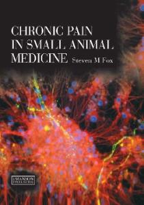
Fig. 1.1 Dorsal T1 weighted MR scan of the adrenal glands of a dog with pituitary-dependent hyperadrenocorticism, showing mild bilateral enlargement. Reproduced with permission of Downs Referrals, Bristol.
Historical Signs
3
Lithium NPK fertilisers Paraquat Phenobarbitone Potassium bromide Primidone Proligestone Terfenadine Theophylline Vitamin D rodenticides Note: Polyuria and polydipsia are considered together here, since one will lead to the other, with only a few exceptions. These include polydipsia in the face of obstructive lower urinary tract disease or oliguric renal failure, and polyuria which is not matched by fluid intake, in which case dehydration will rapidly follow. None of these scenarios are encountered commonly in practice.
References Garrett, L. D. (2003) Insulinomas: A review and what’s new. Proceedings, ACVIM, 2003. Lunn, K. F. (2005) Avoiding the water deprivation test. Proceedings, ACVIM, 2005. Tobias, et al. (2002) Pericardial disorders: 87 cases of pericardial effusion in dogs (January 1, 1999 to December 31, 2001). Proceedings, ACVIM, 2002.
1.1.2 Weight loss Decreased nutrient intake Anorexia q.v. Diet • Poor-quality diet • Underfeeding Dysphagia q.v.
Increased nutrient loss Burns Chronic blood loss • Epistaxis q.v. • Haematemesis q.v. • Haematuria q.v. • Melaena q.v. Diabetes mellitus* Effusions q.v. Fanconi syndrome (D) Intestinal parasites* Neoplasia Protein-losing enteropathy* Protein-losing nephropathy
4
Historical Signs
Increased nutrient use Endocrine, e.g. Hyperthyroidism* (C) Neoplasia* Physiological Cold environment Exercise Fever q.v. Lactation Pregnancy*
Malassimilation Cardiac failure* Exocrine pancreatic insufficiency Hepatic failure/bile salt deficiency* q.v. Hypoadrenocorticism (D) Neoplasia* Renal failure* q.v. Small intestinal disease* q.v.
Regurgitation and vomiting q.v. Reference Rutz, G. M., et al. (2001) Pancreatic acinar atrophy in German Shepherds. Compend Contin Educ Pract Vet, 23:347–56.
1.1.3 Weight gain Fluid accumulation Ascites* q.v. Peripheral oedema q.v. Pleural effusion
Increased body fat Overeating Boredom Excessive appetite (normal in some breeds)* High-calorie diets Overfeeding* Endocrinopathies Acromegaly Hyperadrenocorticism* Hypogonadism Hypothyroidism* (D) Insulinoma
Historical Signs
Increased organ size Hepatomegaly* q.v. Renomegaly q.v. Splenomegaly* q.v. Uterine enlargement q.v. • Pregnancy* • Pyometra*
Neoplasia Large abdominal mass (often associated with poor body condition)* Drugs, e.g. • Corticosteroids
References Garrett, L. D. (2003) Insulinomas: A review and what’s new. Proceedings, ACVIM, 2003. Peterson, M. E., et al. (1990) Acromegaly in 14 cats. JVIM, 4:192–201.
1.1.4 Polyphagia Behavioural/psychological Normal in some breeds* Boredom
Physiological Cold environment Increased exercise Lactation* Pregnancy*
Malassimilation* Increased nutrient loss Increased nutrient use Diet Highly-palatable food* Poor-quality food
Endocrine Diabetes mellitus* Hyperadrenocorticism* Hyperthyroidism* (C) Insulinoma
Miscellaneous Peritoneopericardial diaphragmatic hernia
Drugs/toxins Aminophylline
5
6
Historical Signs
Benzodiazepines Cannabis Cyproheptadine Delmadinone acetate Glucocorticoids Phenobarbitone Potassium bromide Primidone Proligestone
References Garrett, L. D. (2003) Insulinomas: A review and what’s new. Proceedings, ACVIM, 2003. Rexing, J. F. & Coolman, B. R. (2004) A peritoneopericardial diaphragmatic hernia in a cat. Vet Med, 99:314–18.
1.1.5 Anorexia/inappetence Difficulty with prehension Blindness q.v. Myopathy, e.g. Masticatory myositis Tetanus Pain on opening jaw, e.g. Mandibular or maxillary fracture Retrobulbar abscess Skull fractures Soft tissue trauma Temporo-mandibular joint disease Trigeminal nerve disease, e.g. Neoplasia Trigeminal neuritis
Difficulty with mastication Dental disease* Lingual disease Oral neoplasia* Oral ulceration, e.g. • Ingestion of caustic or acidic substances* • Renal disease
Difficulty with swallowing Pharyngeal disease Foreign body* Neoplasia Neurological disease Ulceration
Historical Signs
Oesophageal disease, e.g. Foreign body* Neoplasia Ulceration Megaoesophagus Stricture Vascular ring anomaly
Primary anorexia Intracranial disease, e.g. • Hypothalamic neoplasia
Secondary anorexia Anosmia • Chronic rhinitis q.v. • Nasal neoplasia • Other nasal disease • Neurological disease Endocrine disease, e.g. • Diabetic ketoacidosis • Hypoadrenocorticism (D) Fever* q.v. Gastrointestinal disease q.v., e.g. • Gastritis • Inflammatory bowel disease* Heart disease, e.g. • Cardiac failure* Hepatic disease* q.v. Infection* Metabolic abnormalities, e.g. • Hypercalcaemia q.v. • Hypokalaemia q.v. Pain* Pancreatic disease*, e.g. • Pancreatitis Respiratory disease, e.g. • Airway disease* q.v. • Diaphragmatic hernia • Pleural effusion* q.v. • Pneumonia q.v. Renal disease* q.v. Drugs • Acetazolamide • Amiodarone • Amphotericin B • Bethanechol • Bromocriptine • Butorphanol • Cardiac glycosides • Chlorambucil • Diazoxide • Doxorubicin • Fentanyl
7
8
Historical Signs
• • • • • • • • • • •
Hydralazine Itraconazole Ketoconazole Melphalan Methimazole Mitotane Nicotinamide Oxytetracycline (C) Penicillamine Theophylline Trimethoprim/sulphonamide (C)
Diet Recent dietary changes* Unpalatable diet*
Psychological/behavioural* factors Altered schedule New family members New house New pets
Reference Forman, M. A., et al. (2004) Evaluation of serum feline pancreatic lipase immunoreactivity and helical computed tomography versus conventional testing for the diagnosis of feline pancreatitis. JVIM, 18:807–15.
1.1.6 Failure to grow With good body condition Chondrodystrophy (normal in many breeds)* (D) Endocrine disorders • Congenital hyposomatotropism (pituitary dwarfism) • Congenital hypothyroidism • Hyperadrenocorticism
With poor body condition Dietary intolerance Exocrine pancreatic insufficiency* Inadequate nutrient intake Anorexia q.v. Poor-quality diet Underfeeding Cardiac disorders, e.g. Congenital Endocarditis
Historical Signs
Hepatic disorders, e.g. Hepatitis q.v. Portosystemic shunt Oesophageal disorders, e.g. Megaoesophagus q.v. Vascular ring anomaly (e.g. persistent right aortic arch) Gastrointestinal disease, e.g. Histoplasmosis Obstruction, e.g. • Foreign body* • Intussusception* Parasites* Renal disease Congenital kidney disease Glomerulonephritis Pyelonephritis Inflammatory disease Endocrine disease Diabetes insipidus Diabetes mellitus* Hypoadrenocorticism (D)
Reference Chastain, C. B., et al. (2001) Combined pituitary hormone deficiency in German shepherd dogs with dwarfism. Sm Anim Clin Endocrinol, 11:1–4.
1.1.7 Syncope/collapse (see Table 1.1) Cardiovascular dysfunction Myocardial failure Myocardial infarction Shock q.v. Bradyarrhythmias q.v., e.g. High grade second degree heart block Sick sinus syndrome (D) Third degree heart block Tachyarrhythmias q.v. Supraventricular tachycardia* Ventricular tachycardia*
9
10
Historical Signs
Table 1.1 Differentiating seizures from syncope. This table is a guide to the differentiation of generalised seizures from syncopal episodes. However, there is a lot of overlap between the two: syncopal episodes may involve convulsions; seizures may occur on exercise; tonic–clonic motions may not always be observed with seizures. Syncope
Seizure (generalised)
Precipitating event/ timing
Exercise, excitement, stress, cough, urination, defecation
Often at rest or on waking
Pre-event
Acute weakness, staggering, vocalisation
Anxiety, attention-seeking
Event
Usually flaccid limbs but may be rigid
Jaw motions, hypersalivation, tonic-clonic limb motion or limb rigidity Duration often greater than 1 minute Urination and/or defecation Loss of consciousness
Duration less than 1 minute Rarely urination/defecation Usually retain consciousness, but may lose consciousness Abnormal heart rhythm or rate may or may not be palpatated/auscultated Post-event
Rapid recovery
Obstruction to flow Congenital, e.g. • Aortic stenosis (D) • Pulmonic stenosis (D) Hypertrophic obstructive cardiomyopathy Pericardial effusion* (D) Pulmonary hypertension Arterial obstruction, e.g. • Neoplasia • Thrombosis
Hypoxaemic disease Carboxyhaemoglobinaemia Methaemoglobinaemia Respiratory disease Upper airway, e.g. • Brachycephalic obstructive airway syndrome • Laryngeal paralysis • Tracheal collapse • Tracheal obstruction Lower airway, e.g. • Pneumonia • Small airway disease
Often sinus tachycardia
Slow recovery Prolonged post-event disorientation
Historical Signs
Ventilation-perfusion mismatch, e.g. • Lung collapse Pleural/thoracic disorders, e.g. • Pleural effusion • Pneumothorax • Rib fractures Right-to-left cardiac shunt, e.g. Reverse-shunting patent ductus arteriosus Severe anaemia
Neurological dysfunction Brainstem disease Glossopharyngeal neuralgia Micturition-related collapse Narcolepsy/cataplexy Seizures q.v. Swallowing-related collapse Diffuse cerebral dysfunction, e.g. Encephalopathy Haemorrhage Hydrocephalus Inflammation Oedema Space occupying lesion Trauma Lower motor neurone disorders Endocrine neuropathies, e.g. • Diabetes mellitus* • Hyperadrenocorticism • Hypothyroidism* (D) Lumbosacral disease Paraneoplastic neuropathies, e.g. • Insulinoma Peripheral nerve neoplasia Polyneuropathy Polyradiculoneuropathy Neuromuscular junction disorders Botulism Myasthenia gravis Upper motor neurone disorders Central vestibular disease Cerebellar disease Cerebral disease Peripheral vestibular disease Spinal disease
11
12
Miscellaneous Carotid sinus stimulation, e.g. • Neoplasia • Tight collar Hyperventilation Postural hypotension Tussive syncope
Metabolic disorders Diabetic ketoacidosis Hypercalcaemia/hypocalcaemia q.v. Hypernatraemia/hyponatraemia q.v. Hyperthermia/hypothermia q.v. Hypoglycaemia q.v. Hypokalaemia q.v. Severe acidosis q.v. Severe alkalosis q.v.
Myopathies Corticosteroid myopathy Exertional myopathy Hypocalcaemic myopathy Hypokalaemic myopathy Malignant hyperthermia Mitochondrial myopathy Muscular dystrophy Polymyopathy Polymyositis Protozoal myopathy
Skeletal/joint disorders Bilateral cranial cruciate disease Bilateral hip disease Discospondylitis Intervertebral disc disease Multiple myeloma Osteoarthritis Panosteitis Patellar luxation Polyarthritis
Drugs Anti-arrhythmics, e.g. • Atenolol • Digoxin • Propranolol • Quinidine Sedatives, e.g. • Phenothiazines Vasodilators, e.g. • ACE inhibitors • Hydralazine • Nitroglycerine
Historical Signs
Historical Signs
13
References Berendt, M. (2001) The diagnosis of epilepsy: seizure phenomenology and classification. Proceedings of the World Small Animal Veterinary Association World Congress, 2001. Shelton, G. D. (1998) Myasthenia gravis: lessons from the past 10 years. JSAP, 39:368–72. Ware, W. A. (2002) Syncope. Proceedings, Waltham/OSU Symposium, Small Animal Cardiology, 2002. Wray, J. (2005) Differential diagnosis of collapse in the dog. 1. Aetiology and investigation. In Practice 27:16–28.
1.1.8 Weakness Metabolic disease Renal failure* q.v. Hepatic failure* q.v. Hypoglycaemia q.v. Electrolyte disorders* • Hypercalcaemia*/hypocalcaemia q.v. • Hyperkalaemia/hypokalaemia* q.v. • Hypernatraemia/hyponatraemia q.v. Acid–base disorders • Acidosis q.v. • Alkalosis q.v.
Infectious diseases* Bacterial Viral Fungal Rickettsial Protozoal Other parasitic diseases
Immune-mediated/inflammatory diseases Chronic inflammatory conditions* Immune-mediated haemolytic anaemia* q.v. Immune-mediated polyarthritis
Haematological diseases Anaemia* q.v. Hyperviscosity syndrome
Endocrine diseases Diabetes mellitus* Hyperadrenocorticism Hyperparathyroidism Hypoadrenocorticism (D) Hypoparathyroidism Hypothyroidism* (D) Insulinoma
14
Cardiovascular diseases Bradyarrhythmias q.v., e.g. • High grade second degree heart block • Sick sinus syndrome (D) • Third degree heart block Congestive heart failure* Pericardial effusion* q.v. Hypertension* q.v. Hypotension* q.v. Tachyarrhythmias q.v., e.g. • Ventricular tachycardia*
Respiratory diseases Airway obstruction, e.g. • Feline asthma* (C) • Foreign body* • Neoplasia * Intrathoracic neoplasia* • Pleural effusion* • Pulmonary hypertension • Pulmonary oedema* q.v. • Pulmonary thromboembolism Severe pulmonary parenchymal disease
Neuromuscular diseases Epilepsy* q.v. Myasthenia gravis Myopathies Vestibular disease* q.v. Intracranial disease, e.g. Cerebrovascular accident Infection Inflammation Space-occupying lesions Spinal cord disease q.v., e.g. Infection Inflammation Intervertebral disc disease* (D) Neoplasia Trauma* Peripheral polyneuropathies Endocrine disorders, e.g. • Diabetes mellitus* • Hyperadrenocorticism • Hypothyroidism* (D) Polyradiculoneuritis Paraneoplastic disorders
Historical Signs
Historical Signs
Drugs/toxins, e.g. • Cisplatin • Lead • Vincristine Infections Botulism Tick paralysis
Systemic disorders Dehydration* Fever* q.v. Neoplasia*
Nutritional disorders Cachexia, e.g. Heart failure* Neoplasia* Inadequate calorie intake, e.g. Anorexia* q.v. Poor-quality diet Specific nutrient deficiencies, e.g. Minerals Vitamins
Physiological factors Over-exercise Pain* Stress/anxiety*
Drugs/toxins Alphachloralose Anticoagulant rodenticides Anticonvulsants Antihistamines Blue-green algae Cannabis Diclofenac sodium Glucocorticoids Hypotensive agents, e.g. • Beta-blockers • Vasodilators Ibuprofen Insulin overdosage Iron salts Mistletoe Opioids
15
16
Historical Signs
Organophosphates Petroleum distillates Phenoxy acid herbicides Pyrethrin/pyrethroids Rhododendron Salbutamol Sedatives
References Sadek, D. & Schaer, M. (1996) Atypical Addison’s disease in the dog: a retrospective survey of 14 cases. JAAHA, 32:159–63. Shelton, G. D. (1998) Myasthenia gravis: lessons from the past 10 years. JSAP, 39:368–72.
1.2 Gastrointestinal/abdominal historical signs 1.2.1 Ptyalism/salivation/hypersalivation Physiological factors Appetite stimulation* Fear* Stress*
Oral cavity disease Dental disease* Foreign body* Neoplasia* Inability to close mouth, e.g. Mandibular trauma* Trigeminal nerve disease, e.g. • Idiopathic trigeminal neuritis • Infiltrating neoplasia, e.g. • Lymphoma • Nerve sheath tumours Ulceration*, e.g. Immune-mediated disease Ingestion of irritant substance Renal failure* Inflammation* Faucitis* Gingivitis* Glossitis* Oesophagitis* Stomatitis*
Differential Diagnosis in Small Animal Medicine Alex Gough Copyright © 2007 by Alex Gough 16
Historical Signs
Organophosphates Petroleum distillates Phenoxy acid herbicides Pyrethrin/pyrethroids Rhododendron Salbutamol Sedatives
References Sadek, D. & Schaer, M. (1996) Atypical Addison’s disease in the dog: a retrospective survey of 14 cases. JAAHA, 32:159–63. Shelton, G. D. (1998) Myasthenia gravis: lessons from the past 10 years. JSAP, 39:368–72.
1.2 Gastrointestinal/abdominal historical signs 1.2.1 Ptyalism/salivation/hypersalivation Physiological factors Appetite stimulation* Fear* Stress*
Oral cavity disease Dental disease* Foreign body* Neoplasia* Inability to close mouth, e.g. Mandibular trauma* Trigeminal nerve disease, e.g. • Idiopathic trigeminal neuritis • Infiltrating neoplasia, e.g. • Lymphoma • Nerve sheath tumours Ulceration*, e.g. Immune-mediated disease Ingestion of irritant substance Renal failure* Inflammation* Faucitis* Gingivitis* Glossitis* Oesophagitis* Stomatitis*
Historical Signs
Neurological disease Cataplexy/narcolepsy Hepatic encephalopathy Intracranial neoplasia Partial seizures
Nausea/regurgitation/vomiting q.v. Salivary gland disease q.v. Salivary gland necrosis/sialadenitis Salivary mucocoele Sialadenosis
Normal breed variation, e.g. St Bernards
Drugs/toxins Adder bites Alphachloralose Baclofen Batteries Benzodiazepines Bethanechol Blue-green algae Cannabis Carbamate Chocolate/theobromine Cotoneaster Cyanoacrylate adhesives Daffodil Dieffenbachia Dinoprost tromethamine Glyphosphate Horse chestnut Ivermectin Ketamine Laburnum Levamisole (C) Loperamide Metronidazole Mistletoe NPK fertilisers Organophosphates Paracetamol Paraquat Phenoxy acid herbicides Plastic explosives Pyrethrin/pyrethroids Pyridostigmine Rhododendron Rowan
17
18
Historical Signs
Terfenadine Toads Trimethoprim/sulphonamide (C) Xylazine
References Patterson, E. E., et al. (2003) Clinical characteristics and inheritance of idiopathic epilepsy in Vizslas. JVIM, 17:319–25. Schroeder, H. & Berry, W. L. (1998) Salivary gland necrosis in dogs: a retrospective study of 19 cases. JSAP, 39:121–25. Sozmen, M., et al. (2000) Idiopathic salivary gland enlargement (sialadenosis) in dogs: a microscopic study. JSAP, 41:243–47.
1.2.2 Gagging/retching Congenital disease Achalasia, e.g. • Cricopharyngeal achalasia (D) Cleft palate Hydrocephalus
Neuromuscular disease Brainstem disease Cranial nerve defects (V, VII, IX, XII) Encephalitis Laryngeal paralysis* Muscular dystrophy Myasthenia gravis
Immune-mediated and infectious disease Asthma* (C) Bacterial encephalitis Fungal disease • Granuloma complex Idiopathic glossopharyngitis Laryngitis* Pharyngitis* Rabies Rhinitis* Sialadenitis Viral encephalitis
Systemic disorders Hypocalcaemia Renal failure*
Trauma Foreign body* Pharyngeal haematoma
Historical Signs
19
Styloid apparatus trauma Tracheal rupture
Neoplasia Central nervous system Epiglottis Inner ear Nasal Pharyngeal Tonsillar
Nutrition Food texture and size
Respiratory disease (expectoration), e.g. Bronchitis* Haemorrhage Pulmonary oedema*
Toxic Botulism Ingestion of irritant chemical Smoke
Reference Schroeder, H. & Berry, W. L. (1998) Salivary gland necrosis in dogs: a retrospective study of 19 cases. JSAP, 39:121–25.
1.2.3 Dysphagia Infectious/inflammatory disease Oral disease Dental disease* Osteomyelitis of jaw Periodontitis* Pharyngitis* Rabies Retrobulbar abscess Severe gingivitis* Tooth root abscess* Ulceration, e.g. • Ingestion of irritant substance • Renal disease*
Obstruction Foreign body* Granuloma
20
Historical Signs
Neoplasia Sialocoele
Trauma Fracture* Haematoma Laceration*
Temporomandibular joint disease Neuromuscular disease Cricopharyngeal achalasia Myasthenia gravis Myopathy, e.g. • Masticatory myopathy Trigeminal nerve disease, e.g. • Intracranial disease • Trigeminal neuritis
References Meomartino, L., et al. (1999) Temporomandibular ankylosis in the cat: a review of seven cases. JSAP, 40:7–10. Preifer, R. M. (2003) Cricopharyngeal achalasia in a dog. Can Vet J, 44:993–5.
1.2.4 Regurgitation Salivary gland disease Sialadenitis
Oesophageal disease Foreign body* Megaoesophagus • Idiopathic • Acquired Neoplasia Oesophageal diverticulum Oesophageal fistula Oesophageal inclusion cysts Oesophagitis* Stricture Vascular ring anomaly, e.g. • Persistent right aortic arch
Gastric disease Gastric dilatation-volvulus* (D) Hiatal hernia Pyloric outflow obstruction, e.g. • Foreign body* • Neoplasia • Pyloric stenosis
Historical Signs
21
Neuromuscular disease Peripheral neuropathies, e.g. Giant cell axonal neuropathy (D) Lead poisoning Polyneuritis Polyradiculoneuritis Central nervous system disease, e.g. Brainstem disease Infection Inflammation Intracranial space occupying lesion Trauma Neuromuscular junctionopathies, e.g. Acetylcholinesterase toxicity Botulism Myasthenia gravis Tetanus
Immune-mediated disease Dermatomyositis (D) Polymyositis Systemic lupus erythematosus
Endocrine disease Hypoadrenocorticism (D) Hypothyroidism* (D)
References Han, E., et al. (2003) Feline esophagitis secondary to gastroesophageal reflux disease: clinical: signs and radiographic, endoscopic and histopathological findings. JAAHA, 39:161–7. Hodges, J., et al. (2004) Recurrent regurgitation in a young cat with an unknown history. Vet Med, 99:244–51. Schroeder, H. & Berry, W. L. (1998) Salivary gland necrosis in dogs: a retrospective study of 19 cases. JSAP, 39:121–5. White, R. N., et al. (2003) Vascular ring anomaly with coarctation of the aorta in a cat. JSAP, 44:330–34.
1.2.5 Vomiting ACUTE VOMITING Dietary Dietary indiscretion* Dietary intolerance* Sudden change in diet*
22
Gastrointestinal disease Colitis* Constipation/obstipation* q.v. Foreign body* Gastric dilatation/volvulus* Gastric or duodenal ulceration* Gastritis/enteritis* Haemorrhagic gastroenteritis* Infection, e.g. • Bacterial* • Parasites* • Viral* Inflammatory bowel disease* Intestinal volvulus Intussusception Neoplasia*
Endocrine disease, e.g. Diabetic ketoacidosis* Hypoadrenocorticism (D)
Metabolic/systemic disease Hypercalcaemia/hypocalcaemia q.v. Hyperkalaemia/hypokalaemia* q.v. Hyperthermia* q.v. Liver disease* q.v. Pancreatitis* Peritonitis* Prostatitis* Pyometra* (D) Renal disease* q.v. Septicaemia* Urinary obstruction* Vestibular disease*
Miscellaneous conditions Central nervous system disease Diaphragmatic hernia Motion sickness Psychogenic
Drugs/toxins Acetazolamide Adder bite Allopurinol Alpha-2 agonists Aminophylline Amphotericin B Apomorphine Aspirin Atipamezole Atropine
Historical Signs
Historical Signs
Batteries Benzalkonium chloride Bethanechol Blue-green algae Borax Bromocriptine Calcium edetate Carbimazole Carboplatin Cardiac glycosides Cephalexin Chlorambucil Chloramphenicol Chlorphenamine Clomipramine Colchicine Cotoneaster Cyclophosphamide Cyclosporin Cytarabine Daffodil Dichlorophen Diclofenac sodium Dinoprost tromethamine Dopamine Doxorubicin Doxycycline Dieffenbachia Ethylene glycol Erythryomycin Glipizide Glucocorticoids Glyphosphate Honeysuckle Horse chestnut Hydralazine Ibuprofen Indomethacin Ipecacuanha Iron/iron salts Ivermectin Ketoconazole Laburnum Lead Levamisole Lignocaine Loperamide Medetomidine Melphalan Metaldehyde
23
24
Historical Signs
Methimazole Metronidazole Mexiletine Misoprostol Mistletoe Mitotane Naproxen Nicotinamide Nitroscanate NPK fertilisers NSAIDs Paracetamol Paraquat Penicillamine Pentoxifylline Petroleum distillates Phenoxy acid herbicides Phenytoin Pimobendan Piperazine Plastic explosives Poinsettia Potassium bromide Procainamide Propantheline bromide Pyracantha
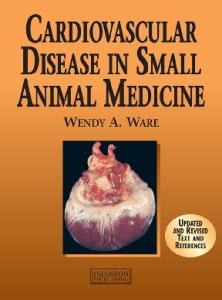
Fig. 1.2 Lateral abdominal radiograph of a dog showing a mineral-density foreign body. Exploratory coeliotomy revealed this to be a large stone within the small intestine. Reproduced with permission of Downs Referrals, Bristol.
Historical Signs
Pyrethrin/pyrethroids Pyridostigmine Rhododendron Rowan Salt Selective serotonin reuptake inhibitors Sildenafil Sotalol Strychnine Sulphasalazine Terfenadine Tetracycline Theobromine Theophylline Tricyclic antidepressants Trimethoprim/sulphonamide Ursodeoxycholic acid Vitamin D rodenticides Xylazine Yew Zinc
CHRONIC VOMITING Gastrointestinal disease Bacterial overgrowth Colitis* Constipation/obstipation* q.v. Enterogastric reflux Gastric motility disorders* Gastric or duodenal ulceration* Gastritis/enteritis* Infection, e.g. • Bacterial • Fungal • Parasites* • Viral Inflammatory bowel disease • Eosinophilic • Lymphocytic • Lymphoplasmacytic • Mixed Irritable bowel syndrome Neoplasia* Obstruction, e.g. • Foreign body* • Inflammatory bowel disease (gastritis or enteritis) • Intussusception* • Neoplasia* • Pyloric stenosis
25
26
Historical Signs
Endocrine disease, e.g. Diabetes mellitus* Hyperthyroidism* (C) Hypoadrenocorticism (D)
Metabolic/systemic disease Heartworm disease Hypercalcaemia/hypocalcaemia q.v. Hyperkalaemia/hypokalaemia q.v. Liver disease* q.v. Pancreatitis* Prostatitis Pyometra* (D) Renal disease* q.v.
Miscellaneous conditions Abdominal neoplasia Diaphragmatic hernia Sialadenitis
References Craven, M., et al. (2004) Canine inflammatory bowel disease: retrospective analysis of diagnosis and outcome in 80 cases (1995–2002). JSAP, 45:336–43. Saxon-Buri, S. (2004) Daffodil toxicosis in an adult cat. Can Vet J, 45:248–50. Schroeder, H. & Berry, W. L. (1998) Salivary gland necrosis in dogs: a retrospective study of 19 cases. JSAP, 39:121–5.
1.2.6 Diarrhoea SMALL INTESTINAL DIARRHOEA Diet Dietary intolerance, e.g. Food hypersensitivity* Food intolerance Gluten-sensitive enteropathy
Extra-gastrointestinal disease Exocrine pancreatic insufficiency* Hepatic disease* q.v. Hyperthyroidism* (C) Hypoadrenocorticism (D) IgA deficiency Nephrotic syndrome Pancreatic duct obstruction Pancreatitis* Renal disease* q.v. Right-sided congestive heart failure*
Historical Signs
27
Systemic lupus erythematosus Uraemia
Infection Bacterial*, e.g. Campylobacter spp Clostridium spp E. coli Salmonella spp Staphylococcus spp Small intestinal bacterial overgrowth Fungal Helminths* Hookworm Roundworm Tapeworm Whipworm Protozoal *, e.g. Cryptosporidiosis Giardia spp Viral*, e.g. Coronavirus Feline leukaemia virus (C) Parvovirus Rickettsial
Inflammatory/immune-mediated disease Basenji enteropathy(D) Duodenal ulceration Haemorrhagic gastroenteritis* Inflammatory bowel disease* • Eosinophilic • Granulomatous • Lymphoplasmacytic Protein-losing enteropathy and nephropathy of the Soft-Coated Wheaten Terrier (D)
Idiopathic disease Lymphangiectasia
Neoplasia*, e.g. Adenocarcinoma Carcinoid tumours Leiomyoma Lymphoma
28
Mast cell tumours Sarcoma
Partial obstruction* Foreign body Intussusception Neoplasia Stricture
Motility disorders, e.g. Dysautonomia Enteritis Functional obstruction (ileus) Hypoalbuminaemia Hypokalaemia
Drugs/toxins (see Large intestinal diarrhoea, below) LARGE INTESTINAL DIARRHOEA Diet* Dietary hypersensitivity Dietary indiscretion
Extra-intestinal conditions Metastatic neoplasia Neurological disease leading to ulcerative colitis Pancreatitis Toxaemia Uraemia
Infection Bacterial*, e.g. Campylobacter spp Clostridium difficile Clostridium perfringens E. coli Salmonella spp Yersinia enterocolitica Viral* Coronavirus Feline immunodeficiency virus (C) Feline infectious peritonitis (C) Feline leukaemia virus (C) Parvovirus Fungal, e.g. Histoplasmosis Protothecosis
Historical Signs
Historical Signs
Parasitic*, e.g. Amoebiasis Ancylostoma spp Balantidium coli Cryptosporidiosis Giardia spp Heterobilharzia americana Roundworm Tapeworm Tritrichomonas foetus (C) Uncinaria spp Whipworm Protozoal, e.g. Toxoplasmosis
Immune-mediated/inflammatory disease Histiocytic ulcerative colitis of Boxers (D) Inflammatory bowel disease*
Idiopathic conditions Fibre-responsive large-bowel diarrhoea Irritable bowel syndrome
Neoplasia* Benign, e.g. Adenomatous polyps Leiomyoma Malignant, e.g. Adenocarcinoma Lymphoma
Obstruction (see Plate 1.2(a) in colour plate section) Caecal inversion Foreign body* Intussusception* Neoplasia Stricture
Miscellaneous Secondary to chronic small intestinal disease Stress
Drugs/toxins Acetazolamide Adder bite Allopurinol
29
30
Aminophylline Amoxicillin Amphotericin B Ampicillin Atenolol Benzalkonium chloride Bethanechol Blue-green algae Borax Calcium edetate Carbamate insecticides Cardiac glycosides Cephalexin Chloramphenicol Chlorphenamine Colchicine Cotoneaster Cyclophosphamide Cyclosporin Cytarabine Daffodil Diazoxide Diclofenac sodium Dieffenbachia Doxycycline Glyphosphate Honeysuckle Horse chestnut Ibuprofen Indomethacin Iron/iron salts Laburnum Lactulose Levamisole Lithium Loperamide Mebendazole Metaldehyde Methiocarb Misoprostol Mistletoe Mitotane Naproxen Nicotinamide NPK fertilisers NSAIDs Organophosphates Oxytetracycline Pamidronate Pancreatic enzyme supplementation
Historical Signs
Historical Signs
31
Paracetamol Paraquat Pentoxifylline Petroleum distillates Phenoxy acid herbicides Piperazine Poinsettia Procainamide Pyracantha Pyrethrin/pyrethroids Pyridostigmine Quinidine Rhododendron Rowan Salt Selective serotonin reuptake inhibitors Sotalol Theobromine Theophylline Vitamin D rodenticides Yew Zinc sulphate Note: Perirectal diseases, e.g. anal sac disease, anal furunculosis, perineal hernia, rectal prolapse, perianal adenoma, may cause signs mimicking large-bowel disease (tenesmus, haematochezia, mucoid stool).
References Chandler, M. (2002) The chronically diarrhoeic dog. 2. Diarrhoea of small intestinal origin. In Practice, 24:18–24. Craven, M., et al. (2004) Canine inflammatory bowel disease: retrospective analysis of diagnosis and outcome in 80 cases (1995–2002). JSAP, 45:336–43. Hostutler, R. A., et al. (2004) Antibiotic-responsive histiocytic ulcerative colitis in 9 dogs. JVIM, 18:499–504. Leib, M. S. (2005) Diagnostic approach to chronic diarrhea I & II. Proceedings, Western Veterinary Conference, 2005. Washabau, R. J. (2005) Infectious GI diseases in dogs and cats. Proceedings, Western Veterinary Conference, 2005.
1.2.7 Melaena Ingestion of blood Nasal disease (see also epistaxis), e.g. Coagulopathy* q.v. Neoplasia* Trauma*
32
Oropharyngeal haemorrhage Coagulopathy* q.v. Neoplasia* Trauma* Respiratory disease (see also haemoptysis), e.g. Coagulopathy* q.v. Exercise-induced pulmonary haemorrhage Parasites Neoplasia* Ruptured aneurysm Trauma*
Gastrointestinal disease Enteritis* Gastritis* Oesophagitis Parasites* Gastrointestinal ulceration* Gastrinoma Helicobacter infection Inflammatory gastroenteric disease* Neurological disease Post foreign body* Stress Uraemia* q.v. Drugs, e.g. • Glucocorticoids* • NSAIDs* Ischaemia, e.g. Mesenteric avulsion Mesenteric thrombosis/infarction Mesenteric volvulus Post gastric-dilatation volvulus* (D) Neoplasia*, e.g. Adenocarcinoma Leiomyoma Leiomyosarcoma Lymphoma
Extra-gastrointestinal disease Hypoadrenocorticism (D) Liver disease* q.v. Mastocytosis Pancreatitis* Septicaemia* Shock* q.v.
Historical Signs
Historical Signs
Systemic hypertension* q.v. Uraemia* q.v. Vasculitis, e.g. • Rocky Mountain Spotted Fever Coagulopathy q.v., e.g. Anticoagulant toxicity* q.v. Congenital clotting factor deficiency q.v. Disseminated intravascular coagulation Thrombocytopenia q.v. von Willebrand’s disease (D)
References Brooks, D. & Watson, G. L. (1997) Omeprazole in a dog with gastrinoma. JVIM, 11:379–81. McTavish, D. (2002) Eosinophilic gastroenteritis in a dog. Can Vet J, 43:463–5. Washabau, R. J. (2004) G. I. hemorrhage: pathogenesis, diagnosis and therapy. Proceedings, Atlantic Coast Veterinary Conference, 2004.
1.2.8 Haematemesis Ingestion of blood Nasal disease (see also epistaxis), e.g. Coagulopathy* q.v. Neoplasia* Trauma* Oropharyngeal haemorrhage Coagulopathy* q.v. Neoplasia* Trauma* Respiratory disease (see also haemoptysis), e.g. Coagulopathy* q.v. Exercise-induced pulmonary haemorrhage Parasites Neoplasia* Ruptured aneurysm Trauma*
Gastrointestinal disease Gastritis* Haemorrhagic gastroenteritis Oesophagitis Gastrointestinal ulceration* Gastrinoma Helicobacter infection*
33
34
Historical Signs
Inflammatory gastroenteric disease* Neurological disease Post foreign body* Stress Systemic mastocytosis Uraemia* Drugs, e.g. • NSAIDs • Glucocorticoids* Ischaemia, e.g. Post gastric-dilatation/volvulus* (D) Neoplasia*, e.g. • Adenocarcinoma • Lymphoma
Extra-gastrointestinal disease Hypoadrenocorticism (D) Liver disease* q.v. Mastocytosis Septicaemia* Shock* Systemic hypertension* q.v. Uraemia* q.v. Coagulopathies q.v., e.g. Anticoagulant toxicity* Congenital clotting factor deficiency Disseminated intravascular coagulation Thrombocytopenia von Willebrand’s disease(D) Pancreatic disease*, e.g. Pancreatitis Vasculitis, e.g. Rocky Mountain Spotted Fever Toxins, e.g. Calcipotriol Paraquat
Reference Brooks, D. & Watson, G. L. (1997) Omeprazole in a dog with gastrinoma. JVIM, 11:379–81.
1.2.9 Haematochezia Extra-gastrointestinal disease Neurological disease leading to ulcerative colitis
Historical Signs
Coagulopathies q.v., e.g. Anticoagulant toxicity* Congenital clotting factor deficiency q.v. Disseminated intravascular coagulation Thrombocytopenia q.v. von Willebrand’s disease (D) Perirectal disease, e.g. Anal furunculosis* Anal sac disease* Perianal adenoma* Perineal hernia* Rectal prolapse*
Gastrointestinal disease Dietary Dietary hypersensitivity Dietary indiscretion Bacterial*, e.g. Campylobacter spp Clostridium spp E. coli Salmonella spp Viral* Coronavirus Feline immunodeficiency virus (C) Feline infectious peritonitis (C) Feline leukaemia virus (C) Parvovirus Fungal, e.g. Histoplasmosis Protothecosis Parasitic*, e.g. Amoebiasis Ancylostoma spp Balantidium coli Cryptosporidiosis Giardia spp Heterobilharzia americana Roundworm Tapeworm Tritrichomonas foetus (C) Uncinaria spp Whipworm
35
36
Historical Signs
Protozoal, e.g. Toxoplasmosis
Immune-mediated/inflammatory disease Histiocytic ulcerative colitis of Boxers (D) Inflammatory bowel disease*
Idiopathic conditions Fibre-responsive large-bowel diarrhoea Haemorrhagic gastroenteritis Irritable bowel syndrome
Neoplasia Benign, e.g. Adenomatous polyps Leiomyoma Malignant, e.g. Adenocarcinoma Lymphoma
Obstructive disease Foreign body* Intussusception*
Drugs Glucocorticoids
References Hostutler, R. A., et al. (2004) Antibiotic-responsive histiocytic ulcerative colitis in 9 dogs. JVIM, 18:499–504. Spielman, B. L. & Garvey, M. S. (1993) Hemorrhagic gastroenteritis in 15 dogs. JAAHA, 29:341–4.
1.2.10 Constipation/obstipation Congenital conditions Atresia ani Atresia coli
Diet Ingestion of hair, bones and foreign material Low-fibre diets
Systemic disease Dehydration* Hypercalcaemia q.v. Hypokalaemia* q.v. Hypothyroidism* (D)
Historical Signs
Neuromuscular disease Feline dysautonomia (C) Lumbosacral disease* Pelvic nerve disease, e.g. • Traumatic*
Obstructive disease (see Plate 1.2(b) in colour plate section) Intraluminal/intramural Diverticulum Foreign body* Neoplasia*, e.g. • Adenoma • Leiomyoma • Leiomyosarcoma • Lymphoma Stricture Extraluminal Granuloma Neoplasia* Pelvic fracture* Perineal hernia* Prostatic disease (D) • Abscess • Benign prostatic hypertrophy* • Neoplasia • Prostatitis* Sublumbar lymph node disease
Prolonged colonic distension, e.g. Narrowing of pelvic canal post fracture*
Painful conditions Anal furunculosis* Anal or rectal inflammation* Anal or rectal mass* Anal or rectal stricture Anal sac disease*, e.g. • Abscess • Anal sacculitis Pelvic trauma (soft tissue or bony)* Spinal cord disease*
Behavioural factors*, e.g. Change of daily routine Dirty litter box Hospitalisation Novel litter substrate
37
38
Historical Signs
Idiopathic conditions Idiopathic megacolon*
Drugs/toxins Aluminium antacids Butylscopolamine (hyoscine) Diphenoxylate Diuretics Loperamide Opioids Propantheline bromide Sucralfate Verapamil Vincristine
References LeRoy, B. E. & Lech, M. E. (2004) Prostatic carcinoma causing urethral obstruction and obstipation in a cat. J Feline Med Surg, 6:397–400. Yam, P. (1997) Decision making in the management of constipation in the cat. In Practice, 19:434–40.
1.2.11 Faecal tenesmus/dyschezia Anal sac disease, e.g. Abscess Anal sacculitis* Neoplasia
Constipation/obstipation q.v. Diet Excess bone Excess fibre
Perianal disease, e.g. Anal furunculosis/perianal fistulas* (D) Perianal adenoma* Perineal hernia* Rectal prolapse*
Caudal abdominal mass* Pelvic narrowing Prostatic disease (D) Abscess Benign prostatic hypertrophy* Neoplasia Prostatitis*
Historical Signs
Trauma, e.g. Pelvic fracture*
Urogenital disease*, e.g. Lower urinary tract disease Urethral obstruction
Colorectal disease, e.g. Colitis q.v. Congenital disease Large intestinal neoplasia
References Hardie, R. J., et al. (2005) Cyclosporin treatment of anal furunculosis in 26 dogs. JSAP, 46:3–9. Simpson, J. (1996) Differential diagnosis of faecal tenesmus in dogs. In Practice, 18:280–87.
1.2.12 Faecal incontinence Anal sphincter incompetence Myopathy Neoplasia* Trauma* Neurological, e.g. Cauda equina syndrome Degenerative myelopathy/CDRM* (D) Distemper encephalomyelitis Dysautonomia Lumbosacral stenosis Myelodysplasia/spinal dysraphism Peripheral neuropathy Sacrocaudal dysgenesis Spinal arachnoid cysts Spinal trauma Perianal disease, e.g. Perianal fistula* Iatrogenic disease, e.g. Damage to anal sphincter during anal sacculectomy
Reservoir incontinence Behavioural CNS disease q.v. Colitis* Diet* Neoplasia*
39
40
Historical Signs
References Guildford, W. G., et al. (1990) Fecal incontinence, urinary incontinence, and priapism associated with multifocal distemper encephalomyelitis in a dog. JAVMA, 197:90–92. Skeen, T. M., et al. (2003) Spinal arachnoid cysts in 17 dogs. JAAHA, 39:271–82.
1.2.13 Flatulence/borborygmus Aerophagia* Competitive/aggressive eating Nervous animal
Diet High fibre diets Milk products/lactase deficiency Spoiled food
Maldigestion, e.g. Exocrine pancreatic insufficiency
Malabsorption, e.g. Inflammatory bowel disease
Drugs/toxins, e.g. Lactulose Metaldehyde
References Roudebush, P. (2001) Flatulence: causes and management options. Compend Contin Educ Pract Vet, 23:1075–81. Rutz, G. M., et al. (2001) Pancreatic acinar atrophy in German Shepherds. Compend Contin Educ Pract Vet, 23:347–56.
1.3 Cardiorespiratory historical signs 1.3.1 Coughing Infection Bacterial, e.g. Bordetellosis* Fungal, e.g. Coccidioidomycosis Viral, e.g. Canine distemper*
Differential Diagnosis in Small Animal Medicine Alex Gough Copyright © 2007 by Alex Gough 40
Historical Signs
References Guildford, W. G., et al. (1990) Fecal incontinence, urinary incontinence, and priapism associated with multifocal distemper encephalomyelitis in a dog. JAVMA, 197:90–92. Skeen, T. M., et al. (2003) Spinal arachnoid cysts in 17 dogs. JAAHA, 39:271–82.
1.2.13 Flatulence/borborygmus Aerophagia* Competitive/aggressive eating Nervous animal
Diet High fibre diets Milk products/lactase deficiency Spoiled food
Maldigestion, e.g. Exocrine pancreatic insufficiency
Malabsorption, e.g. Inflammatory bowel disease
Drugs/toxins, e.g. Lactulose Metaldehyde
References Roudebush, P. (2001) Flatulence: causes and management options. Compend Contin Educ Pract Vet, 23:1075–81. Rutz, G. M., et al. (2001) Pancreatic acinar atrophy in German Shepherds. Compend Contin Educ Pract Vet, 23:347–56.
1.3 Cardiorespiratory historical signs 1.3.1 Coughing Infection Bacterial, e.g. Bordetellosis* Fungal, e.g. Coccidioidomycosis Viral, e.g. Canine distemper*
Historical Signs
Parasitic Aelurostrongylus abstrusus (C) Angiostrongylus vasorum (D) Dirofilaria immitis Oslerus osleri (D) Paragonimiasis
Immune-mediated/inflammatory disease Asthma* (C) Chronic bronchitis* (D)
Miscellaneous conditions Aspiration pneumonia Idiopathic pulmonary fibrosis Inhaled foreign body Laryngeal paralysis Left atrial enlargement* Lung lobe hernia Primary ciliary dyskinesia
Neoplasia Adenocarcinoma Alveolar carcinoma Bronchial gland carcinoma Metastatic disease Squamous cell carcinoma
Pulmonary haemorrhage Coagulopathy q.v. Exercise-induced Neoplasia* Traumatic
Pulmonary oedema Airway obstruction Cardiogenic* Electrocution Hypoglycaemia Hypoproteinaemia q.v. Iatrogenic Ketamine Neurological • Cranial trauma • Seizures Obstruction of lymphatic drainage Primary alveolar–capillary membrane injury Re-expansion
Drugs/toxins/irritants Benzalkonium chloride ingestion
41
42
Historical Signs
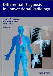
Fig. 1.3 Lateral thoracic radiograph of a dog with pulmonary metastasis secondary to a renal tumour. Reproduced with permission of Downs Referrals, Bristol.
Chemical fume inhalation Potassium bromide (C) Smoke inhalation
References Adamama-Moraitou, K. K., et al. (2004) Feline lower airway disease: a retrospective study of 22 naturally occurring cases from Greece. J Feline Med Surg, 6:227–33. Brownlie, S. E. (1990) A retrospective study of diagnosis in 109 cases of lower respiratory disease. JSAP, 31:371–6. Chapman, P. S., et al. (2004) Angiostrongylus vasorum infection in 23 dogs (1999–2002). JSAP, 45: 435–40. Coleman, M. G. (2005) Dynamic cervical lung hernia in a dog with chronic airway disease. JVIM, 19:103–5. Johnson, L. R., et al. (2003) Clinical, clinicopathologic and radiographic findings in dogs with coccidioidomycosis: 24 cases (1995–2000). JAVMA, 222: 461–6. Kipperman, B. S., et al. (1992) Primary ciliary dyskinesia in a Gordon Setter. JAAHA, 28:375–9. Ogilvie, G. K., et al. (1989) Classification of primary lung tumors in dogs: 210 cases (1975–1985). JAVMA, 195:106–8. Swerczek, T. W. & Lyons, E. T. (2000) Paragonimiasis in a cat in Kentucky. Vet Med, 95:909–11. Welsh, R. D. (1996) Bordetella bronchiseptica infections in cats. JAAHA, 32:153–8.
1.3.2 Dyspnoea/tachypnoea See Section 2.3.1
Historical Signs
1.3.3 Sneezing and nasal discharge Infection Viral Canine distemper virus* (D) Canine infectious tracheobronchitis* (D) Feline calicivirus* (C) Feline herpes virus* (C) Feline immunodeficiency virus* (C) Feline leukaemia virus* (C) Feline pox virus Feline reovirus (C) Fungal Aspergillosis Cryptococcosis Exophiala jeanselmei Penicillium spp Phaeohyphomycosis Rhinosporidium seeberi Parasitic Cuterebra spp Eucoleus böehmi Linguatula serrata Pneumonyssoides caninum Bacterial/mycoplasmal Bordetella bronchiseptica* Chlamydophila spp* Coliforms Mycoplasma spp Pasteurella spp Staphylococcus spp Streptococcus spp
Inflammatory disease Allergic rhinitis* Granulomatous rhinitis Lymphoplasmacytic rhinitis* Nasopharyngeal polyp* (C)
Physical Foreign body* Irritant gases Trauma
Neoplasia Adenocarcinoma*
43
44
Historical Signs
Chondrosarcoma Fibrosarcoma Haemangiosarcoma Lymphoma* Mast cell tumour Melanoma Neuroblastoma Osteosarcoma Squamous cell carcinoma* Transmissible venereal tumour Undifferentiated carcinomas*
Dental disease Tooth root abscess*
Anatomical deformities Acquired nasopharyngeal stenosis Cleft palate Oronasal fistula
Congenital disease Ciliary dyskinesia
Systemic disease (see also epistaxis) Coagulopathy q.v. Hypertension q.v. Hyperviscosity syndrome Vasculitis • Ehrlichiosis • Rocky Mountain Spotted Fever
References Binns, S. & Dawson, S. (1995) Feline infectious upper respiratory disease. In Practice, 17:458–61. Bredal, W. & Vollset, I. (1998) Use of milbemycin oxine in the treatment of dogs with nasal mite (Pneumonyssoides caninum) infection. JSAP, 39:126–30. McEntee, M. C. (2001) Nasal neoplasia in the dog and cat. Proceedings, Atlantic Coast Veterinary Conference, 2001.
1.3.4 Epistaxis Nasal disease Physical Trauma* Neoplasia Adenocarcinoma* Chondrosarcoma
Historical Signs
Fibrosarcoma Haemangiosarcoma Lymphoma* Mast cell tumour Melanoma Osteosarcoma Squamous cell carcinoma* Transmissible venereal tumour Undifferentiated carcinomas* Infection Viral • Canine distemper virus* (D) • Canine infectious tracheobronchitis* (D) • Feline calicivirus* (C) • Feline herpes virus* (C) • Feline immunodeficiency virus* (C) • Feline leukaemia virus* (C) Fungal • Aspergillosis • Cryptococcus spp • Exophiala jeanselmei • Penicillium spp • Phaeohyphomycosis • Rhinosporidium seeberi Parasitic • Cuterebra • Eucoleus böehmi • Linguatula serrata • Pneumonyssoides caninum Bacterial/mycoplasmal • Mycoplasma spp* • Pasteurella spp* Inflammatory disease Allergic rhinitis* Lymphoplasmacytic rhinitis* Dental disease Oronasal fistula Tooth root abscess*
Coagulopathies q.v. Coagulation factor deficiency q.v. Platelet disease • Thrombocytopathia q.v. • Thrombocytopenia q.v.
Miscellaneous conditions Hyperlipidaemia
45
46
Historical Signs
Hypertension q.v. Hyperviscosity syndrome Increased capillary fragility Thromboembolism
References McEntee, M. C. (2001) Nasal neoplasia in the dog and cat. Proceedings, Atlantic Coast Veterinary Conference, 2001. Strasser, J. L. & Hawkins, E. C. (2005) Clinical features of epistaxis in dogs: a retrospective study of 35 cases (1999–2002). JAAHA, 41:179–84. Whitney, B. L., et al. (2005) Four cats with fungal rhinitis. J Feline Med Surg, 7:53–58.
1.3.5 Haemoptysis Pulmonary disease Pulmonary hypertension Pulmonary thromboembolism Infection Parasitic • Aelurostrongylus abstrusus (C) • Angiostrongylus (D) • Capillaria aerophila • Dirofilaria immitis • Paragonimus kellicotti Fungal • Blastomycosis • Coccidioidomycosis • Histoplasmosis Viral • Infectious tracheobronchitis* Bacterial • Nocardiosis • Pneumonia* • Pulmonary abscessation Inflammatory Bronchiectasis Chronic bronchitis* (D) Pulmonary infiltrate with eosinophils Neoplastic Adenocarcinoma Chondrosarcoma Metastatic tumours* Squamous cell carcinoma
Historical Signs
47
Physical Bronchial gland carcinoma Foreign body Lung lobe torsion Trauma
Cardiovascular disease Arteriovenous fistula Bacterial endocarditis Dirofilaria immitis Pulmonary oedema* q.v.
Systemic disease Coagulation factor deficiency q.v. Thrombocytopathia q.v. Thrombocytopenia q.v.
Iatrogenic Diagnostic procedures, e.g. • Bronchoalveolar lavage • Bronchoscopy • Lung aspirate • Trans-tracheal wash Endotracheal intubation*
References Bailiff, N. L. & Norris, C. R. (2002) Clinical signs, clinicopathological findings, etiology, and outcome associated with hemoptysis in dogs: 36 cases (1990–1999). JAAHA, 38:125–33. Chapman, P. S., et al. (2004) Angiostrongylus vasorum infection in 23 dogs (1999–2002). JSAP, 45:435–40.
1.3.6 Exercise intolerance Cardiovascular disease, e.g. Arrhythmias Congestive heart failure* Cyanotic heart disease q.v. Myocardial dysfunction Obstruction to ventricular outflow
Respiratory disease q.v., e.g. Idiopathic pulmonary fibrosis Pleural effusion* Pulmonary oedema* Upper airway obstruction q.v.
Metabolic/endocrine disease, e.g. Anaemia*
48
Historical Signs
Hyperthyroidism* (C) Hypoadrenocorticism (D) Hypoglycaemia q.v. Hypokalaemic polymyopathy Hypothyroidism* (D) Malignant hyperthermia
Neuromuscular/musculoskeletal disease, e.g. Botulism Cervical myelopathy (D) Coonhound paralysis Ischaemic neuromyopathy* (C) Intermittent claudication Lumbosacral pain Myasthenia gravis Myopathies • Congenital • Hypokalaemic • Toxic Peripheral neuropathy q.v. Polyarthritis Polymyositis Protozoal myositis Tick paralysis
Drugs, e.g. Drugs causing hypotension
References Axlund, T. W. (2004) Exercise induced collapse in dogs. Proceedings, Western Veterinary Conference, 2004. Jacques, D., et al. (2002) A retrospective study of 40 dogs with polyarthritis. Vet Surg, 31:428–34.
1.4 Dermatological historical signs 1.4.1 Pruritus Infection Bacterial Deep pyoderma* Surface pyoderma/wet eczema* Superficial bacterial folliculitis* Fungal Candidiasis Dermatophytosis*
Differential Diagnosis in Small Animal Medicine Alex Gough Copyright © 2007 by Alex Gough 48
Historical Signs
Hyperthyroidism* (C) Hypoadrenocorticism (D) Hypoglycaemia q.v. Hypokalaemic polymyopathy Hypothyroidism* (D) Malignant hyperthermia
Neuromuscular/musculoskeletal disease, e.g. Botulism Cervical myelopathy (D) Coonhound paralysis Ischaemic neuromyopathy* (C) Intermittent claudication Lumbosacral pain Myasthenia gravis Myopathies • Congenital • Hypokalaemic • Toxic Peripheral neuropathy q.v. Polyarthritis Polymyositis Protozoal myositis Tick paralysis
Drugs, e.g. Drugs causing hypotension
References Axlund, T. W. (2004) Exercise induced collapse in dogs. Proceedings, Western Veterinary Conference, 2004. Jacques, D., et al. (2002) A retrospective study of 40 dogs with polyarthritis. Vet Surg, 31:428–34.
1.4 Dermatological historical signs 1.4.1 Pruritus Infection Bacterial Deep pyoderma* Surface pyoderma/wet eczema* Superficial bacterial folliculitis* Fungal Candidiasis Dermatophytosis*
Historical Signs
Malassezia dermatitis* Pythiosis Parasitic Cheyletiellosis Demodicosis* Dermanyssus gallinae Dirofilariasis Dracunculiasis Fleas* Hookworm dermatitis Lynxacarus radovsky (C) Notoedres cati (C) Otobius megnini (D) Otodectes cyanotis Pediculosis Pelodera dermatitis Pneumonyssoides caninum (D) Sarcoptic mange* (D) Schistosomiasis Trombiculiasis*
Immune-mediated disease Drug eruptions Discoid lupus erythematosus Systemic lupus erythematosus Allergy/hypersensitivity Atopy* Contact allergy* Food hypersensitivity* Hormonal hypersensitivity (D) Parasite hypersensitivity*, e.g. • Fleas • Mosquitoes Pemphigus complex Pemphigus erythematosus Pemphigus foliaceus Pemphigus vegetans Pemphigus vulgaris Bullous pemphigoid
Keratinisation disorders Acne* Idiopathic facial dermatitis Primary seborrhoea Vitamin A responsive dermatosis
49
50
Historical Signs
Endocrine disorders Calcinosis cutis* Hyperthyroidism* (C) Predisposing to pyoderma • Hyperadrenocorticism • Hypothyroidism* (D)
Environmental Contact irritant dermatitis* Sunburn/solar dermatitis*
Neoplasia Cutaneous T cell lymphoma Mast cell tumour* Mycosis fungoides Other neoplasia with secondary pyoderma Paraneoplastic pruritus
Neurological, e.g. Syringohydromyelia
Miscellaneous Feline hypereosinophilic syndrome (C) Idiopathic sterile granulomatous dermatitis Sterile eosinophilic pustulosis Subcorneal pustular dermatosis Urticaria pigmentosa Waterline disease of black Labradors (D) Zinc responsive dermatosis
Drugs/toxins Methimazole Paracetamol
References Anderson, R. K. & Carpenter, J. L. (1995) Severe pruritus associated with lymphoma in a dog. JAVMA, 207:455–6. Colombini, S. & Dunstan, R. W. (1997) Zinc-responsive dermatosis in northern-breed dogs: 17 cases (1990–1996). JAVMA, 211:451–3. Moriello, K. A. (2004) Acutely pruritic eruptions on a dog’s extremities and trunk. Vet Med, 99:924–7. Omodo-Eluk, A. J., et al. (2003) Comparison of two sampling techniques for the detection of Malassezia pachydermatitis on the skin of dogs with chronic dermatitis. Vet J, 165: 119–21. Rosser, E. J. (1997) German Shepherd dog pyoderma: a prospective study of 12 dogs. JAAHA, 33:355–63. Saevik, B. K., et al. (2004) Cheyletiella infestation in the dog: observations on diagnostic methods and clinical signs. JSAP, 45:495–500. Seavers, A. (1998) Cutaneous syndrome possibly caused by heartworm infestation in a dog. Aust Vet J, 76:18–20.
Differential Diagnosis in Small Animal Medicine Alex Gough Copyright © 2007 by Alex Gough Historical Signs
1.5 Neurological historical signs 1.5.1 Seizures INTRACRANIAL Idiopathic* Congenital Ceroid lipofuscinosis Chiari-like malformation Cortical dysplasia Hydrocephalus Intracranial arachnoid cysts Lissencephaly Lysosomal storage diseases Organic acidurias, e.g. • L-2-hydroxyglutaric aciduria
Infectious Bacterial, e.g. Nocardiosis Pasteurella spp Staphylococcus spp Fungal Aspergillosis Blastomycosis Coccidioidomycosis Cryptococcosis Histoplasmosis Mucormycosis Parasitic Aberrant migration of Cuterebra spp Dirofilariasis Protozoal, e.g. Neosporosis (D) Toxoplasmosis Rickettsial encephalitis Ehrlichiosis/anaplasmosis Rocky Mountain Spotted Fever Viral Canine distemper* (D) Canine herpes virus (D)
51
52
Eastern equine encephalitis Feline immunodeficiency virus* (C) Feline infectious peritonitis* (C) Feline leukaemia virus* (C) Pseudorabies Rabies
Inflammatory/immune-mediated disease Breed-specific necrotising meningoencephalitis Distemper-vaccine-associated (D) Eosinophilic meningoencephalitis Granulomatous meningoencephalomyelitis* (D) Steroid-responsive meningoencephalitis
Physical Trauma
Neoplasia Primary intracranial Astrocytoma Choroid plexus tumours Ependymoma Ganglioblastoma Glioma Medulloblastoma Meningioma Neuroblastoma Oligodendroglioma Local extension Middle-ear tumour Nasal/paranasal sinus tumour Pituitary tumour Skull tumour Metastatic, e.g. Haemangiosarcoma Lymphoma Malignant melanoma Mammary carcinoma Prostatic carcinoma Pulmonary carcinoma Teratoma
Vascular Haemorrhage, e.g. Coagulopathy q.v. Feline ischaemic encephalopathy (C)
Historical Signs
Historical Signs
53
Fig. 1.5(a) Transverse T2 weighted MR scan of the brain of a Boxer with a suspected glioma (arrow). Reproduced with permission of Downs Referrals, Bristol.
Hypertension q.v. Trauma Infarction, e.g. Thromboembolism
EXTRACRANIAL Metabolic Electrolyte imbalances*, e.g. • Hypernatraemia q.v. • Hypocalcaemia q.v. • Hyponatraemia q.v. Hepatic encephalopathy* q.v. Hypoglycaemia q.v. Renal failure* q.v.
Nutritional Thiamine deficiency
Drugs/toxins Alphachloralose Arsenic Baclofen Blue-green algae Borax
54
Historical Signs
Cannabis Carbamate Doxapram Ethylene glycol Glyphosphate Honeysuckle Hymenoptera stings Ibuprofen Iodine-containing myelographic contrast media Laburnum Lead Lignocaine Metaldehyde Metronidazole Mexiletine Mistletoe Organophosphates Paracetamol Petroleum distillates Phenoxy acid herbicides Piperazine Plastic explosives Pyrethrin/pyrethroids Risperidone Salt Selective serotonin reuptake inhibitors Strychnine Terfenadine Theobromine Theophylline Tricyclic antidepressants Vitamin D rodenticides Yew
References Abramson, C. J., et al. (2003) L-2-Hydroxyglutaric aciduria in Staffordshire Bull Terriers. JVIM, 17:551–6. Barnes, H. L., et al. (2004) Clinical signs, underlying cause, and outcome in cats with seizures: 17 cases (1997–2002). JAVMA, 225:1723–6. Duque, C., et al. (2005) Intracranial arachnoid cysts: are they clinically significant? JVIM, 19:772–4. Farrar, M. D. et al (2005) Eastern equine encephalitis in dogs. J Vet Diagn Invest, 17:614–7. Foster, S. F., et al. (2000) Cerebral cryptococcal granuloma in a cat. J Feline Med Surg, 2:201–206. Gough A. (2004) Possible risperidone poisoning in a dog. Vet Rec, 155:156. Inzana, K. D. (2002) Infectious and inflammatory encephalopathies. Proceedings, Western Veterinary Conference, 2002. Jolly, R. D., et al. (1994) Canine ceroid lipofuscinoses: A review and classification. JSAP, 35:299–306.
Historical Signs
55
Kline, K. L., et al. (1994) Feline infectious peritonitis with neurologic involvement: clinical and pathological findings in 24 cats. JAAHA, 30:111–9. O’ Toole, T. E., et al. (2003) Cryptococcosis of the central nervous system in a dog. JAVMA, 222:1722. Parent, J. M. & Quesnel, A. D. (1996) Seizures in cats. Vet Clin North Am Small Anim Pract. 26:811–25. Shell, L. G. (1998) Seizures in cats. Vet Med, 93:541–52. Singh, M. (2003) Inflammatory cerebrospinal fluid analysis in cats: clinical diagnosis and outcome. Proceedings, Australian College of Veterinary Scientists Science Week, 2003.
1.5.2 Trembling/shivering Physiological Ballistocardiographic* Fatigue/weakness* Fear* Reduced environmental temperature*
Neurological Abiotrophies Cerebellar disease q.v. Central nervous system inflammatory disease Cerebrospinal hypomyelinogenesis and dysmyelinogenesis Corticosteroid responsive tremor syndrome (‘white dog shaker disease’) Idiopathic head nod of Dobermanns and bulldogs Lumbosacral disease, e.g. • Disc herniation • Discospondylitis • Neoplasia • Stenosis Lysosomal storage disease Neuroaxonal dystrophy (D) Nerve root compression Niemann-Pick disease (C) Peripheral neuropathies q.v. Primary orthostatic tremor Senility Spongiform encephalopathy
Metabolic Hepatic encephalopathy q.v.* Hyperadrenocorticism/hypoadrenocorticism (D) Hyperkalaemia q.v. Hypocalcaemia q.v. Hypoglycaemia q.v. Primary hyperparathyroidism Uraemia q.v.*
56
Historical Signs
Drugs/toxins 5-fluorouracil Baclofen Benzodiazepines Blue-green algae Bromethalin Caffeine Carbamate Guarana Hexachlorophene Horse chestnut Ivermectin Macadamia nuts Metaldehyde Mexiletine Mycotoxins Risperidone Organochlorines Organophosphates Petroleum distillates Plastic explosives Piperazine Pyrethrin/pyrethroids Rhododendron Salbutamol Salt Strychnine Terbutaline Theobromine Theophylline Tricyclic antidepressants Yew Zinc phosphate
References Garosi, L. S., et al. (2005) Primary orthostatic tremor in Great Danes. JVIM,19:606–609. Gough A. (2004) Possible risperidone poisoning in a dog. Vet Rec, 155:156. Hansen, S. R., et al. (2000) Weakness, tremors, and depression associated with macadamia nuts in dogs. Vet Hum Toxicol, 42:18–21. Kallet, A. J. (1991) Primary hyperparathyroidism in cats: Seven cases (1984–1989). JAVMA, 199:1767–71. Munana, K. R., et al. (1994) Neurological manifestations of Niemann-Pick disease type C in cats. JVIM, 8:117–21. Plumlee, K. H. (2004) Poisons in the medicine cabinet. Proceedings, Western Veterinary Conference, 2004. Wagner, S. O., et al. (1997) Generalized tremors in dogs: 24 cases (1984–1995). JAVMA, 211:731–35. Young, K. L., et al. (2003) Tremorgenic mycotoxin intoxication with Penitrem A and Roquefortine in two dogs. JAVMA, 222:52–3.
Historical Signs
1.5.3 Ataxia/conscious proprioceptive deficits FOREBRAIN Degenerative Leukodystrophy Lysosomal storage disease Mitochondrial encephalopathy Multi-system neuronal degeneration Spongy degeneration
Congenital Dandy-Walker syndrome Hydrocephalus Intra-arachnoid cyst
Metabolic Electrolyte/acid–base disorders q.v.* Hepatic encephalopathy q.v.* Hypoglycaemia q.v. Uraemic encephalopathy q.v.*
Neoplasia Choroid plexus tumours Dermoid cyst Ependymoma Epidermoid cyst Glioma Lymphoma Medulloblastoma Meningioma Metastatic tumour
Immune-mediated disease/infection Encephalitis q.v. Feline spongiform encephalopathy
Vascular Cerebrovascular accident
BRAINSTEM/CENTRAL VESTIBULAR DISORDERS Degenerative Lysosomal storage disorders
Congenital Chiari-like malformation Hydrocephalus Intra-arachnoid cysts
57
58
Historical Signs
Metabolic Electrolyte abnormalities* q.v. Hepatic encephalopathy* q.v. Uraemic encephalopathy* q.v.
Neoplastic Choroid plexus tumours Dermoid cyst Epidermoid cyst Glioma Lymphoma Medulloblastoma Meningioma Metastatic tumour
Nutritional Thiamine deficiency
Immune-mediated/infectious Feline spongiform encephalopathy (C) Meningoencephalitis q.v.
Trauma Vascular Cerebrovascular accident
Drugs Metronidazole
Fig. 1.5(b) Transverse T1 weighted MR scan of a dog showing a cystic tumour in the brain stem (arrow). The contralateral tympanic bulla is filled with a high signal material which in this case was an incidental finding. Reproduced with permission of Downs Referrals, Bristol.
Historical Signs
CEREBELLUM (generally ataxia without conscious proprioceptive deficits) Degenerative Cerebellar cortical degeneration Gangliosidosis Hereditary ataxia of Jack Russell and Smooth-coated Fox Terriers (D) Leukoencephalomalacia (D) Neuroaxonal dystrophy (D) Neuronal vacuolation and spinocerebellar degeneration (D) Storage diseases
Congenital Feline cerebellar hypoplasia (C)
Metabolic Thiamine deficiency
Neoplastic Choroid plexus tumours Dermoid cyst Epidermoid cyst Glioma Lymphoma Medulloblastoma Meningioma Metastatic tumour
Immune-mediated/infectious q.v. In utero infection with feline parvovirus (C)
Vascular Cerebrovascular accident q.v.
Drugs/toxins Heavy metals Organophosphates
PERIPHERAL VESTIBULAR DISEASE Congenital Congenital vestibular disease, e.g. • Lymphocytic labyrinthitis • Non-inflammatory cochlear degeneration
Metabolic Hypothyroidism* (D)
59
60
Historical Signs
Neoplastic Middle- or inner-ear tumours, e.g. Adenocarcinoma Chondrosarcoma Fibrosarcoma Lymphoma Osteosarcoma Squamous cell carcinoma
Immune-mediated/infectious Nasopharyngeal polyps* Otitis media/interna* • Primary secretory otitis media in the Cavalier King Charles Spaniel • Secondary to otitis externa
Idiopathic Canine geriatric vestibular disease Feline idiopathic vestibular disease
Traumatic Drugs/toxins Aminoglycosides Chlorhexidine Topical iodophores
Fig. 1.5(c) Sagittal T2 weighted MR scan of the head of a dog showing high signal material in the tympanic bulla due to otitis media (arrow). Reproduced with permission of Downs Referrals, Bristol.
Historical Signs
SPINE Degenerative Cervical fibrotic stenosis Cervical spondylomyelopathy Degenerative disc disease* (D) Degenerative myelopathy* Leukoencephalomalacia Lumbosacral disease Lysosomal storage disease Neuroaxonal dystrophy Neuronal vacuolation and spinocerebellar degeneration (D) Other leukodystrophies Synovial cysts
Congenital Atlanto-occipital dysplasia Atlantoaxial subluxation Cartilaginous exostoses Dermoid sinus Epidermoid cyst Hereditary myelopathy Meningocoeles Sacral osteochondritis dissecans Sacrocaudal dysgenesis Spina bifida Spinal arachnoid cyst Spinal dysraphism Syringohydromyelia (D) Tethered cord syndrome Vertebral malformations q.v.
Immune-mediated Cauda equina neuritis Granulomatous meningoencephalomyelitis* Steroid-responsive meningitis-arteritis
Infectious Discospondylitis Foreign body Meningomyelitis Spinal epidural empyema
Idiopathic Calcinosis circumscripta Disseminated idiopathic skeletal hyperostosis
Neoplastic Extradural Chondrosarcoma
61
62
Historical Signs
Fibrosarcoma Haemangiosarcoma Lipoma Lymphoma Malignant nerve sheath tumour Meningioma Metastatic disease Myeloma Osteosarcoma Intradural extramedullary Malignant nerve-sheath tumour Meningioma Metastatic Intramedullary Astrocytoma Ependymoma Metastatic tumour Oligodendroglioma
Nutritional Hypervitaminosis A Thiamine deficiency
Fig. 1.5(d) Sagittal T1 weighted MR scan of the cervical spine of a dog showing a spinal meningioma (arrow). Reproduced with permission of Downs Referrals, Bristol.
Historical Signs
Traumatic Brachial plexus avulsion Dural tear Fracture* Gunshot wound Luxation* Sacrocaudal injury Traumatic disc injury*
Vascular Fibrocartilaginous embolism* Fat graft necrosis Myelomalacia Spinal cord haematoma Spinal cord haemorrhage Vascular anomaly
PERIPHERAL NERVES (mono- or polyneuropathies) Degenerative Birman cat distal polyneuropathy (C) Boxer dog progressive axonopathy (D) Giant axonal neuropathy of German Shepherds (D) Globoid cell leukodystrophy Golden Retriever hypomyelinating polyneuropathy (D) Hereditary/idiopathic polyneuropathy of Alaskan Malamutes (D) Hypertrophic neuropathy Hypomyelinating polyneuropathy Laryngeal paralysis–polyneuropathy complex Lysosomal storage diseases • Fucosidosis (D) • Globoid cell leukodystrophy • Glycogen storage disease type IV • Niemann-Pick disease (C) Mucopolysaccharidosis IIIA (D) Sensory neuropathy (D)
Immune-mediated/infectious Chronic inflammatory demyelinating polyneuropathy Feline leukaemia virus associated Polyradiculoneuritis Protozoal Sensory ganglioradiculoneuritis
Neoplastic Lymphoma Malignant nerve-sheath tumours Myelomonocytic neoplasia Paraneoplastic neuropathy
63
64
Traumatic Bite wounds* Iatrogenic Missile injuries Traction injuries
Vascular Ischaemic neuromyopathy* Neurogenic claudication
SYSTEMIC Metabolic Electrolyte/acid–base disorders* Endocrine disease, e.g. • Diabetes mellitus* • Hypothyroidism* (D) Hepatic encephalopathy* Hyperadrenocorticoid neuropathy Hyperchylomicronaemia Insulinoma/hypoglycaemia
Nutritional Vitamin B6 (pyridoxine) overdose
Drugs/toxins Alphachloralose Baclofen Benzodiazepines Blue-green algae Butorphanol Cannabis Carbamate Codeine Daffodil Dichlorophen Diclofenac Ethylene glycol toxicity Fentanyl and other sedatives and tranquillisers Glyphosphate Horse chestnut Ivermectin Loperamide Metaldehyde Methiocarb Metronidazole Naproxen Nitroscanate (C) Organophosphates Paracetamol
Historical Signs
Historical Signs
65
Paraquat Phenobarbitone Phenoxy acid herbicides Phenytoin Piperazine Plastic explosives Potassium bromide Primidone Pyridoxine (Vitamin B6) Selective serotonin reuptake inhibitors Terfenadine Thallium Theobromine Tricyclic antidepressants Vincristine Walker Hound mononeuropathy Yew
References Chrisman, C. L. (2000) Polyneuropathies of cats. JSAP, 41:384–9. Cuddon, P. A. (2002) Canine and Feline Neuropathies. Proceedings, Western Veterinary Conference, 2002. Davies, D. R. & Irwin, P. J. (2003) Degenerative neurological and neuromuscular disease in young Rottweilers. JSAP, 44:388–94. Gandini, G., et al. (2003) Fibrocartilaginous embolism in 75 dogs: clinical findings and factors influencing the recovery rate. JSAP, 44:76–80. Jans, H. E., et al. (1990) An epizootic of peroneal and tibial neuropathy in Walker Hound pups. JAVMA, 197:498–500. Jeffery, N. (1999) Peripheral neuropathies in small animals. In Practice, 21:10–18. Jolly, R. D., et al. (2001) Histological diagnosis of mucopolysaccharidosis IIIA in a wire-haired dachshund. Vet Rec, 148:564–7. Schmid, V., et al. (1992) Dandy-Walker-like syndrome in four dogs: cisternography as a diagnostic aid. JAAHA, 28:355–60. Stern-Bertholtz, W., et al. (2003) Primary secretory otitis media in the Cavalier King Charles spaniel: a review of 61 cases. JSAP, 44:253–56.
1.5.4 Paresis/paralysis SPINAL DISEASE Degenerative Afghan Hound hereditary myelopathy (D) Calcinosis circumscripta Cervical spondylomyelopathy Degenerative disc disease* (D) Degenerative myelopathy* (D) Labrador Retriever axonopathy (D) Lumbosacral disease Lysosomal storage disease
66
Historical Signs
Fig. 1.5(e) Sagittal T1 weighted MR scan of the cervical spine of a dog showing an intervertebral disc protrusion (arrow). Reproduced with permission of Downs Referrals, Bristol.
Neuronal vacuolation and spinocerebellar degeneration (D) Rottweiler leukoencephalomyelopathy (D) Other leukodystrophies Synovial cysts
Congenital Atlantoaxial subluxation Atlanto-occipital dysplasia Cartilaginous exostoses Dermoid sinus Epidermoid cyst Hereditary myelopathy Meningocoeles Osteochondromatosis Sacrocaudal dysgenesis Sacral osteochondritis dissecans Spina bifida Spinal arachnoid cyst Spinal dysraphism Syringohydromyelia (D) Vertebral malformations q.v.
Immune-mediated Cauda equina neuritis Epidural granuloma Granulomatous meningoencephalomyelitis* Steroid-responsive meningitis-arteritis
Historical Signs
Infectious Discospondylitis Infectious meningoencephalomyelitis Spinal epidural empyema
Idiopathic Calcinosis circumscripta Disseminated idiopathic skeletal hyperostosis
Neoplastic Extradural Chondrosarcoma Fibrosarcoma Haemangiosarcoma Lipoma Lymphoma Malignant nerve-sheath tumour Meningioma Metastatic Multiple myeloma Osteosarcoma Plasma cell tumour Intradural extramedullary Malignant nerve-sheath tumour Meningioma Metastatic Intramedullary Astrocytoma Ependymoma Metastatic tumour Oligodendroglioma
Nutritional Hypervitaminosis A Thiamine deficiency
Traumatic Brachial plexus avulsion Dural tear Foreign body Fracture* Gunshot wound Luxation* Sacrocaudal injury Traumatic disc injury*
67
68
Vascular Fibrocartilaginous embolism* Fat-graft necrosis Ischaemic neuromyopathy* Myelomalacia Neurogenic claudication Spinal cord haematoma Spinal cord haemorrhage Vascular anomaly
PERIPHERAL NERVES (MONO- OR POLYNEUROPATHIES) Degenerative Acute idiopathic polyneuropathy Adult onset motor neuron disease Birman cat distal polyneuropathy (C) Boxer dog progressive axonopathy (D) Chronic idiopathic polyradiculoneuropathy (C) Distal denervating disease (D) Giant axonal neuropathy of German Shepherds (D) Golden Retriever hypomyelinating polyneuropathy (D) Hereditary/idiopathic polyneuropathy of Alaskan Malamutes (D) Hypertrophic neuropathy Laryngeal paralysis–polyneuropathy complex Lysosomal storage diseases • Fucosidosis (D) • Globoid cell leukodystrophy • Glycogen storage disease type IV • Niemann-Pick disease (C) Mucopolysaccharidosis IIIA (D) Rottweiler distal sensorimotor polyneuropathy (D) Sensory neuropathy of longhaired Dachshunds (D) Spinal muscular atrophy
Metabolic Diabetic neuropathy* Hyperchylomicronaemia Hypothyroid neuropathy* Primary hyperoxaluria
Immune-mediated/infectious Acute idiopathic polyradiculoneuritis (Coonhound paralysis) (D) Brachial plexus neuritis Chronic inflammatory demyelinating polyneuropathy Protozoal polyradiculoneuritis Sensory ganglioradiculoneuritis
Neoplastic Insulinoma Lymphoma
Historical Signs
Historical Signs
69
Malignant nerve-sheath tumours Myelomonocytic neoplasia Paraneoplastic neuropathy e.g.
Traumatic Bite wounds* Iatrogenic Missile injuries Traction injuries
Vascular Arterial thromboembolism Ischaemic neuromyopathy* Traumatic ischaemic neuromyopathy associated with bottom-hung pivot windows and garage doors
Drugs/toxins Baclofen Blue-green algae Cannabis Daffodil Horse chestnut Ivermectin Methiocarb Organophosphate Petroleum products Phenoxy acid herbicides Pyrethrin/pyrethroids Salinomycin toxicity (C) Thallium Vincristine Vitamin K antagonists Walker Hound mononeuropathy (D)
References Bergman, P. J., et al. (1994) Canine clinical peripheral neuropathy associated with pancreatic islet cell carcinoma. Prog Vet Neurol, 5:57–62. Braund, K. G., et al. (1997) Idiopathic polyneuropathy in Alaskan Malamutes. JVIM, 11:243–9. Cauzinille, L. & Kornegay, J. N. (1996) Fibrocartilaginous embolism of the spinal cord in dogs: review of 36 histologically confirmed cases and retrospective study of 26 suspected cases. JVIM, 10:241–5. Chrisman, C. L. (2000) Polyneuropathies of cats. JSAP, 41:384–389. Davies, D. R. & Irwin, P. J. (2003) Degenerative neurological and neuromuscular disease in young Rottweilers. JSAP, 44:388–94. Fischer, I., et al. (2002) Acute traumatic hind limb paralysis in 30 cats. Tierarztl Prax Ausg K Klientiere Heimtiere, 30:61. Jans, H. E., et al. (1990) An epizootic of peroneal and tibial neuropathy in Walker Hound pups. JAVMA, 197:498–500.
70
Historical Signs
Kraus, K. H., et al. (1989) Paraparesis caused by epidural granuloma in a cat. JAVMA, 194:789–90.
1.5.5 Coma/stupor (see Table 1.5) INTRACRANIAL DISEASE (Note: Especially lesions of midbrain through medulla that impair the ascending reticular activating system.)
Degenerative Inherited neurodegenerative diseases • Multisystem neuronal degeneration of Cocker Spaniels (D) • Multisystemic chromatolytic neuronal degeneration • Spongiform degenerations
Congenital Hydrocephalus
Table 1.5 Modified Glasgow Coma scale. Table reproduced, with permission, from: Platt, S. (2005) Evaluation and treatment of the head trauma patient. In Practice, 27:31–5. Sign
Level
Score
Level of consciousness
Occasional period of alertness and responsiveness Depression or delirium, inappropriate response Semicomatose, responsive to visual stimuli Semicomatose, responsive to auditory stimuli Semicomatose, responsive only to noxious stimuli Comatose, unresponsive
6 5 4 3 2 1
Motor activity
Normal gait and reflexes Hemiparesis, tetraparesis Recumbent, intermittent extensor rigidity Recumbent, constant extensor rigidity Recumbent, constant extensor rigidity, opisthotonus Recumbent, hypotonia, depressed/absent spinal reflexes
6 5 4 3 2 1
Brainstem reflexes
Normal pupillary light reflexes/physiological nystagmus Slow PLR/normal or reduced physiological nystagmus Bilateral unresponsive miosis/normal or reduced physiological nystagmus Pinpoint pupils/reduced or absent physiological nystagmus Unilateral unresponsive mydriasis Bilateral unresponsive mydriasis
6 5
Prognosis: Score 3–8 = grave Score 9–14 = guarded Score 15–18 = good
4 3 2 1
Historical Signs
Neoplastic Primary Choroid plexus papilloma Glioma Meningioma Pituitary tumour Metastatic Carcinoma Haemangiosarcoma Lymphoma Local extension Nasal tumour Skull osteochondroma
Inflammatory/infectious q.v. Trauma Head trauma Intracranial haemorrhage Subdural haematoma
Vascular Cerebrovascular accident Feline ischaemic encephalopathy (C) Hypertension q.v. Intracranial haemorrhage
EXTRACRANIAL DISEASE Metabolic Electrolyte disturbances* q.v. Hepatic encephalopathy* Hypoglycaemia q.v. Hypothyroid myxoedema coma Uraemic encephalopathy* q.v.
CNS perfusion disturbances Anaemia* q.v. Cardiorespiratory disease* Haemoglobin-related toxicity Hyperviscosity Hypovolaemia*
Nutritional Thiamine deficiency
71
72
Historical Signs
Drugs/toxins Alphachloralose Baclofen Barbiturates Benzodiazepines and other sedatives/anaesthetic agents Blue-green algae Borax Cannabis Carbamate insecticides Diclofenac sodium Ethylene glycol Ibuprofen Indomethacin Iron Ivermectin Lead Loperamide Metaldehyde Methiocarb Metronidazole Naproxen Organophosphates Paracetamol Phenoxy acid herbicides Salt Tricyclic antidepressants Vitamin K antagonists Water Xylitol Yew
References Atkinson, K. & Aubert, I. (2004) Myxedema coma leading to respiratory depression in a dog. Can Vet J, 45:318–20. Dunayer, E. K. (2004) Hypoglycaemia following canine ingestion of xylitol-containing gum. Vet Hum Toxicol, 46:87–8. Reidarson, T. H., et al. (1990) Extreme hypernatremia in a dog with central diabetes insipidus: a case report. JAAHA, 26:89–92.
1.5.6 Altered behaviour – general changes (e.g. disorientation, increased aggression, forgetfulness)
INTRACRANIAL DISEASE (see Plate 1.5(a) in colour plate section) Degenerative Cognitive dysfunction
Historical Signs
73
Congenital Hydrocephalus Lissencephaly Lysosomal storage diseases
Neoplastic e.g. Glioma Lymphoma Meningioma Metastatic disease Pituitary
Infectious Viral Canine distemper* (D) Feline immunodeficiency virus* (C) Feline infectious peritonitis* (C) Feline leukaemia virus* (C) Bacterial Fungal Protozoal Neosporosis Toxoplasmosis
Fig. 1.5(f) Sagittal T1 weighted gadolinium-enhanced MR scan of a cat’s brain, showing a large, contrast-enhancing, pituitary tumour. Reproduced with permission of Downs Referrals, Bristol.
74
Historical Signs
Prion Feline spongiform encephalopathy
Inflammatory/immune-mediated Granulomatous meningoencephalomyelitis* Steroid-responsive meningitis-arteritis
Physical Trauma
EXTRACRANIAL DISEASE Metabolic Hepatic encephalopathy q.v. Hypocalcaemia q.v. Hypoglycaemia q.v. Renal failure q.v. Thiamine deficiency
Drugs/toxins Acepromazine Benzodiazepines Other sedatives/tranquillisers Cannabis Ibuprofen Ivermectin Petroleum distillates Phenylpropanolamine Risperidone Salbutamol Selective serotonin reuptake inhibitors Selegiline Terfenadine
1.5.7 Altered behaviour – specific behavioural problems Stereotypy/compulsive behaviour Boredom* Frustration* Genetic predisposition* Physical triggers, e.g. • Anal sac disease (tail chasing)* • Dermatitis in (over-grooming)* Neurological disease • Brainstem lesions q.v. • Forebrain disease q.v. • Lumbosacral disease (tail chasing) • Seizures* q.v. • Sensory neuropathies (self-mutilation)
Historical Signs
75
• Vestibular lesions (circling)* q.v. Stress*
Aggression Dominance* Fear* Hypocholesterolaemia Petting* Play* Possessive* Predatory* Territorial*
Inappropriate urination and defecation Cognitive dysfunction Fear Gastrointestinal disease q.v. Hyperexcitability Litter-box related • Dirty litter • New location of litter box • Unfamiliar litter Separation anxiety Territorial marking Urinary tract disease (see Incontinence/inappropriate urination)
References Gough, A. (2004) Possible risperidone poisoning in a dog. Vet Rec, 155:156. Kelly, D. F., et al. (2005) Neuropathological findings in cats with clinically suspect but histologically unconfirmed feline spongiform encephalopathy. Vet Rec, 156:472–7. Jolly, R. D. (1994) Canine ceroid lipofuscinoses: A review and classification. JSAP, 35:299–306. Penturk, S. & Yalcin, E. (2003) Hypocholesterolaemia in dogs with dominance aggression. J Vet Med A Physiol Pathol Clin Med, 50:339–42. Shull, E. A. (1997) Neurologic disorders in aged dogs. Vet Med, 97:17–19. Sorde, A., et al. (1994) Psychomotor epilepsy associated with metastatic thymoma in a dog. JSAP, 35:377–80.
1.5.8 Deafness Congenital conditions Aplasia/hypoplasia of auditory receptors Hydrocephalus
Infection/inflammation Otitis externa* q.v. Otitis interna* Otitis media*
76
Neoplasia Intracranial Middle ear Nasopharyngeal polyp*
Mechanical Loud noise Trauma
Degenerative disease Presbycussis* (D) • Cochlear conductive defects • Senile ossicle or receptor degeneration
Idiopathic Drugs/toxins Antibiotics Aminoglycosides Amphotericin B Ampicillin Bacitracin Chloramphenicol Colistin Erythromycin Griseofulvin Hygromycin B Minocycline Polymixin B Tetracyclines Vancomycin Antiseptics Benzalkonium chloride Benzethonium chloride Cetrimide Chlorhexidine Ethanol Iodine Iodophores Cancer chemotherapeutics Actinomycin Cisplatin Cyclophosphamide Vinblastine Vincristine
Historical Signs
Historical Signs
77
Diuretics Bumetanide Ethacrynic acid Frusemide Metals/heavy metals Arsenic Gold salts Lead Mercury Triethyl/trimethyl tin Miscellaneous Ceruminolytic agents Danazol Detergents Digoxin Dimethylsulphoxide Diphenylhydrazine Insulin Potassium bromide Prednisolone Propylene glycol Quinidine Salicylates
Reference Strain, G. W. (1996) Aetiology, prevalence and diagnosis of deafness in dogs and cats. British Veterinary Journal, 152:17.
1.5.9 Multifocal neurological disease Degenerative Mitochondrial encephalopathies Organic acidurias Storage diseases
Congenital Hydrocephalus Syringohydromyelia
Metabolic Hepatic disease* q.v. Hyperosmolarity Hypoglycaemia q.v. Hypothyroidism* (D) Renal disease* q.v.
78
Neoplastic Leukaemia Lymphoma Metastatic neoplasia
Nutritional Thiamine deficiency
Infectious Bacterial Bacterial encephalitis/meningitis Tetanus Fungal Aspergillosis Blastomycosis Candidiasis Coccidioidomycosis Cryptococcosis Parasitic Cuterebra spp Toxocariasis Protozoal Neosporosis Toxoplasmosis Rickettsial Ehrlichiosis/anaplasmosis Protothecosis Rocky Mountain Spotted Fever Viral Canine distemper virus (D)* Feline immunodeficiency virus* (C) Feline infectious peritonitis* (C) Feline leukaemia virus* (C) Herpes virus Parainfluenza virus Parvovirus*
Immune-mediated disease Granulomatous meningoencephalomyelitis Necrotising encephalitis Spinal cord vasculitis Steroid-responsive meningitis-arteritis
Idiopathic conditions Dysautonomia
Historical Signs
Historical Signs
Vascular Intracranial haemorrhage • Angiostrongylus vasorum • Coagulopathy • Trauma • Vascular anomaly Hypertension q.v. Spinal haemorrhage Thromboembolism
Drugs/toxins Alphachloralose Baclofen Benzodiazepines Blue-green algae Borax Cannabis Carbamate Daffodil Dichlorophen Diclofenac sodium Ethylene glycol Glyphosphate Horse chestnut Ibuprofen Ivermectin Laburnum Loperamide Metaldehyde Methiocarb Naproxen Organophosphates Paracetamol Petroleum products Piperazine Plastic explosives Pyrethrin/pyrethroids Rhododendron Salbutamol Salt Selective serotonin reuptake inhibitors Terfenadine Theobromine Tricyclic antidepressants Vitamin D2/D3 Vitamin K antagonists Yew See Plate 1.5(b) in colour plate section.
79
80
Historical Signs
References Koenig, A., et al. (2004) Hyperglycemic, hyperosmolar syndrome in feline diabetics: 17 cases (1995–2001). J Vet Emerg Crit Care, 14:30–40. Thomas, J. B. & Eger, C. (1989) Granulomatous meningoencephalomyelitis in 21 dogs. JSAP, 30:287–93.
1.6 Ocular historical signs 1.6.1 Blindness/visual impairment CENTRAL NERVOUS SYSTEM (CNS) Optic nerve disease, e.g. Optic nerve hypoplasia/aplasia Optic neuritis Space-occupying lesion compressing optic nerve Trauma
Brain disease Congenital, e.g. Hydrocephalus Degenerative, e.g. Neuronal ceroid lipofuscinosis Lysosomal storage diseases Immune-mediated/infectious, e.g. Granulomatous meningoencephalomyelitis Toxoplasmosis Metabolic, e.g. Hepatic encephalopathy q.v. Neoplastic, e.g. Lymphoma Meningioma Pituitary tumour Trauma Drugs/toxins, e.g. Ivermectin Lead Levamisole Metaldehyde Vascular, e.g. Cerebrovascular accident
Differential Diagnosis in Small Animal Medicine Alex Gough Copyright © 2007 by Alex Gough 80
Historical Signs
References Koenig, A., et al. (2004) Hyperglycemic, hyperosmolar syndrome in feline diabetics: 17 cases (1995–2001). J Vet Emerg Crit Care, 14:30–40. Thomas, J. B. & Eger, C. (1989) Granulomatous meningoencephalomyelitis in 21 dogs. JSAP, 30:287–93.
1.6 Ocular historical signs 1.6.1 Blindness/visual impairment CENTRAL NERVOUS SYSTEM (CNS) Optic nerve disease, e.g. Optic nerve hypoplasia/aplasia Optic neuritis Space-occupying lesion compressing optic nerve Trauma
Brain disease Congenital, e.g. Hydrocephalus Degenerative, e.g. Neuronal ceroid lipofuscinosis Lysosomal storage diseases Immune-mediated/infectious, e.g. Granulomatous meningoencephalomyelitis Toxoplasmosis Metabolic, e.g. Hepatic encephalopathy q.v. Neoplastic, e.g. Lymphoma Meningioma Pituitary tumour Trauma Drugs/toxins, e.g. Ivermectin Lead Levamisole Metaldehyde Vascular, e.g. Cerebrovascular accident
Historical Signs
81
INTRAOCULAR/PERIOCULAR Congenital Ankyloblepharon Anophthalmia Anterior segment dysgenesis Collie eye anomaly Congenital vitreous opacification Corneal dermoid Entropion (severe) Microphthalmia Persistent hyperplastic primary vitreous Persistent hyperplastic tunica vasculosa lentis Persistent pupillary membranes Posterior segment coloboma Vitreo-retinal dysplasia Retinal disorders Congenital retinal dystrophy Early onset photoreceptor dystrophies • Early retinal degeneration • Photoreceptor dysplasia • Rod–cone dysplasia • Rod dysplasia Hemeralopia Lysosomal storage diseases Primary retinal dysplasia Secondary retinal dysplasia • Idiopathic/inherited • Intra-uterine trauma • Maternal infections • Radiation • Vitamin A deficiency during pregnancy
Acquired Anterior uveitis Cataract* q.v. Chorioretinitis Chronic superficial keratitis/pannus* Chronic uveitis* Corneal lipid dystrophy/degeneration Corneal oedema and endothelial dysfunction* Endophthalmitis Entropion Generalised progressive retinal degeneration Glaucoma* Hypertensive ocular disease* Hyphaema Intraocular haemorrhage* Keratoconjunctivitis sicca*
Lens disorders Aphakia Cataracts Coloboma Lenticonus/lentiglobus Microphakia Spherophakia
82
Historical Signs
Nutritional retinal degeneration • Taurine deficiency • Vitamin A deficiency • Vitamin E deficiency Phthisis bulbi, e.g. • Secondary to ocular trauma or chronic uveitis Pigmentary keratitis Retinal degeneration Retinal detachment* q.v. Retinal haemorrhage Retinal pigment epithelial cell dystrophy Sudden acquired retinal degeneration Superficial keratitis Symblepharon Trauma* Ulcerative keratitis and corneal scarring Vitreal haemorrhage Sequelae to chronic uveitis* Corneal oedema Cyclitic membranes Exudative retinal detachment Hyphaema Intraocular adhesions Lens luxation Phthisis bulbi Secondary cataracts Secondary glaucoma Secondary retinal degeneration*
Reference Sansom, J., et al. (2004) Blood pressure assessment in healthy cats and cats with hypertensive retinopathy. AJVR, 65:245–52.
1.6.2 Epiphora/tear overflow Impaired tear drainage Dacryocystitis Entropion Imperforate/obstructed punctum or canaliculus Lacrimal canicular aplasia Small lacrimal lakes
Painful/irritating ocular conditions Eyelid conditions* Blepharitis Distichiasis/Ectopic cilia Entropion Facial nerve paralysis Lid laceration Neoplasia Trichiasis
Extraorbital conditions Diseases of paranasal sinuses Mechanical or olfactory stimulation of nasal mucosa
Historical Signs
Intraocular conditions Acute uveitis Anterior lens luxation (D) Glaucoma Trauma
83
Ocular surface conditions Conjunctivitis* Corneal ulceration* Foreign body Keratitis*
Third eyelid conditions* Lymphoid hyperplasia Neoplasia Prolapsed nictitans gland Scrolled third eyelid Trauma
1.7 Musculoskeletal historical signs 1.7.1 Forelimb lameness YOUNG ANIMALS Any site Infection* Metaphyseal osteopathy Panosteitis Trauma* • Bruising or strain of soft tissues* • Laceration* • Penetrating wound*
Shoulder Brachial plexus avulsion Fracture of humerus* Fracture of scapula Haemarthrosis Joint capsule rupture Luxation (congenital or acquired) Medially displaced biceps tendon Osteochondrosis* (D) Septic arthritis* Shoulder dysplasia* Traumatic arthritis*
Elbow Avulsion of the medial epicondyle Collateral ligament rupture or avulsion Degenerative joint disease* Elbow incongruity Fracture of humerus* Fracture of radius*
Differential Diagnosis in Small Animal Medicine Alex Gough Copyright © 2007 by Alex Gough Historical Signs
Intraocular conditions Acute uveitis Anterior lens luxation (D) Glaucoma Trauma
83
Ocular surface conditions Conjunctivitis* Corneal ulceration* Foreign body Keratitis*
Third eyelid conditions* Lymphoid hyperplasia Neoplasia Prolapsed nictitans gland Scrolled third eyelid Trauma
1.7 Musculoskeletal historical signs 1.7.1 Forelimb lameness YOUNG ANIMALS Any site Infection* Metaphyseal osteopathy Panosteitis Trauma* • Bruising or strain of soft tissues* • Laceration* • Penetrating wound*
Shoulder Brachial plexus avulsion Fracture of humerus* Fracture of scapula Haemarthrosis Joint capsule rupture Luxation (congenital or acquired) Medially displaced biceps tendon Osteochondrosis* (D) Septic arthritis* Shoulder dysplasia* Traumatic arthritis*
Elbow Avulsion of the medial epicondyle Collateral ligament rupture or avulsion Degenerative joint disease* Elbow incongruity Fracture of humerus* Fracture of radius*
84
Historical Signs
Fracture of ulna* Growth plate disorders Haemarthrosis Luxation (congenital or acquired) Osteochondrosis (D)* • Fragmented medial coronoid process • Osteochondritis dissecans of the medial condyle of the humerus • Ununited anconeal process Septic arthritis Traumatic arthritis*
Carpus Carpal hyperextension Collateral ligament rupture or avulsion Degenerative joint disease* Dysostosis Flexor tendon contracture Fracture of carpal bones* Fracture of metacarpal bones* Fracture of radius* Fracture of ulna* Growth plate disorders
Fig. 1.7 Lateral condylar fracture of the humerus. Reproduced with permission of Downs Referrals, Bristol.
Historical Signs
Luxation Osteochondrosis Septic arthritis Shearing injury Subluxation
Foot Avulsion of deep digital flexor tendon Avulsion of superficial digital flexor tendon Claw disease q.v.* Degenerative joint disease* Fracture of distal metacarpal bones* Fracture of phalanges* Injury to integument, e.g. • Bite wound • Foreign body • Laceration Other pathology of integument* Luxation/subluxation Septic arthritis Sesamoid disease/fracture
ADULT ANIMALS Any site Infection* Trauma* • Bruising or strain of soft tissues • Laceration • Penetrating wound
Shoulder Biceps tendon rupture Bicipital tenosynovitis (D) Degenerative joint disease* Fracture of humerus* Fracture of scapula* Haemarthrosis Infraspinatus contracture/other muscle contractures Joint capsule rupture Luxation (congenital or acquired)* Medially displaced biceps tendon Neoplasia*, e.g. • Metastatic tumour • Nerve root tumour • Primary bone tumour • Soft tissue tumour • Synovial sarcoma Osteochondrosis Septic arthritis Shoulder dysplasia Traumatic arthritis*
85
86
Elbow Collateral ligament rupture or avulsion Degenerative joint disease* Elbow incongruity Fracture of humerus* Fracture of radius* Fracture of ulna* Haemarthrosis Incomplete ossification of humeral condyle Luxation (congenital or acquired) Medial spur Neoplasia* • Bone • Metastatic • Soft tissue Osteochondrosis Septic arthritis Traumatic arthritis*
Carpus Carpal hyperextension Degenerative joint disease* Fracture of radius* Fractures of carpal bones* Fractures of metacarpal bones* Haemarthrosis Luxation or subluxation Neoplasia* • Bone • Metastatic • Soft tissue Septic arthritis Shearing injury Traumatic arthritis*
Foot Avulsion of superficial or deep digital flexor tendon Claw disease q.v. Degenerative joint disease* Fracture of distal metacarpal bones* Fracture of phalanges* Fracture of sesamoid bones* Haemarthrosis Injury to integument*, e.g. • Bite wound • Foreign body • Laceration Other pathology of integument* Luxation Neoplasia • Bone
Historical Signs
Historical Signs
87
• Metastatic • Soft tissue Septic arthritis Sesamoid disease Traumatic arthritis*
References Gilley, R. S., et al. (2002) Clinical and pathologic analyses of bicipital tenosynovitis in dogs. Am J Vet Res, 63:402–407. Mellanby, R. J., et al. (2003) Magnetic resonance imaging in the diagnosis of lymphoma involving the brachial plexus in a cat. Vet Radiol Ultrasound, 44:522–5. Remy, D., et al. (2004) Canine elbow dysplasia and primary lesions in German shepherd dogs in France. JSAP, 45:244–48.
1.7.2 Hind limb lameness YOUNG ANIMALS Any site Infection Metaphyseal osteopathy Panosteitis Trauma • Bruising or strain of soft tissues • Laceration • Penetrating wound
Hip Avascular necrosis of the femoral head (D) Fracture of acetabulum* Fracture of femur* Haemarthrosis Hip dysplasia* Luxation* Septic arthritis Traumatic arthritis*
Stifle Caudal cruciate ligament rupture or avulsion Cranial cruciate ligament rupture or avulsion* Femorotibial luxation Fracture of femur* Fracture of fibula* Fracture of patella* Fracture of tibia* Genu valgum Haemarthrosis Long digital extensor tendon avulsion Meniscal trauma* Osteochondrosis* Patellar ligament rupture or avulsion Patellar luxation*
88
Septic arthritis Stifle hyperextension Traumatic arthritis*
Hock Calcaneal tendon rupture, laceration or avulsion Collateral ligament avulsion Congenital tarsal anomalies Fracture of tibia* Fracture of fibula* Fractures of metatarsal bones* Fractures of tarsal bones* Gastrocnemius tendon rupture, laceration or avulsion Growth plate disorders Haemarthrosis Luxation Osteochondrosis* Septic arthritis Shearing injury Tibial dysplasia Traumatic arthritis*
Foot Avulsion of the superficial or deep digital flexor tendon Claw disease q.v.* Degenerative joint disease* Fractures of distal metatarsal bones* Fractures of phalanges* Fractures of sesamoid bones Haemarthrosis Injury to integument*, e.g. • Bite wound • Foreign body • Laceration Other pathology of integument* Luxation Septic arthritis Sesamoid disease Traumatic arthritis*
ADULT ANIMALS Any site Infection Trauma • Bruising or strain of soft tissues • Laceration • Penetrating wound
Hip Avascular necrosis of the femoral head*
Historical Signs
Historical Signs
Degenerative joint disease* Fracture of acetabulum* Fracture of femur* Haemarthrosis Hip dysplasia* Luxation* Myositis ossificans Neoplasia* • Bone • Soft tissue • Metastatic Septic arthritis Traumatic arthritis*
Stifle Caudal cruciate ligament rupture or avulsion Cranial cruciate ligament rupture or avulsion* Degenerative joint disease* Femorotibial luxation Fracture of femur* Fracture of fibula* Fracture of patella* Fracture of tibia* Haemarthrosis Long digital extensor tendon avulsion Meniscal trauma* Neoplasia* • Bone • Soft tissue • Metastatic Osteochondrosis* Patellar ligament rupture or avulsion Patellar luxation* Septic arthritis Stifle hyperextension Traumatic arthritis*
Hock Calcaneal tendon rupture, laceration or avulsion Collateral ligament avulsion Degenerative joint disease* Fracture of fibula* Fracture of tibia* Fractures of metatarsal bones* Fractures of tarsal bones* Gastrocnemius tendon rupture, laceration or avulsion Growth plate disorders Haemarthrosis Luxation Neoplasia*
89
90
Historical Signs
• Bone • Soft tissue • Metastatic Osteochondrosis* Septic arthritis Shearing injury Superficial digital flexor luxation Tibial dysplasia Traumatic arthritis*
Foot Avulsion of the superficial or deep digital flexor tendon Claw disease* q.v. Degenerative joint disease* Fractures of distal metatarsal bones* Fractures of phalanges* Fractures of sesamoid bones Haemarthrosis Injury to integument*, e.g. • Bite wound • Foreign body • Laceration Other pathology of integument* Luxation* Neoplasia* • Bone • Soft tissue • Metastatic Septic arthritis Sesamoid disease Traumatic arthritis* Traumatic tenosynovitis
References Gibbons, S. E., et al. (2006) Patellar luxation in 70 large breed dogs. JSAP, 47:3–9. Piek, C. J., et al. (1996) Long-term follow-up of avascular necrosis of the femoral head in the dog. JSAP, 37:12–18.
1.7.3 Multiple joint/limb lameness Young animals Borreliosis Chondrodysplasia Drug reaction • Sulponamide • Vaccine Excessive joint laxity Collagen defect • Dietary • Traumatic
Haemarthroses Metaphyseal osteopathy (D) Nutritional secondary hyperthyroidism Osteochondrosis* Polyarthritis Septic arthritis Viral arthritis
Historical Signs
Adult animals Borreliosis Chondrodysplasia Degenerative joint disease* Drug reaction • Sulphonamide • Vaccine Excessive joint laxity • Collagen defect • Dietary • Traumatic Haemarthroses Hyperparathyroidism Neuromuscular disease Osteochondrosis* Nutritional, e.g. • Hypervitimosis A • Copper deficiency Periosteal proliferative arthritis Polyarthritis Septic arthritis Systemic lupus erythematosus Viral arthritis
Reference Cohen, N. D., et al. (1990) Clinical and epizootiologic characteristics of dogs seropositive for Borrelia burgdoferi in Texas: 110 cases (1988). JAVMA. 197:893–98.
1.8 Reproductive historical signs 1.8.1 Failure to observe oestrus Abnormal sex chromosomes Early embryonic death q.v. Idiopathic Immune-mediated oophoritis Inadequate display of oestrus* Inadequate observation of oestrus* Inappropriate photoperiod (C) Lactational anoestrus* Panhypopituitarism Physical/athletic training Poor diet Prepuberty* Previous ovariectomy*
91
Differential Diagnosis in Small Animal Medicine Alex Gough Copyright © 2007 by Alex Gough Historical Signs
Adult animals Borreliosis Chondrodysplasia Degenerative joint disease* Drug reaction • Sulphonamide • Vaccine Excessive joint laxity • Collagen defect • Dietary • Traumatic Haemarthroses Hyperparathyroidism Neuromuscular disease Osteochondrosis* Nutritional, e.g. • Hypervitimosis A • Copper deficiency Periosteal proliferative arthritis Polyarthritis Septic arthritis Systemic lupus erythematosus Viral arthritis
Reference Cohen, N. D., et al. (1990) Clinical and epizootiologic characteristics of dogs seropositive for Borrelia burgdoferi in Texas: 110 cases (1988). JAVMA. 197:893–98.
1.8 Reproductive historical signs 1.8.1 Failure to observe oestrus Abnormal sex chromosomes Early embryonic death q.v. Idiopathic Immune-mediated oophoritis Inadequate display of oestrus* Inadequate observation of oestrus* Inappropriate photoperiod (C) Lactational anoestrus* Panhypopituitarism Physical/athletic training Poor diet Prepuberty* Previous ovariectomy*
91
92
Historical Signs
Pseudohermaphroditism Pseudopregnancy* Seasonal anoestrus (C)* Social factors Spontaneous ovulation Sterile matings True hermaphroditism
Concurrent disease Hyperadrenocorticism Hypoadrenocorticism (D) Hypothyroidism* (D) Poor body condition
Iatrogenic Anabolic steroids Androgens Glucocorticoids Progesterones
Ovarian disease Ovarian aplasia Ovarian cysts and tumours • Granulosa-thecal cell tumours • Luteal cysts • Other neoplasms or cysts causing ovarian atrophy Ovarian hypoplasia Senile ovarian failure
Stress* Frequent showing Frequent travel Overcrowding Temperature extremes
References Chastain, C. B., et al. (2001) Combined pituitary hormone deficiency in German Shepherd dogs with dwarfism. Sm Anim Clin Endocrinol, 11:1–4. Little, S. (2001) Uncovering the cause of infertility in queens. Vet Med, 96:557–68. Switonski, M., et al. (2003) Robertsonian translocation (8;14) in an infertile bitch (Canis familiaris). J Appl Genet, 44:525–7.
1.8.2 Irregular seasons Short pro-oestrus followed by anoestrus Poor diet Shortened inter-pro-oestrus intervals (see below) Stress
Historical Signs
Reduced intensity of visible signs of oestrus Concurrent disease* Drugs* • Anabolic steroids • Androgens • Glucocorticoids • Progesterones
Prolonged pro-oestrus/oestrus Excessive adrenal production of oestrogen (C) Follicular cysts* Hepatic disease Merging of waves of follicular growth (C) Normal in young females* Iatrogenic Drugs used to prevent pregnancy after mating Exogenous gonadotrophins Ovarian tumours Adenocarcinoma Cystadenoma Granulosa cell tumour
Persistence of oestrus behaviour Signs of oestrus in absence of true hormonal oestrus Vaginal foreign body Vaginal tumour Vaginitis* Vulvitis*
Shortened inter-pro-oestrus interval Follicular cysts Frequent episodes of pro-oestrus Ovulatory failure Short anoestrus Split heats Iatrogenic Bromocriptine Cabergoline Prostaglandins
Prolonged inter-pro-oestrus interval Normal in some breeds Hypothyroidism* (D) Idiopathic Ovarian cysts or neoplasia Severe systemic disease Silent heat
93
94
Historical Signs
Reference Little, S. (2001) Uncovering the cause of infertility in queens. Vet Med, 96:557–68.
1.8.3 Infertility in the female with normal oestrus Failure to achieve intromission Male factors* q.v. Congenital defects of the vestibule and vagina Intersexes Vaginal septa Vestibulovaginal strictures Vulval constrictions Acquired vaginal conditions Foreign body Post-partum fibrosis Transmissible venereal tumour Vaginal hyperplasia* Vaginal tumours Vaginal ulceration
Failure of ovulation Idiopathic (D) Inadequate number of matings (C) Incorrect timing of mating* (C)
Miscellaneous Cervical stenosis Cystic endometrial hyperplasia* Early embryonic loss q.v. Endometritis Herpes virus Hypoluteodism Incorrect timing of mating/insemination* Infertile male Non-patent oviducts or uterus Segmental aplasia of the paramesonephric duct Stress Uterine polyps Uterine tumours
References Freshman, J. L. (2002) The dam’s the thing: care of the pregnant bitch. Proceedings, ACVIM, 2002. Kyles, A. E., et al. (1996) Vestibulovaginal stenosis in dogs: 18 cases (1987–1995). JAVMA, 209:1889–93. Miller, M. A., et al. (2003) Uterine neoplasia in 13 cats. J Vet Diagn Invest, 15:515–22.
Historical Signs
Root, M. V., et al. (1995) Vaginal septa in dogs: 15 cases (1983–1992). JAVMA, 206:56–8.
1.8.4 Male infertility Lack of libido Age-related Prepubertal* Senility* Behavioural Inexperience* Previous bad experience when mating* Training not to display sexual interest* Management Overuse* Concurrent/systemic disease*, e.g. Hypoadrenocorticism Hypogonadism Hypothyroidism* (D) Testicular disease Idiopathic testicular degeneration Orchitis Sertoli cell tumour Drugs Anabolic steroids Cimetidine Glucocorticoids Ketoconazole Oestrogens Overuse of testosterone Progestagens Diet Malnutrition Obesity*
Inability to mount the female Prostatic disease q.v. Orthopaedic disease* Hips Spine Stifle
95
96
Historical Signs
Failure to achieve intromission Female factors q.v. Congenital abnormalities, e.g. Diphallus Penile hypoplasia Persistent penile frenulum Preputial stenosis Pseudohermaphroditism Acquired abnormalities Neoplasia of the penis/prepuce Phimosis Trauma of the penis/prepuce Urethral obstruction and subsequent haematoma Miscellaneous Incomplete erection Ineffective thrusting • Experience* • Poor socialisation* • Short os penis • Size discrepancy* • Trauma (desensitised glans) Premature full attainment of erection in inexperienced dog* Premature loss of erection*
Lack of fertility where normal mating/s is/are achieved Failure of/incomplete ejaculation Discomfort or stress during mating* Inadequate tie* Retrograde ejaculation • Disorder of sympathetic nervous system • Urethral sphincter incompetence
Low/absent sperm number or quality Artefact Poor collection technique/analysis* Congenital defects Cryptorchidism Genetic abnormalities in spermatogenesis • Chromosomal abnormalities, e.g. • XXY syndrome (D) • 38,XY/57,XXY (C) • Immotile cilia (Kartagener’s syndrome) Segmental aplasia of the duct system Testicular hypoplasia
Historical Signs
97
Acquired defects Infections causing azoospermia or abnormal sperm/semen • Balanoposthitis • Epididymitis • Orchitis • Prostatitis • Urethritis Increases in testicular temperature • Chemotherapeutics, e.g. • Chlorambucil • Cisplatin • Cyclophosphamide • High environmental temperature • Hyperthermia • Iatrogenic • Orchitis in contralateral testis • Other drugs • Anabolic steroids • Androgens • Glucocorticoids • Radiation therapy/excessive radiography • Scrotal dermatitis Local trauma • Dog bites • Kicks/blows • Lacerations Neoplasia of the testis Overuse* Pain* Prepuberty* Retrograde ejaculation Toxins
References Axner, E., et al. (1996) Reproductive disorders in 10 domestic male cats. JSAP, 37:394–401. Kyles, A. E., et al. (1996) Vestibulovaginal stenosis in dogs: 18 cases (1987–1995). JAVMA, 209:1889–93. Metcalfe, S. S., et al. (1999) Azoospermia in two Labrador retrievers. Aust Vet J, 77:570–73. Neil, J. A., et al. (2002) Kartagener’s syndrome in a Dachshund dog. JAAHA, 38:45–9. Olson, P. N., et al. (1992) Clinical and laboratory findings associated with actual or suspected azoospermia in dogs: 18 cases (1979–1990). JAVMA, 201:478–82.
1.8.5 Vaginal/vulval discharge Pseudopregnancy* Pyometra*
98
Historical Signs
Stump pyometra* Vaginal or uterine neoplasia Vaginitis* Vulvitis*
1.8.6 Abortion Infection Brucella canis (D) Canine adenovirus (D) Canine distemper virus (D)* Canine herpes virus (D) Chlamydophila psittaci (C) Ehrlichiosis Feline herpes virus (C)* Feline infectious peritonitis (C)* Feline leukaemia virus (C)* Feline panleukopenia virus (C)* Leishmaniasis Toxoplasmosis
Habitual abortion Abnormal uterine environment, e.g. • Cystic endometrial hyperplasia Poor luteal function
Drugs, e.g. Cabergoline Corticosteroids Prostaglandins
References Dubey, J. P., et al. (2005) Placentitis associated with leishmaniasis in a dog. JAVMA, 227:1266–9. Sainz, A. (2002) Clinical and therapeutic aspects of canine ehrlichiosis. Proceedings, WSAVA Congress, 2002. Wanke, M. M. (2004) Canine brucellosis. Anim Reprod Sci, 82–83:195–207.
1.8.7 Dystocia MATERNAL CAUSES Uterine inertia* Primary uterine inertia Fatty infiltration of the myometrium
Historical Signs
Hormonal deficiencies Hypocalcaemia* q.v. Inherited Maternal systemic disease Overstretching of myometrium, e.g. • Excessive intra-uterine fluids • Large foetuses* • Large litter* Poor diet Senile changes* Single puppy syndrome* Secondary uterine inertia Exhaustion of myometrium* • Obstruction of birth canal* • Prolonged labour*
Obstruction of the birth canal Congenital uterine malformations • Aplasia of cervix • Aplasia of corpus uteri • Aplasia of uterine horns Fibrosis of the birth canal Narrow pelvic canal • Congenital • Fracture* • Immaturity* Neoplasia Uterine malposition Uterine rupture Uterine torsion Vaginal septa
FOETAL CAUSES Oversized foetuses Physically normal but large puppy* Monstrosities • Duplications • Hydrocephalus • Oedema
Malpresentation* Backward flexion of front legs Breech Lateral or downward deviation of the head Posterior Transverse Two foetuses presenting simultaneously
99
100
Historical Signs
References Ekstrand, C. & Linde-Forsberg, C. (1994) Dystocia in the cat: A retrospective study of 155 cases. JSAP, 35:459–64. Romagnoli, S., et al. (2004) Prolonged interval between parturition of normal live pups in a bitch. JSAP, 45:249–53. Walett Darvelid, A. & Linde-Forsberg, C. (1994) Dystocia in the bitch: A retrospective study of 182 cases. JSAP, 35:402–407.
1.8.8 Neonatal mortality Congenital abnormalities*, e.g. Congenital heart disease Hydrocephalus Hypothyroidism
Infections*, e.g. Feline calicivirus* Feline herpes virus* Feline infectious peritonitis* Feline parvovirus* Septicaemia
Maternal/management factors* Asphyxiation Euthanasia for reasons of congenital deformities or undesirable cosmetic features Hypoglycaemia q.v., e.g. • Secondary to sepsis Hypothermia Inadequate lactation Poor environment, e.g. • Draughts • Heating Poor hygiene Poor mothering Poor nutrition/health of breeding stock
Miscellaneous Fading puppy syndrome* Low birth weight Neonatal isoerythrolysis Stillbirth
References Cave, T. A., et al. (2002) Kitten mortality in the United Kingdom: a retrospective analysis of 274 histopathological examinations (1986–2000). Vet Rec, 151:497–501. Gelens, H. (2003) Fading neonates and failure to thrive. Proceedings, Western Veterinary Conference, 2003.
Historical Signs
101
Nielen, A. L., et al. (1998) Investigation of mortality and pathological changes in a 14-month birth cohort of Boxer puppies. Vet Rec, 142:602–606.
1.9 Urological historical signs 1.9.1 Pollakiuria/dysuria/stranguria Normal urine Behavioural* Idiopathic detrusor-urethral dyssynergia Neuromuscular
With haematuria, pyuria or bacteriuria Diabetes mellitus* Feline lower urinary tract disease* (C) Hyperadrenocorticism/costeroid treatment Iatrogenic disorders Infiltrative urethral diseases Neoplasia Neuromuscular disorders Prostatic disease Renal disease* q.v. Structural abnormalities Trauma/bladder rupture Urolithiasis* Infection Bacterial Fungal Mycoplasmal Viral
References Diaz Espineira, M. M., et al. (1998) Idiopathic detrusor–urethral dyssynergia in dogs: a retrospective analysis of 22 cases. JSAP, 39:264–70. Macintire, D. K. (2004) Feline dysuria. Proceedings, Western Veterinary Conference, 2004. Moroff, S. D., et al. (1991) Infiltrative urethral disease in female dogs: 41 cases (1980–1987). JAVMA, 199:247–51.
1.9.2 Polyuria/polydipsia (see 1.1.1 for full differentials) Diet Congenital lack of ADH receptors Electrolyte disorders Endocrine disease Hepatobiliary disease Hypothalamic disease
Differential Diagnosis in Small Animal Medicine Alex Gough Copyright © 2007 by Alex Gough Historical Signs
101
Nielen, A. L., et al. (1998) Investigation of mortality and pathological changes in a 14-month birth cohort of Boxer puppies. Vet Rec, 142:602–606.
1.9 Urological historical signs 1.9.1 Pollakiuria/dysuria/stranguria Normal urine Behavioural* Idiopathic detrusor-urethral dyssynergia Neuromuscular
With haematuria, pyuria or bacteriuria Diabetes mellitus* Feline lower urinary tract disease* (C) Hyperadrenocorticism/costeroid treatment Iatrogenic disorders Infiltrative urethral diseases Neoplasia Neuromuscular disorders Prostatic disease Renal disease* q.v. Structural abnormalities Trauma/bladder rupture Urolithiasis* Infection Bacterial Fungal Mycoplasmal Viral
References Diaz Espineira, M. M., et al. (1998) Idiopathic detrusor–urethral dyssynergia in dogs: a retrospective analysis of 22 cases. JSAP, 39:264–70. Macintire, D. K. (2004) Feline dysuria. Proceedings, Western Veterinary Conference, 2004. Moroff, S. D., et al. (1991) Infiltrative urethral disease in female dogs: 41 cases (1980–1987). JAVMA, 199:247–51.
1.9.2 Polyuria/polydipsia (see 1.1.1 for full differentials) Diet Congenital lack of ADH receptors Electrolyte disorders Endocrine disease Hepatobiliary disease Hypothalamic disease
102
Infectious disease Neoplasia* Pericardial effusion Physiological Polycythaemia Psychogenic Renal disorders Drugs/toxins
1.9.3 Anuria/oliguria Pre-renal Dehydration* Hypoadrenocorticism (D) Shock q.v.*
Renal Acute renal failure q.v. Chronic renal failure* • Acute • Chronic • End-stage
Post-renal Prostatic disease* Urethral spasm Neoplasia Bladder Extra-urinary tract Urethra Trauma Avulsion of ureters Ruptured bladder/urethra Urolithiasis* Nephroliths Ureteroliths Uroliths in bladder or urethra
1.9.4 Haematuria Physiological Pro-oestrus
Renal disease Cysts
Historical Signs
Historical Signs
Glomerulonephritis Iatrogenic • Biopsy • Fine needle aspirate Idiopathic renal haematuria Infarction, e.g. • Disseminated intravascular coagulation Neoplasia* Parasites • Dioctophyma renale Pyelonephritis Renal telangiectasia Trauma Uroliths*
Ureteral, urinary bladder and urethral disease Feline lower urinary tract disease* Iatrogenic • Cystocentesis* • Forceful catheterisation* Neoplasia Parasites • Capillaria plica Polyps Trauma* Urethritis Uroliths* Drugs • Cyclophosphamide
Prostatic disease Abscess Benign prostatic hyperplasia* (D) Cysts Neoplasia Prostatitis*
Uterine disease Metritis Neoplasia Pyometra* Sub-involution*
Vaginal disease Neoplasia Trauma
Penile disease Neoplasia Trauma
103
104
Historical Signs
Extra-urogenital disease Coagulopathy q.v. Heatstroke Drugs/toxins • Paracetamol
Pseudohaematuria (non-haematuria-related red urine) Bilirubinuria q.v. Food pigments • Blackberries • Beets • Rhubarb Haemoglobinuria q.v. Myoglobinuria q.v. Phenazopyridine Phenolphthalein Phenothiazines
References Charney, S. C., et al. (2003) Risk factors for sterile hemorrhagic cystitis in dogs with lymphoma receiving cyclophosphamide with or without concurrent administration of furosemide: 216 cases (1990–1996). JAVMA, 222:1388–93. Holt. P. E., et al. (1987) Idiopathic renal haemorrhage in the dog. JSAP, 28: 253–63. Moroff, S. D., et al. (1991) Infiltrative urethral disease in female dogs: 41 cases (1980–1987). JAVMA, 199:247–51. Moses, P. A., et al. (2002) Polypoid cystitis in a dog. Aust Vet Pract, 32:12–32. Munday, J. S., et al. (2004) Renal osteosarcoma in a dog. JSAP, 45:618–22.
1.9.5 Urinary incontinence/inappropriate urination With bladder distension Detrusor atony Bladder over-distension Dysautonomia Lower motor neurone disease Neoplastic infiltration of bladder wall Upper motor neurone disease Partial physical obstruction Granulomatous urethritis Neoplasia Prostatic disease* Retroflexion of bladder into a perineal hernia Urethral fibrosis/stricture Urolithiasis* Vestibulovaginal stenosis
Historical Signs
105
Functional obstruction Reflex dyssynergia* Upper motor neurone disease Urethral inflammation* Urethral pain
Without bladder distension Bladder hypercontractility Chronic partial obstruction* Detrusor instability Inflammation* Neoplasia Reduced bladder storage Fibrosis Hypoplasia Neoplasia Urethral sphincter incompetence Congenital Hormone responsive* Intersex Prostatic disease* Urethral inflammation* Urethral neoplasia Urinary tract infection* Miscellaneous Ectopic ureters Ureterocoele Urolithiasis Iatrogenic • Ureterovaginal fistulation Behavioural Secondary to polydipsia/polyuria
References Aaron, A., et al. (1996) Urethral sphincter mechanism incompetence in male dogs: a retrospective analysis of 54 cases. Vet Rec, 139:542–6. Holt, P. E. & Moore, A. H. (1995) Canine ureteral ectopia: an analysis of 175 cases and comparison of surgical treatments. Vet Rec, 136:345–9. Hotston-Moore, A. (2001) Urinary incontinence in adult bitches: 2. Differential diagnosis and treatment. In Practice, 23:588–95. McLoughlin, et al. (1989) Canine ureteroceles: A case report and literature review. JAAHA, 25:699–706.
Differential Diagnosis in Small Animal Medicine Alex Gough Copyright © 2007 by Alex Gough
PART 2
PHYSICAL SIGNS
2.1 General/miscellaneous physical signs 2.1.1 Abnormalities of body temperature – hyperthermia TRUE FEVER Infection Bacterial Localised, e.g. • Abscess*, e.g. • Dental • Lung • Retrobulbar • Cellulitis* • Cholangiohepatitis • Cystitis • Dental disease* • Discospondylitis • Endocarditis • Gastrointestinal infection* • Metritis* • Osteomyelitis* • Peritonitis* • Pneumonia* • Prostatitis* • Pyelonephritis • Pyometra/stump pyometra* • Pyothorax* • Septic arthritis* • Urinary tract infection* Generalised/multifocal, e.g. • Bartonellosis • Brucellosis (D) • Leptospirosis* • Lyme disease • Mycobacterium spp • Plague • Septicaemia from septic focus 106
Physical Signs
Fungal, e.g. Aspergillosis Blastomycosis Coccidioidomycosis Cryptococcosis Histoplasmosis Parasitic, e.g. Aberrant helminth migration Babesiosis Chaga’s disease Cytauxzoon felis Dirofilaria immitis Haemobartonellosis Hepatozoonosis Leishmaniasis Protozoal, e.g. Neosporosis (D) Toxoplasmosis Rickettsial, e.g. Ehrlichiosis Rocky Mountain Spotted Fever (D) Salmon poisoning Viral (many), e.g. Feline calicivirus* (C) Feline herpes virus* (C) Feline immunodeficiency virus* (C) Feline infectious peritonitis* (C) Feline leukaemia virus* (C) Feline panleukopenia virus* (C) Canine distemper virus* (D) Canine hepatitis virus* (D) Canine parainfluenza virus* (D) Canine parvovirus* (D)
Immune-mediated disease Autoimmune skin disease • Bullous pemphigoid • Discoid lupus erythematosus • Pemphigus erythematosus • Pemphigus foliaceus • Pemphigus vulgaris Drug reactions Evan’s syndrome Familial renal amyloidosis Granulomatous meningoencephalomyelitis Immune-mediated haemolytic anaemia*
107
108
Immune-mediated joint disease* • Idiopathic • Periosteal proliferative arthritis • Polyarthritis/meningitis • Polyarthritis/polymyositis • Rheumatoid arthritis • Systemic lupus erythematosus Immune-mediated thrombocytopenia Pemphigus Plasmacytic-lymphocytic gonitis Polyarteritis nodosa Polymyositis Systemic lupus erythematosus
Immunodeficiency syndromes Defects in specific immunity, e.g. Agammaglobulinaemia C3 deficiency Canine leucocyte adhesion deficiency Lethal acrodermatitis Low immunoglobulins in Weimaraners (D) Neutrophil defect of Weimaraners (D) Pneumocystic pneumonia in miniature Dachshunds (D) Transient hypogammaglobulinaemia Selective IgA deficiency Selective IgM deficiency Severe combined immunodeficiency disease Defects in non-specific immunity Bone marrow dyscrasia in Poodles (D) Canine cyclic haematopoiesis (D) Canine granulocytopathy syndrome (D) Chediak-Higashi syndrome (C) Complement deficiency (D) Hypotrichosis with thymic aplasia (C) Immotile cilia syndrome Pelger-Huet anomaly Secondary immunodeficiencies Endocrine • Hyperadrenocorticism Infectious, e.g. • Canine distemper virus* (D) • Demodecosis* • Feline immunodeficiency syndrome* (C) • Feline leukaemia virus* (C) • Parvovirus Metabolic • Uraemia
Physical Signs
Physical Signs
Neoplastic • Haematopoietic Nutritional • Zinc deficiency Drugs • Corticosteroids • Immunosuppressive therapy
Neoplasia Lymphoma* Lymphoproliferative disease Malignant histiocytosis Myeloproliferative disease Solid tumours*
Tissue damage* Surgery* Trauma*
Miscellaneous Metabolic bone disorders • Hypervitaminosis A (C) • Metaphyseal osteopathy • Nutritional secondary hyperthyroidism • Panosteitis Pansteatitis (C) Portosystemic shunt True pyrexia of unknown origin
Inadequate heat dissipation Heat stroke* Hyperpyrexic syndrome
Increased muscular activity Episodic myokymia Hypocalcaemic tetany q.v. Normal exercise* Pain Seizures* q.v. Stress
Pathological hyperthermia Hypermetabolic states • Hyperthyroidism* (C) • Phaeochromocytoma Hypothalamic lesions Malignant hyperthermia
Drugs/toxins Adder bites
109
110
Amphotericin B Aspirin Benzalkonium chloride Benzodiazepines Borax Cannabis Carbamate Daffodil Dichlorophen Diclofenac sodium Dinoprost tromethamine Glyphosphate Horse chestnut Hymenoptera stings Indomethacin Ivermectin Metaldehyde Organophosphates Oxytetracycline Paracetamol Paraquat Penicillamine Petroleum distillates Phenytoin Poinsettia Procainamide Pyrethrin/pyrethroids Salbutamol Theobromine Yew
2.1.2 Abnormalities of body temperature – hypothermia Drugs/toxins Alphachloralose Baclofen Benzodiazepines Cannabis Daffodil Ethylene glycol General anaesthetics Ivermectin Loperamide Paracetamol Sedatives Yew
Physical Signs
Physical Signs
111
Miscellaneous Aortic thromboembolism* (C) Cardiac disease* q.v. Coma q.v. Environmental cold* Hypoadrenocorticism (D) Hypothalamic disorders Hypothyroidism* (D) Loss of thermoregulatory abilities following heat stroke Near drowning Severe sepsis/endotoxaemia*
References Bennet, D. (1995) Diagnosis of pyrexia of unknown origin. In Practice, 17:470–81. Bohnhorst, J. O., et al. (2002) Immune-mediated fever in the dog. Occurrence of antinuclear antibodies, rheumatoid factor, tumor necrosis factor and interleukin-6 in serum. Acta Vet Scand, 43:165–71. Bosak, J. K. (2004) Heat stroke in a great Pyrenees dog. Can Vet J, 45:513–15. Donaldson, C. W. (2002) Marijuana exposure in animals. Vet Med, 97:437–9. Dunn, K. J. & Dunn, J. K. (1998) Diagnostic investigations in 101 dogs with pyrexia of unknown origin. JSAP, 39:574–80. Foale, R. D. (2003) Retrospective study of 25 young weimaraners with low serum immunoglobulin concentrations and inflammatory disease. Vet Rec, 153:553–8. German, A. J., et al. (2003) Sterile nodular panniculitis and pansteatitis in three weimeraners. JSAP, 44:449–55. Lappin, M. R. (2003) Fever of unknown origin. Proceedings, Western Veterinary Conference, 2003. Smith, S. A., et al. (2003) Arterial thromboembolism in cats: acute crisis in 127 cases (1992–2001) and long-term management with low-dose aspirin in 24 cases. JVIM, 17:73–83. Van Ham, L. (2004) ‘Continuous muscle fibre activity’ in six dogs with episodic myokymia, stiffness and collapse.Vet Rec, 155:769–74. Wess, G., et al. (2003) Recurrent fever as the only or predominant clinical sign in four dogs and one cat with congenital portosystemic vascular anomalies. Schweiz Arch Tierheilkd, 145:363–8. Wolf, A. M. (2002) Fever of undetermined origin in the cat. Proceedings, Atlantic Coast Veterinary Conference, 2002.
2.1.3 Enlarged lymph nodes PROLIFERATION/INFLAMMATION Infectious Algal Protothecosis
112
Bacterial Actinomycosis Brucella canis (D) Corynebacterium spp Localised infection Mycobacterium spp Nocardiosis Septicaemia Streptococcus spp Yersinia pestis Fungal Aspergillosis Blastomycosis Coccidioidomycosis Cryptococcosis Histoplasmosis Phycomycosis Sporotrichosis Parasitic Babesiosis Cytauxzoonosis Demodecosis Hepatozoonosis Leishmaniasis Trypanosomiasis Protozoal Neosporosis (D) Toxoplasmosis Rickettsial Ehrlichiosis Rocky Mountain Spotted Fever Salmon poisoning Viral Canine herpes virus* (D) Feline immunodeficiency virus* (C) Feline infectious peritonitis* (C) Feline leukaemia virus* (C) Infectious canine hepatitis* (D)
Non-infectious Dermatopathic lymphadenopathy Drug reactions Idiopathic Immune-mediated
Physical Signs
Physical Signs
113
• Immune-mediated polyarthritides • Mineral associated lymphadenopathy • Puppy strangles* (D) • Rheumatoid arthritis • Systemic lupus erythematosus Localised inflammation* Post-vaccine
INFILTRATION Neoplastic disease Haemolymphatic Leukaemias Lymphoma* Lymphomatoid granulomatosis Malignant histiocytosis Multiple myeloma Systemic mastocytosis Metastatic Adenocarcinomas Carcinomas Malignant melanomas Mast cell tumours Sarcomas
Non-neoplastic disease Eosinophilic granuloma complex Mast cell infiltration
References Bauer, N., et al. (2002) Lymphadenopathy and diarrhea in a miniature schnauzer. Vet Clin Pathol, 31:61–4. Couto, C. G. (1997) Lymphadenopathy in cats. Proceedings, Waltham Feline Medicine Symposium, 1997. Kraje, A. C., et al. (2001) Malignant histiocytosis in 3 cats. JVIM, 15:252–6.
2.1.4 Diffuse pain Gastrointestinal disease, e.g. Cholecystolithiasis/cholecystitis* Gastrointestinal parasitism* Pancreatitis*
Musculoskeletal disease, e.g. Polyarthritis Polymyositis
114
Physical Signs
Neurological disease, e.g. Meningoencephalitis Spinal disease* q.v. Thalamic pain syndrome
Urological disease, e.g. Prostatic disease* Renal parasitism Renal urolithiasis Ureteral urolithiasis Urethral tumour
Other causes of abdominal pain q.v. Reference Holland, C. T., et al. (2000) Hemihyperaesthesia and hyperresponsiveness resembling central pain syndrome in a dog with a forebrain oligodendroglioma. Aust Vet J, 78:676–80.
2.1.5 Peripheral oedema Generalised Hypoalbuminaemia* q.v. Increased central venous pressure • Central venous occlusion • Neoplasia • Thrombosis • Congestive heart failure* Vasculitis
Regional Bilateral forelimb oedema/head and neck oedema Cranial vena cava syndrome • Compression of cranial vena cava, e.g. by mediastinal mass • Granuloma of cranial vena cava • Neoplasia of cranial vena cava • Thrombosis of cranial vena cava Bilateral hind limb oedema Budd-Chiari-like syndrome Obstruction of sublumbar lymph nodes, e.g. neoplasia Increased central venous pressure Central lymph obstruction Central venous occlusion, e.g. • Mediastinal mass • Thrombosis
Physical Signs
115
Localised Arteriovenous fistula Cellulitis* Inflammation* Lymphangitis Lymphoedema Neurogenic or hormonal vasoactive stimuli Proximal venous obstruction Vascular trauma Vasculitis Drugs/toxins • Alphaxalone/alphadolone • Paracetamol • Salbutamol
References Jaffe, M. H., et al. (1999) Extensive venous thrombosis and hind-limb edema associated with adrenocortical carcinoma in a dog. JAAHA, 35:306–10. Kern, M. R. & Black, S. S. (1999) Dyspnea and pitting edema associated with T-cell lymphosarcoma. Canine Pract, 24:6–10. Miller, M. W. (1989) Budd-Chiari-like syndrome in two dogs. JAAHA, 25:277–83. Nicastro, A. & Cote, E. (2002) Cranial vena cava syndrome. Compend Contin Educ Pract Vet, 24:701–10.
2.1.6 Hypertension Adrenal disease Hyperadrenocorticism Hyperaldosteronism Phaeochromocytoma
Anaemia* q.v. CNS disease q.v. Endocrine disease Acromegaly Diabetes mellitus* (D) Hyperoestrogenism Hyperthyroidism* (C)
Hyperviscosity Hyperglobulinaemia q.v. Polycythaemia q.v.
Iatrogenic Overzealous fluid administration
Idiopathic Essential/primary hypertension
Renal disease Renal arterial disease
116
Physical Signs
Renal parenchymal disease • Amyloidosis • Chronic interstitial nephritis* • Glomerulonephritis • Glomerulosclerosis • Pyelonephritis
Thyroid disease Hyperthyroidism* (C)
Drugs/toxins Corticosteroids Cyclosporin A Dobutamine Dopamine Doxapram Erythropoietin Fludrocortisone Phenylpropanolamine Theobromine
References Bodey, A. R. & Sansom, J. (1998) Epidemiological study of blood pressure in domestic cats. JSAP, 39:567–73. Senella, K. A., et al. (2003) Systolic blood pressure in cats with diabetes mellitus. JAVMA, 223:198–201. Struble, A. L., et al. (1998) Systemic hypertension and proteinuria in dogs with diabetes mellitus. JAVMA, 213:822–5.
2.1.7 Hypotension Decreased preload Heatstroke* Hypoadrenocorticism (D) Hypovolaemia* • Blood donation • Burns • Effusions q.v. • Diarrhoea q.v. • Haemorrhage q.v. • Polyuria without polydipsia q.v. • Vomiting q.v.
Decreased venous return Cardiac tamponade Caval syndrome/heartworm disease Gastric dilatation/volvulus* Pneumothorax* q.v.
Physical Signs
Positive pressure ventilation Restrictive pericarditis
Decreased cardiac function Arrhythmias* q.v. Cardiomyopathy* Congenital heart disease Electrolyte/acid–base disorders* q.v. Hypoxia Valvular disease*
Decreased vascular tone Anaphylaxis Babesiosis Electrolyte/acid–base disorders* q.v. Hypoxia Neurological disease q.v. Systemic inflammatory response syndrome
Drugs/toxins ACE inhibitors Adder bites Amiloride Amiodarone Daffodil Diazoxide Dopamine General anaesthetics and sedatives Hydralazine Hymenoptera stings Indomethacin Isosorbide dinitrate Lignocaine Medetomidine Mexiletine Midazolam Mistletoe Nitroprusside Oxytetracycline (intravenous) Phenoxybenzamine Prazosin Procainamide Propofol Pyridostigmine Quinidine Ranitidine (intravenous) Rhododendron Snake venom Sotalol Terbutaline Terfenadine
117
118
Physical Signs
Tricyclic antidepressants Verapamil Xylazine Yew
References Couto, C. G. & Iazbik, M. C. (2005) Effects of blood donation on arterial blood pressure in retired racing Greyhounds. JVIM, 19:845–48. Jacobson, L. S., et al. (2000) Blood pressure changes in dogs with babesiosis. J S Afr Vet Assoc, 71:14–20. Tibballs, J. (1998) The cardiovascular, coagulation and haematological effects of tiger snake (Notechis scutatus) venom. Anaesth Intensive Care, 26:529–35.
2.2 Gastrointestinal/abdominal physical signs 2.2.1 Oral lesions Congenital deformities Neoplasia Oropharyngeal tumours Extramedullary plasmacytoma Fibroma/fibrosarcoma Fibropapilloma Granular cell tumour Haemangiosarcoma Histiocytoma Lymphoma Mast cell tumour Melanoma* Mixed mesenchymal sarcoma Papilloma (D) Rhabdomyosarcoma Squamous cell carcinoma Transmissible venereal tumour (D) Odontogenic tumours Acanthomatous epulides Ameloblastic adenomatoid Ameloblastoma Calcifying epithelial odontogenic tumour Cementoma Dentinoma Fibromatous epulides Fibromyxoma Hamartoma Inductive fibroameloblastoma (C)
Differential Diagnosis in Small Animal Medicine Alex Gough Copyright © 2007 by Alex Gough 118
Physical Signs
Tricyclic antidepressants Verapamil Xylazine Yew
References Couto, C. G. & Iazbik, M. C. (2005) Effects of blood donation on arterial blood pressure in retired racing Greyhounds. JVIM, 19:845–48. Jacobson, L. S., et al. (2000) Blood pressure changes in dogs with babesiosis. J S Afr Vet Assoc, 71:14–20. Tibballs, J. (1998) The cardiovascular, coagulation and haematological effects of tiger snake (Notechis scutatus) venom. Anaesth Intensive Care, 26:529–35.
2.2 Gastrointestinal/abdominal physical signs 2.2.1 Oral lesions Congenital deformities Neoplasia Oropharyngeal tumours Extramedullary plasmacytoma Fibroma/fibrosarcoma Fibropapilloma Granular cell tumour Haemangiosarcoma Histiocytoma Lymphoma Mast cell tumour Melanoma* Mixed mesenchymal sarcoma Papilloma (D) Rhabdomyosarcoma Squamous cell carcinoma Transmissible venereal tumour (D) Odontogenic tumours Acanthomatous epulides Ameloblastic adenomatoid Ameloblastoma Calcifying epithelial odontogenic tumour Cementoma Dentinoma Fibromatous epulides Fibromyxoma Hamartoma Inductive fibroameloblastoma (C)
Physical Signs
Keratinising ameloblastoma (C) Odontogenic fibroma Odontoma Ossifying epulides
Inflammatory masses, e.g. Feline eosinophilic granuloma complex*
Oral ulceration Immune-mediated/inflammatory, e.g. • Eosinophilic granuloma complex* • Lymphoplasmacytic* Infectious, e.g. • Feline calicivirus Ingestion of irritant substances* Metabolic, e.g. • Uraemia* q.v. Traumatic*
Periodontitis/gingivitis Bacterial infection* Diabetes mellitus* Diet (non-abrasive)* Immune deficiency, e.g. • Feline immunodeficiency virus* (C) • Feline leukaemia virus* (C) Immune-mediated disease, e.g. • Lymphoplasmacytic* Periodontal foreign material*, e.g. • Grass • Hair Tooth abnormalities*, e.g. • Crowding • Malocclusion • Rough surfaces
Salivary gland enlargement Infarction Infection Neoplasia • Acinic cell tumour • Adenocarcinoma • Monomorphic adenoma • Mucoepidermoid tumour • Pleomorphic adenoma • Undifferentiated carcinoma Sialadenitis Sialadenosis Sialocoele
119
120
Physical Signs
Stomatitis Immune-mediated/inflammatory, e.g. • Eosinophilic stomatitis • Lymphoplasmacytic stomatitis* Infection, e.g. • Bartonella henselae • Feline calicivirus* (C) • Feline herpes virus* (C) Ingestion of irritant substances Metabolic, e.g. uraemia* Traumatic*
Tooth disease Caries Feline odontoclastic resorptive lesions* (C) Trauma*
References Dhaliwal, R. S., et al. (1998) Oral tumours in dogs and cats. Part I. Diagnosis and clinical signs. Comp Cont Ed, 20:1011–20. Schorr-Evans, E. M., et al. (2003) An epizootic of highly virulent feline calicivirus disease in a hospital setting in New England. J Feline Med Surg, 5:217–26. Sozmen, M., et al. (2000) Idiopathic salivary gland enlargement (sialadenosis) in dogs: a microscopic study. JSAP, 41:243–47.
2.2.2 Abdominal distension Abdominal neoplasia* Ascites* q.v. Bladder distension* q.v. Gastric dilatation* Gastric distension* Obstipation* q.v. Organomegaly* • Enlarged kidney q.v. • Enlarged uterus q.v. • Hepatomegaly q.v. • Splenomegaly q.v. Pneumoperitoneum Weakness of abdominal musculature • Hyperadrenocorticism • Ruptured prepubic tendon
2.2.3 Abdominal pain Gastrointestinal disease Colitis* Constipation* q.v.
Physical Signs
Enteritis* Gastric dilatation/volvulus* (D) Gastric foreign body* Gastric ulceration* Gastritis* Intestinal volvulus Neoplasia* Small intestinal foreign body*
Hepatobiliary disease Cholangitis Cholecystitis* Cholelithiasis Gall bladder obstruction Hepatitis* Liver lobe torsion Portal hypertension
Mechanical factors Dilatation of a hollow viscus Bladder distension* q.v. Gastric dilatation/volvulus* (D) Intestinal dilatation, e.g. • Foreign body • Volvulus Obstruction of outflow Obstruction of bile outflow Urinary tract obstruction
Mesenteric tension/traction/torsion Abscess Bowel incarceration in hernia or mesenteric tear Cryptorchid testicular torsion Foreign body* Haematoma Intestinal volvulus Gastric dilatation/volvulus* (D) Intussusception* Neoplasia Splenic torsion Stenosis/stricture Uterine torsion
Musculoskeletal pain Abdominal muscle rupture Referred spinal pain*
Organ rupture Bile duct Gall bladder
121
122
Intestine Spleen Stomach Urinary tract Uterus, e.g. • Pyometra
Pancreas Pancreatic abscess Pancreatitis*
Peritoneal cavity Ascites q.v. Haemoabdomen Coagulopathy q.v. Neoplasia* Trauma* Peritonitis Blunt trauma* Feline infectious peritonitis* (C) Iatrogenic, e.g. • Post-surgical* Pancreatitis* Penetrating trauma Prostatitis* Rupture or penetration of gastrointestinal tract Ruptured pyometra Uroabdomen Rupture of urinary tract
Reproductive system Labour/dystocia* Metritis* Prostatic disease Pyometra*
Miscellaneous Sterile nodular panniculitis and pansteatitis in Weimaraners
Trauma Fractures* Ruptured viscus
Urinary system Cystitis* Lower urinary tract obstruction* Nephritis
Physical Signs
Physical Signs
123
Pyelonephritis Ureteral obstruction
Drugs/toxins Allopurinol Blue-green algae Borax Daffodil Diclofenac sodium Dieffenbachia Horse chestnut Ibuprofen Indomethacin Itraconazole Loperamide Metaldehyde Misoprostol Naproxen NPK fertilisers Paracetamol Paraquat Petroleum distillates Phenoxy acid herbicides Poinsettia Rhododendron Theobromine Zinc sulphate
References Burrows, C. F. (2002) The acute abdomen. Proceedings, WSAVA Congress, 2002. Downs, M. O., et al. (1998) Liver lobe torsion and liver abscess in a dog. JAVMA, 212:678–80. German, A. J., et al. (2003) Sterile nodular panniculitis and pansteatitis in three weimaraners. JSAP, 44:449–55. Kirpensteijn, J., et al. (1993) Cholelithiasis in dogs: 29 cases (1980–1990). JAVMA, 202:1137–42. Richeter, K. (2002) Diagnostic approach to abdominal pain. Proceedings, Western Veterinary Conference, 2002.
2.2.4 Perianal swelling Anal/rectal prolapse* Faecal tenesmus*
Anal sac disease Anal Anal Anal Anal
sac abscess* sac adenocarcinoma sac impaction* sacculitis*
124
Neoplasia Perianal adenoma* Other perianal neoplasia
Perineal hernia* Idiopathic Secondary to causes of tenesmus q.v.
2.2.5 Jaundice PRE-HEPATIC Haemolytic anaemia q.v. Increased haem liberation • Congenital porphyria • Ineffective erythropoiesis • Internal haemorrhage • Severe myolysis
HEPATIC Intrahepatic cholestasis Hepatic necrosis, e.g. Infections Toxins Infection Bacterial* Fungal Viral • Adenovirus* (D) • Feline immunodeficiency virus* (C) • Feline infectious peritonitis* (C) • Feline leukaemia virus* (C) Inflammation Cholangitis/cholangiohepatitis* Miscellaneous Amyloidosis Cirrhosis Hepatic erythrohaemophagic syndrome Hepatic lipidosis Polycystic kidney disease (C) Neoplasia, e.g. Lymphoma* Mast cell tumour Myeloproliferative disease
Physical Signs
Physical Signs
Drugs/toxins Barbiturates Blue-green algae Carbimazole Diazepam Glipizide Glucocorticoids Glyphosphate Griseofulvin Ketoconazole Methimazole Methyltestosterone Metronidazole Mexiletine NSAIDS, e.g. • Carprofen • Ibuprofen • Paracetamol • Phenylbutazone Phenobarbitone Plastic explosives Primidone Salicylates Sulphasalazine Tetracycline
POST-HEPATIC Bile duct occlusion Extraluminal Choledochal cysts (C) Duodenal disease Pancreatic neoplasia Pancreatitis* Polycystic disease (C) Secondary to peribiliary disease Stricture at porta hepatis Intramural Cholangitis Cholecystitis* Choledochitis Gall bladder/duct neoplasia Intraluminal Choledochal cysts (C) Cholelithiasis Gall bladder mucocoele Haemobilia Inspissated bile Polycystic kidney disease (C)
125
126
Physical Signs
References Center, S. (2002) Icteric dogs and cats. Proceedings, Western Veterinary Conference, 2002. Macphail, C. M., et al. (1998) Hepatocellular toxicosis associated with administration of carprofen in 21 dogs. JAVMA, 212:1895–1901. Marchevsky, A. M., et al. (2000) Pancreatic pseudocyst causing extrahepatic biliary obstruction in a dog. Aust Vet J, 78:99–101. Mayhew, D., et al. (2002) Pathogenesis and outcome of extrahepatic biliary obstruction in cats. JSAP, 43:247–53. Worley, D. R., et al. (2004) Surgical management of gallbladder mucocoeles in dogs: 22 cases (1999–2003). JAVMA, 225: 1418–23.
2.2.6 Abnormal liver palpation Generalised enlargement Endocrine disease Diabetes mellitus* Hyperadrenocorticism Inflammation/infection, e.g. Abscess* Cholangiohepatitis* Feline infectious peritonitis* (C) Fungal infection Granuloma Hepatitis* Lymphocytic cholangitis Miscellaneous Amyloidosis Cholestasis (see Jaundice q.v.) Cirrhosis (early) Hepatic lipidosis Nodular hyperplasia* Storage diseases Neoplasia*, e.g. Lymphoma Malignant histiocytosis Venous congestion Caudal vena cava occlusion (post caval syndrome) • Adhesions • Cardiac neoplasia • Congenital cardiac disease • Diaphragmatic rupture/hernia* • Dirofilariasis
Physical Signs
• Pericardial disease • Thoracic mass* • Thrombosis • Trauma Right sided congestive heart failure, e.g. • Dilated cardiomyopathy* • Pericardial effusion
Drugs Glucocorticoids
Focal enlargement Abscess* Biliary pseudocyst Cyst Granuloma Haematoma* Hepatic arteriovenous fistula Hyperplastic/regenerative nodule* Liver lobe torsion Neoplasia Adenocarcinoma* Biliary cystadenoma Haemangiosarcoma* Hepatocellular carcinoma* Hepatoma Lymphoma* Malignant histiocytosis Metastatic*
Reduced liver size Cirrhosis* Diaphragmatic rupture/hernia* Hypoadrenocorticism (D) Idiopathic hepatic fibrosis Portosystemic shunt • Acquired • Congenital
References Chastain, C. B., et al. (2001) Concurrent disorders in dogs with diabetes mellitus: 221 cases (1993–1998). Sm Anim Clin Endocrinol, 11:14. Huang, H., et al. (1999) Iatrogenic hyperadrenocorticism in 28 dogs. JAAHA, 35:200–207.
127
Differential Diagnosis in Small Animal Medicine Alex Gough Copyright © 2007 by Alex Gough 128
2.3 Cardiorespiratory physical signs 2.3.1 Dyspnoea/tachypnoea Physiological causes Exercise Fear High ambient temperature Pain
Upper airway disorders Cervical tracheal disease Extraluminal compression Foreign body Hypoplasia/stenosis Neoplasia • Extraluminal • Intraluminal • Adenocarcinoma • Chondroma • Chondrosarcoma • Leiomyoma • Lymphoma • Osteochondroma • Osteosarcoma • Plasmacytoma • Polyps • Rhabdomyosarcoma • Squamous cell carcinoma Tracheal collapse* Trauma Pharyngeal disease Elongated or oedematous soft palate* (D) Enlarged tonsils* Laryngeal disease Everted saccules* (D) Laryngeal paralysis* (D) Neoplasia Oedema* Nasal disease, e.g. Aspergillosis Foreign body* Inflammatory disease* Nasopharyngeal polyp
Physical Signs
Physical Signs
Neoplasia Stenotic nares
Lower airway disorders Thoracic tracheal disease, e.g. Extraluminal compression Foreign body Hypoplasia/stenosis Neoplasia (extra- or intraluminal) Tracheal collapse* Trauma Bronchial disease Bronchiectasis Broncho-oesophageal fistula Chronic bronchitis* (D) Cystic-bullous lung disease, e.g. secondary to emphysema Eosinophilic bronchitis* Extraluminal compression • Enlarged left atrium • Hilar lymphadenopathy, e.g. • Fungal disease • Granulomatous disease • Neoplasia • Neoplasia Feline asthma* (C) Foreign body Lungworm Neoplasia Primary ciliary dyskinesia Pulmonary parenchymal disease Foreign body • Abscess • Chronic pulmonary fibrosis • Eosinophilic bronchopneumonopathy • Eosinophilic pneumonitis • Eosinophilic pulmonary granulomatosis • Hilar lymph node enlargement • Inhalation pneumonia Idiopathic pulmonary fibrosis Inflammatory disease Irritating gases Near drowning Neoplasia* Paraquat toxicity Pneumonia/infectious disease* • Bacterial, e.g. • Bordetella bronchiseptica • Chlamydophila psittaci
129
130
• E. coli • Klebsiella pneumoniae • Mycobacterium spp • Mycoplasma pneumoniae • Pasteurellosis • Endogenous lipid pneumonia • Fungal, e.g. • Aspergillosis • Blastomycosis • Coccidioidomycosis • Cryptococcosis • Histoplasmosis • Pneumocystis • Parasitic, e.g. • Aelurostrongylus abstrusus • Angiostrongylus vasorum • Capillaria aerophila • Crenosoma vulpis • Oslerus spp • Paragonimus kellicotti • Visceral larva migrans • Protozoal, e.g. • Toxoplasmosis • Rickettsial • Viral, e.g. • Canine distemper virus* (D) • Feline calicivirus* (C) • Feline immunodeficiency virus* (C) • Feline leukaemia virus* (C) Pulmonary oedema q.v. Pulmonary thromboembolism, e.g. • Cardiac disease • Heartworm disease • Hyperadrenocorticism Smoke inhalation Trauma, e.g. • Pulmonary contusions • Pulmonary haemorrhage
Restrictive disorders Diaphragmatic hernia, e.g. • Peritoneopericardial diaphragmatic hernia • Traumatic* Large intra-abdominal mass Neoplasia • Mediastinal • Thoracic wall Pickwickian syndrome (extreme obesity) Pleural effusion* q.v. Pneumothorax* q.v.
Physical Signs
Physical Signs
131
Fig. 2.3(a) Dorsoventral radiograph showing an adenocarcinoma of the lung. Reproduced with permission of Downs Referrals, Bristol.
Fig. 2.3(b) Ultrasonogram of a disseminated thoracic thymoma. Reproduced with permission of Downs Referrals, Bristol.
Severe ascites q.v. Severe gastric distension Severe hepatomegaly q.v. Thoracic wall abnormalities, e.g.
132
Physical Signs
• Neoplasia • Pectus excavatum • Trauma*
Systemic and miscellaneous disorders Anaemia* q.v. Central neurological disease causing damage to respiratory centres, e.g. • Head trauma • Hyperthermia* q.v. • Hyperthyroidism* (C) • Hypoxia* • Metabolic acidosis q.v. • Neuromuscular weakness, e.g. polyradiculoneuritis • Shock/hypovolaemia* q.v. Acute respiratory distress syndrome Aspiration of acidic substances Drug reaction Inhalation injury Lung lobe torsion Multiple transfusions Pancreatitis Sepsis Shock Surgery Trauma
Drugs/toxins Benzalkonium chloride Blue-green algae Dichlorophen Ibuprofen Metaldehyde Naproxen Paracetamol (methaemoglobinaemia) Paraquat Salbutamol Strychnine Terfenadine
References Chapman, P. S., et al. (2004) Angiostrongylus vasorum infection in 23 dogs (1999–2002). JSAP, 45:435–40. Johnson, L. R., et al. (2003) Clinical, clinicopathologic and radiographic findings in dogs with coccidioidomycosis: 24 cases (1995–2000). JAVMA, 222:461–6. Meiser, H. & Hagedorn, H. W. (2002) Atypical time course of clinical signs in a dog poisoned by strychnine. Vet Rec, 151:21–4. Parent, C. (1996) Clinical and clinicopathologic findings in dogs with acute respiratory distress syndrome: 19 cases (1985–1993). JAVMA, 208:1419–27.
Physical Signs
133
Schermerhorn, T., et al. (2004) Pulmonary thromboembolism in cats. JVIM, 18:533–5. Sherding, R. (2001) Diagnosis and management of bacterial pneumonia. Proceedings, World Small Animal Veterinary Association World Congress, 2001.
2.3.2 Pallor Anaemia q.v. Decreased peripheral perfusion Shock q.v.
Drugs/toxins Adder bites Baclofen Diclofenac sodium Ibuprofen Ivermectin Metaldehyde Naproxen Paracetamol Vitamin D rodenticides
2.3.3 Shock Cardiogenic Decreased systolic function Dilated cardiomyopathy* Myocardial infarction Myocarditis Drugs/toxins, e.g. • Doxorubicin Decreased ventricular filling Hypertrophic cardiomyopathy* (C) Pericardial effusion/tamponade* Restrictive cardiomyopathy* (C) Restrictive pericarditis Obstruction Heartworm disease Intracardiac masses Thrombosis Severe arrhythmia q.v. Valve disease Severe myxomatous degeneration of mitral valve* (D)
134
Physical Signs
Distributive Anaphylactic Septic
Hypoxaemic Anaemia* q.v. Respiratory disease* q.v. Toxins • Carbon monoxide • Paracetamol
Metabolic Heat stroke* Hypoglycaemia Sepsis* Toxins, e.g. • Cyanide
Hypovolaemic Haemorrhage* q.v Hypoadrenocorticism (D) Dehydration, e.g. Diabetes mellitus* Diarrhoea* q.v. Prolonged use of diuretics Renal failure* q.v. Vomiting* q.v. Hypoproteinaemia/plasma loss, e.g. Abdominal surgery Ascites q.v. Burns Peripheral oedema q.v. Pleural effusion
Neurogenic Acute central nervous system disease Electric shock Heat stroke
References Miller, C. W., et al. (1996) Streptococcal toxic shock syndrome in dogs. JAVMA, 209:1421–6. Shafran, N. (2004) Shock overview: Cardiogenic and non-cardiogenic shock syndromes. Proceedings, International Veterinary Emergency and Critical Care Symposium, 2004.
Physical Signs
2.3.4 Cyanosis PERIPHERAL Vasoconstriction Hypothermia* q.v. Reduced cardiac output* Shock* q.v.
Venous obstruction, e.g. Right-sided heart failure* Thrombophlebitis Tourniquet
Arterial obstruction, e.g. Aortic thromboembolism* (C)
CENTRAL Hypoxaemia Respiratory disease* Hypoventilation • Pleural effusion* q.v. • Pneumothorax* q.v. • Respiratory muscle failure • Toxicity Obstruction • Brachycephalic obstructive airway syndrome • Foreign body • Laryngeal • Tracheal • Large airway mass, e.g. • Abscess • Neoplasia • Parasite • Laryngeal paralysis* Ventilation–perfusion mismatch • Acute respiratory distress syndrome • Chronic obstructive pulmonary disease* • Pneumonia • Pulmonary inflammatory conditions • Pulmonary neoplasia* • Pulmonary oedema* q.v. • Pulmonary thromboembolism Reduced inspired oxygen Altitude Anaesthetic
135
136
Physical Signs
Cardiovascular disease (anatomic shunts), e.g. Pulmonary arteriovenous fistula Reverse-shunting patent ductus arteriosus Reverse-shunting ventricular septal defect Tetralogy of Fallot Haemoglobin abnormalities Drugs/toxins Baclofen Blue-green algae Loperamide Metaldehyde Paracetamol (methaemoglobinaemia) Paraquat Theobromine
References Fine, D. M., et al. (1999) Cyanosis and congenital methemoglobinemia in a puppy. JAAHA, 35:33–5. O’Sullivan, S. P. (1989) Paraquat poisoning in the dog. JSAP, 30:361–4.
2.3.5 Ascites (see 3.7.10 for full listing) Bile Blood Chyle Exudate Transudate/modified transudate Urine
2.3.6 Peripheral oedema Generalised Hypoalbuminaemia* q.v. Increased central venous pressure • Central venous occlusion • Neoplasia • Thrombosis • Congestive heart failure*
Regional Bilateral forelimb oedema/head and neck oedema Compression of cranial vena cava, e.g. • Mediastinal mass Thrombosis of cranial vena cava
Physical Signs
137
Bilateral hind limb oedema Budd-Chiari-like syndrome Obstruction of sublumbar lymph nodes, e.g. • Neoplasia Increased central venous pressure Central venous occlusion, e.g. • Mediastinal mass • Thrombosis Central lymph obstruction
Localised Arteriovenous fistula Cellulitis* Inflammation* Lymphoedema Neurogenic or hormonal vasoactive stimuli Proximal venous obstruction Vascular trauma Vasculitis Drugs/toxins • Alphaxalone/alphadolone • Paracetamol • Salbutamol
References Jaffe, M. H., et al. (1999) Extensive venous thrombosis and hind-limb edema associated with adrenocortical carcinoma in a dog. JAAHA, 35:306–10. Kern, M. R. & Black, S. S. (1999) Dyspnea and pitting edema associated with T-cell lymphosarcoma. Canine Pract, 24:6–10. Miller, M. W. (1989) Budd-Chiari-like syndrome in two dogs. JAAHA, 25:277–83. Nicastro, A. & Cote, E. (2002) Cranial vena cava syndrome. Compend Contin Educ Pract Vet, 24:701–10.
2.3.7 Abnormal respiratory sounds Stridor Upper airway obstruction Brachycephalic obstructive airway syndrome Laryngeal obstruction, e.g. • Foreign body • Laryngospasm • Neoplasia • Oedema • Paralysis* Tracheal obstruction, e.g. • Collapse* • Extraluminal compression
138
Physical Signs
• • • • •
Exudate Foreign body Haemorrhage Neoplasia Stenosis
Stertor Nasopharyngeal obstruction, e.g. Brachycephalic obstructive airway syndrome Foreign body* Neoplasia
Crackles Exudate in airways* Haemorrhage in airways Pulmonary fibrosis Pulmonary oedema* q.v.
Wheezes Airway narrowing, e.g. Bronchoconstriction* Extraluminal compression Exudate in airways* Masses in airways
Reference Allen, H. S., et al. (1999) Nasopharyngeal diseases in cats: a retrospective study of 53 cases (1991–1998). JAAHA, 35:457–61.
2.3.8 Abnormal heart sounds TRANSIENT HEART SOUNDS (HEART SOUNDS OF SHORT DURATION) Loud S1 Anaemia* q.v. Intensity varies with arrhythmias, e.g. • Atrial fibrillation • Heart block • Sinus arrhythmia* • Ventricular premature depolarisations* High sympathetic tone* Mitral insufficiency* Systemic hypertension* q.v. Tachycardia* q.v. Thin animals* Young animals*
Physical Signs
139
Quiet S1 Decreased myocardial contractility, e.g. • Dilated cardiomyopathy* Diaphragmatic hernia* Emphysema First degree heart block* Obesity* Pericardial effusion q.v. Pleural effusion* q.v. Shock* q.v.
Split S1 Bundle branch block Cardiac pacing Ectopic beats* Physiological in healthy large-breed dogs* Note: A split S1 should be differentiated from presystolic gallop, ejection sounds and diastolic clicks.
Loud S2 Anaemia* q.v. Fever* q.v. Hyperthyroidism* (C) Intensity varies with arrhythmias, e.g. • Atrial fibrillation • Heart block • Sinus arrhythmia* • Ventricular premature depolarisations* Tachycardia* q.v. Thin animals* Young animals*
Quiet S2 Decreased myocardial contractility, e.g. • Dilated cardiomyopathy* Diaphragmatic hernia* Emphysema Obesity* Pericardial effusion q.v. Pleural effusion* q.v. Thoracic masses* Shock* q.v.
Split S2 Physiological in healthy large-breed dogs* Aortic valve closure follows pulmonic valve closure (A2 follows P2) Aortic stenosis Left bundle branch block
140
Physical Signs
Systemic hypertension Ventricular ectopic beats* Pulmonic valve closure follows aortic valve closure (P2 follows A2) Left to right intracardiac shunt (atrial septal defect) Pulmonary hypertension, e.g. • Heartworm disease Pulmonic stenosis Right bundle branch block Ventricular ectopic beats*
Gallop rhythms Accentuated S3 (protodiastolic) Occasionally noted in healthy animals on phonocardiography Anaemia* q.v. Hyperthyroidism* (C) Mitral regurgitation* Myocardial dysfunction* Patent ductus arteriosus Septal defects Accentuated S4 (presystolic) Inaudible in healthy animals, but may be noted on phonocardiography Hyperthyroidism* (C) Hypertrophic cardiomyopathy* (C) Marked left ventricular hypertrophy Profound heart failure following rupture of chordae tendinae
Early diastolic sounds Opening snaps (rare) • Mitral valve stenosis Pericardial knocks • Constrictive pericarditis Plops • Mobile atrial tumours
Ejection sounds (high frequency sounds in early diastole) Aortic stenosis Dilatation of the great vessels Heartworm disease Hypertension* q.v. Opening of abnormal semilunar valves Pulmonic stenosis Tetralogy of Fallot
Systolic clicks (short, mid- to high-frequency sounds in mid to late systole) Early degenerative valvular disease
Physical Signs
141
Holosystolic plateau-shaped murmur
S1
S2
Holosystolic crescendodecrescendo murmur S1
S2
Continuous waxing waning murmur S1
S2
S1
S2
S1
Fig. 2.3(c) Diagrammatic representation of heart murmur shapes.
MURMURS (HEART SOUNDS OF LONGER DURATION ARISING FROM TURBULENT BLOOD FLOW) Innocent murmurs* Physiological murmurs Anaemia* q.v. Fever* q.v. Hypertension* q.v. Hyperthyroidism* (C) Pregnancy*
Murmurs associated with cardiovascular disease Systolic Holosystolic plateau-shaped • Mitral regurgitation* • Tricuspid regurgitation* • Ventricular septal defect Holosystolic crescendo–decrescendo • Aortic stenosis • Pulmonic stenosis • Ventricular septal defect Diastolic Aortic insufficiency (congenital or associated with bacterial endocarditis) Mitral stenosis
142
Physical Signs
Fig. 2.3(d) Dorsoventral thoracic radiograph of a West Highland White terrier with pulmonic stenosis. Right-sided heart enlargement is evident. Reproduced with permission of Downs Referrals, Bristol.
Fig. 2.3(e) Lateral thoracic radiograph of the same dog as in Figure 2.3(d). Note the lung fields appear underperfused. Reproduced with permission of Downs Referrals, Bristol.
Continuous Coronary arteriovenous fistula Coronary artery or ruptured sinus aneurysm communicating directly with right atrium Patent ductus arteriosus Pulmonary arteriovenous fistula
Physical Signs
143
References Cote, E. (2004) Assessment of the prevalence of heart murmurs in overtly healthy cats. JAVMA, 225:384–8. Haggstrom, J., et al. (1995) Heart sounds and murmurs: changes related to severity of chronic valvular disease in the Cavalier King Charles spaniel. JVIM, 9:75–85. Kvart, C., et al. (1998) Analysis of murmur intensity, duration and frequency components in dogs with aortic stenosis. JSAP, 39:318–24.
2.3.9 Abnormalities in heart rate BRADYCARDIA Normal in athletic dogs, during rest/sleep Cardiac disease/arrhythmias q.v. CNS disease Hypothermia Severe systemic disease
Increased vagal tone*, e.g. Gastrointestinal disease* q.v. Respiratory disease* q.v.
Metabolic disease Hyperkalaemia q.v. Hypoglycaemia q.v. Hypothyroidism* Uraemia*
Drugs/toxins Adder bites Amiodarone Anti-dysrhythmics, e.g. beta blockers Atenolol Baclofen Bethanechol Cannabis Carbamate Clonidine Daffodil Diltiazem Fentanyl Glyphosphate Hypertonic saline Ivermectin Lignocaine Loperamide Medetomidine Mexiletine Organophosphates Paraquat Phenoxy acid herbicides Propranolol Pyridostigmine
144
Rhododendron Sotalol Theobromine Timolol maleate Verapamil Vitamin D rodenticides Xylazine Yew
TACHYCARDIA Sinus tachycardia Physiological Excitement* Exercise* Fear* Pain* Pathological Heart failure* Respiratory disease* Shock* Systemic disease • Anaemia* q.v. • Fever* q.v. • Hyperthyroidism (C)* • Hypoxia* • Sepsis*
Other supraventricular tachycardias* q.v. Ventricular tachycardias* q.v. Drugs/toxins Adder bites Adrenaline Atropine Baclofen Blue-green algae Cannabis Dinoprost tromethamine Dobutamine Dopamine Doxapram Doxorubicin Ethylene glycol Glyceryl trinitrate Glycopyrronium bromide Glyphosphate
Physical Signs
Physical Signs
145
Hydralazine Ibuprofen Isosorbide dinitrate Ketamine Levothyroxine Metaldehyde Paracetamol Paraquat Petroleum distillates Phenoxy acid herbicides Phenoxybenzamine Propantheline bromide Pyrethrins/pyrethroids Salbutamol Selective serotonin reuptake inhibitors Terbutaline Terfenadine Theobromine Theophylline Tricyclic antidepressants Verapamil Vitamin D rodenticides
References Little, C. J. (2005) Hypoglycaemic bradycardia and circulatory collapse in a dog and a cat. JSAP, 46: 445–8. Moise, N. S., et al. (1997) Diagnosis of inherited ventricular tachycardia in German shepherd dogs. JAVMA, 210:403–10. Peterson, M. E., et al. (1989) Primary hypoadrenocorticism in ten cats. JVIM, 3:55–8.
2.3.10 Jugular distension/positive hepatojugular reflux Cardiac disease resulting in right-sided heart failure* Fluid volume overload, e.g. • Iatrogenic* Pericardial disease
2.3.11 Jugular pulse components Cannon a waves Atrioventricular dissociation, e.g. Third-degree heart block
Exaggerated a waves Decreased right ventricular compliance, e.g. Constrictive pericarditis Restrictive right ventricular disease Right ventricular hypertrophy
146
Prominent v waves Tricuspid regurgitation
2.3.12 Alterations in arterial pulse Hypokinetic (weak) pulse Aortic stenosis Increased peripheral resistance Regional loss of pulse (see below) Small stroke volume, e.g. • Hypovolaemia* q.v. • Left-sided heart failure* Tachycardia q.v. Toxins • Alphachloralose • Anticoagulant rodenticides
Hyperkinetic (bounding) pulse Anaemia* q.v. Arteriovenous fistula Bradycardia* q.v. Decreased diastolic blood pressure • Aortic insufficiency • Shunting lesions, e.g. • Increased stroke volume • Increased systolic blood pressure • Patent ductus arteriosus Fever* q.v. Hyperthyroidism* (C)
Pulsus paradoxus Exaggerated in pericardial tamponade Physiological
Pulsus alternans Myocardial failure Tachyarrhythmias q.v.
Pulsus bigeminus Ventricular bigeminy
Pulse deficits Tachyarrhythmias q.v.
Regional loss of pulse Infectious embolus Neoplastic embolus Thromboembolism*
Physical Signs
Physical Signs
147
Normal
Hyperkinetic
Weak
Pulsus alternans
Pulsus paradoxus
Expiration
Inspiration
Expiration
Fig. 2.3(f) Arterial pulse patterns. Modified from Fox, P. R., Sisson, D. & Moise, N. S. (1999) Textbook of Canine and Feline Cardiology: Principles and Clinical Practice, 2nd edn. W.B. Saunders, Philadelphia.
Reference Hogan, D. F. (2002) Diagnosis of congenital heart disease. Proceedings, ACVIM, 2002.
2.4 Dermatological signs 2.4.1 Scaling Primary/inherited disorders of keratinisation Acne* Canine primary idiopathic seborrhoea (D) Ear margin dermatosis Epidermal dysplasia (Armadillo Westie syndrome) (D) Feline idiopathic facial dermatitis (C) Feline primary idiopathic seborrhoea (C) Follicular dysplasia Follicular hyperkeratosis Follicular parakeratosis Footpad hyperkeratosis
Differential Diagnosis in Small Animal Medicine Alex Gough Copyright © 2007 by Alex Gough Physical Signs
147
Normal
Hyperkinetic
Weak
Pulsus alternans
Pulsus paradoxus
Expiration
Inspiration
Expiration
Fig. 2.3(f) Arterial pulse patterns. Modified from Fox, P. R., Sisson, D. & Moise, N. S. (1999) Textbook of Canine and Feline Cardiology: Principles and Clinical Practice, 2nd edn. W.B. Saunders, Philadelphia.
Reference Hogan, D. F. (2002) Diagnosis of congenital heart disease. Proceedings, ACVIM, 2002.
2.4 Dermatological signs 2.4.1 Scaling Primary/inherited disorders of keratinisation Acne* Canine primary idiopathic seborrhoea (D) Ear margin dermatosis Epidermal dysplasia (Armadillo Westie syndrome) (D) Feline idiopathic facial dermatitis (C) Feline primary idiopathic seborrhoea (C) Follicular dysplasia Follicular hyperkeratosis Follicular parakeratosis Footpad hyperkeratosis
148
Ichthyosis Lethal acrodermatitis Lichenoid psoriasiform dermatosis Nasal hyperkeratosis* Nasodigital hyperkeratosis Schnauzer comedo syndrome (D) Sebaceous adenitis Tail gland hyperplasia* Vitamin A responsive dermatosis Zinc responsive dermatosis
Exfoliative dermatoses Contact dermatitis* Drug eruption Epitheliotrophic lymphoma Feline immunodeficiency virus* (C) Feline leukaemia virus* (C) Parapsoriasis Pemphigus foliaceus Systemic lupus erythematosus Thymoma Toxic epidermal necrolysis
Secondary scaling Allergic/immune-mediated Atopy* Contact hypersensitivity Drug hypersensitivity Food hypersensitivity* Hormonal hypersensitivity Pemphigus foliaceus Environmental Low humidity Physical/chemical damage Infectious/parasitic Bacterial pyoderma Cheyletiellosis* Cowpox virus (C) Demodecosis* Dermatophytosis* Endoparasites* Fleas* Leishmaniasis Malassezia spp* Pediculosis* Pyoderma* Scabies* (D)
Physical Signs
Physical Signs
149
Metabolic/endocrine Diabetic dermatopathy Growth hormone-responsive dermatosis Hepatic disease Hyperadrenocorticism Hyperandrogenism Hyperthyroidism* (C) Hypopituitarism Hypothyroidism* (D) Idiopathic male feminising syndrome Intestinal disease Necrolytic migratory erythema Oestrogen-responsive dermatosis Pancreatic disease Renal disease Sertoli cell tumour Sex hormone abnormalities Superficial necrolytic dermatitis • Glucagonoma • Hepatocutaneous syndrome Testosterone-responsive dermatosis Neoplastic Epitheliotrophic lymphoma Nutritional Dietary deficiency of essential fatty acids Malabsorption/malnutrition of essential fatty acids
References Allenspach, K., et al. (2000) Glucagon-producing neuroendocrine tumour associated with hypoaminoacidaemia and skin lesions. JSAP, 41:402–406. Binder, H., et al. (2000) Palmoplanter hyperkeratosis in Irish terriers: evidence of autosomal recessive inheritance. JSAP, 41:52–5. Godfrey, D. R., et al. (2004) Unusual presentations of cowpox infection in cats. JSAP, 45:202–205. March, P. A., et al. (2004) Superficial necrolytic dermatitis in 11 dogs with a history of phenobarbital administration (1995–2002). JVIM, 18: 65–74. McEwan, N. A., et al. (2000) Diagnostic features, confirmation and disease progression in 28 cases of lethal acrodermatitis of bull terriers. JSAP, 41:501–507. Sture, G. (1995) Scaling dermatoses of the dog. In Practice, 17:276–86.
2.4.2 Pustules and papules (including miliary dermatitis) Primary immune-mediated Bullous pemphigoid Pemphigus erythematosus Pemphigus foliaceus
150
Physical Signs
Pemphigus vegetans Pemphigus vulgaris Systemic lupus erythematosus
Immune-mediated diseases causing secondary pyoderma Atopy* Contact allergy* Food hypersensitivity* Hypereosinophilic syndrome
Infectious/parasitic diseases causing secondary pyoderma Cheyletiellosis Demodecosis* Dermatophilosis Dermatophytosis* External parasite bites*, e.g. • Fleas • Mosquitoes Feline immunodeficiency virus* Feline leukaemia virus* Lynxacarus radovsky Malassezia spp* Notoedres cati Pediculosis* Sarcoptic mange* Superficial pustular dermatitis* Trombiculiasis*
Miscellaneous Canine linear IgA pustular dermatosis (D) Contact irritation* Drug eruptions Juvenile cellulitis Sterile eosinophilic pustular dermatosis Subcorneal pustular dermatosis
Neoplasia Epitheliotrophic lymphoma Mast cell tumour*
Nutritional Biotin deficiency Essential fatty acid deficiency
References Beningo, K. E. & Scott, D. W. (2001) Idiopathic linear pustular acantholytic dermatosis in a young Brittany spaniel dog. Vet Dermatol, 12:209–13. Preziosi, D. E., et al. (2003)Feline pemphigus foliaceus: a retrospective analysis of 57 cases. Vet Dermatol, 14:313–21.
Physical Signs
2.4.3 Nodules Inflammation Angiogenic oedema Calcinosis circumscripta Calcinosis cutis Infectious • Bacterial* • Fungal • Parasitic Granuloma, e.g. • Eosinophilic* • Insect bite* Histiocytosis Nodular cutaneous amyloidosis Nodular dermatofibrosis Sterile nodular granuloma Urticaria* Xanthoma Panniculitis Idiopathic • Sterile nodular Immune-mediated • Discoid lupus erythematosus • Systemic lupus erythematosus • Vasculitis Infectious • Bacteria • Fungi • Mycobacteria • Parasites, e.g. insect bites Pancreatic disease Physical • Foreign body • Post-injection • Trauma Vitamin E deficiency
Neoplasia Epithelial Apocrine adenoma/carcinoma* Basal cell tumour* Ceruminous adenoma/carcinoma* Keratoacanthoma* Papilloma* Perianal gland adenoma/carcinoma* Pilomatrixoma*
151
152
Sebaceous adenoma/carcinoma* Squamous cell carcinoma* Sweat gland tumours* Trichoepithelioma* Melanocyte Melanoma Round cell Lymphoma • Epitheliotrophic • Lymphomatoid granulomatosis • Non-epitheliotrophic Histiocytic sarcoma Histiocytoma* Mast cell tumour* Plasmacytoma* Transmissible venereal tumour Mesenchymal Benign fibrous histiocytoma Dermatofibroma Fibrolipoma Fibroma Fibropapilloma Fibrosarcoma Haemangioma/sarcoma Haemangiopericytoma Leiomyoma/sarcoma Lipoma/sarcoma* Lymphangioma/sarcoma Myxosarcoma Schwannoma Metastatic
Non-neoplastic, non-inflammatory Benign nodular sebaceous hyperplasia Cysts* • Dermoid • Epidermoid • Follicular Fibroadnexal dysplasia Haematoma* Naevi/hamartoma • Collagenous • Follicular • Sebaceous • Vascular
Physical Signs
Physical Signs
153
Seroma* Skin polyp* Urticaria pigmentosa
References Malik, R., et al. (2004) Infections of the subcutis and skin of dogs caused by rapidly growing mycobacteria. JSAP, 45:485–94. Mellanby, R. J., et al. (2003) Panniculitis associated with pancreatitis in a cocker spaniel. JSAP, 44:24–8.
2.4.4 Pigmentation disorders (coat or skin) HYPOPIGMENTATION Generalised Age-related greying* Albinism Canine cyclic haematopoiesis (D) Chediak-Higashi syndrome (C) Mucocutaneous hypopigmentation Nutritional deficiencies • Copper • Lysine • Pantothenic acid • Protein • Pyridoxine • Zinc Oculocutaneous albinism Piebaldism Tyrosinase deficiency Waardenburg syndrome Drugs
Localised Trauma Burns Chemical Physical* Radiation Surgical* Immune-mediated Sutton’s halo Uveodermatological syndrome Vitiligo
154
Post-inflammatory Bullous pemphigoid Inflammatory dermatitis* q.v. Lupus erythematosus Infectious Aspergillosis Leishmaniasis Idiopathic Periocular leukotrichia/Aguirre’s syndrome Seasonal nasal hypopigmentation* Neoplastic Basal cell tumour Epitheliotrophic lymphoma Gastric carcinoma Mammary adenocarcinoma* Melanoma Squamous cell carcinoma
HYPERPIGMENTATION Generalised/diffuse Alopecia X Demodecosis* Endocrine disease • Adrenal sex-hormone dermatosis • Growth hormone-responsive dermatosis • Hyperadrenocorticism • Hyperoestrogenism • Hypothyroidism* (D) Iatrogenic • Prolonged glucocorticoid administration Malassezia spp* Recurrent flank alopecia Ultraviolet irradiation of alopecic regions
Multifocal Bowen’s disease (C) Demodecosis* Dermatophytosis* Lentigines Melanoderma Naevus Post-inflammatory Pyoderma* Tumours* Urticaria pigmentosa
Physical Signs
Physical Signs
155
Focal Acanthosis nigrans Demodecosis* Dermatophytosis* Lentigo Naevus Neoplasia* Post-inflammatory Pyoderma* Trauma* Drugs • Minocycline • Mitotane
References Ackerman, L. (2002) Pattern approach to dermatologic diagnosis. In Proceedings, Tufts Animal Expo, 2002. Nelson, R. W., et al. (1988) Hyperadrenocorticism in cats: Seven cases (1978–1987). JAVMA, 193:245–50.
2.4.5 Alopecia (see Plate 2.4 in colour plate section) Failure of hair growth Paraneoplastic alopecia Endocrine disease Diabetes mellitus* Hyperadrenocorticism Hypothyroidism* (D) Systemic diseases Chronic hepatic disease q.v. End-stage renal disease q.v. Feline immunodeficiency virus (C) Feline leukaemia virus (C) Follicular diseases Anagen defluvium • Cancer chemotherapy • Endocrine disease* • Infection • Metabolic disease* Colour-dilution alopecia Congenital follicular dysplasias Congenital hypotrichosis Dark hair follicular dystrophy Hair cycle arrest alopecia Endocrine disease
156
• Alopecia X • Adrenal sex hormone-responsive dermatosis • Castration-responsive dermatosis • Growth hormone-responsive dermatosis • Oestrogen responsive dermatosis • Testosterone responsive dermatosis • Hyperadrenocorticism • Hyperoestrogenism • Hypothyroidism* (D) Idiopathic cyclic flank alopecia Pattern baldness Post-clipping Telogen defluvium* • Stress, e.g. • Anaesthesia • Pregnancy • Shock q.v. • Surgery • Systemic illness
Damage to hair follicle Secondary to pruritus* q.v. Follicular infections Bacterial folliculitis* Demodecosis* Dermatophytosis* Immune-mediated disease Alopecia areata Idiopathic lymphocytic mural folliculitis Pseudopelade Sebaceous adenitis Neoplasia* Trauma/physical Injection site reaction Over-grooming Sensory neuropathy Traction alopecia Trichoptilosis Tricorrhexis nodosa Nutritional Zinc deficiency Zinc responsive dermatosis Miscellaneous Alopecia mucinosis Feline acquired symmetric alopecia (C)
Physical Signs
Physical Signs
Feline pinnal alopecia* (C) Feline pre-auricular alopecia (normal) Follicular lipidosis of Rottweilers (D) Medullary trichomalacia Psychogenic alopecia* Short hair syndrome of Silky breeds (D) Drugs • Carbimazole
References Frank, L. A. (2005) Growth hormone-responsive alopecia in dogs. JAVMA, 226:1494–7. Sawyer, L. S. (1999) Psychogenic alopecia in cats: 11 cases (1993–1996). JAVMA, 214:71–4.
2.4.6 Erosive/ulcerative skin disease Immune-mediated Bullous pemphigoid Discoid lupus erythematosus Epidermolysis bullosa acquisita Erythema multiforme Mucous membrane pemphigoid Perianal fistulae Plasma cell pododermatitis Systemic lupus erythematosus Toxic epidermal necrolysis Ulcerative disease of Shetland Sheepdog and Rough Collie (D)
Idiopathic Feline idiopathic ulcerative dermatosis
Infection Antibiotic responsive ulcerative dermatoses Cowpox virus (C)
Neoplasia* Physical Burns Frostbite Radiation Trauma
Vasculitis Idiopathic Immune-mediated Infectious
157
158
Physical Signs
Drugs/toxins ACE inhibitors Diuretics Fenbendazole Imodium Itraconazole Ivermectin Metoclopramide Metronidazole Phenobarbitone Phenylbutazone Thallium
References Bassett, R. J. (2004) Antibiotic responsive ulcerative dermatoses in German Shepherd Dogs with mucocutaneous pyoderma. Aust Vet J, 82:485–9. Godfrey, D. R., et al. (2004) Unusual presentations of cowpox infection in cats. JSAP, 45: 202–205.
2.4.7 Otitis externa Primary causes Hypersensitivity Atopy* Contact allergy* Drug reactions Food hypersensitivity* Infection Fungal • Dermatophytosis* • Sporothrix schenckii Parasites • Demodecosis* • Fleas* • Otodectes cyanotis* • Pediculosis* • Sarcoptic mange* (D) • Trombiculosis* Pyoderma Endocrine, e.g. Hyperadrenocorticism Hypothyroidism* (D) Physical Foreign body*
Physical Signs
Immune-mediated Bullous pemphigoid Cold agglutinin disease Drug eruption Erythema multiforme Lupus erythematosus Pemphigus erythematosus Pemphigus foliaceus Vasculitis Disorders of keratinisation Primary seborrhoea Sebaceous adenitis Vitamin A responsive dermatosis Miscellaneous Abnormal cerumen production Juvenile cellulitis Neoplasia Adenocarcinoma Adenoma Papilloma Squamous cell carcinoma
Predisposing factors Systemic immunosuppression Ear conformation/structure Ear canal stenosis • Acquired* • Inherited Hypertrichosis* Neoplasia Pendulous pinnae* (D) Polyps* Excessive moisture Humidity Swimming Iatrogenic Irritant ear cleaning products Overuse of cleaning products Trauma
Perpetuating factors Acquired changes secondary to chronic ear disease • Fibrosis* • Hyperplasia*
159
160
Physical Signs
• Mineralisation* • Oedema* • Ulceration* Bacterial infection* • Enterobacter spp • Proteus spp • Pseudomonas spp • Staphylococcus intermedius • Streptococcus spp Candidiasis* Malassezia* spp Otitis media
References Jacobson, L. S. (2002) Diagnosis and medical treatment of otitis externa in the dog and cat. J S Afr Vet Assoc, 73:162–70. Little, C. (1996) A clinician’s approach to the investigation of otitis externa. In Practice, 18:9–16.
2.4.8 Pododermatitis Asymmetric pododermatitis Foreign body* Irritant* Neoplasia Trauma Infection Bacterial* • Actinomyces spp • Nocardia spp • Proteus spp • Pseudomonas spp • Staphylococcus intermedius Fungal • Blastomycosis • Candidiasis • Cryptococcosis • Dermatophytosis* • Eumycotic mycetoma • Malassezia* spp Parasitic, e.g. • Demodecosis* Miscellaneous Acral lick dermatitis* Arteriovenous fistula Calcinosis circumscripta Osteomyelitis Sensory neuropathy
Physical Signs
Symmetric pododermatitis Congenital Acrodermatitis of Bull Terriers (D) Familial hyperkeratosis in Irish Terriers (D) Familial vasculopathy of German Shepherd (D) Idiopathic footpad hyperkeratosis Tyrosinaemia Vasculitis of Jack Russell Terriers (D) Immune-mediated/allergic Atopy* Bullous pemphigoid Cold agglutinins Contact allergy* Dermatomyositis (D) Drug eruption Food allergy* Pemphigus foliaceus Pemphigus vulgaris Plasma cell pododermatitis (C) Sterile granuloma/pyogranuloma Systemic lupus erythematosus Vasculitis Immunodeficiencies Acquired Congenital Infection Bacterial, e.g. • Staphylococcus intermedius Fungal, e.g. • Malassezia spp Parasitic, e.g. • Demodecosis • Hookworm • Leishmaniasis • Pelodera Irritant Metabolic Calcinosis circumscripta Superficial necrolytic dermatitis Miscellaneous Dermatofibrosis Distemper* (D) Neoplasia
161
162
Physical Signs
Nutritional Zinc responsive dermatosis Psychogenic/neurogenic Acral mutilation of German Short-Haired Pointers (D) Sensory neuropathy
References Boord, M. J. (2002) Canine pododermatitis. In Proceedings, Western Veterinary Conference, 2002. Pereira, P. D. & Faustine, A. M. R. (2003) Feline plasma cell pododermatitis: a study of 8 cases. Vet Dermatol, 14:333–7. Rosychuk, R. A. (2002) Pododermatitis in dogs and cats. In Proceedings, ACVIM, 2002.
2.4.9 Disorders of the claws Idiopathic conditions Idiopathic onychodystrophy Idiopathic onychogryphosis Idiopathic onychomadesis
Immune-mediated disease Bullous pemphigoid Cryoglobulinaemia Discoid lupus erythematosus/Symmetric lupoid onychodystrophy Drug eruption Eosinophilic granuloma complex Pemphigus complex Systemic lupus erythematosus Vasculitis
Infection Bacterial • Secondary to trauma or virus* Fungal • Blastomycosis • Candidiasis • Cryptococcosis • Dermatophytosis • Geotrichosis • Malassezia spp • Sporothricosis Parasitic • Ascarids • Demodecosis • Hookworm dermatitis Protozoal • Leishmaniasis Viral • Canine distemper virus* (D)
Physical Signs
163
• Feline immunodeficiency virus* (C) • Feline leukaemia virus* (C)
Inherited/primary disease Anonychia Dermatomyositis Epidermolysis bullosa Naevus Primary seborrhoea Supernumerary claws
Metabolic/endocrine disease Acromegaly Diabetes mellitus* Hyperadrenocorticism Hyperthyroidism* (C) Hypothyroidism* (D) Necrolytic migratory erythema
Neoplasia, e.g. Metastatic lung carcinoma Squamous cell carcinoma
Nutrition Lethal acrodermatitis Zinc responsive dermatosis
Drugs/toxins Thallotoxicosis
Trauma Irritant chemical* Physical injury*
Vascular Disseminated intravascular coagulation Raynaud-like disease
References Carlotti, D. N. (1999) Claw diseases in dogs and cats. Eur J Comp An Prac, IX:21–33. Mueller, R. S., et al. (2003) A retrospective study regarding the treatment of lupoid onychodystrophy in 30 dogs and literature review. JAAHA, 39:139–50. Scott, D. W., et al. (1995) Symmetrical lupoid onychodystrophy in dogs: a retrospective analysis of 18 cases (1989–1993). JAAHA, 31:194–201.
2.4.10 Anal sac/perianal disease Perianal/caudal pruritus Anal sac impaction*
164
Physical Signs
Anal sacculitis* Atopy* Flea bite hypersensitivity* Food hypersensitivity* Intertrigo* • Perineal • Tail fold • Vulval fold Parasitism*, e.g. • Cheyletiellosis • Sarcoptic mange
Perianal swelling Anal sac abscess* Anal sac neoplasia* Perianal adenoma* Other perianal neoplasia Perineal hernia* Rectal prolapse*
Perianal fistula Anal furunculosis* Ruptured anal sac abscess*
Reference Esplin, D. G. (2003) Squamous cell carcinoma of the anal sac in five dogs. Vet Pathol, 40:332–4.
2.5 Neurological signs 2.5.1 Abnormal cranial nerve (CN) responses The anatomical localisation of lesions associated with the abnormal test are listed, together with other disorders that can produce alterations in the cranial nerve tests. Differentiating intracranial disease from peripheral neuropathy can be aided by the fact that intracranial disease is more likely to involve multiple cranial nerves and other neurological signs are usually present. Specific disorders of selected cranial nerves are also listed below.
Anisocoria (see Plate 2.5(a) in colour plate section) Abnormal pupil–constricted Corneal ulcers/lacerations Horner’s syndrome Posterior synechiae Previous inflammation Uveitis*
Differential Diagnosis in Small Animal Medicine Alex Gough Copyright © 2007 by Alex Gough 164
Physical Signs
Anal sacculitis* Atopy* Flea bite hypersensitivity* Food hypersensitivity* Intertrigo* • Perineal • Tail fold • Vulval fold Parasitism*, e.g. • Cheyletiellosis • Sarcoptic mange
Perianal swelling Anal sac abscess* Anal sac neoplasia* Perianal adenoma* Other perianal neoplasia Perineal hernia* Rectal prolapse*
Perianal fistula Anal furunculosis* Ruptured anal sac abscess*
Reference Esplin, D. G. (2003) Squamous cell carcinoma of the anal sac in five dogs. Vet Pathol, 40:332–4.
2.5 Neurological signs 2.5.1 Abnormal cranial nerve (CN) responses The anatomical localisation of lesions associated with the abnormal test are listed, together with other disorders that can produce alterations in the cranial nerve tests. Differentiating intracranial disease from peripheral neuropathy can be aided by the fact that intracranial disease is more likely to involve multiple cranial nerves and other neurological signs are usually present. Specific disorders of selected cranial nerves are also listed below.
Anisocoria (see Plate 2.5(a) in colour plate section) Abnormal pupil–constricted Corneal ulcers/lacerations Horner’s syndrome Posterior synechiae Previous inflammation Uveitis*
Physical Signs
Drugs, e.g. • Pilocarpine Abnormal pupil–dilated Iris, retina, CN II, CN III • Chorioretinitis • Glaucoma • Iris atrophy/hypoplasia • Iris trauma • Posterior synechiae • Unilateral blindness • Drugs, e.g. • Atropine • Phenylephrine
Auditory response reduced CN VIII External auditory canal* Middle* or inner ear
Corneal reflex reduced Brainstem CN V CN VII
Gag reflex reduced Brainstem CN IX CN X
Facial asymmetry (see Plate 2.5(b) in colour plate section) Facial paralysis • CN VII • Idiopathic neuritis • Neoplasia of the middle ear • Otitis media* Masticatory muscle wastage • CN V • Idiopathic trigeminal neuritis • Malignant trigeminal nerve sheath tumour • Masticatory myositis
Jaw tone reduced/inability to close jaw CN • • •
V Idiopathic trigeminal neuritis Lymphoma* Neosporosis
Lack of response to non-irritant smell CN I Nasal disease
165
166
Menace response reduced Brainstem Cerebellum CN II CN VII Forebrain Immature animal Retina
Palpebral reflex reduced Brainstem CN V CN VII
Pupillary light reflex reduced Brainstem CN II CN III Retina
Response to stimulation of nasal mucosa reduced Brainstem CN V Forebrain
Response to vagal manoeuvres reduced CN X
Spontaneous nystagmus Brainstem CN VIII Toxic, e.g. • Cannabis • Metaldehyde Vestibular disease q.v., e.g. • Canine idiopathic geriatric vestibular disease* • Congenital vestibular disease • Middle ear disease
Strabismus Ventrolateral CN III Dorsolateral CN IV Medial CN VI
Vestibulo-ocular reflex reduced Brainstem
Physical Signs
Physical Signs
CN CN CN CN
167
III IV VI VIII
Diseases of CN V Idiopathic trigeminal neuritis Infiltrating neoplasia, e.g. • Lymphoma • Nerve sheath tumours
Diseases of CN VII Idiopathic Insulinoma Otitis media/interna Trauma of middle ear Tumour of middle ear
References: Bagley, R. S. (2002) Differential diagnosis of animals with intracranial disease, Part 2: diseases of the brainstem, cranial nerves, and cerebellum. In Proceedings, Atlantic Coast Veterinary Conference, 2002. Braund, K. G., et al. (1987) Insulinoma and subclinical peripheral neuropathy in two dogs. JVIM, 1:86–90. Mayhew, P. D., Bush, W. W. & Glass, E. N. (2002) Trigeminal neuropathy in dogs: a retrospective study of 29 cases (1991–2000). JAAHA, 38:262–70.
2.5.2 Vestibular disease (Signs include: head tilt, nystagmus, circling, leaning, falling, rolling)
PERIPHERAL VESTIBULAR SYSTEM Congenital vestibular disease Metabolic disease Hypothyroidism* (D)
Neoplasia Ceruminous gland adenocarcinoma Chondrosarcoma Fibrosarcoma Osteosarcoma Schwannoma Squamous cell carcinoma
Idiopathic conditions Idiopathic geriatric vestibular disease*
168
Infection Extension of otitis externa* q.v. Foreign bodies* Haematogenous spread of infection Otitis media/interna* Polyps*
Trauma Drugs/toxins Antibiotics Aminoglycosides Amphotericin B Ampicillin Bacitracin Chloramphenicol Colistin Erythromycin Griseofulvin Hygromycin B Metronidazole Minocycline Polymixin B Tetracyclines Vancomycin Antiseptics Benzalkonium chloride Benzethonium chloride Cetrimide Chlorhexidine Ethanol Iodine Iodophores Cancer chemotherapeutics Actinomycin Cisplatin Cyclophosphamide Vinblastine Vincristine Diuretics Bumetanide Ethacrynic acid Frusemide Metals/heavy metals Arsenic Gold salts
Physical Signs
Physical Signs
169
Lead Mercury Triethyl/trimethyl tin Miscellaneous Ceruminolytic agents Danazol Detergents Digoxin Dimethylsulphoxide Diphenylhydrazine Insulin Mexiletine Potassium bromide Prednisolone Propylene glycol Quinidine Salicylates
CENTRAL VESTIBULAR SYSTEM Trauma
Degeneration Lysosomal storage disorders
Congenital conditions Chiari-like malformation Hydrocephalus
Fig. 2.5(a) Transverse T1 weighted MR scan of the head of a dog, showing a large neoplasm in the middle ear. Reproduced with permission of Downs Referrals, Bristol.
170
Physical Signs
Metabolic disease Electrolyte abnormalities* q.v. Hepatic encephalopathy* q.v. Uraemic encephalopathy* q.v.
Neoplasia Choroid plexus tumours Dermoid cyst Epidermoid cyst Glioma Lymphoma Medulloblastoma Meningioma Metastatic tumour
Nutrition Thiamine deficiency
Immune-mediated/Infection Feline spongiform encephalopathy (C) Meningoencephalitis
Idiopathic conditions Arachnoid cysts
Drugs/toxins Metronidazole
Fig. 2.5(b) Sagittal T1 weighted MR scan of the brain and cervical spine of a Cavalier King Charles Spaniel, showing syringohydromyelia (arrow). Reproduced with permission of Downs Referrals, Bristol.
Physical Signs
171
Vascular disorders Cerebrovascular accident
References: Dewey, C. W. (2003) Chiari-like malformation in the dog. Proceedings, ACVIM, 2003. Forbes, S. & Cook, J. R. (1991) Congenital peripheral vestibular disease attributed to lymphocytic labyrinthitis in two related litters of Dobermann Pinscher pups. JAVMA, 198:447–9. Troxel, M. T., et al. (2005) Signs of neurologic dysfunction in dogs with central versus peripheral vestibular disease. JAVMA, 227:570–4.
2.5.3 Horner’s syndrome 1st order (hypothalamus, rostral midbrain, spinal cord to T3) Intracranial disease, e.g. • Neoplasia Spinal disease q.v. Thoracic disease, e.g. • Cranial mediastinal mass
2nd order (pre-ganglionic) (T1–T3, vagosympathetic trunk, caudal and cranial cervical ganglia) Brachial plexus avulsion Cervical soft tissue disease, e.g. • Mass • Neoplasia • Trauma Cervical surgery, e.g. • Thyroidectomy
3rd order (post-ganglionic) (middle ear, cranial cavity, eye) Feline immunodeficiency virus* (C) Iatrogenic, e.g. • Bulla osteotomy Idiopathic* Middle ear • Mass • Neoplasia Otitis media/interna* • Under middle ear Retrobulbar • Injury • Mass* • Neoplasia
Reference Kern T.J., et al. (1989) Horner’s syndrome in dogs and cats: 100 cases (1975–1985). JAVMA, 195:369–73.
172
Physical Signs
2.5.4 Hemineglect syndrome (Forebrain dysfunction q.v.) 2.5.5 Spinal disorders (see Fig. 2.5(c) for neurolocalisation) C1–C5 Acute Atlantoaxial subluxation Cervical spondylomyelopathy (D) Degenerative disc disease* (D) Discospondylitis Fibrocartilaginous embolism* Fracture* Granulomatous meningoencephalomyelitis Haematoma Ischaemic myelopathy Luxation Neoplasia Chronic Atlanto-occipital dysplasia Atlantoaxial subluxation Calcinosis circumscripta Cervical fibrotic stenosis Cervical spondylomyelopathy* (D) Feline infectious peritonitis (C) Hypervitaminosis A Neoplasia
L4-S3
T3-L3
T/L LMN P/L LMN
T/L Normal P/L UMN
C6-T2 C1-C5
Brain
T/L LMN T/L UMN T/L UMN P/L UMN P/L UMN P/L UMN
Fig. 2.5(c) Localisation of spinal lesions.
Key T/L - Thoracic limb P/L - Pelvic limb UMN - Upper motor neurone LMN - Lower motor neurone
Physical Signs
Spinal arachnoid cysts Synovial cysts Syringohydromyelia*
C6–T2 Acute Brachial plexus avulsion Cervical spondylomyelopathy* (D) Degenerative disc disease* (D) Discospondylitis Fibrocartilaginous embolism* Fracture* Granulomatous meningoencephalomyelitis Haematoma Luxation Neoplasia Chronic (see Plate 2.5(c) in colour plate section) Cervical spondylomyelopathy* (D) Dermoid sinus Neoplasia Spinal arachnoid cysts Synovial cysts
T3–L3 Acute Ascending myelomalacia Degenerative disc disease* (D) Discospondylitis Fibrocartilaginous embolism Fracture* Granulomatous meningoencephalomyelitis Luxation Neoplasia Chronic Calcinosis circumscripta Degenerative disc disease* (D) Degenerative myelopathy* Neoplasia Spinal arachnoid cyst Synovial cysts
L4–S3 Acute Ascending myelomalacia Cauda equina neuritis* (D) Degenerative disc disease* (D)
173
174
Physical Signs
Discospondylitis Fibrocartilaginous embolism Fracture* Granulomatous meningoencephalomyelitis Ischaemic neuromyopathy Luxation Neoplasia Psoas muscle injury Chronic Degenerative myelopathy* Dermoid sinus Lumbosacral disc disease* (D) Neoplasia Sacral osteochondritis dissecans Sacrocaudal dysgenesis Spina bifida Tethered cord syndrome
References Jurina, K. & Grevel, V. (2004) Spinal arachnoid pseudocysts in 10 Rottweilers. JSAP, 45:9–15. Knipe, M. F., et al. (2001) Intervertebral disc extrusion in six cats. J Feline Med Surg, 3:161–8. Salvadori, C., et al. (2003) Degenerative myelopathy associated with cobalamin deficiency in a cat. J Vet Med A Physiol Pathol Clin Med, 50:292–6.
2.6 Ocular signs 2.6.1 Red eye CONJUNCTIVITIS Chemical Acid Alkali Antiseptics Shampoos
Immune-mediated Allergic Arthropod bites* Atopy* Drug reaction Food hypersensitivity* Idiopathic Keratoconjunctivitis sicca*
Differential Diagnosis in Small Animal Medicine Alex Gough Copyright © 2007 by Alex Gough 174
Physical Signs
Discospondylitis Fibrocartilaginous embolism Fracture* Granulomatous meningoencephalomyelitis Ischaemic neuromyopathy Luxation Neoplasia Psoas muscle injury Chronic Degenerative myelopathy* Dermoid sinus Lumbosacral disc disease* (D) Neoplasia Sacral osteochondritis dissecans Sacrocaudal dysgenesis Spina bifida Tethered cord syndrome
References Jurina, K. & Grevel, V. (2004) Spinal arachnoid pseudocysts in 10 Rottweilers. JSAP, 45:9–15. Knipe, M. F., et al. (2001) Intervertebral disc extrusion in six cats. J Feline Med Surg, 3:161–8. Salvadori, C., et al. (2003) Degenerative myelopathy associated with cobalamin deficiency in a cat. J Vet Med A Physiol Pathol Clin Med, 50:292–6.
2.6 Ocular signs 2.6.1 Red eye CONJUNCTIVITIS Chemical Acid Alkali Antiseptics Shampoos
Immune-mediated Allergic Arthropod bites* Atopy* Drug reaction Food hypersensitivity* Idiopathic Keratoconjunctivitis sicca*
Physical Signs
Infectious Bacterial* Fungal, e.g. • Blastomycosis Mycoplasmal Parasitic, e.g. • Thelazia spp Rickettsial Viral, e.g. • Canine distemper virus* (D)
Neurological Lack of blink reflex • Lesions of facial nerve q.v. • Lesions of trigeminal nerve q.v. Lack of tear production • Neurogenic keratoconjunctivitis sicca
Physical Cilia* Dust* Foreign body* Masses* Poor eyelid anatomy* • Ectropion • Entropion
Radiation therapy Neoplastic, e.g. Mast cell tumour Melanoma Squamous cell carcinoma
Systemic diseases Hepatozoonosis Leishmaniasis Listeriosis Multiple myeloma Systemic histiocytosis Tyrosinaemia (D)
ANTERIOR UVEITIS Idiopathic
Ionising radiation Infection Algae Protothecosis
175
176
Bacteria Bartonella Borreliosis Brucellosis (D) Leptospirosis Septicaemia • Abscesses* • Bacterial endocarditis • Dental infections* • Neonatal umbilical infections Fungal Blastomycosis Candidiasis Coccidioidomyocosis Cryptococcosis Histoplasmosis Parasitic Angiostrongylosis Baylisascaris procyonis Diptera Dirofilariasis Toxocariasis Protozoa Leishmaniasis Neosporosis (D) Toxoplasmosis Rickettsia Ehrlichiosis Rocky Mountain Spotted Fever Viruses Canine adenovirus-1 (D) Canine distemper virus Canine herpes virus (D) Feline immunodeficiency virus (C)* Feline infectious peritonitis (C)* Feline leukaemia virus (C)* Rabies
Neoplasia Adenocarcinomas Ciliary body Ciliary body adenoma Medulloepitheliomas Melanoma Metastatic neoplasia, especially
Physical Signs
• • • •
Prostatitis* Pyelonephritis Pyometra* Pyothorax
Physical Signs
• Haemangiosarcoma • Lymphoma Sarcoma Systemic histiocytosis
Non-infectious inflammatory Lens-associated anterior uveitis • Cataract* • Luxation* • Penetrating trauma* Granulomatous meningoencephalomyelitis Idiopathic Immune-mediated vasculitis Pigementary uveitis Uveodermatological syndrome
Systemic, e.g. Coagulopathy Hyperlipidaemia q.v. Systemic hypertension* q.v. Toxaemia
Trauma Blunt trauma* Penetrating trauma*/Intraocular foreign bodies Drugs, e.g. • Miotics
BULBAR HYPERAEMIA/VASCULAR CONGESTION Anterior scleritis Trauma*
Episcleritis Nodular Simple
Glaucoma Primary Goniodysgenesis Primary open angle glaucoma Secondary Cataract* q.v. Intraocular haemorrhage* q.v. Lens luxation* Neoplasia Neovascular tissue overlying pectinate ligament Pigmentary glaucoma Trauma
177
178
Physical Signs
Uveitis* q.v. Vitreous prolapse post-lentectomy Drugs • Atropine • Sildenafil INSIDE RED EYE Anterior uveitis Hyphaema Iris mass Retinal detachment Vitreal haemorrhage
CORNEA RED Neovascularisation Granulation tissue Haemorrhage
References Pena, M. T., et al. (2000) Ocular and periocular manifestations of leishmaniasis in dogs: 105 cases (1993–1998). Vet Ophthalmol, 3:35–41. Sansom, J. (2000) Diseases involving the anterior chamber of the dog and cat. In Practice, 22:58–70. Whitley, R. D. (2000) Canine and feline primary ocular bacterial infections. Vet Clin North Am Small Anim Pract, 30:1151–67.
2.6.2 Corneal opacification Corneal oedema Anterior uveitis* q.v. Canine adenovirus-1 (D) Corneal ulceration* q.v. Endophthalmitis Endothelial dystrophy Glaucoma q.v. Historic use of canine adenovirus-1 live vaccine Intraocular neoplasia Mechanical trauma*/iatrogenic Neovascularisation Persistent pupillary membranes Drugs/toxins • Tocainide
Pigmentation Anterior synechiae Chronic corneal insult* Congenital endothelial pigmentation Corneal sequestrum Limbal melanoma Persistent pupillary membranes Pigmentary glaucoma
Corneal vascularisation Endophthalmitis Glaucoma q.v. Intraocular neoplasia Keratitis* Pannus* Uveitis* q.v.
Physical Signs
179
Miscellaneous Calcium deposition Cellular infiltration Degenerative changes Foreign bodies* Lipid deposition Neoplastic infiltration Scarring* Xerosis
References Adam, S. & Crispin, S. (1995) Differential diagnosis of keratitis in cats. In Practice, 17:355–63. Pentlarge, V. W. (1989) Corneal sequestration in cats. Compend Contin Educ Pract Vet, 11:24–32.
2.6.3 Corneal ulceration/erosion Degeneration Corneal calcific degeneration Lipid keratopathy
Dystrophic Bullous keratopathy Corneal endothelial dystrophy Corneal sequestrum (C) Epithelial basement membrane dystrophy (indolent ulcer)
Infection Bacterial (secondary invaders) Bacillus spp Corynebacterium spp Escherichia coli Pseudomonas spp Staphylococcus spp Streptococcus spp Fungal Acremonium spp Alternaria spp Aspergillosis Candidiasis Cephalosporium spp Curvalia spp Pseudallescheria spp Scedosporium spp
180
Physical Signs
Protozoal Viral Feline herpes virus* (C)
Inflammation/immune-mediated Feline eosinophilic keratitis Keratoconjunctivitis sicca* Punctate keratopathy (D)
Mechanical/irritant trauma Aberrant hairs* Distichiasis* Ectopic cilia* Eyelid abnormalities* • Ectropion • Entropion Heat Irritant chemicals Self-trauma* Shampoos Smoke* Trichiasis* Ultraviolet light*
Neurological conditions Ionising radiation Lack of blink reflex • Lesions of facial nerve q.v. • Lesions of trigeminal nerve q.v. Lack of tear production • Neurogenic keratoconjunctivitis sicca
References Adam, S. & Crispin, S. (1995) Differential diagnosis of keratitis in cats. In Practice, 17:355–63. Morgan, R. V., et al. (1996) Feline eosinophilic keratitis: a retrospective study of 54 cases: (1989–1994). Vet Comp Ophthalmol, 6:131–4. Nasisse, M. (2002) Corneal ulcers. In Proceedings, Tufts Animal Expo, 2002.
2.6.4 Lens lesions Cataract Age-related* Electrocution Glaucoma q.v. Lens luxation (see below) Non-hereditary developmental Post-inflammation Radiation Retinal degeneration
Physical Signs
181
Hereditary, e.g. Congenital with microphththalmos and rotatory nystagmus Early onset and progressive Posterior polar subcapsular cataract Metabolic Diabetes mellitus* Hypocalcaemia Nutritional secondary hyperparathyroidism Nutritional Hand rearing on milk substitutes Traumatic* Blunt Penetrating Drugs/toxins Diazoxide Dimethyl sulfoxide Dinitrophenol Hydroxymethylglutaryl-coenzyme A reductase inhibitors Ketoconazole Pefloxacin Phenylpiperazine Progesterone-based contraceptives Sulfonylurea glimepiride Topical dexamethasone
Luxation/subluxation Primary Secondary Chronic uveitis q.v. Glaucoma q.v. Lens shape/size abnormalities Trauma
References Beam, S., et al. (1999) A retrospective-cohort study on the development of cataracts in dogs with diabetes mellitus: 200 cases. Vet Comp Ophthalmol, 2:169–72. Crispin, S., Bedford, P., Yellowley, J. & Warren, C. (1995) Hereditary eye disease and the BVA/KC/ISDS Eye scheme. In Practice, 17:254–64. Da Costa, P. D., et al. (1996) Cataracts in dogs after long-term ketoconazole therapy. Vet Comp Ophthalmol, 6:176–80.
182
2.6.5 Retinal lesions Retinal detachment Fibrous vitreoretinal adhesions Trauma* Congenital, e.g. Collie eye anomaly Persistent hyperplastic primary vitreous and retinal dysplasia Iatrogenic Complication of lens surgery Space-occupying lesions Extraocular Intraocular Systemic disease Hypertension* q.v. Severe systemic inflammatory disease Uveodermatological syndrome
Swollen optic disc Papilloedema, e.g. Acute glaucoma Hypertension q.v. Neoplasia of optic nerve Orbital space-occupying lesion Raised intracranial pressure • Brain tumours • Intracranial haemorrhage Optic neuritis Inflammatory • Granulomatous meningoencephalomyelitis Infectious • Blastomycosis • Canine distemper virus* (D) • Cryptococcosis • Histoplasmosis • Toxoplasmosis Idiopathic Local disease • Orbital abscess* • Orbital cellulitis* Neoplasia Trauma* Toxins Pseudopapilloedema Congenital defects
Physical Signs
Physical Signs
183
Disc oedema Glaucoma q.v. Post-operative hypotony Uveitis q.v. Neoplasia Metastatic Primary
Retinal haemorrhage*, e.g. Coagulopathy Hypertensive retinopathy Hyperviscosity Inflammatory/infectious chorioretinitis Neoplastic chorioretinitis
References Crispin, S., Bedford, P., Yellowley, J. & Warren, C. (1995) Hereditary eye disease and the BVA/KC/ISDS Eye scheme. In Practice, 17:254–64. Grahn, B. H., et al. (2004) Inherited retinal dysplasia and persistent hyperplastic primary vitreous in Miniature Schnauzer dogs. Vet Ophthalmol, 7:151–8. Sansom, J. & Bodey, A. (1997) Ocular signs in four dogs with hypertension. Vet Rec, 140:593–8.
2.6.6 Intraocular haemorrhage/hyphaema Chronic glaucoma Coagulopathy Congenital disease Collie eye anomaly Persistent hyaloid artery Persistent hyperplastic primary vitreous Vitreoretinal dysplasia
Hyperviscosity syndrome Hyperglobulinaemia Polycythaemia q.v.
Iatrogenic Post surgery
Inflammation, e.g. Uveitis
Neoplasia Neovascularisation Retinal Uveal
184
Physical Signs
Retinal detachment q.v. Systemic hypertension* q.v. Trauma* References Friedman, D. S., et al. (1989) Malignant canine anterior uveal melanoma. Vet Pathol, 26:523–5. Nelms, S. R. (1993) Hyphema associated with retinal disease in dogs: 17 cases (1986–1991). JAVMA, 202:1289–92. Sansom, J., et al. (1994) Ocular disease associated with hypertension in 16 cats. JSAP, 35:604–11.
2.6.7 Abnormal appearance of anterior chamber Anterior synechia Anterior uveitis q.v. Congenital lesions Coloboma Iris cysts Persistent pupillary membranes
Hyphaema q.v. Hypopyon Deep corneal ulceration Uveitis q.v.
Infiltration by neoplastic cells Lipaemic aqueous Masses Foreign body* Iris cysts Luxated lens Organised fibrin post inflammation* Uveal tumours • Adenocarcinoma • Adenoma • Medulloepithelioma • Melanoma • Metastatic
References Bedford, P. G. (1998) Collie eye anomaly in the Lancashire heeler. Vet Rec, 143:354–6.
Physical Signs
Friedman, D. S. (1989) Malignant canine anterior uveal melanoma. Vet Pathol, 26:523–5.
2.7 Musculoskeletal signs 2.7.1 Muscular atrophy or hypertrophy ATROPHY Disuse atrophy* Orthopaedic disease* q.v. Restricted exercise*
Metabolic/endocrine/systemic disease Cachexia* • Cardiac disease* • Neoplasia* Glycogen storage diseases Hyperadrenocorticism Hyperthyroidism* (C) Hypothyroid myopathy (D) Lipid storage myopathy Mitochondrial myopathy Poor nutritional states • Gastrointestinal disease q.v. • Inadequate protein-calorie intake
Myopathies Degenerative/inherited Distal myopathy of Rottweilers (D) Fibrotic myopathy Labrador Retriever myopathy (D) Merosin-deficient myopathy Muscular dystrophy Nemaline myopathy Inflammatory/infectious Bacterial Dermatomyositis Extra-ocular myositis Leptospirosis Masticatory myositis Polymyositis Protozoal • Neosporosis (D) • Toxoplasmosis Tetanus
185
Differential Diagnosis in Small Animal Medicine Alex Gough Copyright © 2007 by Alex Gough Physical Signs
Friedman, D. S. (1989) Malignant canine anterior uveal melanoma. Vet Pathol, 26:523–5.
2.7 Musculoskeletal signs 2.7.1 Muscular atrophy or hypertrophy ATROPHY Disuse atrophy* Orthopaedic disease* q.v. Restricted exercise*
Metabolic/endocrine/systemic disease Cachexia* • Cardiac disease* • Neoplasia* Glycogen storage diseases Hyperadrenocorticism Hyperthyroidism* (C) Hypothyroid myopathy (D) Lipid storage myopathy Mitochondrial myopathy Poor nutritional states • Gastrointestinal disease q.v. • Inadequate protein-calorie intake
Myopathies Degenerative/inherited Distal myopathy of Rottweilers (D) Fibrotic myopathy Labrador Retriever myopathy (D) Merosin-deficient myopathy Muscular dystrophy Nemaline myopathy Inflammatory/infectious Bacterial Dermatomyositis Extra-ocular myositis Leptospirosis Masticatory myositis Polymyositis Protozoal • Neosporosis (D) • Toxoplasmosis Tetanus
185
186
Physical Signs
Neurogenic Neoplasia, e.g. • Malignant nerve sheath tumour Peripheral neuropathies q.v. Spinal cord disease q.v.
HYPERTROPHY/MUSCULAR SWELLING Athletic training* Breed related* Myositis ossificans Myotonia (D) Muscular dystrophy Traumatic ischaemic neuromyopathy associated with bottom-hung pivot windows and garage doors (C)
References Bley, T., et al. (2002) Genetic aspects of Labrador retriever myopathy. Res Vet Sci, 73:231–6. Evans, J., et al. (2004) Canine inflammatory myopathies: a clinicopathologic review of 200 cases. JVIM, 18:679–91. Fischer, I., et al. (2002) Acute traumatic hind limb paralysis in 30 cats. Tierarztl Prax Ausg K Klientiere Heimtiere, 30:61. Hickford, F. H., et al. (1998) Congenital myotonia in related kittens. JSAP, 39:281–5.
2.7.2 Trismus (‘lockjaw’) Temporomandibular joint ankylosis Infection Systemic arthropathies Trauma* Tumours
Pain on opening jaw Foreign body* Retrobulbar cellulitis or abscess* Temporomandibular joint arthritis* Tooth root abscess* Trauma to buccal cavity or temporomandibular joint*
Inflammatory Dermatomyositis Granulomatous meningoencephalomyelitis Infectious • Neosporosis • Tetanus • Toxoplasmosis Masticatory myositis Trigeminal neuritis
Physical Signs
187
Mechanical Foreign body Malicious, e.g. placement of rubber band Neoplasia • Mandibular • Maxillary • Oral • Orbital • Retrobulbar
Drugs/toxins, e.g. Cocaine
References Gilmour, M. A., et al. (1992) Masticatory myopathy in the dog: A retrospective study of 18 cases. JAAHA, 28:300–306. Meomartino, L., et al. (1999) Temporomandibular ankylosis in the cat: a review of seven cases. JSAP, 40:7–10. Polizopoulou, Z. S. (2002) Presumed localized tetanus in two cats. J Feline Med Surg, 4:209–12.
2.7.3 Weakness (see 1.1.8 for full listings) Cardiovascular disease* Endocrine disease* Haematological disease* Immune-mediated disease Infectious disease* Metabolic disease Neuromuscular disease Nutritional disorders Physiological Respiratory disease Systemic disorders* Drugs/toxins
2.8 Urogenital physical signs 2.8.1 Kidneys abnormal on palpation Enlarged kidneys (see Plate 2.8 in colour plate section) Irregular surface Feline infectious peritanitis (C) Infarcts Neoplasia* Pericapsular abscess Pericapsular haematoma
Differential Diagnosis in Small Animal Medicine Alex Gough Copyright © 2007 by Alex Gough Physical Signs
187
Mechanical Foreign body Malicious, e.g. placement of rubber band Neoplasia • Mandibular • Maxillary • Oral • Orbital • Retrobulbar
Drugs/toxins, e.g. Cocaine
References Gilmour, M. A., et al. (1992) Masticatory myopathy in the dog: A retrospective study of 18 cases. JAAHA, 28:300–306. Meomartino, L., et al. (1999) Temporomandibular ankylosis in the cat: a review of seven cases. JSAP, 40:7–10. Polizopoulou, Z. S. (2002) Presumed localized tetanus in two cats. J Feline Med Surg, 4:209–12.
2.7.3 Weakness (see 1.1.8 for full listings) Cardiovascular disease* Endocrine disease* Haematological disease* Immune-mediated disease Infectious disease* Metabolic disease Neuromuscular disease Nutritional disorders Physiological Respiratory disease Systemic disorders* Drugs/toxins
2.8 Urogenital physical signs 2.8.1 Kidneys abnormal on palpation Enlarged kidneys (see Plate 2.8 in colour plate section) Irregular surface Feline infectious peritanitis (C) Infarcts Neoplasia* Pericapsular abscess Pericapsular haematoma
188
Physical Signs
Polycystic kidney disease Renal cyst Smooth surface Acute renal failure q.v. Amyloidosis Compensatory hypertrophy Hydronephrosis Neoplasia* Perinephric pseudocyst Polycystic kidney disease Pyelonephritis Pyogranulomatous nephritis Renal cyst
Normal-sized kidneys – irregular surface Infarcts Neoplasia* Pericapsular haematoma Polycystic kidney disease Renal cyst Subcapsular haematoma
Fig. 2.8 Dorsoventral abdominal radiograph of a dog with right-sided renomegaly, due to a suspected renal adenocarcinoma. Reproduced with permission of Downs Referrals, Bristol.
Physical Signs
189
Small kidneys Irregular surface Chronic generalised glomerulo- or tubulo-interstitial disease* q.v. Hypoplastic kidneys Multiple infarcts Smooth surface Hypoplasia
Absent kidneys Aplasia Nephrectomy
References Cuypers, M. D., et al. (1997) Renomegaly in dogs and cats. Part I. Differential diagnoses. Compend Contin Educ Pract Vet, 19:1019–32. Ochoa, V. B., et al. (1999) Perinephric pseudocysts in the cat: A retrospective study and review of the literature. JVIM, 13:47–55. Rentko, V. T., et al. (1992) Canine leptospirosis: A retrospective study of 17 cases J Vet Intern Med, 6:235–44. Zatelli, A. & D’Ippolito, P. (2004) Bilateral perirenal abscesses in a domestic neutered shorthair cat. JVIM, 18:902–903.
2.8.2 Bladder abnormalities Palpable mass Neoplasia* Urolith*
Large bladder, difficult to express Mechanical obstruction Matrix-crystalline plugs* Neoplasia* • Bladder • Urethra Prostatomegaly* Urethral stricture Uroliths* • Bladder neck • Urethra Functional obstruction Neurological disease • Upper motor neurone bladder* • Spinal disorders cranial to L7 q.v.
190
Psychogenic* • Pain • Stress Reflex dyssynergia Drugs/toxins, e.g. • Atropine • Glycopyrronium bromide • Propantheline bromide • Tricyclic antidepressants
Large bladder, easy to express Normal Neurological disease, e.g. Dysautonomia Lower motor neurone bladder* • Cauda equina syndrome • Lesion of sacral spinal cord • Lesions of pelvic/lumbosacral plexus
Small/difficult to palpate bladder Congenital hypoplasia Ectopic ureters Non-distensible bladder • Diffuse bladder-wall neoplasia • Severe cystitis, e.g. • Calculi • Infection • Trauma Oliguric/anuric renal failure q.v. Recent voiding* Ruptured bladder Ruptured ureters
2.8.3 Prostate abnormal on palpation Enlargement Diffuse Bacterial prostatitis Benign prostatic hyperplasia* Neoplasia Focal lesions Abscess Cysts • Paraprostatic • Prostatic Neoplasia
Physical Signs
Physical Signs
191
2.8.4 Uterus abnormal on palpation Enlargement on palpation Haemometra Hydrometra Mucometra Neoplasia* • Adenocarcinoma • Adenoma • Leiomyoma • Leiomyosarcoma Post partum* Pregnancy* Pyometra*
2.8.5 Testicular abnormalities Single palpable testis Castration of single descended testis with subsequent descent of unilateral cryptorchid testis Unilateral cryptorchid* Unilateral testicular agenesis
No palpable testis Bilateral cryptorchid* Bilateral testicular agenesis Intersex abnormalities Previous castration*
Large testis Acute infection Inguinoscrotal hernia Neoplasia Sperm granuloma Testicular torsion
Small testis Chronic inflammation Cryptorchidism Degeneration Hypoplasia Intersex Sertoli cell tumour in contralateral testis
Reference Yates, D. (2003) Incidence of cryptorchidism in dogs and cats. Vet Rec, 152:502–504.
192
Physical Signs
2.8.6 Penis abnormalities Paraphimosis Chronic balanoposthitis Foreign bodies in prepuce Fracture of the os penis Idiopathic Obstruction of the preputial opening by long hair* Small preputial opening • Congenital • Post-surgical • Traumatic Soft tissue trauma* Spinal lesions
Penile bleeding Haematuria* q.v. Herpes virus Transmissible venereal tumour Other tumours Trauma Prostatic disease, e.g. Benign hyperplasia Urethral disease, e.g. Urethral prolapse
Reference Papazoglou, L. G. (2001) Idiopathic chronic penile protrusion in the dog: a report of six cases. JSAP, 42:510–13.
Differential Diagnosis in Small Animal Medicine Alex Gough Copyright © 2007 by Alex Gough
PART 3
RADIOGRAPHIC AND ULTRASONOGRAPHIC SIGNS
3.1 Thoracic radiography 3.1.1 Artefactual causes of increased lung opacity Chemical stains/dirty cassettes Dirty or wet fur Forelimbs not pulled sufficiently forward Movement blur Obesity Poorly inflated lungs • Abdominal distension • Expiratory film • Upper airway obstruction Underdevelopment Underexposure
3.1.2 Increased bronchial pattern Normal variation* Chondrodystrophic breeds Older dogs
Bronchial wall oedema, e.g. Congestive heart failure*
Bronchiectasis Chronic bronchitis* Primary ciliary dyskinesia (D) Infection Bacterial* Fungal, e.g. • Pneumocystis carinii Parasitic, e.g. • Crenosoma vulpis (D) Protozoal, e.g. • Toxoplasmosis Viral 193
194
Radiographic and Ultrasonographic Signs
Inflammation, e.g. Eosinophilic bronchopneumonopathy (Pulmonary infiltrate with eosinophils) (D) Feline asthma (C) Endocrine Hyperadrenocorticism
Figure 3.1(a) Dorsoventral thoracic radiograph of a cat with feline asthma, showing a predominantly bronchial pattern. A microchip is also visible. Reproduced with permission of Downs Referrals, Bristol.
Figure 3.1(b) Lateral thoracic radiograph of the same case as in Figure 3.1(a). Reproduced with permission of Downs Referrals, Bristol.
Radiographic and Ultrasonographic Signs
195
Neoplasia Bronchogenic carcinoma Lymphoma
References Clercx, C. (2002) Is canine eosinophilic bronchopneumopathy an asthmatic disease? Proceedings, 12th ECVIM-CA/ESVIM Congress, 2002. Foster, S. F. (2004) Twenty-five cases of feline bronchial disease (1995–2000). J Feline Med Surg, 6:181–8. Kirberger, R. M. & Lobetti, R. G. (1998) Radiographic aspects of Pneumocystis carinii pneumonia in the miniature Dachshund. Vet Radiol Ultrasound, 39:313–17. Mantis, P., et al. (1998) Assessment of the accuracy of thoracic radiography in the diagnosis of canine chronic bronchitis. JSAP, 39:518–20. McCarthy, G. (1999) Investigation of lower respiratory tract disease in the dog. In Practice, 21:521–7. Unterer, S., et al. (2002) Spontaneous Crenosoma vulpis infection in 10 dogs: laboratory, radiographic and endoscopic findings. Schweiz Arch Tierheilkd, 144:174–9.
3.1.3 Increased alveolar pattern Atelectasis Airway obstruction Chronic pleural or pulmonary disease* Collapse of lung lobes under general anaesthesia* Extra-pulmonary thoracic mass Feline asthma* (C) Lack of surfactant (newborn, acute respiratory distress syndrome) Lung lobe torsion Pleural effusion* q.v. Pneumothorax* q.v. Recumbency
Neoplasia Malignant histiocytosis Primary lung tumour, e.g. • Bronchoalveolar carcinoma Pulmonary lymphomatoid granulomatosis
Pulmonary oedema Acute dyspnoea in Swedish Hunting Dogs Acute pancreatitis* Airway obstruction Brain trauma Congestive heart failure* Electrocution Hypoalbuminaemia Hypostatic congestion* Iatrogenic • Aspirated hypertonic contrast media
196
Radiographic and Ultrasonographic Signs
• IV contrast media • Overhydration Inhalation of irritant gases/smoke Lung lobe torsion Near drowning Obstruction of pulmonary drainage mechanisms, e.g. • Hilar mass Post-ictal Re-expansion, e.g. • Post pneumothorax Seizures Other CNS disease Uraemia q.v. Acute respiratory distress syndrome Iatrogenic, e.g. • Overhydration • Oxygen therapy Infection Inhalation pneumonia Pancreatitis Trauma Toxins Alphanapthylthiourea Endotoxin Ethylene glycol Paracetamol Snake venom
Pneumonia Aspiration pneumonia Aspirated foreign body* Cleft palate Gastrobronchial fistula Generalised weakness Iatrogenic, e.g. • Anaesthetic complication • Force feeding • Incorrectly placed stomach tube Oesophagotracheal/bronchial fistula Regurgitation, e.g. • Megaoesophagus Swallowing disorders Vomiting Bronchopneumonia, e.g. Canine distemper virus with secondary bacterial infection* (D) Tracheobronchitis*
Radiographic and Ultrasonographic Signs
197
Bacterial, e.g. Tuberculosis Tularaemia Fungal, e.g. Pneumocystis carinii Parasitic, e.g. Aelurostrongylus abstrusus (C) Angiostrongylus vasorum (D) Dirofilaria immitis Oslerus osleri (D) Miscellaneous Kartagener’s syndrome Primary ciliary dyskinesia Radiation therapy
Pulmonary haemorrhage Coagulopathy q.v. Exercise-induced Idiopathic Neoplasia* Trauma*
Inflammation/immune-mediated Eosinophilic bronchopneumonopathy (Pulmonary infiltrate with eosinophilia)
Pulmonary thromboembolism
Figure 3.1(c) Lateral thoracic radiograph showing an alveolar pattern due to pulmonary oedema. The enlarged cranial lobar pulmonary veins suggest that this is secondary to left-sided congestive heart failure. Reproduced with permission of Downs Referrals, Bristol.
198
Radiographic and Ultrasonographic Signs
Figure 3.1(d) Dorsoventral thoracic radiograph of a cat with chylothorax. A microchip is present. Reproduced with permission of Downs Referrals, Bristol.
Figure 3.1(e) Lateral thoracic radiograph of the same cat as in Fig. 3.1(d). Reproduced with permission of Downs Referrals, Bristol.
References Ballegeer, E. A., et al. (2002) Radiographic appearance of bronchoalveolar carcinoma in nine cats. Vet Radiol Ultrasound, 43:267–71. Boag, A. K. (2004) Radiographic findings in 16 dogs infected with Angiostrongylus vasorum. Vet Rec, 154:426–30. Drobatz, K. J. (1995) Noncardiogenic pulmonary edema in dogs and cats: 26 cases (1987–1993). JAVMA, 206:1732–6. Egenvall, A., et al. (2003) Pulmonary oedema in Swedish hunting dogs. JSAP, 44:209–17.
Radiographic and Ultrasonographic Signs
199
Forrest, L. J. & Graybush, C. A. (1998) Radiographic patterns of pulmonary metastasis in 25 cats. Vet Radiol Ultrasound, 39:4–8. Kirberger, R. M. & Lobetti, R. G. (1998) Radiographic aspects of Pneumocystis carinii pneumonia in the miniature Dachshund. Vet Radiol Ultrasound, 39:313–17. McCarthy, G. (1999) Investigation of lower respiratory tract disease in the dog. In Practice, 21:521–7. Sherding, R. (2001) Bronchopulmonary parasite infections. Proceedings, World Small Animal Veterinary Association World Congress, 2001.
3.1.4 Increased interstitial pattern Nodular Artefact End-on view of blood vessels Nipples Objects adhering to coat Ossification of costochondral junctions Thoracic wall nodules Infection Abscesses Feline infectious peritonitis* (C) Granulomata • Bacterial • Foreign body* • Fungal Hydatid cysts Parasitic • Aelurostrongylus abstrusus (C) • Crenosoma vulpis (D) • Oslerus osleri (D) • Paragonimus kellicotti (D) • Tularaemia • Visceral larva migrans Pneumonia • Fungal pneumonia • Haematogenous bacterial pneumonia • Mycobacterial pneumonia Protozoal, e.g. • Toxoplasmosis Neoplasia Lymphoma* Metastatic tumours* Primary lung tumours Miscellaneous Calcified pleural plaques* Disseminated intravascular coagulation
200
Haematomata Idiopathic mineralisation Pulmonary osteomata (heterotopic bone)*
Diffuse/unstructured Artefact, e.g. • Expiratory film Neoplasia Oedema (early) q.v. Endocrine Hyperadrenocorticism Infection Bacterial Fungal, e.g. • Blastomycosis • Coccidioidomycosis • Cryptococcosis • Histoplasmosis • Pneumocystis carinii (D) Mycoplasmosis Parasitic • Aelurostrongylus abstrusus (C) • Angiostrongylus vasorum (D) • Babesiosis • Dirofilariasis Protozoal, e.g. Rickettsial, e.g. • Rocky Mountain Spotted Fever (D) Toxoplasmosis Viral, e.g. • Canine distemper virus* (D) • Feline infectious peritonitis* (C) Inhalation Dust Irritant gases Pulmonary fibrosis Idiopathic Secondary to chronic respiratory disease Pulmonary haemorrhage Coagulopathy q.v. Exercise-induced Idiopathic Neoplasia Trauma
Radiographic and Ultrasonographic Signs
Radiographic and Ultrasonographic Signs
201
Miscellaneous Acute respiratory distress syndrome Pancreatitis Pulmonary thromboembolism Radiation therapy Uraemia* q.v. Very old animals Very young animals Drugs/toxins Chronic glucocorticoid administration Paraquat
Reticular pattern Normal ageing* Chronic fibrosis Fungal pneumonia Lymphoma* Metastatic neoplasia*
References Boag, A. K. (2004) Radiographic findings in 16 dogs infected with Angiostrongylus vasorum. Vet Rec, 154:426–30. Forrest, L. J. & Graybush, C. A. (1998) Radiographic patterns of pulmonary metastasis in 25 cats. Vet Radiol Ultrasound, 39:4–8. Kirberger, R. M. & Lobetti, R. G. (1998) Radiographic aspects of Pneumocystis carinii pneumonia in the miniature Dachshund. Vet Radiol Ultrasound, 39:313–17. Lobetti, R. G. (2001) Chronic idiopathic pulmonary fibrosis in five dogs. JAAHA, 37:119–27. McCarthy, G. (1999) Investigation of lower respiratory tract disease in the dog. In Practice, 21:521–7.
3.1.5 Increased vascular pattern Increased size of pulmonary arteries Aelurostrongylus abstrusus (C) Angiostrongylus vasorum (D) Dirofilariasis Large left-to-right shunts, e.g. • Atrial septal defect • Endocardial cushion defects • Patent ductus arteriosus • Ventricular septal defect Pulmonary hypertension Pulmonary thromboembolism
202
Radiographic and Ultrasonographic Signs
Increased size of pulmonary veins Left-sided heart failure* Left-to-right shunts, in some cases
Increased size of pulmonary arteries and veins Left-to-right shunts, e.g. • Atrial septal defect • Endocardial cushion defects • Patent ductus arteriosus • Ventricular septal defect
References Hayward, N. J., et al. (2004) The radiographic appearance of the pulmonary vasculature in the cat. Vet Rad & Ultrasound, 45:501–504. McCarthy, G. (1999) Investigation of lower respiratory tract disease in the dog. In Practice, 21:521–7.
3.1.6 Decreased vascular pattern Generalised Pericardial disease, e.g. Pericardial effusion* q.v. Restrictive pericarditis Pulmonary hypoperfusion Hypoadrenocorticism (D) Localised hypoperfusion due to pulmonary thromboembolism Pulmonic stenosis Severe dehydration* Shock* Tetralogy of Fallot Pulmonary over-inflation Air trapping • Chronic bronchitis* (D) • Feline asthma* (C) • Upper respiratory tract obstruction, e.g. • Foreign body* • Nasopharyngeal polyp* (C) Compensatory • Following lobectomy • Secondary to atelectasis of another lobe • Secondary to congenital lobar atresia/agenesis Emphysema Iatrogenic • Anaesthesia
Radiographic and Ultrasonographic Signs
203
Right-to-left cardiac shunts, e.g. Atrial septal defect Reverse-shunting patent ductus arteriosus Tetralogy of Fallot Ventricular septal defect
Localised Emphysema Pulmonary thromboembolism
Reference McCarthy, G. (1999) Investigation of lower respiratory tract disease in the dog. In Practice, 21:521–7.
3.1.7 Cardiac diseases that may be associated with a normal cardiac silhouette Bacterial endocarditis Congestive heart failure overzealously treated with diuretics Constrictive pericarditis Functional murmurs* Hypertrophic cardiomyopathy* (C) Neoplasia Small atrial septal defect Small ventricular septal defect
3.1.8 Increased size of cardiac silhouette Generalised cardiomegaly Normal variation, e.g. • Greyhound* Artefact • Bacterial endocarditis • Bradycardia* q.v. • Chronic anaemia* q.v. • Concurrent mitral and tricuspid valve deficiency • Dysplasia • Intrapericardial fat • Mediastinal fat • Myxomatous degeneration* (D) Congenital cardiac disease, e.g. • Peritoneopericardial diaphragmatic hernia Enlargement of specific chamber sizes q.v. Pericardial effusion* q.v. Myocardial disease Inflammatory • Immune-mediated, e.g. rheumatoid arthritis
204
Radiographic and Ultrasonographic Signs
• Infectious, e.g. • Bacterial • Fungal • Parvovirus • Protozoal Ischaemic • Arteriosclerosis Non-inflammatory • Dilated cardiomyopathy* • Hypertrophic cardiomyopathy (C)* • Restrictive cardiomyopathy (C) Secondary • Acromegaly • Amyloidosis • End-stage mitral valve insufficiency* (D) • Glycogen storage disease • Hypertension* q.v. • Hyperthyroidism* (C) • Mucopolysaccharidosis • Neoplasia • Neuromuscular disease • Nutrition • L-carnitine deficiency • Taurine deficiency • Trauma • Drugs/toxins • Doxorubicin • Heavy metals Volume overload Iatrogenic Left-sided heart failure
Figure 3.1(f) Dorsoventral thoracic radiograph of a dog, demonstrating a very large cardiac silhouette due to pericardial effusion. Reproduced with permission of Downs Referrals, Bristol.
Radiographic and Ultrasonographic Signs
• • • •
205
Bacterial endocarditis Dilated cardiomyopathy* Mitral valve dysplasia Myxomatous degeneration of the mitral valve* (D)
References Dark, R. D. (2002) Radiology of cardiac diseases. Proceedings, Western Veterinary Conference, 2002. Ferasin, L., et al. (2002) Feline idiopathic cardiomyopathy. A retrospective study of 106 cats (1994–2001). Proceedings, ACVIM, 2002. Yaphe, W., et al. (1993) Severe cardiomegaly secondary to anemia in a kitten. JAMVA, 202:961–4.
3.1.9 Decreased size of cardiac silhouette Atrophic myopathies Constrictive pericarditis Hypoadrenocorticism (D) Post thoracotomy Artefact Deep-chested dogs Deep inspiration Heart displaced from sternum, e.g. • Mediastinal shift • Pneumothorax Pulmonary over-inflation, e.g. • Emphysema • Hyperventilation Decrease in muscle mass Chronic systemic disease Malnutrition Myopathies Shock* q.v., e.g. Hypovolaemia, e.g. • Blood loss • Severe dehydration
Reference Melian, C., et al. (1999) Radiographic findings in dogs with naturally-occurring primary hypoadrenocorticism. JAAHA, 35:208–12.
3.1.10 Abnormalities of the ribs Congenital disorders Absence of xiphisternum Agenesis/hypoplasia of 13th rib* Pectus excavatum Supernumerary ribs
206
Radiographic and Ultrasonographic Signs
New bone Cartilaginous exostoses Healed fractures Mineralisation of the costal cartilages* Neoplasia Non-union fractures Periosteal reaction to soft tissue mass
Osteolysis Metastatic tumours Osteomyelitis Primary tumours • Chondrosarcoma • Fibrosarcoma • Haemangiosarcoma • Multiple myeloma • Osteoma • Osteosarcoma
Thoracic wall trauma* References Fossum, T. W. (1989) Pectus excavatum in eight dogs and six cats. JAAHA, 25:595–605. Franch, J., et al. (2005) Multiple cartilaginous exostosis in a golden retriever crossbred puppy. Clinical, radiographic and backscattered scanning microscopy findings. Vet Comp Ortho Trauma, 18:189–93.
3.1.11 Abnormalities of the oesophagus OESOPHAGEAL DILATATION Generalised Transient megaoesophagus Hiatal hernia Respiratory infection Sedation/anaesthesia* Acquired megaoesophagus Idiopathic Immune-mediated myopathies • Myasthenia gravis • Polymyositis • Polyradiculoneuritis • Systemic lupus erythematosus Metabolic/endocrine • Diabetes mellitus* • Glucocorticoid administration* • Hyperadrenocorticism*
Radiographic and Ultrasonographic Signs
• Hypoadrenocorticism (D) • Hypothyroidism* (D) • Insulinoma • Renal failure* q.v. Miscellaneous • Dysautonomia • Gastric dilatation/volvulus* • Hypertrophic muscular dystrophy • Oesophageal foreign body • Reflux oesophagitis • Thiamine deficiency Toxic • Botulinum toxin • Chlorinated hydrocarbons • Heavy metals • Herbicides • Organophosphates • Snake venom • Tetanus Congenital megaoesophagus Canine giant axonal neuropathy (D) Glycogen storage disease Hereditary megaoesophagus Hereditary myopathy Vascular ring anomaly, e.g. • Double aortic arch • Normal aorta with aberrant right subclavian artery • Persistent right aortic arch • Persistent right ductus arteriosus • Right aortic arch with aberrant right subclavian artery
Localised Redundant oesophagus Transient Aerophagia* Dyspnoea* Swallowing* Congenital Dilatation cranial to a congenital stenosis Dilatation cranial to oesophageal hiatal hernia Segmental oesophageal hypomotility Vascular ring anomaly, e.g. • Double aortic arch • Normal aorta with aberrant right subclavian artery • Persistent right aortic arch • Persistent right ductus arteriosus • Right aortic arch with aberrant right subclavian artery • Oesophageal diverticulum
207
208
Radiographic and Ultrasonographic Signs
Acquired Dilatation cranial to a gastro-oesophageal intussusception Dilatation cranial to acquired stricture, e.g. • Extraluminal compression • Granuloma • Mucosal adhesion • Neoplasia • Post general anaesthesia Dilatation cranial to an oesophageal foreign body* Oesophagitis Scar tissue post trauma
INCREASED OESOPHAGEAL OPACITY Soft tissue density Megaoesophagus with collection of food/water Normal variation, e.g. • Fluid in oesophagus* • Superimposition of trachea* Soft tissue mass Intraluminal • Food-containing oesophageal diverticulum • Foreign body* • Gastro-oesophageal intussusception • Oesophageal hiatal hernia Intramural • Abscess • Foreign body • Granuloma, e.g. • Spirocerca lupi (D) • Neoplasia • Metastatic • Primary oesophageal, e.g. Leiomyoma/sarcoma Squamous cell carcinoma • Secondary to Spirocerca lupi (D) Extraluminal • Abscess • Neoplasia • Paraoesophageal hiatal hernia
Bony density Foreign body* Megaoesophagus with collection of food Osteosarcoma, e.g. • Secondary to Spirocerca lupi (D)
References Beasley, J. N. (1988) Gastrointestinal parasites in dogs and cats: Some common and unusual complications. Companion Anim Pract, 2:27–30.
Radiographic and Ultrasonographic Signs
209
Buchanan, J. W. (2004) Tracheal signs and associated vascular anomalies in dogs with persistent right aortic arch. JVIM, 18:510–14. Kornegay, J. N. (2003) Feline neuromuscular diseases. Proceedings, ACVIM, 2003. Mears, E. A. (1997) Canine and feline megaesophagus. Compend Contin Educ Pract Vet, 19:313–26. Ranen, E., et al. (2004) Spirocercosis-associated esophageal sarcomas in dogs. A retrospective study of 17 cases (1997–2003). Vet Parasitol, 119:209–21. Shelton, G. D. (1998) Myasthenia gravis: lessons from the past 10 years. JSAP, 39:368–72. Spielman, B. L., et al. (1992) Esophageal foreign body in dogs: A retrospective study of 23 cases. JAAHA, 28:570–74.
3.1.12 Abnormalities of the trachea Dorsal displacement Artefact • Expiration • Rotation • Ventroflexion Breed variation* Cardiomegaly* Cranioventral mediastinal mass Heart base tumour Tracheobronchial lymphadenopathy*
Ventral displacement Craniodorsal mediastinal mass Megaoesophagus Oesophageal foreign body* Post-stenotic aortic dilatation Vertebral spondylosis
Lateral displacement Artefact • Expiration • Rotation • Ventroflexion Breed variation* Cranial mediastinal mass Heart base tumour Mediastinal shift q.v. Megaoesophagus Vascular ring anomaly
Narrowing Congenital hypoplasia Artefact Hyperextension of neck Superimposition of muscle/oesophagus
210
Radiographic and Ultrasonographic Signs
External compression Cranial mediastinal mass Megaoesophagus Oesophageal foreign body* Vascular ring anomaly Mucosal thickening Feline infectious peritonitis* (C) Inflammation, e.g. • Allergy* • Infection* • Irritant gases Submucosal haemorrhage, e.g. • Coagulopathy Stricture/stenosis Congenital Excessive pressure from the cuff of endotracheal tube Focal intramural mass Post-traumatic injury Tracheal collapse* Acquired, e.g. • Secondary to chronic bronchitis Congenital
Opacification of lumen Abscess Aspiration of positive contrast agents Foreign body* Granuloma Oslerus osleri Polyp Neoplasia Adenocarcinoma Chondrosarcoma Leiomyoma Lymphoma Mast cell tumour Osteochondroma Osteosarcoma
References Brown, M. Q. & Rogers, K. S. (2003) Primary tracheal tumors in dogs and cats. Compend Contin Educ Pract Vet, 25:854–60. Buchanan, J. W. (2004) Tracheal signs and associated vascular anomalies in dogs with persistent right aortic arch. JVIM, 18:510–14. Coyne, B. E. (1992) Hypoplasia of the trachea in dogs: 103 cases (1974–1990). JAVMA, 201:768–72.
Radiographic and Ultrasonographic Signs
3.1.13 Pleural effusion Bile pleuritis Ruptured biliary tree with diaphragmatic hernia
Blood Autoimmune disorders, e.g. • Immune mediated thrombocytopenia Coagulopathy Neoplasia, e.g. • Haemangiosarcoma Trauma
Chyle Congenital duct malformation (D) Constrictive pleuritis Cranial mediastinal mass Diaphragmatic rupture* Feline dirofilariasis (C) Idiopathic* Lung lobe torsion Neoplasia Peritoneopericardial diaphragmatic hernia Post pacemaker implantation (C) Rupture of thoracic duct Heart disease* Dilated cardiomyopathy (C) Hypertrophic cardiomyopathy (C)* Pericardial disease Right-sided heart failure (C) Obstruction of thoracic duct Intraluminal • Granuloma • Neoplasia Extraluminal • Increased intrathoracic pressure
Exudate Actinomycosis Autoimmune disorders, e.g. • Rheumatoid arthritis • Systemic lupus erythematosus Feline infectious peritonitis* (C) Fungal infection Neoplasia* Nocardiosis Pneumonia*
211
212
Radiographic and Ultrasonographic Signs
Pyothorax* • Foreign body • Haematogenous spread • Penetrating thoracic wound • Penetration of trachea/oesophagus Tuberculosis
Transudate/modified transudate Congestive heart failure* Diaphragmatic rupture* Foreign body Hyperthyroidism* (C) Hypoproteinaemia q.v.* • Liver disease* • Protein-losing enteropathy* • Protein-losing nephropathy* Idiopathic Lung lobe torsion Neoplasia, e.g. • Lymphoma* Pneumonia* Thromboembolism
References Demetriou, J. L., et al. (2002) Canine and feline pyothorax: a retrospective study of 50 cases in the UK and Ireland. JSAP, 43:388–94. Rebar, A. H. (2003) Cytology of pleural and peritoneal effusions. Proceedings, Western Veterinary Conference, 2003. Sturgess, K. (2001) Diagnosis and management of chylothorax in dogs and cats. In Practice, 23:506–13.
3.1.14 Pneumothorax Artefact Overdevelopment Overexposure* Overinflation of the lungs Skin folds* Undercirculation
Iatrogenic Cardiopulmonary resuscitation Leaking chest drain Lung aspiration/biopsy Thoracocentesis Thoracotomy
Spontaneous Bacterial pneumonia Parasites
Radiographic and Ultrasonographic Signs
213
• Dirofilariasis • Oslerus osleri • Paragonimus Pleural adhesions Rupture of congenital or acquired bullae, cysts or blebs Tumours*
Trauma Perforation Perforation Perforation Perforation
of of of of
lung* oesophagus thoracic wall* trachea/bronchi*
References Lipscomb, V. J., et al. (2003) Spontaneous pneumothorax caused by pulmonary blebs and bullae in 12 dogs. JAAHA, 39:435–45. Smith, J. W., et al. (1998) Pneumothorax secondary to Dirofilaria immitis infection in two cats. JAVMA, 213:91–3.
3.1.15 Abnormalities of the diaphragm Cranial displacement Diaphragmatic rupture/hernia* Abdominal causes Abdominal neoplasia* Ascites* Gastric dilatation* Obesity* Organomegaly*, e.g. • Liver • Spleen Pneumoperitoneum Pregnancy* Pyometra* Thoracic causes Atelectasis Diaphragmatic paralysis Diaphragmatic tumour Expiratory film* Lung lobectomy Pleural adhesions Pulmonary fibrosis
Caudal displacement Abdominal causes Abdominal body wall rupture/hernia leading to abdominal organ displacement Poor body condition
214
Radiographic and Ultrasonographic Signs
Thoracic causes Chronic dyspnoea* Deep inspiration* Intrathoracic mass* Pleural effusion* Pneumothorax*
Irregular diaphragmatic contour Diaphragmatic rupture/hernia* Hypertrophic muscular dystrophy Pleural masses, e.g. • Granuloma • Neoplasia Severe lung hyperinflation
Lack of visualisation of diaphragmatic border Artefact, e.g. • Expiratory film Diaphragmatic hernia* Increased lung density, e.g. • Alveolar pattern* Neoplasia adjacent to diaphragm* Peritoneopericardial diaphragmatic hernia Pleural effusion*
References Hyun, C. (2004) Radiographic diagnosis of diaphragmatic hernia: review of 60 cases in dogs and cats. J Vet Sci, 5:157–62. Rexing, J. F. & Coolman, B. R. (2004) A peritoneopericardial diaphragmatic hernia in a cat. Vet Med, 99:314–18. Smelstoys, J. A., et al. (2004). Outcome of and prognostic indicators for dogs and cats with pneumoperitoneum and no history of penetrating trauma: 54 cases (1988–2002). JAVMA, 225:251–5.
3.1.16 Mediastinal abnormalities Mediastinal shift Away from affected hemithorax Diaphragmatic rupture/hernia* Lobar emphysema Lung mass* Oblique view Pleural mass* Unilateral pleural effusion* Unilateral pneumothorax* Towards affected hemithorax Atelectasis • Feline asthma* (C)
Radiographic and Ultrasonographic Signs
• Foreign body* • Mass* • Radiation Hypostatic congestion*, e.g. • General anaesthesia • Illness resulting in prolonged lateral recumbency Lobar agenesis/hypoplasia Lobectomy Lung lobe torsion Oblique view Radiation-induced fibrosis Unilateral phrenic nerve paralysis
Pneumomediastinum Emphysematous mediastinitis Iatrogenic Secondary to severe dyspnoea* Air from neck Gas-forming bacteria Trauma*, e.g. • Jugular venipuncture • Oesophagus • Pharynx • Soft tissue • Trachea Air from bronchi/lungs, e.g. Lung lobe torsion Spontaneous Trauma*
Widened mediastinum Normal variation* • Bulldogs Abscess • Foreign body Masses (see below) Megaoesophagus q.v. Obesity* Mediastinal effusions, e.g. Chylomediastinum Haemorrhage • Coagulopathy • Neoplasia • Trauma* Mediastinitis/mediastinal abscess Feline infectious peritonitis (C) Lymphadenitis
215
216
Radiographic and Ultrasonographic Signs
Oesophageal/tracheal perforation Penetrating neck wound* Pleuritis* Pneumonia* Oedema* Congestive heart failure* Hypoproteinaemia* q.v. Neoplasia* Trauma*
Mediastinal masses Aortic aneurysm Cyst Granuloma • Actinomycosis • Nocardiosis Haematoma Hiatal hernia Oesophageal dilatation Oesophageal foreign body* Oesophageal granuloma • Spirocerca lupi (D) Thymus Artefact Left or right atrial enlargement Lung lobe tip Pleural fluid Post-stenotic dilatation of aorta or pulmonary artery Lymphadenopathy Neoplasia • Lymphoma* • Malignant histiocytosis • Metastatic neoplasia* Bacterial • Actinomycosis • Nocardiosis • Tuberculosis Eosinophilic pulmonary granulomatosis Fungal • Blastomycosis • Coccidioidomycosis • Cryptococcosis • Histoplasmosis Neoplasia Ectopic parathyroid tumour Ectopic thyroid tumour
Radiographic and Ultrasonographic Signs
217
Fibrosarcoma Heart base tumours Lipoma* Lymphoma* Malignant histiocytosis Rib tumour Thymoma
References Mason, G. D., et al. (1990) Fatal mediastinal hemorrhage in a dog. Vet Radiol Ultrasound, 31:214–16. Mellanby, R. J., et al. (2002) Canine pleural and mediastinal effusions: a retrospective study of 81 cases. JSAP, 43:447–51. Zekas, L. J. & Adams, W. M. (2002) Cranial mediastinal cysts in nine cats. Vet Radiol Ultrasound, 43:413–18.
3.2 Abdominal radiography 3.2.1 Liver Generalised enlargement Endocrine disease Diabetes mellitus* Hyperadrenocorticism Infection/inflammation Abscess Feline infectious peritonitis* (C) Fungal infection Granuloma Hepatitis* Lymphocytic cholangitis* Neoplasia, e.g. Haemangiosarcoma Lymphoma* Malignant histiocytosis Metastatic tumours* Venous congestion Caudal vena cava occlusion (post caval syndrome) • Adhesions • Cardiac neoplasia • Congenital cardiac disease • Diaphragmatic rupture/hernia* • Dirofilariasis • Pericardial disease
Differential Diagnosis in Small Animal Medicine Alex Gough Copyright © 2007 by Alex Gough Radiographic and Ultrasonographic Signs
217
Fibrosarcoma Heart base tumours Lipoma* Lymphoma* Malignant histiocytosis Rib tumour Thymoma
References Mason, G. D., et al. (1990) Fatal mediastinal hemorrhage in a dog. Vet Radiol Ultrasound, 31:214–16. Mellanby, R. J., et al. (2002) Canine pleural and mediastinal effusions: a retrospective study of 81 cases. JSAP, 43:447–51. Zekas, L. J. & Adams, W. M. (2002) Cranial mediastinal cysts in nine cats. Vet Radiol Ultrasound, 43:413–18.
3.2 Abdominal radiography 3.2.1 Liver Generalised enlargement Endocrine disease Diabetes mellitus* Hyperadrenocorticism Infection/inflammation Abscess Feline infectious peritonitis* (C) Fungal infection Granuloma Hepatitis* Lymphocytic cholangitis* Neoplasia, e.g. Haemangiosarcoma Lymphoma* Malignant histiocytosis Metastatic tumours* Venous congestion Caudal vena cava occlusion (post caval syndrome) • Adhesions • Cardiac neoplasia • Congenital cardiac disease • Diaphragmatic rupture/hernia* • Dirofilariasis • Pericardial disease
218
Radiographic and Ultrasonographic Signs
• Thoracic mass • Thrombosis • Trauma* Right-sided congestive heart failure, e.g. • Dilated cardiomyopathy* • Pericardial effusion q.v. • Tricuspid regurgitation Miscellaneous Amyloidosis Cholestasis q.v.* Cirrhosis (early)* Hepatic lipidosis (C) Nodular hyperplasia* Storage diseases Drugs Glucocorticoids
Focal enlargement Infection/inflammation Abscess Granuloma Neoplasia* Biliary cystadenoma Haemangiosarcoma Hepatocellular carcinoma* Hepatoma
Figure 3.2(a) Lateral abdominal radiograph of a young Labrador demonstrating hepatomegaly. Cytology revealed this to be due to hepatic lymphoma. Reproduced with permission of Downs Referrals, Bristol.
Radiographic and Ultrasonographic Signs
219
Lymphoma* Malignant histiocytosis Metastatic* Miscellaneous Biliary pseudocyst Cyst Haematoma Hepatic arteriovenous fistula Hyperplastic/regenerative nodule* Liver lobe torsion
Reduced liver size Cirrhosis Diaphragmatic rupture/hernia* Hypoadrenocorticism (D) Idiopathic hepatic fibrosis Portosystemic shunt • Acquired • Congenital
References Farrar, E. T., et al. (1996) Hepatic abscesses in dogs: 14 cases (1982–1994). JAVMA, 208:243–7. Liptak, J. M. (2004) Massive hepatocellular carcinoma in dogs: 48 cases (1992–2002). JAVMA, 225:1225–30. Melian, C., et al. (1999) Radiographic findings in dogs with naturally-occurring primary hypoadrenocorticism. JAAHA, 35:208–12.
3.2.2 Spleen Enlargement Normal, e.g. Breed related* Congestion Gastric dilatation/volvulus* Portal hypertension Right-sided congestive heart failure Sedation and general anaesthesia* Splenic thrombosis Splenic torsion Haematoma* Idiopathic Secondary to neoplasia Trauma
220
Hyperplasia* Chronic anaemia q.v. Chronic infection Lymphoid Inflammation/immune-mediated Hypereosinophilic syndrome Immune-mediated haemolytic anaemia Systemic lupus erythematosus Infection Abscess Babesiosis Bacteraemia Ehrlichiosis Feline infectious peritonitis* (C) Fungal infections Haemobartonellosis Infectious canine hepatitis (D) Leishmaniasis Mycobacteria Toxoplasmosis Salmonellosis Septicaemia* Neoplasia Fibrosarcoma Haemangioma Haemangiosarcoma* Leiomyosarcoma Leukaemia Lymphoma* Malignant histiocytosis Multiple myeloma Systemic mastocytosis Miscellaneous Amyloidosis Extramedullary haematopoiesis* Infarction Splenic myeloid metaplasia Trauma Foreign body Penetrating wound
Reduction in size Dehydration* Shock* q.v.
Radiographic and Ultrasonographic Signs
Radiographic and Ultrasonographic Signs
221
Absence Artefact Displacement though hernia/rupture Splenectomy
References O’ Brien, R. T. (2004) Sonographic features of drug-induced splenic congestion. Vet Radiol Ultrasound, 45:225–7. Shaiken, L. C., et al. (1991) Radiographic findings in canine malignant histiocytosis. Vet Radiol Ultrasound, 32:237–42. Spangler, W. L. & Kass, P. H. (1999) Splenic myeloid metaplasia, histiocytosis, and hypersplenism in the dog (65 cases). Vet Pathol, 36:583–93.
3.2.3 Stomach Cranial displacement Diaphragmatic hernia/rupture* Hiatal hernia Late pregnancy* Microhepatica Neoplasia/mass, e.g. • Colonic • Mesenteric • Pancreatic Peritoneopericardial diaphragmatic hernia
Caudal displacement Enlargement of thoracic cavity, e.g. • Overinflation of lungs • Pleural effusion* q.v. Hepatomegaly* q.v.
Distended Acute gastritis* Gastric dilatation volvulus* Pancreatitis* Aerophagia* Bolting food Dyspnoea Pain Iatrogenic Anticholinergic drugs Endoscopic inflation Misplaced endotracheal tube Stomach tube
222
Radiographic and Ultrasonographic Signs
Outflow obstruction Fibrosis/scarring Foreign body* Granuloma Muscular or mucosal hypertrophy Neoplasia Pylorospasm Ulceration
Abnormal contents Gas Aerophagia* Gastric dilatation/volvulus* Mineral opacity Foreign body* Gravel sign (outflow obstruction)* Iatrogenic • Barium • Bismuth • Kaolin Soft tissue opacity Blood clot Food/ingested liquid* Foreign body* Intussusception Neoplasia Polyp
Increased wall thickness (contrast radiography) Focal Artefact • Empty stomach Hypertrophy • Mucosal • Muscular Inflammation • Eosinophilic • Fungal infection • Granulomatous Neoplasia • Adenocarcinoma • Leiomyoma • Leiomyosarcoma • Lymphoma
Radiographic and Ultrasonographic Signs
223
Diffuse Inflammation • Chronic gastritis* • Eosinophilic gastritis* Neoplasia • Lymphoma • Pancreatic tumour Chronic hyperplastic gastropathy
Delayed gastric emptying Gastritis* General anaesthesia/sedation* Functional disorders Adynamic ileus* Dysautonomia Pancreatitis* Primary dysmotilities Uraemia* q.v. Pylorospasm Anxiety Stress Pyloric outflow obstruction Chronic hyperplastic gastropathy Fibrosis/scar tissue Foreign body* Granuloma Neoplasia • Biliary • Duodenal • Gastric • Pancreatic Pyloric hypertrophy • Mucosal • Muscular Ulceration Ulceration Duodenal Gastric
References Guildford, G. W. (2005) Motility disorders: Approach and management. Proceedings, BSAVA Congress, 2005. Swann, H. M., et al. (2002) Canine gastric adenocarcinoma and leiomyosarcoma: A retrospective study of 21 cases (1986–1999) and literature review. JAAHA, 38:157–64.
224
Radiographic and Ultrasonographic Signs
3.2.4 Intestines SMALL INTESTINE Increased number of small intestinal loops visible Normal distension with fluid, food or gas* Functional obstruction Abdominal pain* Acute gastroenteritis* Adynamic ileus/pseudo-obstruction* Amyloidosis Neurogenic disease Oedema Post surgery Vascular disease Drugs Physical obstruction Adhesions* Foreign body* Intussusception Localised inflammation* Neoplasia
Decreased number of small intestinal loops visible Body wall/diaphragmatic hernia/rupture* Enterectomy Intussusception Linear foreign body* Loss of serosal detail q.v. Normal empty small intestine Obesity*
Displacement Diaphragmatic disorders Peritoneopericardial diaphragmatic hernia Rupture/hernia* Cranial displacement Empty stomach* Enlarged urinary bladder* q.v. Enlarged uterus* • Pregnancy* • Pyometra* Microhepatica Caudal displacement Distended stomach*
Radiographic and Ultrasonographic Signs
Empty urinary bladder* Hepatomegaly* q.v. Hernias* • Inguinal* • Perineal* Lateral displacement Hepatomegaly* q.v. Prolonged lateral recumbency* Renomegaly* q.v. Splenomegaly* q.v.
Bunching Adhesions* Linear foreign body* Obesity*
Increased width of small intestinal loops Artefact Mistaking colon for small intestine Mechanical obstruction Abscess Adhesions* Caecal impaction Constipation* Foreign body* Granuloma Intestinal volvulus Intussusception Neoplasia, e.g. • Adenocarcinoma • Leiomyoma • Leiomyosarcoma • Lymphoma Polyps Strangulation in hernia/mesenteric tear Stricture Functional obstruction Dysautonomia Electrolyte imbalances* q.v. Pancreatitis* Peritonitis* Recent abdominal surgery* Secondary to chronic mechanical obstruction* Severe gastroenteritis*
225
226
Radiographic and Ultrasonographic Signs
Variation in small intestinal contents Gas density Normal* Adhesions* Aerophagia* Enteritis* Functional obstruction • Dysautonomia • Electrolyte imbalances* q.v. • Pancreatitis* • Peritonitis* • Recent abdominal surgery* • Secondary to chronic mechanical obstruction* • Severe gastroenteritis* Mechanical obstruction • Abscess • Adhesions • Caecal impaction • Constipation* • Foreign body* • Granuloma • Intestinal volvulus • Intussusception • Neoplasia, e.g. • Adenocarcinoma • Leiomyoma • Leiomyosarcoma • Lymphoma • Polyps • Strangulation in hernia/mesenteric tear Partial obstruction* Prolonged recumbency* Fluid/soft tissue density Normal* Diffuse infiltrative neoplasia Functional obstruction • Dysautonomia • Electrolyte imbalances* q.v. • Pancreatitis* • Peritonitis* • Recent abdominal surgery* • Secondary to chronic mechanical obstruction* • Severe gastroenteritis* Mechanical obstruction • Abscess • Adhesions* • Caecal impaction
Radiographic and Ultrasonographic Signs
• • • • • •
Constipation* Foreign body* Granuloma Intestinal volvulus Intussusception Neoplasia, e.g. • Adenocarcinoma • Leiomyoma • Leiomyosarcoma • Lymphoma • Polyps • Strangulation in hernia/mesenteric tear Mistaking colon or enlarged uterus for small intestine Bony/mineral density Food* Foreign body* Iatrogenic • Contrast media • Medications
Delayed intestinal transit time Diffuse neoplasia Enteritis* Inflammatory bowel disease* Sedation/general anaesthesia* Functional obstruction Dysautonomia Electrolyte imbalances* q.v. Pancreatitis* Peritonitis* Recent abdominal surgery* Secondary to chronic mechanical obstruction* Severe gastroenteritis* Mechanical obstruction (partial) Abscess Adhesions* Caecal impaction Constipation* Foreign body* Granuloma Intussusception Neoplasia, e.g. • Adenocarcinoma • Leiomyoma • Leiomyosarcoma • Lymphoma
227
228
Radiographic and Ultrasonographic Signs
Polyps Strangulation in hernia/mesenteric tear
Luminal filling defects on contrast radiography Foreign body* Intussusception Neoplasia Parasitism* Polyp
Increased wall thickness (contrast radiography) Inflammatory bowel disease* Fungal infections Lymphangiectasia Neoplasia, e.g. • Adenocarcinoma • Leiomyoma • Leiomyosarcoma • Lymphoma
LARGE INTESTINE Displacement Ascending colon Adrenal mass Duodenal dilatation* Hepatomegaly* q.v. Lymphadenopathy* q.v. Pancreatic mass Renomegaly q.v. Transverse colon Diaphragmatic rupture/hernia* Dilatation of stomach* Enlarged bladder* q.v. Enlarged uterus* Hepatomegaly* q.v. Lymphadenopathy* q.v. Microhepatica q.v. Mid-abdominal mass* Pancreatic mass Descending colon Adrenal mass Enlarged bladder* q.v. Enlarged uterus* q.v. Hepatomegaly* q.v. Lymphadenopathy* q.v. Prostatomegaly*
Radiographic and Ultrasonographic Signs
229
Renomegaly* q.v. Retroperitoneal fluid Splenomegaly* q.v. Rectum Paraprostatic cyst Perineal hernia* Prostatomegaly* Sacral or vertebral mass Urethral mass Vaginal mass Other pelvic/intrapelvic mass
Dilatation Constipation/obstipation* q.v.
Variation in contents Empty Normal Caecal inversion Enema Gastric/small intestinal obstruction* q.v. Large intestinal diarrhoea* q.v. Intussusception Neoplasia Typhlitis Soft tissue/mineral density Caecal impaction Constipation/obstipation* q.v. Undigested dietary material*
Luminal filling defects on contrast radiography Caecal inversion Faeces* Foreign body* Intussusception Masses • Neoplasia • Polyps
Increased wall thickness (contrast radiography) Colitis* Fibrosis from previous trauma/surgery Neoplasia
References Bowersox, T. S. (1991) Idiopathic, duodenogastric intussusception in an adult dog. JAVMA, 199:1608–1609. Cohn, L. A. (2002) What is your diagnosis? JAVMA, 220:169–70.
230
Radiographic and Ultrasonographic Signs
Junius, G., et al. (2004) Mesenteric volvulus in the dog: a retrospective study of 12 cases. JSAP, 45:104–107. Paoloni, M. C., et al. (2002) Ultrasonographic and clinicopathological findings in 21 dogs with intestinal adenocarcinoma. Vet Rad and Ult, 43:562–7. Patsikas, M. N., et al. (2003) Ultrasonographic signs of intestinal intussusception associated with acute enteritis or gastroenteritis in 19 young dogs. JAAHA, 39:57–66. Prosek, R., et al. (2000) Using radiographs to diagnose the cause of vomiting in a dog. Vet Med, 95:688–90.
3.2.5 Ureters Dilated Ascending infection Ectopic ureter • Congenital • Iatrogenic, e.g. • Post ovariohysterectomy External compression, e.g. • Abdominal mass* Hydroureter • Iatrogenic • Neoplasia • Stricture following ureterolith or other trauma • Ureterolith Ureteral diverticula Ureterocoele
Reference Sutherland, J. (2004) Ectopic ureters and ureteroceles in dogs: Presentation, cause, and diagnosis. Compend Contin Educ Pract Vet, 26:303–10.
3.2.6 Bladder Non-visualisation Ascites Bladder hypoplasia Bladder rupture Empty bladder • Bilateral ectopic ureters • Cystitis* • Post voiding* Lack of abdominal fat Positioning fault
Displacement Abdominal hernia/rupture* Constipation/obstipation* q.v. Enlarged uterus* q.v. Lymphadenopathy* q.v.
Radiographic and Ultrasonographic Signs
Obesity* Perineal hernia* Prepubic tendon rupture Prostatomegaly* Short urethra Traumatic urethral injury
Enlarged bladder Normal* Functional obstruction Neurological • Cauda equina syndrome • Dysautonomia • Upper motor neurone spinal cord lesion q.v., e.g. • Intervertebral disc disease* (D) • Trauma • Tumour Psychogenic* • Lack of outside/litter access • Pain • Stress Mechanical obstruction Crystalline–matrix plugs* Neoplasia • Bladder • Urethra Prostatomegaly* Urethral stricture Uroliths* • Bladder neck • Urethra
Small bladder Anuria Congenital hypoplasia Ectopic ureters Non-distensible bladder • Diffuse bladder-wall neoplasia • Severe cystitis, e.g. • Calculi* • Infection* • Trauma* Recent voiding* Ruptured bladder Ruptured ureters
Abnormal shape Diverticula
231
232
Radiographic and Ultrasonographic Signs
Herniation Neoplasia Patent urachus Positioning errors Rupture
Increased opacity Chronic cystitis* Foreign body Neoplasia Radio-opaque calculi* • Oxalate • Silica • Struvite Superimposition of other organs
Decreased opacity Emphysematous cystitis Iatrogenic
Abnormal bladder contents (contrast cystography) Filling defects Artefact • Air bubbles* Blood clots* Calculi* Neoplasia Polyps Severe cystitis* Increased opacity Blood clots* Neoplasia Polyps Uroliths*
Thickening of bladder wall (contrast cystography) Chronic cystitis* Chronic outflow obstruction Polyps Small bladder* Neoplasia Adenocarcinoma Leiomyoma Leiomyosarcoma Metastatic neoplasia
Radiographic and Ultrasonographic Signs
Rhabdomyosarcoma Squamous cell carcinoma Transitional cell carcinoma
Failure of bladder to distend (contrast radiography) Congenital defects, e.g. • Ectopic ureters • Hypoplasia Cystitis* Neoplasia Rupture
References Labato, M. A. (2002) Management of micturition disorders. Proceedings, Tufts Animal Expo, 2002. Norris, A. M., et al. (1992) Canine bladder and urethral tumors: A retrospective study of 115 cases (1980–1985). JVIM, 6:145–53.
3.2.7 Urethra Filling defects (contrast urethrography) Air bubbles* Blood clots Neoplasia Uroliths*
Strictures/irregular surface Neoplasia Previous surgery Previous uroliths Prostatic disease* Urethritis*
Displacement Adjacent neoplasia Bladder displacement Prostatic disease*
Contrast medium leakage Hypospadia Normal Previous urethrotomy/urethrostomy Prostatic disease* Urethral rupture • Iatrogenic • Trauma
Reference Moroff, S. D. (1991) Infiltrative urethral disease in female dogs: 41 cases (1980–1987). JAVMA, 199:247–51.
233
234
Radiographic and Ultrasonographic Signs
3.2.8 Kidneys Non-visualisation Artefact/technical factors Nephrectomy Obscured by gastrointestinal tract contents* Reduced intra-abdominal contrast* q.v. Retroperitoneal effusion • Haemorrhage • Urine Unilateral renal agenesis Very small kidneys
Enlargement Smooth outline Acute pyelonephritis Acute renal failure q.v. Amyloidosis Compensatory renal hypertrophy Congenital conditions • Ectopic ureter • Ureterocoele Feline infectious peritonitis* (C) Hydronephrosis • Extrinsic mass • Neoplasia, e.g. • Bladder • Prostate • Trigone • Paraureteral pseudocyst • Ureteral blood clot • Ureteral inflammation • Ureterolith • Ureteral stricture Neoplasia, e.g. • Lymphoma* Nephritis* Perirenal pseudocysts Portosystemic shunts Subcapsular abscess Subcapsular haematoma Irregular outline Abscess Cyst Granuloma Haematoma Infarction Neoplasia • Adenoma • Anaplastic sarcoma
Radiographic and Ultrasonographic Signs
235
• Cystadenocarcinoma • Haemangioma • Metastatic neoplasia • Nephroblastoma • Papilloma • Renal cell carcinoma • Transitional cell carcinoma Polycystic kidney disease
Small kidneys Chronic glomerulonephritis Chronic interstitial nephritis* Chronic pyelonephritis
Increased radio-opacity Nephroliths Artefact Superimposition Dystrophic mineralisation Abscess Granuloma Haematoma Neoplasia Osseous metaplasia Nephrocalcinosis Chronic renal failure* q.v. Ethylene glycol toxicity Hyperadrenocorticism Hypercalcaemia q.v. Nephrotoxic drugs Renal telangiectasia
Figure 3.2(b) Dorsoventral abdominal radiograph taken during intravenous urography. The right kidney is enlarged, and the ureter fails to opacify, due to a right ureterolith. Reproduced with permission of Downs Referrals, Bristol.
236
Radiographic and Ultrasonographic Signs
Dilatation of the renal pelvis (contrast radiography) Chronic pyelonephritis Diuresis Ectopic ureter Nephrolithiasis Renal neoplasia Hydronephrosis Extrinsic mass Neoplasia • Bladder • Prostate • Trigone Paraureteral pseudocyst Ureteral blood clot Ureteral inflammation Ureteral stricture Ureterolith Renal pelvic blood clot Coagulopathy Iatrogenic (post biopsy) Idiopathic renal haemorrhage Neoplasia Trauma
References Diez-Prieto, I., et al. (2001) Diagnosis of renal agenesis in a beagle. JSAP, 42:599–602. Grooters, A. M., et al. (1997) Renomegaly in dogs and cats. Part II. Diagnostic approach. Compend Contin Educ Pract Vet, 19:1213–29. Hansen, N. (2003) Bilateral hydronephrosis secondary to anticoagulant rodenticide intoxication in a dog. J Vet Emerg Crit Care, 13:103–107.
3.2.9 Loss of intra-abdominal contrast Artefact Ultrasound gel on coat* Wet hair coat*
Ascites/peritoneal fluid Bile Ruptured biliary tract • Neoplasia • Post surgery, e.g. • Cholecystectomy • Severe cholecystitis • Trauma
Radiographic and Ultrasonographic Signs
Blood Coagulopathy q.v. Neoplasia*, e.g. • Haemangiosarcoma Trauma Chyle Lymphangiectasia Ruptured cisterna chyli • Neoplasia • Trauma Exudate Feline infectious peritonitis* (C) Septic peritonitis, e.g. • Iatrogenic/nosocomial • Neoplasia* • Pancreatitis* • Penetrating wound • Ruptured viscus • Neoplasia* • Post surgery, e.g. Enterotomy wound dehiscence* • Trauma* Transudate/modified transudate, e.g. Cardiac tamponade Caudal vena caval obstruction Hepatic disease • Cholangiohepatitis* • Chronic hepatitis* • Cirrhosis* • Fibrosis* Hypoalbuminaemia* q.v. Neoplasia Portal hypertension Right-sided heart failure* Urine Lower urinary tract rupture • Bladder • Ureter • Urethra
Diffuse peritoneal neoplasia Lack of abdominal fat Emaciation* Immaturity*
Peritonitis Neoplasia*
237
238
Radiographic and Ultrasonographic Signs
Irritant Bile Urine Septic Bile leakage Gastrointestinal tract leakage • Devitalisation • Foreign body* • Gastric dilatation/volvulus* • Intestinal volvulus • Intussusception • Perforation • Enterotomy wound dehiscence* • Gastroduodenal ulceration • Penetrating wound Hepatic abscess Ruptured prostatic abscess Ruptured uterus Septicaemia* Splenic abscesses Urinary tract disruption Viral Feline infectious peritonitis* (C) Miscellaneous Pancreatitis*
References Costello, M. F., et al. (2004) Underlying cause, pathophysiologic abnormalities and response to treatment in cats with septic peritonitis:51 cases (1990–2001). JAVMA, 225:897–902. King, L. G. & Gelens, H. C. J. (1992) Ascites. Compend Contin Educ Pract Vet, 14:1063–75.
3.2.10 Prostate Displacement Abdominal weakness Full bladder* Perineal hernia* Prostatomegaly*
Enlargement Benign prostatic hyperplasia* Paraprostatic cysts
Radiographic and Ultrasonographic Signs
Prostatic cysts Prostatic neoplasia Prostatitis* Testicular neoplasia*
Reference Caney, S. M., et al. (1998) Prostatic carcinoma in two cats. JSAP, 39:140–3.
3.2.11 Uterus Enlargement Haemometra Hydrometra Mucometra Neoplasia Post partum* Pregnancy* Pyometra* Torsion
3.2.12 Abdominal masses Cranial abdomen Adrenal mass Hepatomegaly/hepatic mass* q.v. Pancreatic mass Stomach distension/mass*
Mid abdomen Cryptorchidism* Mesenteric lymphadenopathy* Ovarian masses* Renomegaly/renal mass* q.v. Small intestine • Foreign body* • Neoplasia* • Obstruction* Splenomegaly/splenic mass* q.v.
Caudal abdomen Distended urinary bladder* q.v. Enlarged uterus* q.v. Large intestine • Foreign body* • Neoplasia • Obstruction* Prostatomegaly*
239
240
Radiographic and Ultrasonographic Signs
3.2.13 Abdominal calcification/mineral density Abdominal fat Idiopathic Pansteatitis
Adrenal glands Idiopathic Neoplasia
Arteries Arteriosclerosis
Gastrointestinal tract Foreign bodies and ingesta* Iatrogenic • Contrast media • Medication Uraemic gastritis* q.v.
Genital tract Chronic prostatitis* Cryptorchidism* Neoplasia Ovarian neoplasia Ovarian or prostatic cyst* Pregnancy*
Liver Abscess Cholelithiasis Chronic cholecystitis* Chronic hepatopathy* Cyst Granuloma Haematoma Neoplasia Nodular hyperplasia*
Lymph nodes Inflammation* Neoplasia*
Pancreas Chronic pancreatitis* Fat necrosis Neoplasia Pancreatic pseudocyst
Plate 1.2(a) An intussusception in a cat. Reproduced with permission of Downs Referrals, Bristol.
Plate 1.2(b) A large perineal hernia in a dog, causing chronic constipation. Reproduced with permission of Downs Referrals, Bristol.
Plate 1.5(a) A Dalmatian dog showing head pressing behaviour due to an intracranial space occupying lesion. Reproduced with permission of Downs Referrals, Bristol.
Plate 1.5(b) Post-mortem dissection of the brain of a dog that showed multiple intracranial neurological signs. There is massive dilation of the lateral ventricle and a very thin cerebral cortex. Reproduced with permission of Downs Referrals, Bristol.
Plate 2.4 Alopecia secondary to a severe flea infestation.
Plate 2.5(a) Anisocoria in a cat. Reproduced with permission of Downs Referrals, Bristol.
Plate 2.5(b) Unilateral masticatory muscle atrophy due to a malignant nerve sheath tumour of the trigeminal nerve. Reproduced with permission of Downs Referrals, Bristol.
Plate 2.5(c) A Dermoid sinus in a Rhodesian Ridgeback. Reproduced with permission of D. Bush, Downs Referrals, Bristol.
Plate 2.8 Post-mortem dissection of the kidneys of a Persian cat with polycystic kidney disease.
Plate 4.1(a) Peripheral oedema in a dog, secondary to hypoalbuminaemia, demonstrating pitting. Reproduced with permission of Downs Referrals, Bristol.
Plate 4.1(b) Abdominal distension in a dog, due to ascites caused by cirrhosis of the liver. Reproduced with permission of Downs Referrals, Bristol.
Plate 4.1(c) Skin tenting in a severely dehydrated cat. Reproduced with permission of Downs Referrals, Bristol.
Plate 4.3 A parathyroid adenoma in a dog with hypercalcaemia. Reproduced with permission of Downs Referrals, Bristol.
Plate 4.5 A large number of eosinophils detected in a bronchoalveolar lavage from a dog with eosinophilic bronchitis. Reproduced with permission of Abbey Veterinary Services.
Plate 6.12 Measuring buccal mucosal bleeding time.
Radiographic and Ultrasonographic Signs
241
Spleen Abscess Haematoma* Histoplasmosis
Urinary tract Chronic inflammation* Neoplasia Nephrocalcinosis • Chronic renal failure* q.v. • Hyperadrenocorticism • Hypercalcaemia* q.v. • Nephrotoxic drugs q.v. Urolithiasis*
Miscellaneous Calcinosis cutis Chronic hygroma Foreign body* Mammary gland neoplasia* Myositis ossificans
References Lamb, C. R., et al. (1991) Diagnosis of calcification on abdominal radiographs. Vet Rad and Ultrasound, 32:211–20. Lefbom, B. K., et al. (1996) Mineralized arteriosclerosis in a cat. Vet Radiol, 37:420–23.
3.3 Skeletal radiography 3.3.1 Fractures Congenital/inherited weakness, e.g. Incomplete ossification of the humeral condyle
Pathological Bone cyst Osteopenia q.v. Neoplasia Chondrosarcoma Fibrosarcoma Haemangiosarcoma Metastatic neoplasia Multilobular osteochondrosarcoma Multiple myeloma Osteosarcoma*
Differential Diagnosis in Small Animal Medicine Alex Gough Copyright © 2007 by Alex Gough Radiographic and Ultrasonographic Signs
241
Spleen Abscess Haematoma* Histoplasmosis
Urinary tract Chronic inflammation* Neoplasia Nephrocalcinosis • Chronic renal failure* q.v. • Hyperadrenocorticism • Hypercalcaemia* q.v. • Nephrotoxic drugs q.v. Urolithiasis*
Miscellaneous Calcinosis cutis Chronic hygroma Foreign body* Mammary gland neoplasia* Myositis ossificans
References Lamb, C. R., et al. (1991) Diagnosis of calcification on abdominal radiographs. Vet Rad and Ultrasound, 32:211–20. Lefbom, B. K., et al. (1996) Mineralized arteriosclerosis in a cat. Vet Radiol, 37:420–23.
3.3 Skeletal radiography 3.3.1 Fractures Congenital/inherited weakness, e.g. Incomplete ossification of the humeral condyle
Pathological Bone cyst Osteopenia q.v. Neoplasia Chondrosarcoma Fibrosarcoma Haemangiosarcoma Metastatic neoplasia Multilobular osteochondrosarcoma Multiple myeloma Osteosarcoma*
242
Radiographic and Ultrasonographic Signs
Osteomyelitis Bacterial* Fungal Protozoal, e.g. • Leishmaniasis
Iatrogenic Bone biopsy Complication of orthopaedic surgery
Traumatic* References Banks, T., et al. (2003) Repair of three pathologic fractures in a dog with multiple myeloma. Aust Vet Pract, 33:98–102. Higginbotham, M. L. (2003) Primary bone tumors in dogs. Proceedings, Western Veterinary Conference, 2003. Marcellin-Little, D. J., et al. (1994) Incomplete ossification of the humeral condyle in Spaniels. Vet Surg, 23:475–87.
3.3.2 Altered shape of long bones Abnormally straight Premature closure of growth plate
Angulation Fractures*
Bowing Asymmetric growth plate bridging • Iatrogenic, e.g. • Plating • Metaphyseal osteopathy Chondrodysplasia Chondrodystrophy • May be normal breed variation* Congenital hypothyroidism Rickets Tension • Quadriceps contracture • Shortening of ulna
Irregular margination Calcifying tendinopathy Bone cyst • Enchondromatosis Metaphyseal osteopathy Neoplasia • Chondrosarcoma
Radiographic and Ultrasonographic Signs
243
• Multiple cartilaginous exostoses • Osteosarcoma* Periosteal remodelling q.v.
Reference Watson, C. L. & Lucroy, M. D. (2002) Primary appendicular bone tumors in dogs. Compend Contin Educ Pract Vet, 24:128–38.
3.3.3 Dwarfism Proportionate Hypothyroidism (D) Pituitary dwarfism
Disproportionate Chondrodysplasia Hypervitaminosis A Hypothyroidism (D) Mucolipidosis type II Mucopolysaccharidosis Rickets
Reference Tanner, E. & Langley-Hobbs, S. J. (2005) Vitamin D-dependent rickets type 2 with characteristic radiographic changes in a 4-month-old kitten. J Feline Med Surg, 7:307–11.
3.3.4 Delayed ossification/growth plate closure Chondrodysplasia Copper deficiency Early neutering Hypervitaminosis D Hypothyroidism (D) Mucopolysaccharidosis Pituitary dwarfism
3.3.5 Increased radiopacity Artefact Bone infarcts Folding fractures* Growth arrest lines Lead poisoning Metaphyseal osteopathy Neoplasia Panosteitis Skeletal immaturity* (metaphyseal condensation)
244
Radiographic and Ultrasonographic Signs
Osteomyelitis Bacterial* Fungal Protozoal, e.g. • Leishmaniasis
Osteopetrosis Acquired • Chronic excess dietary intake of calcium • Chronic hypervitaminosis D • Feline leukaemia virus* (C) • Idiopathic • Myelofibrosis Congenital
Reference Buracco, P., et al. (1997) Osteomyelitis and arthrosynovitis associated with Leishmania donovani infection in a dog. JSAP, 38:29–30.
3.3.6 Periosteal reactions Craniomandibular osteopathy Hip dysplasia* Hypertrophic osteopathy Hypervitaminosis A Metaphyseal osteopathy Mucopolysaccharidosis Neoplasia Panosteitis Trauma* Infection Bacterial* Fungal Protozoal • Hepatozoonosis • Leishmaniasis Tuberculosis
References Gawor, J. P. (2004) Case reports of four cases of craniomandibular osteopathy. Eur J Comp An Pract, 14:209–13. Tyrrel, D. (2004) Hypertrophic osteodystrophy. Aust Vet Pract, 34:124–6.
Radiographic and Ultrasonographic Signs
3.3.7 Bony masses Neoplasia Benign Chondroma Endochondroma Monostotic osteochondroma Multiple osteochondroma (C) Osteoma Polyostotic osteochondroma/multiple cartilaginous exostoses Malignant Locally invasive soft tissue • Malignant melanoma of digit • Soft tissue sarcomas • Squamous cell carcinoma of digit Primary bone • Chondrosarcoma • Fibrosarcoma • Giant cell tumour • Haemangiosarcoma • Liposarcoma • Lymphoma • Multiple myeloma • Multilobular osteochondrosarcoma • Osteosarcoma • Parosteal osteosarcoma • Plasma cell tumour • Undifferentiated sarcoma Tumours which metastasise to bone • Mammary carcinoma • Prostatic carcinoma • Pulmonary carcinoma • Sarcomas of rib/chest wall
Proliferative joint disease Disseminated skeletal hyperostosis Feline periosteal proliferative polyarthropathy (C) Hypervitaminosis A Osteoarthritis*
Trauma Callus* Hypertrophic non-union Periosteal reaction
Miscellaneous Craniomandibular osteopathy Enthesiopathies
245
246
Radiographic and Ultrasonographic Signs
References Blackwood, L. (1999) Bone tumours in small animals. In Practice, 21:31–7. Franch, J., et al. (2005) Multiple cartilaginous exostosis in a Golden Retriever crossbred puppy. Clinical, radiographic and backscattered scanning microscopy findings. Vet Comp Ortho Trauma, 18:189–93. Gawor, J. P. (2004) Case reports of four cases of craniomandibular osteopathy. Eur J Comp An Pract, 14:209–13.
3.3.8 Osteopenia Artefact Disuse Fracture* Lameness* Paralysis
Iatrogenic Chronic anticonvulsant therapy, e.g. • Phenobarbitone • Phenytoin • Primidone Chronic glucocorticoid administration Stress protection from plating/casting
Metabolic/endocrine/systemic Diabetes mellitus* Hyperadrenocorticism Hyperthyroidism* (C) Lactation* Mucopolysaccharidosis Pregnancy* Primary hyperparathyroidism Renal secondary hyperparathyroidism*
Neoplasia Multiple myeloma Pseudohyperparathyroidism (see below)
Nutrition Chronic protein malnutrition Hypervitaminosis A Hyper-/hypovitaminosis D Nutritional secondary hyperparathyroidism Pseudohyperparathyroidism • Adenocarcinoma of apocrine glands of anal sacs • Gastric squamous cell carcinoma • Lymphoma* • Mammary adenocarcinoma • Multiple myeloma
Radiographic and Ultrasonographic Signs
247
• Testicular interstitial cell tumour • Thyroid adenocarcinoma Rickets
Miscellaneous Ageing changes Osteogenesis imperfecta Panosteitis
Toxins Lead poisoning
References Schwarz, T., et al. (2000) Osteopenia and other radiographic signs in canine hyperadrenocorticism. JSAP, 41:491–5. Seeliger, F., et al. (2003) Osteogenesis imperfecta in two litters of dachshunds. Vet Pathol, 40:530–39. Tomsa, K., et al. (1999) Nutritional secondary hyperparathyroidism in six cats. JSAP, 40:533–9.
3.3.9 Osteolysis Avascular necrosis of the femoral head* (D) Bone cysts Feline femeral metaphyseal osteopathy (C) Fibro-osseous dysplasia Fibrous dysplasia Infarct Intraosseous epidermoid cysts Metaphyseal osteopathy Pressure atrophy Retained cartilaginous core Trauma* Infection Bacterial • Bone abscess • Iatrogenic, e.g. around surgical implants* • Osteomyelitis* • Sequestra Fungal Protozoal • Leishmaniasis Neoplasia Enchondroma Malignant soft tissue tumour Metastatic tumour Multiple myeloma Osteochondroma/multiple cartilaginous exostoses Osteoclastoma
248
Radiographic and Ultrasonographic Signs
Reference Piek, C. J., et al. (1996) Long-term follow-up of avascular necrosis of the femoral head in the dog. JSAP, 37:12–18.
3.3.10 Mixed osteolytic/osteogenic lesions Neoplasia Chondrosarcoma Fibrosarcoma Haemangiosarcoma Liposarcoma Malignant soft tissue tumour* Metastatic* Osteosarcoma*
Infection Bacterial Osteomyelitis* Sequestrum Fungal Aspergillosis Blastomycosis Coccidioidomycosis Cryptococcosis Histoplasmosis Protozoal Leishmaniasis
Reference Johnson, K. A. (1994) Osteomyelitis in dogs and cats. JAVMA, 204:1882–7.
3.3.11 Joint changes Soft tissue swelling – joint effusion Haemarthrosis Ligament injury Osteoarthrosis Osteochondrosis Shar Pei fever (D) Soft tissue callus Synovial cyst Trauma* Villonodular synovitis
Radiographic and Ultrasonographic Signs
Arthritis Iatrogenic • Drugs, e.g. • Sulphonamides • Vaccine reactions Idiopathic polyarthritis Immune-mediated disease • Arthritis of the Akita (D) • Gastrointestinal disease associated • Idiopathic • Neoplasia associated • Polyarteritis nodosa • Polyarthritis/meningitis • Polyarthritis/polymyositis • Systemic lupus erythematosus • Vaccine reaction Infection • Borreliosis • Ehrlichiosis • Sepsis (bacterial)* Periarticular swelling Abscess* Cellulitis* Haematoma Neoplasia Oedema*
Reduced size of joint space Degenerative joint disease* Erosive rheumatoid arthritis Erosive septic arthritis Periarticular fibrosis Positioning artefact*
Increased size of joint space Degenerative joint disease Intra-articular soft tissue mass Joint effusion* Juvenile animal Positioning artefact/traction Subluxation Epiphyseal dysplasia Chondrodysplasia Congenital hypothyroidism Mucopolysaccharidosis Pituitary dwarfism Subchondral osteolysis Neoplasia
249
250
Radiographic and Ultrasonographic Signs
Osteochondrosis Rheumatoid arthritis Septic arthritis*
Osteolytic joint disease Avascular necrosis of the femoral head* (D) Chronic haemarthrosis Epiphyseal dysplasia causing apparent osteolysis Incomplete ossification in juveniles Osteochondrosis Osteopenia q.v. Rheumatoid arthritis Subchondral cysts Villous nodular synovitis Infection Feline tuberculosis (C) Leishmaniasis Mycoplasmosis Septic arthritis* Neoplasia Metastatic digital carcinoma Synovial sarcoma Other soft tissue neoplasia
Proliferative joint disease Disseminated idiopathic skeletal hyperostosis Enthesiopathies Hypervitaminosis A Mucopolysaccharidosis Systemic lupus erythematosus Neoplasia Osteoma Osteosarcoma* Synovial osteochondroma Osteoarthritis Ageing* Angular limb deformities Chondrodysplasia Elbow dysplasia* Hip dysplasia* Post articular fractures* Post surgery* Other chronic joint stresses Repeated haemarthroses Soft tissue damage, e.g. • Ruptured cranial cruciate ligament*
Radiographic and Ultrasonographic Signs
251
Mixed osteolytic/proliferative joint disease Avascular necrosis of the femoral head* (D) Feline periosteal proliferative polyarthropathy (C) Feline tuberculosis (C) Leishmaniasis Neoplasia Non-infectious erosive polyarthritis Osteochondromatosis Periosteal proliferative polyarthritis Repeated haemarthroses Rheumatoid arthritis Septic arthritis* Villonodular synovitis
References Nieves, M. A. (2002) Differential diagnosis for ‘swollen joints’. Proceedings, Western Veterinary Conference, 2002. Roush, J. K. (1989) Rheumatoid arthritis subsequent to Borrelia burgdorferi infection in two dogs. JAVMA, 195:951–3.
3.4 Radiography of the head and neck 3.4.1 Increased radiopacity/bony proliferation of the maxilla Healing/healed fracture* Neoplasia Osteomyelitis*
3.4.2 Decreased radiopacity of the maxilla Granuloma Nasolacrimal duct cysts
Hyperparathyroidism Nutritional secondary Primary Renal secondary*
Neoplasia Fibrosarcoma Local extension of tumour, e.g. • From nasal cavity* Malignant melanoma Osteosarcoma* Squamous cell carcinoma
Odontogenic cysts Adamantinoma
Differential Diagnosis in Small Animal Medicine Alex Gough Copyright © 2007 by Alex Gough Radiographic and Ultrasonographic Signs
251
Mixed osteolytic/proliferative joint disease Avascular necrosis of the femoral head* (D) Feline periosteal proliferative polyarthropathy (C) Feline tuberculosis (C) Leishmaniasis Neoplasia Non-infectious erosive polyarthritis Osteochondromatosis Periosteal proliferative polyarthritis Repeated haemarthroses Rheumatoid arthritis Septic arthritis* Villonodular synovitis
References Nieves, M. A. (2002) Differential diagnosis for ‘swollen joints’. Proceedings, Western Veterinary Conference, 2002. Roush, J. K. (1989) Rheumatoid arthritis subsequent to Borrelia burgdorferi infection in two dogs. JAVMA, 195:951–3.
3.4 Radiography of the head and neck 3.4.1 Increased radiopacity/bony proliferation of the maxilla Healing/healed fracture* Neoplasia Osteomyelitis*
3.4.2 Decreased radiopacity of the maxilla Granuloma Nasolacrimal duct cysts
Hyperparathyroidism Nutritional secondary Primary Renal secondary*
Neoplasia Fibrosarcoma Local extension of tumour, e.g. • From nasal cavity* Malignant melanoma Osteosarcoma* Squamous cell carcinoma
Odontogenic cysts Adamantinoma
252
Radiographic and Ultrasonographic Signs
Ameloblastoma Complex odontoma Dentigerous cyst
Periodontal disease* Reference Watanabe, K. (2004) Odontogenic cysts in three dogs: one odontogenic keratocyst and two dentigerous cysts. J Vet Med Sci, 66:1167–70.
3.4.3 Increased radiopacity/bony proliferation of the mandible Acromegaly Canine leukocyte adhesion deficiency (D) Craniomandibular osteopathy Healing/healed fracture* Neoplasia Osteomyelitis*
Reference Trowald-Wigh, G., et al. (2000) Clinical, radiological and pathological features of 12 Irish Setters with canine leukocyte adhesion deficiency. JSAP, 41:211–17.
3.4.4 Decreased radiopacity of the mandible Granuloma Periodontal disease
Hyperparathyroidism Nutritional secondary Primary Renal secondary*
Neoplasia Fibrosarcoma Malignant melanoma Osteosarcoma* Squamous cell carcinoma
Odontogenic cysts Adamantinoma Ameloblastoma Complex odontoma Dentigerous cyst
Reference Watanabe, K. (2004) Odontogenic cysts in three dogs: one odontogenic keratocyst and two dentigerous cysts. J Vet Med Sci, 66:1167–70.
Radiographic and Ultrasonographic Signs
253
3.4.5 Increased radiopacity of the tympanic bulla Positioning artefact Abnormal contents Cholesteatoma Granuloma Neoplasia Otitis media* Polyp*
Thickening of bulla wall Canine leukocyte adhesion deficiency (D) Craniomandibular osteopathy Neoplasia Otitis media* Polyp*
References Griffiths, L. G., et al. (2003) Ultrasonography versus radiography for detection of fluid in the canine tympanic bulla. Vet Radiol Ultrasound, 44:210–13. Trowald-Wigh, G., et al. (2000) Clinical, radiological and pathological features of 12 Irish Setters with canine leukocyte adhesion deficiency. JSAP, 41:211–17.
3.4.6 Decreased radiopacity of the nasal cavity Artefact Turbinate destruction Aspergillosis Congenital defect of hard palate Destruction of palatine or maxillary bone, e.g. • Neoplasia* Foreign body* Previous rhinotomy Viral rhinitis*
References Henderson, S. M., et al. (2004) Investigation of nasal disease in the cat – a retrospective study of 77 cases. J Feline Med Surg, 6:245–57. Tomsa, K., et al. (2003) Fungal rhinitis and sinusitis in three cats. JAVMA, 222:1380–84.
254
Radiographic and Ultrasonographic Signs
3.4.7 Increased radiopacity of the nasal cavity Artefact Epistaxis q.v. Neoplasia Nasal cavity* Adenocarcinoma* Chondrosarcoma Esthesioneuroblastoma Fibrosarcoma Haemangiosarcoma Histiocytoma Leiomyosarcoma Liposarcoma Lymphoma* Malignant fibrous histiocytoma Malignant melanoma Malignant nerve sheath tumour Mast cell tumour Myxosarcoma Neuroendocrine tumours Osteosarcoma Paranasal meningioma Rhabdomyosarcoma Squamous cell carcinoma* Transitional cell carcinoma Transmissible venereal tumour Undifferentiated carcinomas* Undifferentiated sarcoma Nasal planum Cutaneous lymphoma Fibroma Fibrosarcoma Haemangioma Mast cell tumour* Melanoma Squamous cell carcinoma
Miscellaneous Foreign body Hyperparathyroidism Kartagener’s syndrome Polyp Primary ciliary dyskinesia
Radiographic and Ultrasonographic Signs
Rhinitis* q.v. Reference Henderson, S. M., et al. (2004) Investigation of nasal disease in the cat – a retrospective study of 77 cases. J Feline Med Surg, 6:245–7.
3.4.8 Increased radiopacity of the frontal sinuses Neoplasia Carcinoma* Local extension, e.g. • Nasal tumour* Osteoma Osteosarcoma
Obstruction of drainage Neoplasia* Trauma*
Sinusitis Allergic* Bacterial* Fungal Kartagener’s syndrome Viral*
Miscellaneous Canine leukocyte adhesion deficiency (D) Craniomandibular osteopathy
3.4.9 Increased radiopacity of the pharynx Foreign body* Mineralisation of laryngeal cartilages Nasopharyngeal stenosis Obesity* Pharyngeal paralysis Salivary calculi
Pharyngeal soft tissue mass Abscess* Granuloma Nasopharyngeal polyp* Neoplasia • Carcinoma • Lymphoma
255
256
Radiographic and Ultrasonographic Signs
Retropharyngeal mass Abscess* Enlarged lymph nodes* Neoplasia, e.g. • Lymphoma*
Soft palate thickening Brachycephalic obstructive airway syndrome* (D) Mass • Cyst • Granuloma • Neoplasia
3.4.10 Thickening of the soft tissues of the head and neck Focal Abscess* Cyst* Foreign body* Granuloma Haematoma* Iatrogenic, e.g. • Subcutaneous fluid administration* Neoplasia*
Figure 3.4 Transverse T2 weighted MR scan of a dog with a large facial sarcoma. Reproduced with permission of Downs Referrals, Bristol.
Radiographic and Ultrasonographic Signs
Diffuse Acromegaly Cellulitis* Cranial vena cava syndrome Neoplasia* Obesity* Oedema*
Reference Peterson, M. E., et al. (1990) Acromegaly in 14 cats. JVIM, 4:192–201.
3.4.11 Decreased radiopacity of the soft tissues of the head and neck Gas Abscess* Perforation • Oesophagus • Pharynx • Skin • Trachea Pneumomediastinum
Fat Lipoma* Obesity*
3.4.12 Increased radiopacity of the soft tissues of the head and neck Artefact Calcification Calcinosis circumscripta Calcinosis cutis Calcification of: Abscess Granuloma Haematoma Tumour
Foreign body* Neoplasia Iatrogenic Barium Microchip
257
258
Radiographic and Ultrasonographic Signs
References Kooistra, H. S. (2005) Growth hormone disorders: diagnosis & treatment: the veterinary perspective. Proceedings, ACVIM, 2005. McEntee, M. C. (2001) Nasal neoplasia in the dog and cat. Proceedings, Atlantic Coast Veterinary Conference, 2001. Nicastro, A. & Cote, E. (2002) Cranial vena cava syndrome. Compend Contin Educ Pract Vet, 24:701–10.
3.5 Radiography of the spine 3.5.1 Normal and congenital variation in vertebral shape and size Normal variation C7 may be shorter than adjacent vertebrae L7 may be shorter than adjacent vertebrae Ventral L3 and L4 may be poorly defined
Congenital variation Abnormal dorsal angulation of the dens of C2 Agenesis/incomplete development of dens of C2 Anomalous development of a transverse process of a lumbar vertebra Block vertebrae Butterfly vertebrae Cervical vertebral malformation malarticulation syndrome (Wobbler syndrome)* (D) Chondrodystrophic dwarfism Congenital metabolic disease • Congenital hypothyroidism • Pituitary dwarfism Fused dorsal spinal processes Hemivertebrae Mucopolysaccharidosis Narrowed vertebral canal • Cervical vertebral malformation malarticulation syndrome (Wobbler syndrome) (D) • Congenital lumbosacral stenosis • Secondary to hemivertebrae or block vertebrae • Thoracic stenosis Occipital dysplasia Perocormus Sacrococcygeal dysgenesis Scoliosis Shortened dens of C2 Spina bifida Spinal stenosis Transitional vertebrae
Differential Diagnosis in Small Animal Medicine Alex Gough Copyright © 2007 by Alex Gough 258
Radiographic and Ultrasonographic Signs
References Kooistra, H. S. (2005) Growth hormone disorders: diagnosis & treatment: the veterinary perspective. Proceedings, ACVIM, 2005. McEntee, M. C. (2001) Nasal neoplasia in the dog and cat. Proceedings, Atlantic Coast Veterinary Conference, 2001. Nicastro, A. & Cote, E. (2002) Cranial vena cava syndrome. Compend Contin Educ Pract Vet, 24:701–10.
3.5 Radiography of the spine 3.5.1 Normal and congenital variation in vertebral shape and size Normal variation C7 may be shorter than adjacent vertebrae L7 may be shorter than adjacent vertebrae Ventral L3 and L4 may be poorly defined
Congenital variation Abnormal dorsal angulation of the dens of C2 Agenesis/incomplete development of dens of C2 Anomalous development of a transverse process of a lumbar vertebra Block vertebrae Butterfly vertebrae Cervical vertebral malformation malarticulation syndrome (Wobbler syndrome)* (D) Chondrodystrophic dwarfism Congenital metabolic disease • Congenital hypothyroidism • Pituitary dwarfism Fused dorsal spinal processes Hemivertebrae Mucopolysaccharidosis Narrowed vertebral canal • Cervical vertebral malformation malarticulation syndrome (Wobbler syndrome) (D) • Congenital lumbosacral stenosis • Secondary to hemivertebrae or block vertebrae • Thoracic stenosis Occipital dysplasia Perocormus Sacrococcygeal dysgenesis Scoliosis Shortened dens of C2 Spina bifida Spinal stenosis Transitional vertebrae
Radiographic and Ultrasonographic Signs
3.5.2 Acquired variation in vertebral shape and size Altered vertebral shape Hyperparathyroidism • Nutritional secondary • Primary • Renal secondary* Hypervitaminosis A Mucopolysaccharidosis Spondylosis deformans Trauma • Fracture* Neoplasia Chondrosarcoma Fibrosarcoma Haemangiosarcoma Metastatic neoplasia* • Haemangiosarcoma • Lymphosarcoma • Prostatic carcinoma Multiple cartilaginous exostoses Multiple myeloma Osteochondroma Osteosarcoma*
Increased vertebral size Baastrup’s disease Bone cyst Callus formation secondary to trauma/pathological fracture Disseminated idiopathic skeletal hyperostosis Hypervitaminosis A Mucopolysaccharidosis Neoplasia Chondrosarcoma Fibrosarcoma Haemangiosarcoma Metastatic neoplasia*, e.g. • Haemangiosarcoma • Lymphosarcoma • Prostatic carcinoma Multiple cartilaginous exostoses Osteochondroma Osteosarcoma* Spondylitis Bacterial, e.g. • Foreign body*
259
260
Radiographic and Ultrasonographic Signs
• Haematogenous • Puncture wound Fungal, e.g. • Actinomycosis • Aspergillosis • Coccidioidomyocosis Parasitic, e.g. • Spirocerca lupi Protozoal, e.g. • Hepatozoonosis Spondylosis deformans Cervical vertebral malformation malarticulation syndrome (Wobbler syndrome)* (D) Chronic disc disease* (D) Degeneration of annulus fibrosis Discospondylitis Hemivertebrae Post surgery Trauma*
Decreased vertebral size Discospondylitis Fracture* Intervertebral disc herniation* (D) Mucopolysaccharidosis Nutritional secondary hyperparathyroidism
Vertebral canal changes Widened Arachnoid cyst Syringohydromyelia Tumour Narrowed Adjacent bone pathology, e.g. • Callus Cervical vertebral malformation malarticulation syndrome (Wobbler syndrome)* (D) Lumbosacral stenosis
References Bailey, C. S. & Morgan, J. P. (1992) Congenital spinal malformations. Vet Clin North Am Small Anim Pract, 22:985–1015. Morgan, J. P. (1999) Transitional lumbosacral vertebral anomaly in the dog: a radiographic study. JSAP, 40:167–72. Sturges, B. K. (2003) Congenital spinal malformations. Proceedings, Western Veterinary Conference, 2003. Tomsa, K., et al. (1999) Nutritional secondary hyperparathyroidism in six cats. JSAP, 40:533–9.
Radiographic and Ultrasonographic Signs
261
3.5.3 Changes in vertebral radiopacity Generalised decrease in radiopacity Disuse atrophy Hyperadrenocorticism Hyperparathyroidism • Nutritional secondary • Primary • Pseudohyperparathyroidism* • Renal secondary* Hyperthyroidism* (C) Hypothyroidism* (D) Osteogenesis imperfecta Senile osteoporosis
Generalised increase in radiopacity Osteopetrosis
Focal or multifocal decrease in radiopacity Discospondylitis Osteomyelitis* Vertebral physitis Neoplasia Chondrosarcoma Fibrosarcoma Haemangiosarcoma Metastatic neoplasia Multiple myeloma Osteochondroma Osteosarcoma*
Focal or multifocal increase in radiopacity Neoplasia Chondrosarcoma Fibrosarcoma Haemangiosarcoma Metastatic neoplasia*, e.g. • Haemangiosarcoma • Lymphosarcoma • Prostatic carcinoma Osteochondroma Osteosarcoma*
References Bertoy, R. W. & Umphlet, R. C. (1989) Vertebral osteosarcoma in a dog : Pathologic fracture resulting in acute hind limb paralysis. Companion Anim Pract, 19:7–10.
262
Radiographic and Ultrasonographic Signs
Jimenez, M. M. & O’Callaghan, M. W. (1995) Vertebral physitis: a radiographic diagnosis to be separated from discospondylitis: a preliminary report. Vet Radiol, 36:188–95.
3.5.4 Abnormalities in the intervertebral space Widened disc space Normal variation Adjacent to hemivertebra Artefact (traction) End-plate erosion • Discospondylitis • Neoplasia Mucopolysaccharidosis Trauma • Luxation • Subluxation
Decreased size of disc space Adjacent hemivertebra Adjacent neoplasia Artefact • Divergence of X-ray beam at periphery of radiograph • Positioning artefact Cervical vertebral malformation malarticulation syndrome)* (D) Degenerative canine lumbosacral stenosis Discospondylitis Hansen type I disc extrusion* (D) Hansen type II disc protrusion* (D) Post surgery Spondylosis deformans* Subluxation Within block vertebra
Irregular margination of disc space Ageing in cats Degenerative intervertebral disc disease Discospondylitis Mucopolysaccharidosis Nutritional secondary hyperparathyroidism Spondylosis deformans*
Increased radiopacity of disc space Artefact • Superimposition of normal bone/soft tissue Incidental mineralisation Intervertebral disc disease* (D)
syndrome
(Wobbler
Radiographic and Ultrasonographic Signs
Reference Dickinson, P. J. (2003) Non-Contrast Spinal Radiography. Proceedings, Western Veterinary Conference, 2003.
3.5.5 Contrast radiography of the spine (myelography) Artefact Contrast medium in soft tissues outside vertebral canal Contrast medium in spinal parenchyma Epidural leakage Injection of contrast into central canal Injection of gas into subarachnoid space Subdural injection
Extradural lesions Congenital abnormalities Foreign body Neoplasia Degenerative Hansen type I disc extrusion* (D) Hansen type II disc protrusion* (D) Hansen type III disc high velocity low volume extrusion Hypertrophied ligamentum flavum Arachnoid cysts Inflammatory Abscess Granuloma Trauma Fracture* Luxation* Vascular Haematoma Haemorrhage
Intradural/extramedullary Degenerative Disc disease Neoplasia Lymphoma Meningioma Nerve root tumour Nerve sheath tumour
263
264
Radiographic and Ultrasonographic Signs
Idiopathic Intra-arachnoid cyst Inflammatory Subdural granuloma Vascular Subarachnoid haematoma Subarachnoid haemorrhage
Intramedullary Degenerative Disc disease* (D) Congenital Syringohydromyelia* (D) Neoplastic Ependymoma Glioma Lymphoma Metastatic tumours
Figure 3.5(a) Dorsoventral myelogram of the thoracolumbar spine of a dog, demonstrating loss of contrast at T13–L1, suggesting a prolapsed intervertebral disc. Reproduced with permission of Downs Referrals, Bristol.
Radiographic and Ultrasonographic Signs
265
Figure 3.5(b) Lateral myelogram of the same dog as in Figure 3.5(a). Reproduced with permission of Downs Referrals, Bristol.
Inflammatory Granulomatous meningoencephalomyelitis Traumatic Cord swelling • Concussion • Disc extrusion Vascular Ischaemic myelopathy* Myelomalacia secondary to infarction Contrast column splitting Lateralised extradural compression(s) Midline extradural compression
References Diaz, F. L. (2005) Practical contrast radiography. 4. Myelography. In Practice, 27:502–10. Tanaka, H., et al. (2004) Usefulness of myelography with multiple views in diagnosis of circumferential location of disc material in dogs with thoracolumbar intervertebral disc herniation. J Vet Med Sci, 66:827–33.
3.6 Thoracic ultrasonography 3.6.1 Pleural effusion (See 3.1.13 for full listings) Bile pleuritis Blood Chyle Exudate Transudate/modified transudate
Differential Diagnosis in Small Animal Medicine Alex Gough Copyright © 2007 by Alex Gough Radiographic and Ultrasonographic Signs
265
Figure 3.5(b) Lateral myelogram of the same dog as in Figure 3.5(a). Reproduced with permission of Downs Referrals, Bristol.
Inflammatory Granulomatous meningoencephalomyelitis Traumatic Cord swelling • Concussion • Disc extrusion Vascular Ischaemic myelopathy* Myelomalacia secondary to infarction Contrast column splitting Lateralised extradural compression(s) Midline extradural compression
References Diaz, F. L. (2005) Practical contrast radiography. 4. Myelography. In Practice, 27:502–10. Tanaka, H., et al. (2004) Usefulness of myelography with multiple views in diagnosis of circumferential location of disc material in dogs with thoracolumbar intervertebral disc herniation. J Vet Med Sci, 66:827–33.
3.6 Thoracic ultrasonography 3.6.1 Pleural effusion (See 3.1.13 for full listings) Bile pleuritis Blood Chyle Exudate Transudate/modified transudate
266
Radiographic and Ultrasonographic Signs
3.6.2 Mediastinal masses Granuloma Idiopathic mediastinal cysts Neoplasia • Lymphoma* • Mast cell tumour • Melanoma • Thymoma* • Thyroid carcinoma Reactive lymphadenopathy* Thymic branchial cysts
Reference Malik, R., et al. (1997) Benign cranial mediastinal lesions in three cats. Aust Vet J, 75:183–7.
3.6.3 Pericardial effusion Secondary to cardiomyopathy (C)*
Haemorrhagic Coagulopathy q.v. Left atrial rupture
Idiopathic*(D) Neoplastic* Haemangiosarcoma Heart base tumours • Chemodectoma • Metastatic parathyroid tumour • Metastatic thyroid tumour • Other metastatic tumours* • Nonchromaffin paraganglioma Lymphoma Mesothelioma
Pericarditis Bacterial • Bite wounds • Extension of pulmonary infection • Foreign bodies • Oesophageal perforation Fungal Uraemic Viral • Feline infectious peritonitis* (C)
Radiographic and Ultrasonographic Signs
267
Figure 3.6(a) Right parasternal short axis echocardiogram at the level of the chordae tendinae showing a pericardial effusion (arrowed). Reproduced with permission of Downs Referrals, Bristol.
References Miller, M. W. (2002) Pericardial diseases. Proceedings, Waltham/OSU Symposium, Small Animal Cardiology, 2002. Stafford Johnson, M., et al. (2004) A retrospective study of clinical findings, treatment and outcome in 143 dogs with pericardial effusion. JSAP, 45:546–52.
3.6.4 Altered chamber dimensions LEFT HEART Enlarged left atrium Chronic bradycardia Dilated cardiomyopathy* Hyperthyroidism* (C) Hypertrophic cardiomyopathy* (C) Left-to-right shunt Mitral dysplasia Myxomatous degeneration of the mitral valve* (D) Primary atrial disease Restrictive cardiomyopathy (C)
Left ventricle Dilatation Anaemia Arteriovenous fistula Chronic bradycardia q.v. Chronic tachyarrhythmia q.v. Dilated cardiomyopathy
268
Radiographic and Ultrasonographic Signs
Figure 3.6(b) Right parasternal short axis view of the left atrium at the level of the aortic valve, showing left atrial dilation and an atrial thrombus. Reproduced with permission of Downs Referrals, Bristol.
• • • •
Idiopathic* Parvovirus Taurine deficiency Drugs/toxins, e.g. • Doxorubicin High output states • Anaemia* q.v. • Hyperthyroidism* (C) Myocarditis Volume overload • Aortic insufficiency • Left-to-right shunts • Arteriovenous fistulas • Atrial septal defects • Patent ductus arteriosus • Ventricular septal defects • Mitral regurgitation, e.g. • Mitral dysplasia • Myxomatous degeneration of the mitral valve* (D) Hypertrophy Cardiomyopathy • Hypertrophic* (C) Coarctation of the aorta Endomyocardial fibrosis Hyperthyroidism* (C) Infiltrative cardiac disease, e.g. • Lymphoma
Radiographic and Ultrasonographic Signs
Pressure overload • Aortic/subaortic stenosis • Systemic arterial hypertension* Pseudohypertrophy from volume depletion* Reduction Hypovolaemia q.v.* Wall thinning Aneurysm Dilated cardiomyopathy* Infarction Prior myocarditis
RIGHT HEART Right atrium Anaemia q.v. Arteriovenous fistula Atrial septal defect Chronic bradycardia Cor pulmonale Dilated cardiomyopathy* Heartworm disease Hyperthyroidism* (C) Hypertrophic cardiomyopathy* (C) Myxomatous degeneration of the tricuspid valve* (D) Primary atrial myocardial diseases Pulmonary hypertension Restrictive cardiomyopathy (C) Right-to-left shunts Tricuspid dysplasia Tricuspid stenosis/atresia
Right ventricle Dilatation Right ventricular volume overload • Atrial septal defects • Cardiomyopathy • Dilated cardiomyopathy* (D) • Hypertrophic cardiomyopathy* (C) • Restrictive cardiomyopathy (C) • Pulmonic insufficiency • Tricuspid insufficiency • Myxomatous degeneration of the tricuspid valve* (D) • Tricuspid dysplasia Hypertrophy Hypertrophic cardiomyopathy* (C)
269
270
Radiographic and Ultrasonographic Signs
Figure 3.6(c) Right parasternal short axis echocardiogram at the level of the pulmonic valve in a dog with pulmonic stenosis (stenotic pulmonic valve arrowed). Reproduced with permission of Downs Referrals, Bristol.
Pressure overload • Cor pulmonale • Heartworm disease • Large ventricular septal defect • Pulmonary hypertension • Pulmonary thromboembolism • Pulmonic stenosis • Tetralogy of Fallot Restrictive cardiomyopathy (C) Reduction Cardiac tamponade Hypovolaemia* q.v.
References Guglielmini, C., et al. (2002) Atrial septal defect in five dogs. JSAP, 43:317–22. Luis-Fuentes, V. (2003) Echocardiography: Canine & feline case vignettes. Proceedings, ACVIM, 2003. Washizu, M., et al. (2003) Hypertrophic cardiomyopathy in an aged dog. J Vet Med Sci, 65:753–6.
3.6.5 Changes in ejection phase indices of left ventricular performance (fractional shortening – FS%, ejection fraction – EF)
Apparently reduced performance (decreased FS%, decreased EF) Decreased preload, e.g. Hypovolaemia* q.v.
Radiographic and Ultrasonographic Signs
271
Figure 3.6(d) M-mode view of the left ventricle, showing ventricular dilatation and a poor fractional shortening, due to dilated cardiomyopathy. Reproduced with permission of Downs Referrals, Bristol.
Increased afterload, e.g. Aortic stenosis Systemic arterial hypertension* q.v. Reduced systolic function Canine X-linked muscular dystrophy Chronic valvular heart disease* (D) Dilated cardiomyopathy*
Apparently increased performance (increased FS%, increased EF) Decreased afterload, e.g. Hypotension Mitral valve regurgitation* Increased preload, e.g. Iatrogenic fluid overload* Myocardial disease, e.g. Hypertrophic cardiomyopathy* (C)
Reference Vollmar, A. C. (1999) Use of echocardiography in the diagnosis of dilated cardiomyopathy in Irish Wolfhounds. JAAHA, 35:279–83.
Differential Diagnosis in Small Animal Medicine Alex Gough Copyright © 2007 by Alex Gough 272
Radiographic and Ultrasonographic Signs
3.7 Abdominal ultrasonography 3.7.1 Renal disease Diffuse abnormalities Renomegaly q.v. Small kidneys q.v. Increased cortical echogenicity with normal or enhanced corticomedullary definition End-stage renal disease* q.v. Ethylene glycol toxicity Fat in cortex* Feline infectious peritonitis* (C) Glomerulonephritis Interstitial nephritis* Nephrocalcinosis Renal lymphoma Squamous cell carcinoma Medullary rim sign May be normal* Chronic interstitial nephritis* Ethylene glycol toxicity Feline infectious peritonitis* (C) Hypercalcaemic nephropathy Idiopathic acute tubular necrosis Leptospirosis* Increased cortical echogenicity with reduced corticomedullary definition Chronic inflammatory disease* Congenital renal dysplasia End-stage kidneys* Reduced cortical echogenicity Lymphoma
Focal abnormalities Anechoic/hypoechoic lesions Abscess Acquired cysts secondary to nephropathies Congenital cysts Cystadenocarcinoma Haematoma Lymphoma Perirenal pseudocyst Polycystic kidney disease* Tumour necrosis
Radiographic and Ultrasonographic Signs
Hyperechoic lesions Calcified abscess Calcified cyst wall Calcified haematoma Calculi Chronic renal infarcts Fibrosis Gas Granuloma Neoplasia • Chondrosarcoma • Haemangioma • Haemangiosarcoma • Metastatic thyroid adenocarcinoma • Osteosarcoma Mixed echogenicity lesions Abscess Acute infarct Granuloma Haematoma Neoplasia • Adenocarcinoma • Haemangioma • Lymphoma
Pelvic dilatation Contralateral renal disease/absence (mild dilatation) Polyuria/diuresis Pyelonephritis Renal neoplasia Congenital conditions Ectopic ureter Ureterocoele Hydronephrosis Extrinsic mass Neoplasia • Bladder • Prostate • Trigone Paraureteral pseudocyst Ureteral blood clot Ureteral inflammation Ureteral stricture Ureterolith
273
274
Radiographic and Ultrasonographic Signs
Figure 3.7(a) Renal ultrasonogram. The kidney is enlarged, and the renal architecture is disrupted by a presumed neoplastic lesion. Reproduced with permission of Downs Referrals, Bristol.
References Cannon, M. J., et al. (2001) Prevalence of polycystic kidney disease in Persian cats in the United Kingdom. Vet Rec, 149:409–11. Hansen, N. (2003) Bilateral hydronephrosis secondary to anticoagulant rodenticide intoxication in a dog. J Vet Emerg Crit Care, 13:103–107. Mantis, P. & Lamb, C. R. (2000) Most dogs with medullary rim sign on ultrasonography have no demonstrable renal dysfunction. Vet Radiol Ultrasound, 41:164–6. Matton, J. S. (2003) Upper urinary ultrasonography. Proceedings, Western Veterinary Conference, 2003.
3.7.2 Hepatobiliary disease Focal or multifocal hepatic parenchymal abnormalities Nodular hyperplasia (D)* Abscess Biliary disease* Chronic glucocorticoid administration Diabetes mellitus* Liver lobe torsion Neoplasia* Pancreatitis* Penetrating foreign body Cysts Acquired cysts • Biloma • Polycystic renal disease* Congenital cysts
Radiographic and Ultrasonographic Signs
Cyst-like masses Biliary pseudocyst Inflammation Necrosis Neoplasia* Trauma Haematoma Coagulopathy q.v. Trauma* Hepatic necrosis Chemical insult Immune-mediated* Infection* Toxin Neoplasia Biliary cystadenoma Cholangiocellular adenocarcinoma Cholangiocellular adenoma Hepatocellular adenocarcinoma* Hepatocellular adenoma* Lymphoma* Metastatic tumours*
Diffuse hepatic disease Hepatomegaly q.v.* Microhepatica q.v. Decreased echogenicity Amyloidosis Congestion* Hepatitis* Leukaemia Lymphoma* Increased echogenicity Chronic hepatitis* Cirrhosis* Fatty infiltration • Diabetes mellitus* • Obesity* Lymphoma* Steroid hepatopathy* Mixed echogenicity Cirrhosis* Diffuse neoplasia* Hepatocutaneous syndrome
275
276
Radiographic and Ultrasonographic Signs
Biliary obstruction (see also Jaundice) Abscess Biliary calculi Gastrointestinal disease* q.v. Granuloma Hepatobiliary disease* q.v. Lymphadenopathy* q.v. Neoplasia* Pancreatitis*
Focal/multifocal increased echogenicity of gall bladder Biliary calculi Gall bladder mucocoele Gall bladder sludge* Neoplasia Polyps
Gall bladder wall thickening Acute hepatitis* q.v. Cholangiohepatitis* Cholecystitis* q.v. Chronic hepatitis* q.v. Gall bladder mucocoeles Hypoalbuminaemia* q.v. Neoplasia* Right-sided congestive heart failure* Sepsis*
Dilatation of caudal vena cava and hepatic veins Haematological disorders Systemic infection* Obstruction of caudal vena cava/hepatic veins Budd-Chiari syndrome Liver disease* q.v. Neoplasia* Strictures Thrombosis Trauma* Right-sided heart failure* Cardiac tamponade Dirofilariasis Myocardial disease Pulmonary hypertension Pulmonic stenosis Tricuspid insufficiency
Radiographic and Ultrasonographic Signs
277
Figure 3.7(b) Hepatic ultrasonogram showing a hypoechoic mass. Cytology revealed this to be a lymphoma. Reproduced with permission of Downs Referrals, Bristol.
References Henry, G. (2003) Hepatic ultrasonography. Proceedings, Western Veterinary Conference, 2003. Lamb, C. R. & Cuccovillo, A. (2002) Cellular features of sonographic target lesions of the liver and spleen in 21 dogs and a cat. Vet Radiol Ultrasound, 43:275–8. Liptak, J. M. (2004) Massive hepatocellular carcinoma in dogs: 48 cases (1992–2002) JAVMA, 225:1225–30. Sergeeff, J. S., et al.(2004) Hepatic abscesses in cats: 14 cases (1985–2002). JVIM, 18:205–300.
3.7.3 Splenic disease Diffuse splenic disease – splenomegaly Abscess Amyloidosis Extramedullary haematopoiesis Immune-mediated disease* Infarction Parenchymal necrosis Portal hypertension Splenic vein thrombosis Congestion Anaesthetic agents* Haemolytic anaemia* Portal vein obstruction Right-sided heart failure* Torsion of splenic pedicle • Gastric dilatation/volvulus • Isolated Toxaemia* Tranquillizers*
278
Radiographic and Ultrasonographic Signs
Infection Bacterial* Fungal Neoplasia Lymphoma* Lymphoproliferative disease Malignant histiocytosis Mastocytosis Myeloproliferative disease Parasites Babesiosis Ehrlichiosis Haemobartonellosis
Focal or multifocal splenic disease Abscess Fat deposits Nodular hyperplasia Haematoma Abdominal trauma Coagulopathy Infarcts Cardiovascular disease* Hyperadrenocorticism Hypercoagulability Inflammatory diseases • Endocarditis • Pancreatitis* • Septicaemia* Liver disease* q.v. Neoplasia* • Fibrosarcoma • Haemangioma • Haemangiosarcoma • Leiomyosarcoma • Lymphoma Renal disease* q.v. Neoplasia Chondrosarcoma Fibrosarcoma Fibrous histiocytoma Haemangioma* Haemangiosarcoma* Leiomyosarcoma Liposarcoma
Radiographic and Ultrasonographic Signs
279
Lymphoma* Metastatic tumours* Myxosarcoma Osteosarcoma Rhabdomyosarcoma Undifferentiated sarcoma
References Henry, G. (2003) Splenic ultrasonography. Proceedings, Western Veterinary Conference 2003. O’ Brien, R. T., et al. (2004) Sonographic features of drug-induced splenic congestion. Vet Radiol Ultrasound, 45:225–7.
3.7.4 Pancreatic disease Focal pancreatic lesions Abscess (D) Cyst-like structures • Congenital cysts • Pseudocysts • Retention cysts Neoplasia Nodular changes
Diffuse enlargement Pancreatic neoplasia Pancreatic oedema Pancreatitis*
References Coleman, M. G. (2005) Pancreatic masses following pancreatitis: pancreatic pseudocysts, necrosis, and abscesses. Compend Contin Educ Pract Vet, 27:147–54. Coleman, M. G., et al. (2005) Pancreatic cyst in a cat. N Z Vet J, 53:157–9. Saunders, H. M., et al. (2002) Ultrasonographic findings in cats with clinical, gross pathologic, and histologic evidence of acute pancreatitis necrosis: 20 cases (1994–2001). JAVMA, 221:1724–30.
3.7.5 Adrenal disease Adrenomegaly Unilateral Adrenal tumour • Adrenocortical adenocarcinoma* • Adrenocortical adenoma* • Blastoma • Metastatic tumours • Phaeochromocytoma
280
Radiographic and Ultrasonographic Signs
Bilateral Adrenal tumours • Adrenocortical adenocarcinoma* • Adrenocortical adenoma* • Metastatic tumours Hyperplasia Pituitary dependent hyperadrenocorticism* Stressful non-adrenal illness* Drugs • Trilostane
References Besso, J. G., et al. (1997) Retrospective ultrasonographic evaluation of adrenal lesions in 26 dogs. Vet Radiol, 38:448–55. Mantis, P., et al. (2003) Changes in ultrasonographic appearance of adrenal glands in dogs with pituitary dependent hyperadrenocorticism treated with trilostane. Vet Rad & Ult, 44:682–5.
3.7.6 Urinary bladder disease Increased wall thickness Diffuse Chronic cystitis* Emphysematous cystitis • Clostridial infection • Diabetes mellitus Empty bladder* Fibrosis/calcification of bladder wall Focal or multifocal Mural haematomas • Coagulopathies q.v. • Iatrogenic • Infection • Neoplasia • Trauma Neoplasia • Adenocarcinoma • Chemodectoma • Fibroma • Fibrosarcoma • Haemangioma • Haemangiosarcoma • Leiomyoma • Leiomyosarcoma • Lymphoma • Myxoma • Rhabdomyosarcoma • Squamous cell carcinoma
Radiographic and Ultrasonographic Signs
281
Figure 3.7(c) Ultrasonogram of the bladder of a cat, demonstrating a mass at the cranial pole (arrowed). Reproduced with permission of Downs Referrals, Bristol.
• Transitional cell carcinoma • Undifferentiated carcinoma
Focal wall defects Acquired diverticulum Patent urachus Urachal diverticulum Ureterocoele
Intraluminal lesions, e.g. Blood clots* Foreign bodies Gas bubbles Sediment* Uroliths*
References Biller, D. S. (1990) Diagnostic ultrasound of the urinary bladder. JAAHA, 26:397–402. Norris, A. M., et al. (1992) Canine bladder and urethral tumors: A retrospective study of 115 cases (1980–1985). JVIM, 6:145–53. Nyland, T. G. (2002) Sonograms of the urinary tract. Proceedings, Western Veterinary Conference, 2002.
3.7.7 Gastrointestinal disease Increased wall thickness Diffuse Acute haemorrhagic gastroenteritis* Colitis* q.v.
282
Radiographic and Ultrasonographic Signs
Gastritis* • Dietary* • Infectious* • Parvovirus* • Inflammatory* • Uraemic* q.v. Inflammatory bowel disease* Neoplasia • Lymphoma* Focal/multifocal Benign adenomatous polyps Chronic hypertrophic gastropathy Congenital hypertrophic pyloric stenosis Inflammatory bowel disease* Intussusception (apparent) Neoplasia • Adenocarcinoma • Adenoma • Carcinoid tumours • Carcinoma • Leiomyoma • Leiomyosarcoma • Lymphoma • Neurilemmoma
Decreased intestinal motility (ileus) Functional Abdominal pain* Acute gastroenteritis* Amyloidosis
Figure 3.7(d) Abdominal ultrasonogram of a palpable abdominal mass (arrowed). Exploratory coeliotomy revealed the mass to be a retained swab from previous abdominal surgery. Reproduced with permission of Downs Referrals, Bristol.
Radiographic and Ultrasonographic Signs
283
Figure 3.7(e) Abdominal radiograph of a cat with a palpable abdominal mass. There is a thickening of the small intestine wall (arrowed), with loss of normal layering, due to severe inflammatory bowel disease. Reproduced with permission of Downs Referrals, Bristol.
Neurogenic disease Oedema Postoperative abdomen* Vascular disease Drugs Mechanical Adhesions* Foreign body* Intussusception Localised inflammation* Neoplasia
References Beck, C., et al. (2001) The use of ultrasound in the investigation of gastric carcinoma in a dog. Aust Vet J, 79:332–4. Guilford, W. G. (2005) Motility disorders: approach and management. Proceedings, BSAVA Congress, 2005. Paoloni, M. C., et al. (2002) Ultrasonographic and clinicopathologic findings in 21 dogs with intestinal adenocarcinoma. Vet Radiol Ultrasound, 43:562–7. Penninck, D. (2003) Diagnostic value of ultrasonography in differentiating enteritis from intestinal neoplasia in dogs. Vet Radiol Ultrasound, 44:570–5.
3.7.8 Ovarian and uterine disease Ovarian masses Ovarian stump granuloma
284
Radiographic and Ultrasonographic Signs
Cysts* Follicular Luteinising Neoplasia Adenoma Adenocarcinoma Dysgerminoma Granulosa cell tumour Luteoma Teratoma Thecoma
Uterine enlargement Haemometra Hydrometra Mucometra Post partum* Pregnancy* Pyometra*
Uterine wall thickening Neoplasia Adenocarcinoma Adenoma Fibroma Fibrosarcoma Leiomyoma Leiomyosarcoma Lymphoma
References Bigliardi, E., et al. (2004) Ultrasonography and cystic hyperplasia-pyometra complex in the bitch. Reprod Domest Anim, 39:136–40. Yeager, A. E., et al. (1992) Ultrasonographic appearance of the uterus, placenta, fetus, and fetal membranes throughout accurately timed pregnancy in beagles. Am J Vet Res, 53:342–51.
3.7.9 Prostatic disease Prostatic enlargement Diffuse Bacterial prostatitis* Benign prostatic hyperplasia* Neoplasia Squamous metaplasia
Radiographic and Ultrasonographic Signs
285
Figure 3.7(f) Ultrasonogram of the prostate, showing prostatic adenocarcinoma. Reproduced with permission of Downs Referrals, Bristol.
Focal lesions Abscessation Cysts • Paraprostatic • Prostatic Neoplasia • Adenocarcinoma • Fibroma • Leiomyoma • Leiomyosarcoma • Squamous cell carcinoma • Transitional cell carcinoma • Undifferentiated carcinoma
References Stowater, J. L. (1989) Ultrasonographic features of paraprostatic cysts in nine dogs. Vet Radiol Ultrasound, 30:232–9. Williams, J. & Niles, J. (1999) Prostatic disease in the dog. In Practice, 21:558–75.
3.7.10 Ascites Bile – ruptured biliary tract Neoplasia Post surgery, e.g. • Cholecystectomy Severe cholecystitis* Trauma
Blood Coagulopathy
286
Radiographic and Ultrasonographic Signs
Neoplasia, e.g. • Haemangiosarcoma* Organ or major blood vessel rupture Thrombosis Trauma Vasculitis
Chyle Congestive heart failure Feline infectious peritonitis (C) Lymphangiectasia Lymphangiosarcoma Lymphoma Mesenteric root strangulation Ruptured cisterna chyli • Neoplasia • Trauma Steatitis
Exudate Diaphragmatic hernia Feline infectious peritonitis* (C) Hepatitis Neoplasia Organ torsion Pancreatitis Pericardiodiaphragmatic hernia Septic peritonitis Abscess Haematogenous spread Iatrogenic/nosocomial Local extension of infection from elsewhere Migrating foreign body Neoplasia* Pancreatitis* Penetrating wound Ruptured viscus, e.g. • Neoplasia • Post surgery, e.g. • Enterotomy wound dehiscence* • Pyometra • Trauma Steatitis
Transudate/modified transudate Cardiac tamponade q.v. Caudal vena caval obstruction Hepatic disease • Cholangiohepatitis* q.v.
Radiographic and Ultrasonographic Signs
287
Figure 3.7(g) Abdominal ultrasonogram demonstrating ascites. The bladder wall is clearly visible. The apparent hole in its cranial pole is artefactual. Reproduced with permission of Downs Referrals, Bristol.
• Chronic hepatitis* q.v. • Cirrhosis* • Fibrosis* • Portal hypertension Hypoalbuminaemia* q.v. Inflammation • Feline infectious peritonitis Neoplasia* Portal hypertension Right-sided heart failure* Ruptured cyst Splenic disease
Urine – lower urinary tract rupture Bladder Ureter Urethra
References Monteiro, C. B. & O’ Brien, R. T. (2004) A retrospective study on the sonographic findings of abdominal carcinomatosis in 14 cats. Vet Rad & Ult, 45:559–64. Savary, C. M., et al. (2001) Chylous abdominal effusion in a cat with feline infectious peritonitis. JAAHA, 37:35–40. Tasker, S. & Gunn-Moore, D. (2000) Differential diagnosis of ascites in cats. In Practice, 22:472–9.
Differential Diagnosis in Small Animal Medicine Alex Gough Copyright © 2007 by Alex Gough 288
Radiographic and Ultrasonographic Signs
3.8 Ultrasonography of other regions 3.8.1 Testes Enlargement Neoplasia* Orchitis Torsion
Focal lesions – neoplasia Interstitial cell tumour* Seminoma* Sertoli cell tumour*
Reference England, G. C. (1995) Ultrasonographic diagnosis of non-palpable Sertoli cell tumours in infertile dogs. JSAP, 36:476–80.
3.8.2 Eyes Intraocular masses Foreign body* Inflammation* Infection* Bacteria Fungi • Blastomycosis • Coccidioidomyocosis • Cryptococcosis • Histoplasmosis Viral • Feline infectious peritonitis* (C) Neoplasia Ciliary body adenocarcinoma Ciliary body adenoma Lymphoma Medulloepithelioma Melanoma Metastatic cancer Squamous cell carcinoma Organised haemorrhage* Chronic glaucoma Coagulopathy q.v. Diabetes mellitus*
Radiographic and Ultrasonographic Signs
289
Hypertension* q.v. Neoplasia Neovascularisation Persistent hyaloid artery Trauma* Vitreoretinal disease
Point-like and membranous lesions of vitreous chamber Asteroid hyalosis Endophthalmitis Foreign body Haemorrhage (see above) Persistent hyperplastic primary vitreous Posterior vitreal detachment Vitreous floaters Vitreous membrane formation
Retinal detachment q.v. Retrobulbar masses Abscess/cellulitis* Extension from nasal cavity Extension from paranasal sinuses Extension from tooth-root infection* Extension from zygomatic salivary gland Foreign body Haematogenous spread Oral inflammatory disease Penetrating wound Neoplasia Metastatic tumours • Chondrosarcoma • Haemangiosarcoma • Lacrimal gland tumour • Lymphoma • Meningioma • Nasal adenocarcinoma • Neurofibrosarcoma • Osteosarcoma • Rhabdomyosarcoma • Squamous cell carcinoma • Zygomatic gland tumour Primary epithelial and mesenchymal tumours
References Bayon, A., et al. (2001) Ocular complications of persistent hyperplastic primary vitreous in three dogs. Vet Ophthalmol, 4:35–40. Homco, L. D. & Ramirez, O. (1995) Retrobulbar abscesses. Vet Radiol, 36:240–42.
290
Radiographic and Ultrasonographic Signs
3.8.3 Neck Enlarged parathyroid gland(s) Neoplasia Adenocarcinoma Adenoma Hyperplasia Nutritional secondary hyperparathyroidism Renal secondary hyperparathyroidism
Enlarged thyroid gland(s) Neoplasia Adenocarcinoma* Adenoma* Miscellaneous Thyroid cyst Thyroiditis
Lymph node enlargement Inflammation/infection Abscess* Inflammation* Neoplasia Lymphoma* Metastatic neoplasia*
Salivary gland enlargement Salivary cysts • Retention cyst • True cyst Salivary gland abscess* Salivary gland neoplasia Sialitis Sialocoele* Sialolithiasis
Neck masses at other sites Inflammation/infection Abscess* Cellulitis Granuloma Neoplasia Lipoma*
Radiographic and Ultrasonographic Signs
291
Metastatic neoplasia Primary neoplasia Miscellaneous Arteriovenous malformation Cyst* Haematoma*
References Sueda, M. T. & Stefanacci, J. D. (2000) Ultrasound evaluation of the parathyroid glands in two hypercalcemic cats. Vet Radiol Ultrasound, 41:448–51. Wisner, E. R., et al. (1994) Ultrasonographic examination of cervical masses in the dog and cat. Vet Radiol Ultrasound, 35:310–15.
Differential Diagnosis in Small Animal Medicine Alex Gough Copyright © 2007 by Alex Gough
PART 4
LABORATORY FINDINGS
In order to avoid repetition, ‘laboratory error’ has been omitted from the differential diagnoses in this chapter. However, it should always be borne in mind that factors such as mislabelling or misidentification of samples, errors introduced by the laboratory machinery (especially certain in-house laboratories where quality control is inadequate), errors due to ageing samples or incorrect collection techniques can all cause apparent abnormalities. Where a test result is unexpectedly abnormal it should be repeated, preferably by a different method. It is also important to remember that normal ranges are usually based on the values into which 95% of the healthy population would fall, so small changes outside these values may not be significant. Finally, different laboratories use different reference ranges, due to differences in testing methodology.
4.1 Biochemical findings 4.1.1 Albumin Increased Artefact • Lipaemia Haemoconcentration* • Dehydration
Decreased Relative (dilutional) Decreased protein intake Malabsorption* Maldigestion Malnutrition Decreased production Chronic inflammatory disease* Hepatic failure* q.v. Increased loss Cutaneous lesions, e.g. • Burns External haemorrhage*, e.g. • Coagulopathy q.v. • Gastrointestinal neoplasia 292
Laboratory Findings
293
• Gastrointestinal ulceration • Trauma Protein-losing enteropathy* • Acute viral infection • Cardiac disease • Inflammatory bowel disease • Gastrointestinal neoplasia • Gastrointestinal parasitism • Gastrointestinal ulceration • Lymphangiectasia • Intestinal inflammation • Intestinal neoplasia • Lymphangitis • Primary/congenital • Venous hypertension Protein-losing nephropathy q.v. Sequestration Body cavity effusion* q.v. See Plate 4.1(a) in colour plate section.
References King, L. G. (1994) Postoperative complications and prognostic indicators in dogs and cats with septic peritonitis: 23 cases (1989–1992). JAVMA, 204:407–14. McGrotty, Y. & Knottenbelt, C. (2002) Significance of plasma protein abnormalities in dogs and cats. In Practice, 24:512–17. Simpson, J. W. (2005) Protein-losing enteropathies. Proceedings, BSAVA Congress, 2005.
4.1.2 Alanine transferase Decreased (see Plate 4.1(b) in colour plate section) Chronic liver disease Normal variation* Nutritional deficiency • Vitamin B6 • Zinc
Increased Artefact Haemolysis Lipaemia Liver disease Cholangiohepatitis* q.v. Cholangitis* q.v. Chronic hepatitis* q.v. Cirrhosis*
294
Laboratory Findings
Copper storage disease (D) Feline infectious peritonitis* (C) Hepatotoxin Neoplasia, e.g. • Hepatocellular adenocarcinoma* • Lymphoma* Trauma* Extrahepatic disease Anoxia Endocrine disease, e.g. • Hyperadrenocorticism • Hyperthyroidism (C) Inflammatory disease, e.g. • Pancreatitis Drugs/toxins Barbiturates Cimetidine Colchicine Cyclophosphamide Danazol Diazepam (C) Glucocorticoids Griseofulvin Itraconazole Ketoconazole Methamizole Methotrexate Metronidazole Mexiletine Nandrolone NSAIDs, e.g. • Ibuprofen • Paracetamol • Phenylbutazone Oxytetracycline Phenobarbitone Phenylbutazone Phenytoin Primidone Procainamide Salicylates Tetracycline Trimethoprim/sulphonamide
References Foster, S. F., et al. (2000) Effects of phenobarbitone on serum biochemical tests in dogs. Aust Vet J, 78:23–6. Kaufman, A. C. & Greene, C. E. (1993) Increased alanine transaminase activity associated with tetracycline administration in a cat. JAVMA, 202:628–30.
Laboratory Findings
4.1.3 Alkaline phosphatase INCREASED Normal in young growing animals*
Artefact Haemolysis Hyperbilirubinaemia Lipaemia
Hepatic disease Cholangiohepatitis* q.v. Chronic hepatitis* q.v. Cirrhosis* q.v. Copper storage disease (D) Feline infectious peritonitis* (C) Hepatic lipidosis (C) Hepatic neoplasia*, e.g. • Haemangiosarcoma • Hepatocellular carcinoma • Lymphoma • Metastatic carcinoma
Extrahepatic disease Bile duct neoplasia Bone disease, e.g. • Fracture • Osteomyelitis Cholecystitis* Cholelithiasis Diabetes mellitus* Diaphragmatic hernia* Ehrlichiosis Gall bladder mucocoele Hyperadrenocorticism Hyperthyroidism (C)* Pancreatic neoplasia Pancreatitis* Right-sided congestive heart failure* Septicaemia*
Drugs/toxins Aflatoxin Barbiturates Cimetidine Colchicine Cyclophosphamide Danazol Diazepam (C) Glucocorticoids
295
296
Laboratory Findings
Griseofulvin Itraconazole Ketoconazole Methamizole Methotrexate Metronidazole Mexiletine Nandrolone NSAIDs, e.g. • Ibuprofen • Paracetamol • Phenylbutazone Oxytetracycline Phenobarbitone Phenoxy acid herbicides Phenylbutazone Phenytoin Primidone Procainamide Salicylates Trimethoprim/sulphonamide
References Foster, S. F., et al. (2000) Effects of phenobarbitone on serum biochemical tests in dogs. Aust Vet J, 78:23–6. Komnenou, A., et al. (2005) Correlation of serum alkaline phosphatase activity with the healing process of long bone fractures in dogs. Vet Clin Pathol, 34:35–8. Worley, D. R., et al. (2004) Surgical management of gallbladder mucocoeles in dogs: 22 cases (1999–2003). JAVMA, 225:1418–23.
4.1.4 Ammonia Decreased Drugs Diphenhydramine Enemas Lactulose Oral antibiotics, e.g. • Aminoglycosides • Probiotics
Increased Artefact Delay in sample analysis Fluoride/oxalate anti-coagulants Strenuous exercise
Laboratory Findings
297
Hepatic insufficiency, e.g. Acquired portosystemic shunt Congenital portosystemic shunt Miscellaneous High protein diet* Intestinal haemorrhage Portosystemic shunts Urea cycle disorders Drugs Ammonium salts Asparaginase Diuretics
Reference Winkler, J. T., et al. (2003) Portosystemic shunts: diagnosis, prognosis and treatment of 64 cases (1993–2001). JAAHA, 39:169–85.
4.1.5 Amylase INCREASED Intestinal disease* Pancreatic disease* Necrosis Neoplasia Pancreatic duct obstruction Pancreatitis*
Reduced glomerular filtration q.v. Pre-renal disease* Renal disease* Post-renal disease*
Drugs/toxins Azathioprine Carbamate Diazoxide Frusemide Glucocorticoids L-asparaginase Metronidazole Oestrogens Potassium bromide Sulphonamides Tetracyclines Thiazide diuretics
Reference Mansfield, C. S., et al. (2003) Assessing the severity of canine pancreatitis. Res Vet Sci, 74: 137–44.
298
Laboratory Findings
4.1.6 Aspartate aminotransferase INCREASED Artefact Haemolysis Lipaemia
Haemolysis* Hepatic disease* q.v. Muscle damage* Exercise Inflammation Intramuscular injection Ischaemia Necrosis Neoplasia Trauma
Drugs/toxins Barbiturates Carbamate Glucocorticoids Griseofulvin Ketoconazole NSAIDs, e.g. • Ibuprofen • Paracetamol • Phenobarbitone • Phenylbutazone • Primidone • Salicylates
Reference Evans, J., et al. (2004) Canine inflammatory myopathies: A clinicopathologic review of 200 cases. JVIM, 18:679–91.
4.1.7 Bilirubin Decreased Artefact Prolonged exposure to sunlight or fluorescent light
Increased (see also Jaundice) Artefact Haemolysis Lipaemia
Laboratory Findings
299
Pre-hepatic Haemolysis* Hepatic, e.g. Cholestatic liver disease* q.v. Post-hepatic, e.g. Biliary obstruction* q.v. Drugs/toxins Barbiturates Blue-green algae Glucocorticoids Glyphosphate Griseofulvin Ketoconazole Metronidazole Phenobarbitone Plastic explosives Primidone NSAIDs, e.g. • Ibuprofen • Paracetamol • Phenylbutazone Salicylates
References Mayhew, P. D., et al. (2002) Pathogenesis and outcome of extrahepatic biliary obstruction in cats. JSAP, 43:247–53. Worley, D. R., et al. (2004) Surgical management of gallbladder mucocoeles in dogs: 22 cases (1999–2003). JAVMA, 225:1418–23.
4.1.8 Bile acids/dynamic bile acid test Failure to stimulate Cholestyramine Delayed gastric emptying Failure to feed a sufficiently high fat meal for bile acid stimulation test Malabsorption Rapid intestinal transit time Normal
Increased Artefact • Haemolysis • Lipaemia Cholestatic disease* q.v. Hepatic parenchymal disease* q.v.
300
Laboratory Findings
Fig. 4.1 Ventrodorsal radiograph of an intraoperative mesenteric venogram, showing an extrahepatic portosystemic shunt. Reproduced with permission of Downs Referrals, Bristol.
Portosystemic shunt • Acquired • Congenital Secondary hepatic disease* Drugs • Ursodeoxycholic acid
References Charles, J. (2005) An update on bile acids. Proceedings, BSAVA Congress, 2005. Winkler, J. T., et al. (2003) Portosystemic shunts: diagnosis, prognosis and treatment of 64 cases (1993–2001). JAAHA, 39:169–85.
4.1.9 C-reactive protein Increased Inflammation* Neoplasia* Parturition* Tissue trauma*
Reference Kjelgaard-Hansen, M., et al. (2006) Measurement of serum interleukin-10 in the dog. Vet J, Feb 2006.
Laboratory Findings
301
4.1.10 Cholesterol Decreased Artefact Intravenous dipyrone Gastrointestinal Hepatic insufficiency* q.v. Maldigestion/malabsorption* q.v. Protein-losing enteropathy* q.v. Drugs Azathioprine Oral aminoglycosides
Increased Idiopathic hyperlipidaemia Postprandial hyperlipidaemia Artefact Hyperbilirubinaemia Lipaemia Breed-related Hypercholesterolaemia of the Briard, Rough Collie, Shetland Sheepdog (D) Secondary hyperlipidaemia Cholestatic disease* q.v. Diabetes mellitus* Hyperadrenocorticism Hypothyroidism* (D) Nephrotic syndrome Drugs Corticosteroids Phenytoin Thiazide diuretics
References Jeusette, I., et al. (2004) Hypercholesterolaemia in a family of rough collie dogs. JSAP, 45:319–24. Sato, K., et al. (2000) Hypercholesterolemia in Shetland sheepdogs. J Vet Med Sci, 62:1297–1301.
4.1.11 Creatinine Decreased Poor body condition
302
Laboratory Findings
Increased Heavily muscled dogs Pre-renal azotaemia* Renal failure* • Acute renal failure • Chronic renal failure • Post-renal failure* (see urea, q.v.)
Reference Elliott, J. & Barber, P. J. (1998) Feline chronic renal failure: clinical findings in 80 cases diagnosed between 1992 and 1995. JSAP, 39:78–85.
4.1.12 Creatine kinase Mild increases Intramuscular injections* Muscle biopsy Muscle damage Physical activity* Prolonged recumbency* Restraint*
Moderate increases Anorexia Convulsions* Masticatory myopathy Muscle damage Neuropathies Trauma* Tremors/shivering q.v. Toxins, e.g. • Carbamate • Lily poisoning • Phenoxy acid herbicides
Marked increases Feline obstructive urethral syndrome* Thromboembolic disease Inherited myopathies Hereditary Labrador Retriever myopathy Muscular dystrophy Myotonia Myositis Infectious • Neurosporosis • Toxoplasmosis Immune-mediated • Polymyosistis
Endocrine Hyperadrenocorticism Hypothyroidism* (D) Toxic Monensin
Laboratory Findings
303
Nutritional myopathy Selenium deficiency Vitamin E deficiency
References Fascetti, A. J., et al. (1997) Correlation between serum creatine kinase activities and anorexia in cats. JVIM, 11:9–13. Rumbeiha, W. K., et al. (2004) A comprehensive study of Easter lily poisoning in cats. J Vet Diagn Invest, 16:527–41.
4.1.13 Ferritin Decreased Iron deficiency disorders q.v.
Increased Haemolysis* Inflammation* Liver disease* Neoplasia* • Lymphoma Repeated blood transfusions
References Kazmierski, K. J., et al. (2001) Serum zinc, chromium, and iron concentrations in dogs with lymphoma and osteosarcoma. JVIM, 15:585–8. Sprague, W. S., et al. (2003) Hemochromatosis secondary to repeated blood transfusions in a dog. Vet Pathol, 40:334–7.
4.1.14 Fibrinogen Decreased Artefact • Clot • Incorrect anticoagulant Disseminated intravascular coagulation* Excessive blood loss* Hereditary fibrinogen deficiency Severe hepatic insufficiency
Increased Breed • Cavalier King Charles Spaniels
304
Laboratory Findings
Inflammation* Parturition* Pregnancy* Renal disease*
References McGrotty, Y. & Knottenbelt, C. (2002) Significance of plasma protein abnormalities in dogs and cats. In Practice, 24:512–17. Sjodahl-Essen, T. (2001) Fibrinogen deficiency and other haemostatic disorders in dogs. Eur J Comp An Prac, XI: 81–8. Tarnow, I., et al. (2004) Assessment of changes in hemostatic markers in Cavalier King Charles Spaniels with myxomatous mitral valve disease. Am J Vet Res, 65:1644–52.
4.1.15 Folate Decreased Dietary deficiency Proximal small intestinal disease*
Increased Dietary supplementation Exocrine pancreatic insufficiency Small intestinal bacterial overgrowth*
Reference Rutgers, H. C., et al. (1995) Small intestinal bacterial overgrowth in dogs with chronic intestinal disease. JAVMA, 206:187–93.
4.1.16 Fructosamine Decreased Hyperthyroidism (C) Insulin overdosage Persistent hypoglycaemia q.v., e.g. • Insulinoma
Increased Hypothyroidism (D)* Persistent hyperglycaemia, e.g. • Diabetes mellitus*
References Chastain, C. B. (2003) Serum fructosamine concentrations in dogs with hypothyroidism. Sm Anim Clin Endocrinol, 13:11–12. Mellanby, R. J. & Herrtage, M. E. (2002) Insulinoma in a normoglycaemic dog with low serum fructosamine. JSAP, 43:506–508.
Laboratory Findings
305
4.1.17 Gamma-glutamyl transferase INCREASED Artefact Lipaemia
Hepatic disease Cholangiohepatitis* q.v. Chronic hepatitis* q.v. Cirrhosis* q.v. Copper storage disease (D) Feline infectious peritonitis* (C) Hepatic lipidosis (C) Hepatic neoplasia*, e.g. • Haemangiosarcoma • Hepatocellular carcinoma • Lymphoma • Metastatic carcinoma
Extrahepatic disease Bile duct neoplasia Cholecystitis* Cholelithiasis Diabetes mellitus* Diaphragmatic hernia* Gall bladder mucocoele Hyperadrenocorticism Hyperthyroidism (C)* Pancreatic neoplasia Pancreatitis* Right-sided congestive heart failure* Septicaemia*
Drugs Barbiturates Glucocorticoids Griseofulvin Ketoconazole NSAIDs, e.g. • Ibuprofen • Paracetamol • Phenylbutazone Phenobarbitone Primidone Salicylates
Reference Aitken, M. M., et al. (2003) Liver-related biochemical changes in the serum of dogs being treated with phenobarbitone. Vet Rec, 153:13–16.
306
Laboratory Findings
4.1.18 Gastrin Increased Antral G-cell hyperplasia Atrophic gastritis Chronic omeprazole administration Gastric outlet obstruction Gastrinoma Hyperparathyroidism Renal failure* q.v. Short bowel syndrome
Reference Fukushima, R., et al. (2004) A case of canine gastrinoma. J Vet Med Sci, 66:993–5.
4.1.19 Globulins INCREASED Polyclonal Dehydration Infectious Bacterial disease*, e.g. • Bacterial endocarditis • Brucellosis • Pyoderma* Fungal disease, e.g. • Blastomycosis • Coccidioidomycosis • Histoplasmosis Parasitic disease*, e.g. • Demodicosis* • Dirofilariasis • Scabies* Protozoal disease Rickettsial disease, e.g. • Ehrlichiosis Viral disease*, e.g. • Feline immunodeficiency virus* (C) • Feline infectious peritonitis* (C) • Feline leukaemia virus* (C) Immune-mediated/inflammatory Acute inflammatory response, e.g. • Hepatitis* • Nephritis * • Suppurative diseases*
Laboratory Findings
307
Allergies* Autoimmune polyarthritis Bullous pemphigoid Immune-mediated haemolytic anaemia Immune-mediated thrombocytopenia Pemphigus complex Systemic lupus erythematosus Neoplasia Lymphoma
Monoclonal/Oligoclonal Cutaneous amyloidosis Idiopathic Macroglobulinaemia Plasmacytic gastroenterocolitis Infectious Ehrlichiosis Leishmaniasis Neoplastic Extramedullary plasmacytoma Lymphoma* Multiple myeloma
DECREASED Normal in greyhounds External haemorrhage, e.g. • Coagulopathy q.v. • Gastrointestinal neoplasia • Gastrointestinal ulceration • Trauma* Hepatic insufficiency* q.v. Neonate* Protein-losing enteropathies* q.v.
References McGrotty, Y. & Knottenbelt, C. (2002) Significance of plasma protein abnormalities in dogs and cats. In Practice, 24:512–17. Savary, C. M., et al. (2001) Chylous abdominal effusion in a cat with feline infectious peritonitis. JAAHA, 37:35–40.
4.1.20 Glucose Decreased Polycythaemia q.v.
308
Renal failure* q.v. Sepsis* Artefact Prolonged contact of serum/plasma with erythrocytes Endocrine Hypoadrenocorticism (D) Hypopituitarism Insulinoma Hepatic Hepatic failure • Cirrhosis* • Hepatic necrosis, e.g. • Infection • Toxin • Trauma • Portosystemic shunts (acquired or congenital) Idiopathic Juvenile Neonatal Neoplastic* Hepatic leiomyoma/leiomyosarcoma Hepatic/splenic haemangiosarcoma Hepatocellular carcinoma Pancreatic Substrate deficiency Glycogen storage disease Hunting dog hypoglycaemia Juvenile hypoglycaemia Neonatal hypoglycaemia Reduced dietary intake of glucose or its precursors Drugs/toxins Anabolic steroids Beta blockers, e.g. • Propranolol Ethanol Ethylene glycol Insulin Salicylates
Laboratory Findings
Laboratory Findings
309
Sulfonylurea Xylitol
Increased Pancreatitis* Parenteral nutrition Post-prandial Renal insufficiency* q.v. Stress hyperglycaemia* Artefact Azotaemia Endocrine Acromegaly Diabetes mellitus* Hyperadrenocorticism Phaeochromocytoma Progesterone-induced*, e.g. Dioestrus Lactation Pregnancy Drugs/toxins Daffodil Glucocorticoids Hydrochlorothiazide Megestrol acetate Oestrogens Phenytoin Progestagens Snake venom Thiazide diuretics Xylazine
References Dunayer, E. K. (2004) Hypoglycaemia following canine ingestion of xylitol-containing gum. Vet Hum Toxicol, 46:87–8. Segev, G., et al. (2004) Vipera palaestinae envenomation in 327 dogs: a retrospective cohort study and analysis of risk factors for mortality. Toxicon, 43:691–9.
4.1.21 Iron Decreased Acute phase inflammatory reactions* Chronic inflammatory disease* Hypothyroidism (D)
310
Laboratory Findings
Portosystemic shunt Renal disease* q.v. Chronic external blood loss* Chronically bleeding external masses* External parasites, e.g. • Heavy flea burden* Gastrointestinal*, e.g. • Clotting disorder q.v. • Neoplasia • Parasitism • Ulceration Decreased intake Milk-only diet in immature animals Neoplasia Lymphoma Osteosarcoma
Increased Haemolysis* q.v. Ingestion of iron supplements/parental overdose Liver disease* q.v. Refractory anaemia
References Bunch, S. E., et al. (1995) Characterization of iron status in young dogs with portosystemic shunt. Am J Vet Res, 56:853–8. Kazmierski, K. J., et al. (2001) Serum zinc, chromium, and iron concentrations in dogs with lymphoma and osteosarcoma. JVIM, 15:585–8.
4.1.22 Lactate dehydrogenase INCREASED Artefact Haemolysis Sample ageing
Cardiac muscle disorders Degeneration Ischaemia • Aortic thromboembolism* • Bacterial endocarditis • Dirofilariasis • Myocardial infarction Neoplasia Trauma
Respiratory disease* Necrosis Thromboembolism
Laboratory Findings
311
Skeletal muscle disorders Exertional rhabdomyolysis Neoplasia* Seizures* Trauma* Endocrine Hyperadrenocorticism* Hypothyroidism* (D) Inflammatory/infectious Bacterial* Protozoal* Idiopathic Idiopathic polymyositis Masticatory myopathy Inherited myopathies Hereditary Labrador Retriever myopathy Muscular dystrophy Myotonia Metabolic Glycogen storage diseases Mitochondrial myopathy Nutritional Vitamin E deficiency Vascular Aortic thromboembolism* (C)
Miscellaneous Hepatocellular damage* q.v. Hyperthyroidism* (C)
References Alleman, A. R. (2003) Laboratory profiling in dogs/cats. Western Veterinary Conference, 2003. Haynes, J. S. & Wade, P. R. (1995) Hepatopathy associated with excessive hepatic copper in a Siamese cat. Vet Pathol, 32:427–9.
4.1.23 Lipase Decreased Artefact Haemolysis Hyperbilirubinaemia Lipaemia
312
Laboratory Findings
Increased Pancreatic disease Necrosis Neoplasia Pancreatic duct obstruction Pancreatitis* Reduced glomerular filtration Pre-renal disease* q.v. Renal disease* q.v. Post-renal disease* q.v. Drugs Azathioprine Diazoxide Frusemide Glucocorticoids L-asparaginase Metronidazole Oestrogens Potassium bromide Sulphonamides Tetracyclines Thiazide diuretics
References Mansfield, C. S., et al. (2003) Assessing the severity of canine pancreatitis. Res Vet Sci, 74: 137–44. Mohr, A. J., et al. (2000) Acute pancreatitis: a newly recognised potential complication of canine babesiosis. J S Afr Vet Assoc, 71:232–9.
4.1.24 Triglycerides Decreased Artefact • Intravenous dipyrone Hyperthyroidism* (C) Protein-losing enteropathy* Drugs • Ascorbic acid therapy
Increased Artefact • Hyperbilirubinaemia Post-prandial* Primary/idiopathic hyperlipidaemia Familial hyperchylomicronaemia in the cat
Laboratory Findings
313
Idiopathic hyperchylomicronaemia of the Miniature Schnauzer Idiopathic hypertriglyceridaemia Lipoprotein lipase deficiency (C) Transient hyperlipidaemia and anaemia in kittens (C) Secondary hyperlipidaemia Acute pancreatitis* Cholestasis* Diabetes mellitus* Hepatic insufficiency* q.v. Hyperadrenocorticism Hypothyroidism* (D) Nephrotic syndrome Drugs Glucocorticoids Megestrol acetate
References Chikamune, T., et al. (1998) Lipoprotein profile in canine pancreatitis induced with oleic acid. J Vet Med Sci, 60:413–21. Gunn-Moore, D. A., et al. (1997) Transient hyperlipidaemia and anaemia in kittens. Vet Rec, 140:355–9.
4.1.25 Trypsin-like immunoreactivity Decreased Exocrine pancreatic insufficiency Very low protein diet
Increased High-protein diet Pancreatitis* Post-pancreatic obstruction Reduced glomerular filtration rate
References Carro, T. & Williams, D. A, (1989). Relationship between dietary protein concentration and serum trypsin-like immunoreactivity in dogs. Am J Vet Res, 50:2105–2107. Mansfield, C. S., et al. (2003) Assessing the severity of canine pancreatitis. Res Vet Sci, 74:137–44.
4.1.26 Urea (see Plate 4.1(c) in colour plate section) INCREASED Pre-renal Dehydration*
314
Laboratory Findings
Gastro-intestinal haemorrhage Heart failure* High protein diet* Hypoadrenocorticism (D) Increased catabolic state, e.g. • Fever* Shock* q.v. Tetracyclines
Renal (see Table 4.1) Acute renal failure Diabetes mellitus* Hypercalcaemia Immune-mediated diseases, e.g. • Glomerulonephritis • Systemic lupus erythematosus Infection, e.g. • Leptospirosis • Pyelonephritis Ischaemia • Decreased cardiac output* • Extensive burns • Hyper-/hypothermia* q.v. • Prolonged anaesthesia* • Renal vessel thrombosis • Shock, e.g. • Hypovolaemia • Sepsis*
Table 4.1 Differentiating acute and chronic renal failure. Acute
Chronic
Signalment
Any age, breed.
Usually older, unless breed predisposed to congenital kidney disease.
History
Toxin exposure, trauma, ischaemic insult, acute course.
PUPD, weight loss, chronic course.
Physical findings
Normal or large kidneys; other clinical signs often more severe than in CRF.
Often small irregular kidneys. Oral ulceration, mucous membrane pallor.
Clinical pathology
Hyperkalaemia may be seen, especially in oliguric or obstructed cases.
Potassium may be normal or low; non-regenerative anaemia often present; PTH may be elevated.
Urinalysis
Proteinuria, glucosuria, granular casts may be seen; may be anuric, oliguric or polyuric.
May be bacterial infection; polyuria usually seen unless ‘acute on chronic’.
Laboratory Findings
• Transfusion reactions • Trauma* Urinary tract obstruction* Drugs/toxins • ACE inhibitors • Anaesthetics • Antibiotics, e.g. • Aminoglycosides • Amphotericin B • Cephalosporins • Tetracyclines • Borax • Calcium edetate • Chemotherapeutics, e.g. • Cisplatin • Cimetidine • Corticosteroids • Dipyrone (metamizole) • Heavy metals, e.g. • Arsenic • Lead • Mercury • Hymenoptera stings • Intravenous radiographic contrast agents • Iron/iron salts • Methylene blue • NSAIDs • Organic compounds, e.g. • Ethylene glycol • Herbicides • Pesticides • Pigments, e.g. • Myoglobin/haemoglobin • Paraquat • Plastic explosives • Salt • Snake venom Chronic renal failure, e.g. Subsequent to acute renal failure Glomerulonephritis* Interstitial nephritis* Nephrotoxins
Post-renal Bladder obstruction*, e.g. • Blood clot • Neoplasia • Polyp* • Urolith*
315
316
Laboratory Findings
Bladder trauma Ureteral obstruction (may need to be bilateral to cause azotaemia) Urethral obstruction, e.g. • Neoplasia • Urolith Urethral trauma Uroabdomen
DECREASED Normal in neonates* Dialysis/over-hydration Diuresis, e.g. • Fluid and drug therapy* Liver failure, e.g. • Cirrhosis • Portosystemic shunt* Low-protein diet/malnutrition* Polyuria q.v., e.g. • Diabetes insipidus • Hyperadrenocorticism Pregnancy* Urea cycle enzyme deficiency
References Birnbaum, N., et al. (1998) Naturally acquired leptospirosis in 36 dogs: serological and clinicopathological features. JSAP, 39:231–6. Elliott, J. & Barber, P. J. (1998) Feline chronic renal failure: clinical findings in 80 cases diagnosed between 1992 and 1995. JSAP, 39:78–85. Spreng, D. (2004) Urinary tract trauma. Proceedings, WSAVA World Congress, 2004.
4.1.27 Vitamin B12 (cobalamin) Increased Vitamin B12 supplementation
Decreased Exocrine pancreatic insufficiency Hepatic lipidosis (C) Inflammatory biliary tract disorders Inherited defect of absorption Intestinal mucosal disease* Pancreatitis
Reference Simpson, K. W., et al. (2001) Subnormal concentrations of serum cobalamin (vitamin b12) in cats with gastrointestinal disease. JVIM, 15:26–32.
Laboratory Findings
317
4.1.28 Zinc Decreased Decreased dietary intake Zinc-responsive dermatosis
Increased Ingestion of zinc-containing objects, e.g. • Coins
Reference Hammond, G. M., et al. (2004) Diagnosis and treatment of zinc poisoning in a dog. Vet Hum Toxicol, 46:272–5.
4.2 Haematological findings 4.2.1 Regenerative anaemia (see Table 4.2(a)) HAEMORRHAGE Internal Bleeding tumour* Coagulopathy q.v. Traumatic injury*
External Bleeding tumour* Coagulopathy q.v. Epistaxis q.v.
Table 4.2(a) Differentiating regenerative from non-regenerative anaemia.
MCV MCHC RPI
Regenerative
Non-regenerative
Iron deficiency
N/↑ ↓ >2
N N <1
↓ ↓ <1
Key: MCV = Mean corpuscular volume MCHC = Mean corpuscular haemoglobin concentration RPI = Reticulocyte production index RPI is calculated according to the following formula: RPI = [% reticulocytes × (patient haematocrit/species haematocrit)]/correction factor Species haematocrit = 45% (dog), 35% (cat) Correction factor: PCV > 35% = 1; PCV 25 − 35% = 1.5; PCV 15 − 25% = 2; PCV < 15% = 2.5
Differential Diagnosis in Small Animal Medicine Alex Gough Copyright © 2007 by Alex Gough Laboratory Findings
317
4.1.28 Zinc Decreased Decreased dietary intake Zinc-responsive dermatosis
Increased Ingestion of zinc-containing objects, e.g. • Coins
Reference Hammond, G. M., et al. (2004) Diagnosis and treatment of zinc poisoning in a dog. Vet Hum Toxicol, 46:272–5.
4.2 Haematological findings 4.2.1 Regenerative anaemia (see Table 4.2(a)) HAEMORRHAGE Internal Bleeding tumour* Coagulopathy q.v. Traumatic injury*
External Bleeding tumour* Coagulopathy q.v. Epistaxis q.v.
Table 4.2(a) Differentiating regenerative from non-regenerative anaemia.
MCV MCHC RPI
Regenerative
Non-regenerative
Iron deficiency
N/↑ ↓ >2
N N <1
↓ ↓ <1
Key: MCV = Mean corpuscular volume MCHC = Mean corpuscular haemoglobin concentration RPI = Reticulocyte production index RPI is calculated according to the following formula: RPI = [% reticulocytes × (patient haematocrit/species haematocrit)]/correction factor Species haematocrit = 45% (dog), 35% (cat) Correction factor: PCV > 35% = 1; PCV 25 − 35% = 1.5; PCV 15 − 25% = 2; PCV < 15% = 2.5
318
Haematemesis q.v. Haematuria q.v. Intestinal blood loss q.v. Traumatic injury* Parasitism* Ancylostoma spp Fleas Lice Ticks Uncinaria spp
HAEMOLYSIS Immune-mediated Primary (auto-immune haemolytic anaemia)* Immunological Anti-lymphocyte globulin therapy Neonatal isoerythrolysis Systemic lupus erythematosus Transfusion reactions Infectious Ancylostoma spp Babesiosis Cytauxzoonosis Dirofilariasis Ehrlichiosis Feline leukaemia virus* (C) Haemobartonellosis Leishmaniasis Leptospirosis* Trypanosomiasis (D) Uncinaria spp Neoplastic Haemangiosarcoma Lymphoproliferative disease, e.g. • Leukaemia • Lymphoma* Drugs/toxins Antiarrhythmics Anticonvulsants Cephalosporins Chlorpromazine Copper Dipyrone Levamisole
Laboratory Findings
Laboratory Findings
Methimazole Methylene blue NSAIDs, e.g. • Paracetamol Penicillins Propylthiouracil Quinidine Trimethoprim/sulphonamide
Mechanical injury of red cells Dirofilariasis Disseminated intravascular coagulation* Enlarged spleen Glomerulonephritis Haemolytic–uraemic syndrome Neoplasia causing microangiopathic haemolytic anaemia, e.g. • Splenic haemangiosarcoma* Patent ductus arteriosus Vasculitis
Genetic defects of red cells Feline porphyria Hereditary elliptocytosis Hereditary haemolysis in Abyssinian and Somali cats (C) Hereditary stomatocytosis Methaemoglobin reductase deficiency Non-spherocytic haemolytic anaemia of Beagles (D) Phosphofructokinase deficiency (D) Pyruvate kinase deficiency
Acquired defects of red cells Hypophosphataemia Chemical damage Copper Cyclic hydrocarbons Heavy metals Propylene glycol Oxidative damage (Heinz body anaemia) Benzocaine toxicity D-L methionine toxicity Garlic toxicity Glycol toxicity High doses of vitamin K Lymphoma Metabolic disease • Diabetes mellitus* • Hyperthyroidism* (C) • Renal failure*
319
320
Laboratory Findings
Methylene blue Onion toxicity Paracetamol toxicity Phenazopyridine (C) Phenolic-compound toxicity, e.g. • Mothballs Propylene toxicity Vitamin K3 toxicity Zinc toxicity
References DeLong, D., et al. (1990) Immune mediated hemolytic anemia associated with antilymphocyte globulin therapy in dogs. Lab Anim Sci, 40, 415–18. Lobettie, R. (2002) Infectious causes of anaemia. Proceedings, WSAVA Congress, 2002. MacWilliams, P. (2003) Red cell responses in disease. Proceedings, Western Veterinary Conference, 2003. Skibild, E. (2001) Haemolytic anaemia and exercise intolerance due to phosphofructokinase deficiency in related Springer spaniels. JSAP, 42:298–300.
4.2.2 Poorly-/non-regenerative anaemia (see Table 4.2(a)) Normal Young animals
Acute, pre-regenerative anaemia Anaemia of chronic disease/associated with systemic disease Chronic inflammatory disease* Chronic renal failure* q.v. Cytauxzoonosis Feline immunodeficiency virus* (C) Feline infectious peritonitis* (C) Feline leukaemia virus* (C) Hepatic disease* q.v. Histoplasmosis Hypoadrenocorticism (D) Hypothyroidism* (D) Leishmaniasis Malignant neoplasia Trypanosomiasis (D)
Bone marrow disorders – reduced red cell production Aplastic anaemia Hyperoestrogenism, e.g. • Iatrogenic • Sertoli cell tumour Infection • Ehrlichiosis • Viruses, e.g.
Laboratory Findings
• Feline leukaemia virus* (C) • Parvovirus* Irradiation Drugs/toxins • Albendazole • Anti-cancer chemotherapeutics • Chloramphenicol • Cyclic hydrocarbons • DDT • Diazoxide • Oestrogens • Phenylbutazone • Sulpha drugs • Trichloroethylene • Trimethoprim-sulphadiazine Myelodysplasia Primary Secondary • Cobalamin or folate deficiencies • Drug-induced toxicosis • Immune-mediated diseases • Neoplastic diseases Myelophthisis Granulomatous inflammation • Fungi • Histoplasmosis • Tuberculosis Myelofibrosis • Idiopathic • Lymphoproliferative • Myeloproliferative • Other types of neoplasia • Prolonged marrow stimulation, e.g. • Chronic haemolytic anaemia • Radiation Neoplasia • Leukaemia • Metastatic neoplasia, e.g. • Carcinoma • Melanoma Pure red cell aplasia Feline leukaemia virus* (C) Immune-mediated Haematopoietic neoplasia Lymphoproliferative • Lymphoid leukaemia
321
322
• Acute lymphoblastic leukaemia • Chronic lymphocytic leukaemia • Granular lymphocytic leukaemia • Multiple myeloma Myeloproliferative • Acute monocytic leukaemia • Acute myeloid leukaemia • Acute myelomonocytic leukaemia • Chronic myeloid/granulocytic leukaemia
Defects in haemoglobin synthesis Copper deficiency Erythropoietic porphyria Hereditary porphyria Iron deficiency anaemia q.v. Lead poisoning Vitamin B6 deficiency
Defects in nucleotide synthesis Nutrient deficiencies Cobalt Folic acid Vitamin B12
Erythropoietin deficiency Chronic renal failure* q.v.
Iron deficiency Inadequate intake Dietary deficiency, e.g. • Milk diet Inadequate stores Neonates* Chronic external haemorrhage Bleeding tumour* Coagulopathy q.v. Epistaxis q.v. Haematemesis q.v. Haematuria q.v. Intestinal blood loss q.v. Parasitism* • Ancylostoma spp • Fleas • Lice • Ticks • Uncinaria spp
Laboratory Findings
Laboratory Findings
323
Rapid erythropoiesis Erythropoietin therapy of anaemia Neonates Repeat phlebotomy Blood donors* Frequent blood sampling of small patients* Therapeutic phlebotomy, e.g. • Polycythaemia Traumatic injury
Sideroblastic anaemia References Comazzi, S., et al. (2004) Haematological and biochemical abnormalities in canine blood: frequency and associations in 1022 samples. JSAP, 45:343–9. Lobettie, R. (2002) Infectious causes of anaemia. Proceedings, WSAVA Congress, 2002. Thrall, M, A, (2002) Interpretation of bone marrow aspirates. Proceedings, Western Veterinary Conference, 2002. Weiss, D. J. (2005) Sideroblastic anemia in 7 dogs (1996–2002). JVIM, 19:325–8.
4.2.3 Polycythaemia Relative polycythaemia Dehydration* Burns Diarrhoea Heat stroke Polyuria without matching polydipsia Vomiting Water deprivation Splenic contraction* Excitement Exercise Stress
Primary polycythaemia Myeloproliferative disease (polycythaemia vera)
Secondary polycythaemia Physiologically appropriate Altitude Chronic respiratory disease, e.g. • Feline asthma* • Neoplasia*
324
Laboratory Findings
Haemoglobinopathies Right-to-left congenital cardiac shunt, e.g. • Atrial septal defect with pulmonic stenosis • Pulmonary arteriovenous fistula • Reverse-shunting patent ductus arteriosus • Reverse-shunting ventricular septal defect • Tetralogy of Fallot Physiologically inappropriate Extra-renal neoplasia • Caecal leiomyosarcoma • Hepatic carcinoma • Hepatoblastoma • Nasal fibrosarcoma Hyperadrenocorticism Hyperthyroidism* (C) Non-neoplastic renal diseases • Fatty infiltration of the kidney • Hydronephrosis • Renal capsular effusion • Renal cysts Renal neoplasia • Adenocarcinoma • Fibrosarcoma • Lymphoma • Nephroblastoma Toxins, e.g. • Carbamate
References Couto, C. G. (1989) Tumor-associated erythrocytosis in a dog with nasal fibrosarcoma. JVIM, 3:183–5. Giger, U. (2003) Polycythemia: Is it P. vera? Proceedings, ACVIM, 2003. Hasler, A. H. & Giger, U. (1996) Serum erythropoietin values in polycythemic cats. JAAHA, 32:294–301. Jarvinen, A. K. (2001) Leukaemias and myeloproliferative disorders in the dog. Eur J Comp An Prac, XI:53–8. Sato, K., et al. (2002) Secondary erythrocytosis associated with high plasma erythropoietin concentrations in a dog with cecal leiomyosarcoma. JAVMA, 220:486–90.
4.2.4 Thrombocytopenia Decreased production Bone marrow neoplasia, e.g. Lymphoproliferative disease Metastatic disease Myeloproliferative disease
Laboratory Findings
325
Infection Bacterial • Endotoxaemia* Fungal • Blastomycosis • Coccidioidomycosis • Cryptococcosis • Histoplasmosis Parasitic • Cytauxzoonosis • Hepatozoonosis Rickettsial • Ehrlichiosis • Rocky Mountain Spotted Fever Viral • Canine distemper virus* (D) • Canine parvovirus* (D) • Feline immunodeficiency virus* (C) • Feline infectious enteritis* (C) • Feline leukaemia virus* (C) Drugs Albendazole Antibiotics, e.g. • Chloramphenicol • Trimethoprim/sulphonamide Chemotherapeutic/cytotoxic drugs Chloramphenicol Diazoxide Griseofulvin Methimazole Oestrogens Phenylbutazone Phenytoin Propylthiouracil Ribavirin Thiazide diuretics Miscellaneous Haemophagocytic syndrome Myelofibrosis • Idiopathic • Neoplasia, e.g. • Myeloproliferative disease • Prolonged marrow stimulation • Secondary to sepsis
Immune-mediated destruction Primary immune-mediated thrombocytopenia Concurrent immune-mediated thrombocytopenia and immune-mediated haemolytic anaemia (Evan’s syndrome)
326
Secondary immune-mediated thrombocytopenia Infections • Babesiosis • Dirofilariasis • Ehrlichiosis • Feline immunodeficiency virus* (C) • Feline leukaemia virus* (C) • Leptospirosis Neonatal alloimmune thrombocytopenia Neoplasia, e.g. • Lymphoma* • Solid tumours Systemic lupus erythematosus Transfusion reactions Drugs/toxins Cephalosporins Chlorpromazine Colchicine Cytotoxic drugs Dipyrone Heparin Levamisole Methimazole Modified live vaccines NSAIDs Oestrogens Penicillins Propylthiouracil Quinidine Trimethoprim/sulphonamide
Increased utilisation/non-immune destruction Disseminated intravascular coagulation Haemolytic uraemic syndrome Microangiopathic destruction Septicaemia Snake venom Chronic/severe haemorrhage Coagulopathy Neoplasia Vasculitis Canine adenovirus-1 Canine herpes virus Dirofilariasis Ehrlichiosis Feline infectious peritonitis* (C) Neoplasia
Laboratory Findings
Laboratory Findings
327
Polyarteritis nodosa Rocky Mountain Spotted Fever Septicaemia Systemic lupus erythematosus
Sequestration Hepatomegaly* q.v. Sepsis* Splenomegaly* q.v. Chronic infection* Haematoma* Immune-mediated haemolytic anaemia* Neoplasia • Haemangioma • Haemangiosarcoma • Mast cell • Metastatic Portal hypertension Splenic torsion Splenitis Systemic lupus erythematosus
References Andrews, D. A. (2002) Primary platelet disorders. Proceedings, Western Veterinary Conference, 2002. Dell’Orco, M., et al. (2005) Hemolytic-uremic syndrome in a dog. Vet Clin Pathol, 34:264–9. Feldman, B. F. (2003) Primary hemostasis: the vessel wall and platelets. Proceedings, ACVIM, 2003. Prater, M. R. (2003) Focus on platelet problems: too few, too many, and too defunct. Proceedings, ACVIM, 2003. Raskin, R. E. (2002) Hematologic parasites. Proceedings, Western Veterinary Conference, 2002.
4.2.5 Thrombocytosis Normal May be normal in older animals
Splenic contraction Excitement* Exercise* Stress*
Post splenectomy Primary Essential thrombocytosis
328
Laboratory Findings
Reactive Bradycardia q.v. Chronic haemorrhage* q.v. Fractures* Gastrointestinal disease* q.v. Hyperadrenocorticism Hypercoagulability/disseminated intravascular coagulation Hyperviscosity syndromes Hypotension* Infection Inflammation/immune-mediated disease* Metastatic carcinoma Non-specific bone marrow stimulation Paraneoplastic • Bronchoalveolar carcinoma • Chronic myeloid leukaemia • Gingival carcinoma • Metastatic squamous cell carcinoma • Osteosarcoma Polycythaemia q.v. Shock* q.v.
Rebound Secondary to resolution of previous thrombocytopenia
References Chisholm-Chait, A. (1999) Essential thrombocytopenia in dogs and cat. Part I. Comp Cont Ed, 21:158–67. Comazzi, S., et al. (2004) Haematological and biochemical abnormalities in canine blood: frequency and associations in 1022 samples. JSAP, 45:343–9. Favier, R. P. (2004) Essential thrombocythaemia in two dogs. Tijdschr Diergeneeskd, 129:360–64. Jarvinen, A. K. (2001) Leukaemias and myeloproliferative disorders in the dog. Eur J Comp An Prac, XI:53–8. Prater, M. R. (2003) Focus on platelet problems: too few, too many, and too defunct. Proceedings, ACVIM, 2003.
4.2.6 Neutrophilia Immunodeficiency syndromes Canine leukocyte adhesion deficiency (D) Weimaraner immunodeficiency (D)
Laboratory Findings
Inflammatory conditions – acute or chronic*, e.g. Chemical exposure Immune-mediated disease*, e.g. Haemolytic anaemia* Polyarthritis Systemic lupus erythematosus Infections Bacterial* Fungal Protozoal Viral* Neoplasia Necrosis* Secondary bacterial infection* Ulceration* Tissue necrosis, e.g. Large tumours* Pancreatitis* Pansteatitis Toxins Endotoxin* Snakebite
Physiological Stress • Adrenaline release • Corticosteroid (endogenous or exogenous)
Reactive Haemolysis* q.v. Haemorrhage* Neoplasia* Oestrogen toxicity Recent surgery* Trauma*
Primary Myeloproliferative disease • Acute myeloid leukaemia • Chronic myeloid leukaemia
References Day, M. J. (2003) Recurrent infection in the Weimaraner. Proceedings, Western Veterinary Conference, 2003.
329
330
Laboratory Findings
Jarvinen, A. K. (2001) Leukaemias and myeloproliferative disorders in the dog. Eur J Comp An Prac, XI:53–8. Lobettie, R. G. & Joubert, K. (2004) Retrospective study of snake envenomation in 155 dogs from the Onderstepoort area of South Africa. J S Afr Vet Assoc, 75:169–72. Trowald-Wigh, G., et al. (2000) Clinical, radiological and pathological features of 12 Irish Setters with canine leucocyte adhesion deficiency. JSAP, 41:211–17.
4.2.7 Neutropenia Decreased neutrophil survival Haemophagocytic syndromes Immune-mediated neutropenia (D) Parvovirus enteritis* Sepsis/endotoxaemia*, e.g. Acute salmonellosis* Aspiration pneumonia* Peritonitis* Pyometra* Pyothorax*
Reduced neutrophil production Canine cyclic haematopoiesis Acute viral infections* Canine parvovirus* (D) Feline immunodeficiency virus* (C) Feline leukaemia virus* (C) Feline panleukopenia virus* (C) Infectious canine hepatitis* (D) Bone marrow disease Aplastic anaemia • Ehrlichiosis • Idiopathic • Toxicity • Oestrogen • Phenylbutazone Bone marrow neoplasia, e.g. • Lymphoproliferative disease • Metastatic neoplasia • Myeloproliferative disease Disseminated granulomatous disease Immune-mediated destruction of neutrophil precursors Myelodysplasia Myelophthisis Bone marrow suppression Oestrogen toxicity, e.g.
Laboratory Findings
• Iatrogenic • Sertoli cell tumour Radiation therapy Drugs • Albendazole • Azathioprine • Busulphan • Carbimazole • Carboplatin • Chlorambucil • Chloramphenicol • Cyclophosphamide • Cytarabine • Diazoxide • Doxorubicin • Frusemide • Griseofulvin • Hydroxyurea • Lomustine • Melphalan • Methimazole • Phenobarbitone • Phenylbutazone • Trimethoprim/sulphonamide (C) • Vinblastine
References Jacobs, G., et al. (1998) Neutropenia and thrombocytopenia in three dogs treated with anticonvulsants. JAVMA, 212:681–4. McManus, P. M., et al (1999) Immune-mediated neutropenia in 2 dogs. JVIM, 13:372–4.
4.2.8 Lymphocytosis Physiological* Excitement* Exercise* Immature animal* Post vaccination* Stress (adrenaline response)*
Neoplasia Leukaemia • Acute lymphoblastic leukaemia • Chronic lymphocytic leukaemia Stage V lymphoma
Miscellaneous Chronic infection*
331
332
Laboratory Findings
Hypoadrenocorticism (D) Recent vaccination*
References Comazzi, S., et al. (2004) Haematological and biochemical abnormalities in canine blood: frequency and associations in 1022 samples. JSAP, 45:343–9. Jarvinen, A. K. (2001) Leukaemias and myeloproliferative disorders in the dog. Eur J Comp An Prac, XI:53–8.
4.2.9 Lymphopenia Physiological Stress (corticosteroid response)*
Hyperadrenocorticism Immunodeficiency syndromes Loss of lymph Chylothorax Lymphangiectasia Protein-losing enteropathy* q.v.
Infectious/inflammatory Septicaemia* Viral infections, e.g. Canine distemper virus* (D) Coronavirus* Feline immunodeficiency virus* (C) Feline leukaemia virus* (C) Infectious canine hepatitis* (D) Parvovirus
Drugs/therapy Albendazole Azathioprine Busulphan Carbimazole Carboplatin Chlorambucil Chloramphenicol Corticosteroids Cyclophosphamide Cyclosporine Cytarabine
Laboratory Findings
Diazoxide Doxorubicin Frusemide Griseofulvin Hydroxyurea Lomustine Melphalan Phenylbutazone Trimethoprim/sulphonamide (C) Vinblastine
References Adamo, F. P., et al. (2004) Use of cyclosporine to treat granulomatous meningoencephalitis in three dogs. JAVMA, 225:1211–16. Alleman, A. R. (2003) White cell responses in disease II. Proceedings, Western Veterinary Conference, 2003. Faldyna, M., et al. (2001) Immunosuppression in bitches with pyometra. JSAP, 42:5–10.
4.2.10 Monocytosis Chronic inflammation Granulomatous inflammation Pyogranulomatous inflammation Suppuration* Tissue necrosis*
Corticosteroids Hyperadrenocorticism Iatrogenic Stress
Infections Viral, e.g. Feline immunodeficiency virus* (C) Fungal, e.g. Coccidioidomycosis Parasitic, e.g. Leishmaniasis
Haemolytic/haemorrhagic diseases* q.v. Immune-mediated disease, e.g. Immune-mediated haemolytic anaemia* Immune-mediated polyarthritis
333
334
Laboratory Findings
Neoplasia Tumours with necrotic centres* Monocytic leukaemia Myelomonocytic leukaemia
References Johnson, L. R., et al. (2003) Clinical, clinicopathologic, and radiographic findings in dogs with coccidioidomycosis: 24 cases (1995–2000). JAVMA, 222:461–6. Leiva, M., et al. (2005) Therapy of ocular and visceral leishmaniasis in a cat. Vet Ophthalmol, 8:71–5.
4.2.11 Eosinophilia Immune-mediated Allergies * • Atopy* • Feline asthma* (C) • Flea allergy* • Food allergies* Canine panosteitis (D) Eosinophilic gastroenteritis* Eosinophilic granuloma complex* Eosinophilic myositis Feline hypereosinophilic syndrome (C) Pemphigus foliaceus Pulmonary infiltrate with eosinophilia (D)
Infection Bacterial* Fungal, e.g. Aspergillosis Cryptococcosis Parasites*, e.g. Aelurostrongylus abstrusus Ancylostoma spp Angiostrongylus vasorum Capillaria aerophila Dirofilaria immitis Oslerus osleri Pneumonyssoides caninum Trichuris vulpis
Hormonal Oestrus in some bitches
Neoplastic Eosinophilic leukaemia
Laboratory Findings
335
Tumour-associated eosinophilia Fibrosarcoma Myeloproliferative disease Lymphoma Mast cell tumour Mucinous carcinomas Transitional cell carcinoma
References Mackay, B. (2005) Eosinophils as a marker of systemic disease. Proceedings, Australian College of Veterinary Scientists Science Week, 2005. Lilliehook, I., et al. (2000) Diseases associated with pronounced eosinophilia: a study of 105 dogs in Sweden. JSAP, 41:248–53.
4.2.12 Eosinopenia Acute infection* Acute inflammation* Glucocorticoid therapy* Hyperadrenocorticism Stress*
Reference Huang, H., et al. (1999) Iatrogenic hyperadrenocorticism in 28 dogs. JAAHA, 35:200–7.
4.2.13 Mastocythaemia Disseminated mast cell neoplasia Mast cell leukaemia Mast cell tumour*, e.g. • Intestinal tract • Spleen Severe inflammation
4.2.14 Basophilia Chronic granulocytic leukaemia Hyperlipoproteinaemia Hypersensitivity reactions Lymphoma Lymphoplasmacytic gastroenteritis Mast cell tumours* Parasitism, especially dirofilariasis
Reference Dennis, J. S., et al. (1992) Lymphocytic/plasmacytic gastroenteritis in cats: 14 cases (1985–1990). JAVMA, 200:1712–18.
336
Laboratory Findings
4.2.15 Increased buccal mucosal bleeding time (disorders of primary haemostasis) (see Table 4.2(b)) Thrombocytopenia q.v. Thrombocytopathia Inherited Basset Hound thrombopathia (D) Canine thrombasthenic thrombopathia of Otter Hounds and Great Pyrenees (D) Chediak-Higashi syndrome (C) Cocker Spaniel bleeding disorders (D) Glanzmann’s thrombasthenia (D) von Willebrand’s disease* (D) Acquired Chronic anaemia Disseminated intravascular coagulation Hepatic disease* Infection • Ehrlichiosis • Feline leukaemia virus* (C) Neoplasia*, e.g. • Lymphocytic leukaemia • Multiple myeloma Paraproteinaemias • Benign macroglobulinaemia • Polyclonal gammopathies Uraemia* q.v. Drugs/toxins • Antibiotics • Barbiturates • Calcium channel blockers • Heparin • Hetastarch • NSAIDS, especially aspirin • Propranolol • Theophylline • Snake venom
References Prater, M. R. (2003) Focus on platelet problems: too few, too many, and too defunct. Proceedings, ACVIM, 2003. Varela, F., et al. (1997) Thrombocytopathia and light-chain proteinuria in a dog naturally infected with Ehrlichia canis. JVIM, 11:309–11.
Laboratory Findings
337
4.2.16 Increased prothrombin time (disorders of extrinsic and common pathways) (see Table 4.2(b)) Artefact, e.g. Deficiency of Factor II, V, VII or X Disseminated intravascular coagulation Hypo- or dysfibrinogenaemia Liver disease*, e.g. • Portosystemic shunt • Vitamin K antagonism*
References Andrews, D. A. (2002) Secondary hemostasis & coagulopathies. Proceedings, Western Veterinary Conference, 2002. Niles, J. D., et al. (2001) Hemostatic profiles in 39 dogs with congenital portosystemic shunts. Vet Surg, 30:97–104.
Table 4.2(b) Test profiles for common congenital and acquired bleeding disorders. Reproduced, with permission of the British Veterinary Association, from: Ian Johnstone (2002) Bleeding disorders in dogs. 1. Inherited disorders. In Practice, 24 (1), 2–10 and: Ian Johnstone (2002) Bleeding disorders in dogs. 2. Acquired disorders. In Practice, 24 (2), 62–68. Condition
PC
BMBT
ACT
PTT
PT
TCT
Other
Vitamin K antagonism Immune-mediated thrombocytopenia Disseminated intravascular coagulation Platelet dysfunction Deficiencies of FVIII, FIX, FXI, FXII Deficiency of FVII Deficiencies of FII, FX Deficiency of FI von Willebrand’s disease
N ↓
N ↑
↑ N/↑
↑ N
↑ N
N N
PIVKA
↓
↑
↑
↑
↑
↑
FDPs
N N
↑ N
N ↑
N ↑
N N
N N
N N N N
N N N ↑
N ↑ ↑ N/↑
N ↑ ↑ N/↑
↑ ↑ ↑ N
N N ↑ N
Key: PC = platelet count BMBT = buccal mucosal bleeding time ACT = activated clotting time PTT = partial thromboplastin time PT = prothrombin time TCT = thrombin clotting time PIVKA = proteins induced by vitamin K antagonism FDPs = fibrin degradation products
338
Laboratory Findings
4.2.17 Increased partial thromboplastin time or activated clotting time (disorders of intrinsic and common pathways) (see Table 4.2(b)) Colloid administration Disseminated intravascular coagulation Factor II, V, X, XI or XII deficiency Haemophilia A (Factor VIII deficiency) Haemophilia B (Factor IX deficiency) Haemorrhage Hypo- or dysfibrinogenaemia Liver disease* q.v. Vitamin K antagonism* Vitamin K-dependent coagulopathy
References Andrews, D. A. (2002) Secondary hemostasis & coagulopathies. Proceedings, Western Veterinary Conference, 2002. Johnstone, I. (2002) Bleeding disorders in dogs: 1. Inherited disorders. In Practice, 24:2–10. Johnstone, I. (2002) Bleeding disorders in dogs: 2. Acquired disorders. In Practice, 24:62–8. Mason, D. J. (2002) Vitamin K-dependent coagulopathy in a black Labrador Retriever. JVIM, 16:485–8.
4.2.18 Increased fibrin degradation products Disseminated intravascular coagulation Hepatic disease* q.v. Internal haemorrhage Thrombosis* Vitamin K antagonism*
References Scott-Moncrieff, J. C., et al. (2001) Hemostatic abnormalities in dogs with primary immune-mediated hemolytic anemia. JAAHA, 37:220–7. Wardrop, K. J. (2004) Diagnosis of bleeding disorders. Proceedings, Western Veterinary Conference, 2004.
4.2.19 Decreased fibrinogen levels Artefact • Clot • Incorrect anticoagulant Disseminated intravascular coagulation* Excessive blood loss*
Laboratory Findings
339
Hereditary fibrinogen deficiency Immune-mediated haemolytic anaemia Severe hepatic deficiency
Reference Scott-Moncrieff, J. C., et al. (2001) Hemostatic abnormalities in dogs with primary immune-mediated hemolytic anemia. JAAHA, 37:220–7.
4.2.20 Decreased antithrombin III levels Heparin therapy Hepatic disease* q.v. Hypercoagulability, e.g. • Disseminated intravascular coagulation Protein-losing enteropathy* q.v., e.g. • Parvovirus enteritis Protein-losing nephropathy* q.v.
Reference Otto, C. M., et al. (2000) Evidence of hypercoagulability in dogs with parvoviral enteritis. JAVMA, 217:1500–1504.
4.3 Electrolyte and blood gas findings 4.3.1 Total calcium Increased (Table 4.3) Acute renal failure q.v. Artefact • Lipaemia
Table 4.3 Differentiating causes of hypercalcaemia by laboratory methods. Table modified with permission from Elsevier from: Feldman, E.C. & Nelson, R.W. (2004) Canine and Feline Endocrinology and Reproduction, 3rd edn. WB Saunders Co, Philadelphia.
Primary hyperparathyroidism Lymphoma Chronic renal failure Apocrine gland tumour of the anal sac Hypervitaminosis D
PTH
PTHRP
iCa2+
1,25DHCC
↑/N ↓ ↑/N ↓
↓/N ↑ ↑/N ↑
↑ ↑ ↓/N ↑
↑/N ↓ ↓ ↓
↓
↓/N
↑
↑
Key: PTH = parathyroid hormone PTHRP = parathyroid hormone related peptide iCa2+ = ionised calcium 1,25DHCC = 1,25 dihydroxycholecalciferol (Vitamin D)
Differential Diagnosis in Small Animal Medicine Alex Gough Copyright © 2007 by Alex Gough Laboratory Findings
339
Hereditary fibrinogen deficiency Immune-mediated haemolytic anaemia Severe hepatic deficiency
Reference Scott-Moncrieff, J. C., et al. (2001) Hemostatic abnormalities in dogs with primary immune-mediated hemolytic anemia. JAAHA, 37:220–7.
4.2.20 Decreased antithrombin III levels Heparin therapy Hepatic disease* q.v. Hypercoagulability, e.g. • Disseminated intravascular coagulation Protein-losing enteropathy* q.v., e.g. • Parvovirus enteritis Protein-losing nephropathy* q.v.
Reference Otto, C. M., et al. (2000) Evidence of hypercoagulability in dogs with parvoviral enteritis. JAVMA, 217:1500–1504.
4.3 Electrolyte and blood gas findings 4.3.1 Total calcium Increased (Table 4.3) Acute renal failure q.v. Artefact • Lipaemia
Table 4.3 Differentiating causes of hypercalcaemia by laboratory methods. Table modified with permission from Elsevier from: Feldman, E.C. & Nelson, R.W. (2004) Canine and Feline Endocrinology and Reproduction, 3rd edn. WB Saunders Co, Philadelphia.
Primary hyperparathyroidism Lymphoma Chronic renal failure Apocrine gland tumour of the anal sac Hypervitaminosis D
PTH
PTHRP
iCa2+
1,25DHCC
↑/N ↓ ↑/N ↓
↓/N ↑ ↑/N ↑
↑ ↑ ↓/N ↑
↑/N ↓ ↓ ↓
↓
↓/N
↑
↑
Key: PTH = parathyroid hormone PTHRP = parathyroid hormone related peptide iCa2+ = ionised calcium 1,25DHCC = 1,25 dihydroxycholecalciferol (Vitamin D)
340
Laboratory Findings
Chronic renal failure* q.v. Dehydration/hyperalbuminaemia* q.v. Granulomatous disease Hypervitaminosis A Hypervitaminosis D Hypoadrenocorticism (D) Idiopathic hypercalcaemia of cats (C) Physiological • Post prandial • Young dog* Tertiary hyperparathyroidism Hypercalcaemia of malignancy (Fig. 4.3) Carcinoma • Bronchogenic • Mammary • Nasal cavity • Prostatic • Squamous cell • Thyroid Haematological malignancies • Lymphoma* • Multiple myeloma • Myeloproliferative disease
Fig. 4.3 Transverse T2 weighted MR scan of the neck of a dog, showing a thyroid carcinoma (arrow). Reproduced with permission of Downs Referrals, Bristol.
Laboratory Findings
Metastatic or primary bone neoplasia q.v. Pseudohyperparathyroidism • Apocrine gland adenocarcinoma* • Lymphoma* Primary hyperparathyroidism Hereditary neonatal hyperparathyroidism Multiple endocrine neoplasia Parathyroid gland adenoma Parathyroid gland carcinoma Primary hyperplasia of the parathyroid glands Skeletal lesions Bone metastases Hypertrophic osteodystrophy Osteomyelitis Systemic mycoses Drugs/toxins Anabolic steroids Calcipotriol Cholecalciferol rodenticides Hydralazine Jasmine Oestrogen Oral or intravenous calcium Oral phosphate binders Paracetamol Parenteral calcium administration Progesterone Testosterone Trilostane Vitamin D analogues
Decreased (see Plate 4.3 in colour plate section) Acute pancreatitis* Acute renal failure q.v. Canine distemper virus* (D) Chronic renal failure* q.v. Hypoalbuminaemia* q.v. Hypomagnesaemia q.v. Hypoproteinaemia Iatrogenic (post thyroidectomy)* Idiopathic Infarction of parathyroid gland adenomas Intestinal malabsorption* Medullary carcinoma of the thyroid (C-cell tumour) Nutritional secondary hyperparathyroidism Primary hypoparathyroidism Puerperal tetany (eclampsia)*
341
342
Laboratory Findings
Rhabdomyolysis Tumour lysis syndrome Artefact Haemolysis Incorrect anticoagulant Drugs/toxins Anticonvulsants EDTA Ethylene glycol Frusemide Glucagon Intravenous phosphate administration Mithramycin Pamidronate Phosphate-containing enemas Sodium bicarbonate Transfusion using citrated blood
References Barber, P. (2001) Disorders of calcium homeostasis in small animals. In Practice, 23:262–9. Chastain, C. B. (2001) Eclampsia in dogs: 31 cases (1995–1998). Sm Anim Clin Endocrinol, 11:9. Fan, T. M., et al. (1998) Calcipotriol toxicity in a dog. JSAP, 39:581–6. Gear, R. N. A., et al. (2005) Primary hyperparathyroidism in 29 dogs: diagnosis, treatment, outcome and associated renal failure. JSAP, 46:10–16. Piek, C. J. & Teske, E. (1996) Tumor lysis syndrome in a dog. Tijdschr Diergeneeskd, 121:64–6. Rosol, T. J., et al. (1988) Acute hypocalcaemia associated with infarction of parathyroid gland adenomas in two dogs. JAVMA, 192:212. Tomsa, K., et al. (1999) Nutritional secondary hyperparathyroidism in six cats. JSAP, 40:533–9. Weisbrode, S. E. & Krakowka, S. (1979) Canine distemper-virus associated hypocalcemia. Am J Vet Res, 40:147–9.
4.3.2 Chloride Note: Most causes of hyperchloraemia also cause concurrent hypernatraemia, and if changes are proportionate it is usually easier to look for causes of hypernatraemia. Formulae to correct chloride to account for sodium changes have been suggested as follows: Dogs:
Cats:
Cl− (corrected) = Cl− Reference ranges: Cl− Cl− − Cl (corrected) = Cl− Reference ranges: Cl− Cl−
(measured) × [146/Na+ (measured)] (measured) = 100 − 116 mmol/l (corrected) = 107 − 113 mmol/l (measured) × [156/Na+ (measured)] (measured) = 100 − 124 mmol/l (corrected) = 117 − 123 mmol/l
Note: Reference ranges may vary depending on the instruments used to perform the measurement.
Laboratory Findings
343
Increased Artefact Hypotonic water loss Lipaemia Potassium bromide therapy Pure water loss Corrected hyperchloraemia Chronic respiratory alkalosis q.v. Diabetes mellitus* Fanconi syndrome Hyperaldosteronism Hypoadrenocorticism (D) Renal failure* q.v. Renal tubular acidosis Small intestinal diarrhoea* Drugs/toxins • Acetazolamide • Fluid therapy with saline • Potassium chloride supplementation • Salt poisoning • Spironolactone • Total parenteral nutrition • Urinary acidifiers, e.g. ammonium chloride
Decreased Artefact Lipaemia Corrected hypochloraemia Chronic respiratory acidosis q.v. Exercise* Hyperadrenocorticism Vomiting* Drugs • Frusemide • Sodium bicarbonate • Thiazide diuretics
References de Morais, H. S. A. (1992) Chloride ion in small animal practice: the forgotten ion. J Vet Emerg Crit Care, 2:11–24. Settles, E. L. & Schmidt, D. (1994) Fanconi syndrome in a Labrador Retriever JVIM, 8:390–3.
4.3.3 Magnesium Increased Artefact • Sample haemolysis
344
Haemolysis Hypoadrenocorticism (D) Obstructive uropathy* Renal failure* q.v. Thoracic neoplasia/pleural effusion (C) Drugs • Oral antacids • Parenteral administration • Progesterones
Decreased Acute pancreatitis* Cholestasis* q.v. Decreased intake Hypercalcaemia q.v. Hypokalaemia q.v. Artefact Haemolysis Endocrine Diabetic ketoacidosis* Hyperthyroidism* (C) Hypoparathyroidism (ionised hypomagnesaemia) Primary hyperaldosteronism Primary hyperparathyroidism Intestinal loss Bowel resection Enteropathies* Redistribution Hypothermia* q.v. Sepsis* Trauma* Renal Acute tubular necrosis Drug-induced tubular injury • Aminoglycosides • Cisplatin Post-obstructive diuresis* Drugs/iatrogenic Amino acids Aminoglycosides Blood transfusion Cisplatin Digitalis Diuretics, e.g. • Frusemide • Thiazides
Laboratory Findings
Laboratory Findings
345
Haemodialysis Insulin Nasogastric suction Pamidronate Peritoneal dialysis Prolonged intravenous fluid therapy Total parenteral nutrition
References Kimmel, S. E., et al. (2000) Hypomagnesemia and hypocalcemia associated with protein-losing enteropathy in Yorkshire terriers: five cases (1992–1998). JAVMA, 217:703–6. Schenck, P. A. (2005) Serum ionized magnesium concentrations in dogs and cats with hypoparathyroidism. Proceedings, ACVIM, 2005. Toll, J., et al. (2002) Prevalence and incidence of serum magnesium abnormalities in hospitalized cats. JVIM, 16:217–21.
4.3.4 Potassium Increased Artefact/pseudohyperkalaemia Contamination of sample with potassium EDTA Haemolysis (especially Japanese Akita) Marked leukocytosis/thrombocytosis with delay in separating serum Thrombocytosis Decreased urinary excretion Acute renal failure q.v. Chylothorax with repeated drainage Gastrointestinal diseases* • Perforated duodenal ulcer • Salmonellosis • Trichuriasis Hyporeninaemic hypoaldosteronism Post-renal failure* q.v. Ruptured bladder/uroperitoneum Hypoadrenocorticism (D) Increased intake Iatrogenic Translocation Acidosis q.v. Diabetes mellitus/diabetic ketoacidosis* Reperfusion injury, e.g. • Aortic thromboembolism • Crush Tumour lysis syndrome
346
Drugs/toxins ACE inhibitors Amiloride Beta blockers Cardiac glycosides Ethylene glycol NSAIDs Oral or parenteral potassium supplementation Paraquat Prostaglandin inhibitors Salbutamol Spironolactone Succinylcholine Tricyclic antidepressants Trilostane
Decreased Diet Decreased dietary intake High protein acidifying diets Endocrine Diabetes mellitus* Hyperadrenocorticism Mineralocorticoid excess Primary hyperaldosteronism Increased loss Chronic renal failure* q.v. Diuresis, e.g. • Diabetes mellitus* • Diuretic therapy Gastrointestinal loss (vomiting, diarrhoea)* q.v. Post obstructive diuresis* Renal tubular acidosis Translocation Alkalosis Hypothermia* q.v. Idiopathic hypokalaemia of Burmese cats (C) Drugs/iatrogenic Albuterol Amphotericin B Catecholamines Dialysis
Laboratory Findings
Laboratory Findings
Diuretics, e.g. • Frusemide • Mineralocorticoids • Penicillins • Thiazides Fludrocortisone Frusemide Glucose Hydrochlorothiazide Inadequate potassium supplementation during fluid therapy Insulin Terbutaline Total parenteral nutrition
References Hodson, S. (1998) Feline hypokalaemia. In Practice, 20:135–44. Rijnberk, A., et al. (2001) Hyperaldosteronism in a cat with metastasised adrenocortical tumour. Vet Q, 23:38–43.
4.3.5 Phosphate Decreased Decreased dietary intake Decreased intestinal absorption Diarrhoea* q.v. Eclampsia* Hypercalcaemia of malignancy* Hypothermia* q.v. Hypovitaminosis D Increased urinary excretion* Metabolic acidosis* q.v. Renal tubular defects, e.g. • Fanconi syndrome Respiratory alkalosis q.v. Vomiting* q.v. Endocrine disorders Diabetic ketoacidosis* Hyperadrenocorticism Hyperinsulinism/insulinoma Primary hyperparathyroidism Drugs/iatrogenic Bicarbonate Diuretics Fluid therapy Glucocorticoids Glucose Insulin
347
348
Laboratory Findings
Pamidronate Phosphate binding antacids Salicylates Vitamin D deficiency
Increased Acute or chronic renal failure* q.v. Haemolysis* q.v. Metabolic acidosis* q.v. Muscle trauma/necrosis* Normal juvenile animal Osteolytic bone lesions Pre-renal failure* q.v. Post-renal failure q.v. Tumour lysis syndrome Artefact Haemolysis Endocrine disorders Acromegaly Hyperthyroidism* (C) Nutritional secondary hyperparathyroidism Primary hypoparathyroidism Renal secondary hyperparathyroidism* Drugs/toxins Cholecalciferol rodenticides Hypervitaminosis D Jasmine toxicity Phosphate-containing enemas Phosphate supplementation
References Comazzi, S., et al. (2004) Haematological and biochemical abnormalities in canine blood: frequency and associations in 1022 samples. JSAP, 45:343–9. Tomsa, K., et al. (1999) Nutritional secondary hyperparathyroidism in six cats. JSAP, 40:533–9.
4.3.6 Sodium Decreased Congestive heart failure with effusion* Diarrhoea* Hyperglycaemia* q.v. Hyperlipidaemia q.v. Hypoadrenocorticism (D) Liver disease with ascites* q.v. Marked hyperproteinaemia q.v.
Laboratory Findings
Nephrotic syndrome with effusion Over-hydration Pancreatitis* Renal failure* q.v. Vomiting* q.v. Effusions Peritonitis* Pleural effusion* q.v. Uroabdomen Dehydration/hypovolaemia Cutaneous loss, e.g. • Burns Gastrointestinal loss* Hypoadrenocorticism (D) Third space loss Chylothorax with repeated drainage Pancreatitis* Peritonitis* Uroabdomen Normal hydration Inappropriate antidiuretic hormone secretion Inappropriate fluid therapy Myxoedema coma of hypothyroidism Psychogenic polydipsia* Drugs Cyclophosphamide Diuretics, e.g. • Amiloride • Frusemide • Mannitol • Spironolactone • Thiazides NSAIDs Vincristine
Increased Hypotonic fluid loss Cutaneous, e.g. • Burns Diabetes mellitus (secondary to osmotic diuresis)* Gastrointestinal (vomiting, diarrhoea, small intestinal obstruction)* q.v. Post-obstructive diuresis* Renal failure* q.v. Third space loss, e.g.
349
350
Laboratory Findings
• Pancreatitis* • Peritonitis* Increased intake Hyperadrenocorticism Hyperaldosteronism Iatrogenic Salt poisoning Pure water loss Hypodypsia or adipsia, e.g. • Cranial trauma • Diabetes insipidus • Inflammatory brain disease • Intracranial neoplasia Hyperthermia q.v. Lack of free access to water with normal or increased insensible losses Drugs/toxins Fludrocortisone Salt-containing products, e.g. • Playdough Sodium bicarbonate Sodium phosphate enemas
References Barr, J. M., et al. (2004) Hypernatremia secondary to homemade play dough ingestion in dogs: a review of 14 cases from 1998 to 2001. J Vet Emerg Crit Care, 14:196–202. Peterson, M. E., et al. (1996) Pretreatment clinical and laboratory findings in dogs with hypoadrenocorticism: 225 cases (1979–1993). JAVMA, 208:85–91.
4.3.7 pH ACIDAEMIA Metabolic acidosis Diabetic ketoacidosis* Hypoadrenocorticism (D) Post-hypocapnic metabolic acidosis Renal failure* q.v. Renal tubular acidosis Lactic acid production Diarrhoea* q.v. Hypoxaemia Pancreatitis* Sepsis* Shock* q.v.
Laboratory Findings
Drugs/toxins Acetazolamide Ammonium chloride Ethylene glycol Methanol Methionine Paraldehyde Salicylic acid
Respiratory acidosis Cardiopulmonary arrest CNS disease (brain stem/high cervical spinal lesion), e.g. Intracranial space occupying lesion Trauma Neuromuscular defects Botulism Idiopathic hypokalaemia of Burmese (C) Myasthenia gravis Polymyositis Polyradiculoneuritis Tetanus Tick paralysis Severe respiratory disease Acute respiratory distress syndrome Airway obstruction* Aspiration pneumonia Chest wall trauma Diaphragmatic hernia* Haemothorax* Neoplasia* Pleural effusion* q.v. Pneumonia* q.v. Pneumothorax* q.v. Pulmonary fibrosis Pulmonary oedema* q.v. Pulmonary thromboembolism Pyothorax* Smoke inhalation Iatrogenic respiratory depression Anaesthesia Opiates Organophosphates Pancuronium Succinylcholine
351
352
Laboratory Findings
ALKALAEMIA Metabolic alkalosis Hyperadrenocorticism Post hypercapnia Primary hyperaldosteronism Vomiting* Drugs Acetate Bicarbonate Citrate Diuretics Exogenous steroid therapy Gluconate Lactate
Respiratory alkalosis Overzealous ventilator therapy Hypoxaemia, e.g. Congestive heart failure* High altitude Pulmonary disease* Right-to-left cardiac shunts Severe anaemia* q.v. Panting/hyperventilation Anxiety* Fever* Heat stroke* Hyperthyroidism* (C) Pain* Direct stimulation of medullary respiratory centre (neurogenic hyperventilation) CNS disease q.v. Hepatic disease q.v. Sepsis* Drugs • Methyl xanthines • Salicylate intoxication
References Elliott, J., et al. (2003) Assessment of acid-base status of cats with naturally occurring chronic renal failure. JSAP, 44:65–70. Shaffran, N. (2003) Blood gas interpretation. Proceedings, ACVIM, 2003.
Laboratory Findings
4.3.8 pa02 Decreased CNS disease (brain stem/high cervical spinal lesion), e.g. Intracranial space occupying lesion Trauma Heart disease Pulmonary oedema* q.v. Right-to-left shunting Iatrogenic respiratory depression Anaesthesia Opiates Organophosphates Pancuronium Succinylcholine Inadequate oxygen in inspired air Failure of oxygen supply during anaesthesia High altitude Neuromuscular defects Botulism Idiopathic hypokalaemia of Burmese cats (C) Myasthenia gravis Polymyositis Polyradiculoneuritis Tetanus Tick paralysis Severe respiratory disease Acute respiratory distress syndrome Airway obstruction* Aspiration pneumonia* Chest wall trauma* Diaphragmatic hernia* Haemothorax* Neoplasia* Pleural effusion* q.v. Pneumonia* q.v. Pneumothorax* q.v. Pulmonary fibrosis Pulmonary oedema* q.v. Pulmonary thromboembolism Pyothorax* Smoke inhalation
353
354
Laboratory Findings
Increased Oxygen supplementation
Reference Joubert, K. E. & Lobetti, R. (2002) The cardiovascular and respiratory effects of medetomidine and thiopentone anaesthesia in dogs breathing at an altitude of 1486 m. J S Afr Vet Assoc, 73:104–10.
4.3.9 Total C02 Increased Respiratory acidosis q.v.
Decreased Respiratory alkalosis q.v.
4.3.10 Bicarbonate Increased Metabolic alkalosis q.v.
Decreased Metabolic acidosis q.v.
4.3.11 Base excess Increased Metabolic alkalosis q.v.
Decreased Metabolic acidosis q.v.
4.4 Urinalysis findings 4.4.1 Alterations in specific gravity HYPOSTHENURIA Increased water loss but no increased loss of solutes Polyuria due to decreased ADH secretion Insulinoma Overhydration Phaeochromocytoma Primary central diabetes insipidus
Differential Diagnosis in Small Animal Medicine Alex Gough Copyright © 2007 by Alex Gough 354
Laboratory Findings
Increased Oxygen supplementation
Reference Joubert, K. E. & Lobetti, R. (2002) The cardiovascular and respiratory effects of medetomidine and thiopentone anaesthesia in dogs breathing at an altitude of 1486 m. J S Afr Vet Assoc, 73:104–10.
4.3.9 Total C02 Increased Respiratory acidosis q.v.
Decreased Respiratory alkalosis q.v.
4.3.10 Bicarbonate Increased Metabolic alkalosis q.v.
Decreased Metabolic acidosis q.v.
4.3.11 Base excess Increased Metabolic alkalosis q.v.
Decreased Metabolic acidosis q.v.
4.4 Urinalysis findings 4.4.1 Alterations in specific gravity HYPOSTHENURIA Increased water loss but no increased loss of solutes Polyuria due to decreased ADH secretion Insulinoma Overhydration Phaeochromocytoma Primary central diabetes insipidus
Laboratory Findings
Psychogenic polydipsia* Drugs, e.g. • Adrenaline • Phenytoin Polyuria due to ADH inhibition/resistance Hyperadrenocorticism Hypercalcaemia* q.v. Hyperthyroidism* (C) Hypokalaemia* q.v. Liver disease* q.v. Primary hyperparathyroidism Primary nephrogenic diabetes insipidus Toxaemia, e.g. • Pyometra* Drugs Anticonvulsants Carbonic anhydrase inhibitors Corticosteroids Frusemide Spironolactone Thiazide diuretics
Inability of kidneys to concentrate urine Acute renal failure q.v. Chronic renal failure* q.v. Pyelonephritis
HYPERSTHENURIA Polyuria with excess solute loss Acromegaly Diabetes mellitus* Diet • High protein • High salt Fanconi syndrome Hyperviscosity Osmotic diuretics • Dextrose • Mannitol Primary renal glucosuria
Decreased loss of water and no decreased loss of solutes Cardiac failure* Dehydration* Haemorrhage* Renal infarction Shock* q.v.
355
356
Laboratory Findings
References Feldman, E. C. (2004) Polyuria and polydipsia. Proceedings, Western Veterinary Conference, 2004. Feldman, E. C. (2005) Diagnosis and treatment of canine and feline PD/PU. Proceedings, Western Veterinary Conference, 2005. von Vonderen, I. K., et al. (2004) Vasopressin response to osmotic stimulation in 18 young dogs with polyuria and polydipsia. JVIM, 18:800–806.
4.4.2 Abnormalities in urine chemistry Glucose – increased Hyperglycaemia q.v. Diabetes mellitus* Hyperadrenocorticism Iatrogenic Phaeochromocytoma Primary hyperaldosteronism Stress* Renal tubular disorders Fanconi syndrome Primary renal glucosuria Urinary tract haemorrhage with mild hyperglycaemia
References Flood, S. M., et al. (1999) Primary hyperaldosteronism in two cats. JAAHA, 35:411–16. Hostutler, R. A. (2004) Transient proximal renal tubular acidosis and Fanconi syndrome in a dog. JAVMA, 224:1611–14.
Blood See Haematuria q.v.
Haemoglobin Haematuria q.v. Haemolysis q.v. Disseminated intravascular coagulation Haemoplasmosis Immune-mediated haemolytic anaemia* Incompatible blood transfusion Neonatal isoerythrolysis Physical causes • Burns • Intravenous hypotonic solutions • Radiation Splenic torsion
Laboratory Findings
357
Toxins • Benzocaine • Chlorate • Dimethyl sulphoxide • Nitrate • Paracetamol • Propylthiouracil • Snake venom
Reference Klag, A. R., et al. (1993) Idiopathic immune-mediated hemolytic anemia in dogs: 42 cases (1986–1990). JAVMA, 202:783–8.
Bilirubin False positive, e.g. pigmenturia Fever* q.v. Hyperbilirubinaemia* q.v. Normal in small quantities in dogs* Starvation*
Myoglobin – muscle injury/necrosis Athletic performance Exercise-induced rhabdomyolysis Heat stroke* Ischaemia, e.g. • Aortic thromboembolism* Trauma • Crush injury* Toxins • Snake bites
Reference Taylor, R. A. (1988) Metabolic and physiologic effects of athletic competition in the Greyhound. Companion Anim Pract, 2:7–11.
Urobilinogen (Note: Of limited use in veterinary medicine.) Re-establishment of bile flow after an episode of biliary obstruction
Reference MacWilliams, P. (2003). Profiling the urinary system I. Proceedings, Western Veterinary Conference, 2003.
Nitrite (Note: Many false negatives in dogs and cats.) Gram negative bacteriuria
Reference MacWilliams, P. (2003). Profiling the urinary system I. Proceedings, Western Veterinary Conference, 2003.
358
Protein – increased False positives (strip test) Contamination, e.g. • Benzalkonium Chloride • Cetrimide • Chlorhexidine Stale urine False positives (20% sulphosalicylic acid test) Cephalosporins Penicillins Radiographic contrast media Sulphafurazole Thymol Tolbutamide Pre-renal Haemoglobinuria, e.g. • Haemolytic anaemia* Hyperproteinaemia q.v. Myoglobinuria, e.g. • Muscle trauma* • Rhabdomyolysis Physiological, e.g. • Exercise* • Stress* Renal Mild to moderate • Acute renal failure q.v. • Amyloidosis • Breed-associated nephropathy (D) • Chronic renal failure* q.v. • Fanconi syndrome • Glomerulonephritis • IgA nephropathy • Primary renal glucosuria • Secondary glomerular disease • Bacterial endocarditis • Borreliosis • Brucellosis • Chronic bacterial infection* • Chronic skin disease* q.v. • Diabetic glomerulosclerosis • Dirofilariasis • Ehrlichiosis • Feline infectious peritonitis* (C) • Feline leukaemia virus* (C) • Hyperthermia* q.v.
Laboratory Findings
Laboratory Findings
359
• Hypothermia* q.v. • Immune-mediated haemolytic anaemia* • Infectious canine hepatitis* (D) • Inflammatory bowel disease* • Leishmaniasis • Leptospirosis* • Mycoplasma polyarthritis • Pancreatitis* • Polyarthritis • Prostatitis* • Pyometra* • Pyrexia* q.v. • Rocky Mountain Spotted Fever (D) • Septicaemia* • Sulphonamide hypersensitivity • Systemic lupus erythematosus Severe • Amyloidosis • Glomerulonephritis Post-renal Genital tract inflammation • Prostatitis* • Vaginitis* Genital tract secretions Urinary tract inflammation • Trauma* • Urinary tract infection* • Urolithiasis* Urogenital neoplasia • Bladder neoplasia • Ureteral neoplasia • Urethral neoplasia • Vaginal or prostatic neoplasia
References Grauer, G. F. (2005) Canine glomerulonephritis: new thoughts on proteinuria and treatment. JSAP, 46:469–78. Jacob, F., et al. (2005) Evaluation of the association between initial proteinuria and morbidity rate or death in dogs with naturally occurring chronic renal failure. JAVMA, 226:393–400. Senior, D. F. (2005) Proteinuria. Proceedings, WSAVA, 2005.
pH DECREASED (<7) Acidifying diets* Metabolic acidosis* q.v. Respiratory acidosis* q.v. Drugs
360
Laboratory Findings
• • • • •
Ammonium chloride Frusemide Methionine Sodium acid phosphate Sodium chloride
INCREASED Diet • Low protein* • Post-prandial alkaline tide* Metabolic alkalosis q.v. Urinary tract disease • Proximal renal tubular acidosis • Urinary retention* • Urinary tract infection with urea-producing bacteria* Drugs • Acetazolamide • Chlorthiazides • Potassium citrate • Sodium bicarbonate • Sodium lactate Artefact Contamination with ammonia, detergents Old sample
Reference Elliot, J., et al. (2003) Assessment of acid-base status of cats with naturally occurring chronic renal failure. JSAP, 44:65–70.
Ketones – shift of energy production from carbohydrate to fat Hypoglycaemia, e.g. • Insulinoma q.v. Low carbohydrate, high fat diet Starvation Uncontrolled diabetes mellitus/diabetic ketoacidosis*
Reference Bruskiewicz, K. A., et al. (1997) Diabetic ketosis and ketoacidosis in cats: 42 cases (1980–1995). JAVMA, 211:188–92.
4.4.3 Abnormalities in urine sediment Increased white blood cells Low numbers normal Neoplasia Urinary tract infection* Urinary tract inflammation* Urolithiasis*
Laboratory Findings
361
Increased red blood cells Haematuria q.v.
Casts Bilirubin • Bilirubinuria Broad casts • Chronic pyelonephritis • Dilated renal tubules Epithelial cell, fatty, granular and waxy casts • Acute renal failure q.v. • Chronic renal failure* q.v. • Degeneration/necrosis of tubular epithelial cells • Degeneration of white cells • Glomerulopathy Haemoglobin • Haemoglobinuria q.v. Hyaline • Associated with proteinuria q.v. Myoglobin • Myoglobinuria q.v. Red blood cell • Renal tubular haemorrhage White cell • Tubulointerstitial inflammation
Reference Morton, L. D., et al. (1990) Juvenile renal disease in miniature schnauzer dogs. Vet Pathol, 27:455–8.
Crystals (predisposing factors) Bilirubin (see Bilirubinuria, Hyperbilirubinaemia) Calcium oxalate Diet • Excess calcium • Excess oxalic acid • Excess Vitamin C • Excess Vitamin D Ethylene glycol poisoning Hyperadrenocorticism Hypercalcuria • Hypercalcaemia q.v. Calcium phosphate Alkaline urine Primary hyperparathyroidism Renal tubular acidosis
362
Laboratory Findings
Cystine Acid pH Inherited defect of renal tubular cells Silica Dietary • Gluten • Soya bean hulls Soil ingestion Xanthine Allopurinol administration Hereditary Struvite Alkaline urine* Urinary bladder foreign body Urinary tract infection* Urate Acid urine Breed-associated • Dalmatian* • English Bulldog Portosystemic shunts Urinary tract infection*
References Feldman, E. C., et al. (2005) Pretreatment clinical and laboratory findings in dogs with primary hyperparathyroidism: 210 cases (1987–2004). JAVMA, 227:756–61. Hess, R. S., et al. (1998) Association between hyperadrenocorticism and development of calcium-containing uroliths in dogs with urolithiasis. JAVMA, 212:1889–91. Houston, D. M., et al. (2004) Canine urolithiasis: A look at over 16 000 urolith submissions to the Canadian Veterinary Urolith Centre from February 1998 to April 2003. Can Vet, 45:225–30.
4.4.4 Infectious agents Bacteria Contamination* • Catheterised sample* • Failure of sterile collection technique • Voided sample* Urinary tract infection*
Fungi Blastomycosis Candidiasis Contaminants*
Laboratory Findings
Cryptococcosis Prolonged antibiotic therapy
Parasites Capillaria ova Dioctophyma renale ova Dirofilaria microfilaria Faecal contamination*
Predisposing factors to urinary tract infection Alteration of urothelium Changes in normal flora of distal urogenital tract Metaplasia • Oestrogens • Exogenous • Sertoli cell tumours* Neoplasia* Trauma • External* • Iatrogenic, e.g. • Catheterisation* • Palpation • Surgery* • Urolithiasis* Drugs • Cyclophosphamide • Oestrogens Alterations in urine Decreased frequency of urination • Involuntary retention* • Voluntary retention* Decreased volume • Decreased water consumption* • Increased fluid loss* • Oliguric/anuric renal failure q.v. Dilute urine* Glucosuria* Anatomic defects Acquired • Chronic lower urinary tract disease* • Secondary vesicoureteral reflux • Surgical procedures Congenital • Ectopic ureters • Persistent urachal diverticula • Primary vesicoureteral reflux • Urethral
363
364
Laboratory Findings
Immunodeficiency Congenital diseases Hyperadrenocorticism Iatrogenic, e.g. • Corticosteroids* Uraemia* q.v. Interference with normal micturition Outflow obstruction • Neoplasia* • Prostatic disease* • Strictures • Urinary bladder herniation • Urolithiasis* Incomplete emptying of bladder • Anatomic defects • Diverticula • Vesicoureteral reflux • Neurogenic • Reflex dyssynergia* • Spinal disease
References Hitt, M. E. (1986) Hematuria of renal origin. Compend Contin Educ Pract Vet, 8:14–19. Torres, S. M. F. (2005) Frequency of urinary tract infection among dogs with pruritic disorders receiving long-term glucocorticoid treatment. JAVMA, 227:239–43.
4.5 Cytological findings 4.5.1 Tracheal/bronchoalveolar lavage Increased neutrophils Aspiration pneumonia* Bacterial bronchitis* Bronchopneumonia* Canine tracheobronchitis* (D) Chronic bronchitis* Foreign body* Parasites, e.g. • Angiostrongylus vasorum
Increased eosinophils (see Plate 4.5 in colour plate section) Drugs • Potassium bromide (C)T Eosinophilic bronchitis* Feline asthma* (C)
Differential Diagnosis in Small Animal Medicine Alex Gough Copyright © 2007 by Alex Gough 364
Laboratory Findings
Immunodeficiency Congenital diseases Hyperadrenocorticism Iatrogenic, e.g. • Corticosteroids* Uraemia* q.v. Interference with normal micturition Outflow obstruction • Neoplasia* • Prostatic disease* • Strictures • Urinary bladder herniation • Urolithiasis* Incomplete emptying of bladder • Anatomic defects • Diverticula • Vesicoureteral reflux • Neurogenic • Reflex dyssynergia* • Spinal disease
References Hitt, M. E. (1986) Hematuria of renal origin. Compend Contin Educ Pract Vet, 8:14–19. Torres, S. M. F. (2005) Frequency of urinary tract infection among dogs with pruritic disorders receiving long-term glucocorticoid treatment. JAVMA, 227:239–43.
4.5 Cytological findings 4.5.1 Tracheal/bronchoalveolar lavage Increased neutrophils Aspiration pneumonia* Bacterial bronchitis* Bronchopneumonia* Canine tracheobronchitis* (D) Chronic bronchitis* Foreign body* Parasites, e.g. • Angiostrongylus vasorum
Increased eosinophils (see Plate 4.5 in colour plate section) Drugs • Potassium bromide (C)T Eosinophilic bronchitis* Feline asthma* (C)
Laboratory Findings
Parasites • Aelurostrongylus abstrusus • Angiostrongylus vasorum • Capillaria aerophila • Crenosoma vulpis • Oslerus spp Pulmonary infiltrate with eosinophils/eosinophilic bronchopneumonopathy
Organisms visible on microscopy/detectable on culture Upper respiratory tract Aelurostrongylus abstrusus Bordetella bronchiseptica Capillaria aerophila Malassezia pachydermatis Mycobacteria spp Mycoplasma spp Oslerus osleri Lower respiratory tract Aelurostrongylus abstrusus Aspergillus spp Blastomyces dermatitidis Bordetella bronchiseptica* Capillaria aerophila Coccidioides immitis Crenosoma vulpis (D) Cryptococcus neoformans Eucoleus aerophilus Haemophilus felis Histoplasma capsulatum Mycobacteria spp Mycoplasma spp Opportunist bacteria* • Pasteurella spp • Pseudomonas spp • Salmonella Typhimurium Oslerus spp Paragonimus kellicotti (D) Penicillium spp Pneumocystis carinii (D) Toxocara canis Toxoplasma gondii Yersinia pestis
References Chapman, P. S., et al. (2004) Angiostrongylus vasorum infection in 23 dogs (1999–2002). JSAP, 45:435–40. Clercx, C., et al. (2000) Eosinophilic bronchopneumopathy in dogs. JVIM, 14:282–91.
365
366
Laboratory Findings
Foster, S. F., et al. (2004) A retrospective analysis of feline bronchoalveolar lavage cytology and microbiology (1995–2000). J Feline Med Surg, 6:189–98.
4.5.2 Nasal flush cytology Neoplasia Adenocarcinoma* Chondrosarcoma Esthesioneuroblastoma Fibrosarcoma Haemangiosarcoma Histiocytoma Leiomyosarcoma Liposarcoma Lymphoma* Malignant fibrous histiocytoma Malignant melanoma Malignant nerve sheath tumour Mast cell tumour Myxosarcoma Neuroendocrine tumour Osteosarcoma Paranasal meningioma Rhabdomyosarcoma Squamous cell carcinoma* Transitional cell carcinoma Transmissible venereal tumour Undifferentiated carcinoma* Undifferentiated sarcoma
Inflammation Acute or chronic inflammation secondary to foreign body or dental disease* Allergic rhinitis* Granulomatous rhinitis Lymphoplasmacytic rhinitis* Nasopharyngeal polyp* Oronasal fistula
Organisms visible on microscopy/detectable on culture Fungal disease Aspergillosis Cryptococcosis Penicillium spp Rhinosporidium spp Bacterial/mycoplasmal disease Bordetella bronchiseptica* Chlamydophila felis* (C)
Laboratory Findings
367
Haemophilus felis Mycoplasma spp* Parasites Capillaria aerophila Cuterebra spp Eucoleus böehmi Linguatula serrata Pneumonyssoides caninum (D)
References Ballwener, L. R. (2004) Respiratory parasites. Proceedings, Western Veterinary Conference, 2004. Windsor, R. C., et al. (2004) Idiopathic lymphoplasmacytic rhinitis in dogs: 37 cases (1997–2002). JAVMA, 224:1952–7.
4.5.3 Liver cytology Amyloidosis Hyperplasia Nodular hyperplasia*
Increased bile pigment Cholestasis* q.v.
Increased copper Copper-associated hepatopathy
Infectious hepatopathies Babesiosis Bacillus piliformis Bacterial cholangiohepatitis* Canine adenovirus-1* (D) Canine herpes virus (D) Capillaria hepatica Cytauxzoonosis Ehrlichiosis Extrahepatic sepsis Feline coronavirus* (C) Hepatozoon canis Leishmaniasis Leptospirosis* Liver abscess Metorchis conjunctus Mycobacteriosis Neosporosis Opisthorchis felineus
368
Laboratory Findings
Rhodococcus equi Toxoplasmosis Yersiniosis
Inflammatory hepatopathies Cholangiohepatitis* q.v. Chronic hepatitis* q.v. Copper retention/storage disease Granulomatous hepatitis • Bartonella henselae • Fungal disease • Intestinal lymphangitis/lymphangiectasia • Leishmaniasis Idiosyncratic drug reaction Lobular dissecting hepatitis Drugs • Anticonvulsants • NSAIDs
Neoplastic cells, e.g. Bile duct carcinoma Haemangiosarcoma Hepatocellular adenocarcinoma* Leiomyosarcoma Lymphoma* Mast cell Metastatic tumour*
Vacuolar hepatopathies Chronic infections, e.g. • Dental disease* • Pyelonephritis Diabetes mellitus* Exogenous glucocorticoid administration* Hyperadrenocorticism Hyperlipidaemia Hypothyroidism* (D) Inflammatory bowel disease* Lipid storage disease Neoplasia* Pancreatitis*
References Rutgers, H. C. & Haywood, S. (1988) Chronic hepatitis in the dog. JSAP, 29:679–90. Thrall, M. A. (2002) Cytology of intra-abdominal organs and masses. Proceedings, Western Veterinary Conference, 2002. Washabau, R. J. (2004) Common canine liver diseases Proceedings, Western Veterinary Conference, 2004.
Laboratory Findings
369
4.5.4 Kidney cytology Neoplastic cells Adenocarcinoma Chondrosarcoma Haemangioma Haemangiosarcoma Lymphoma* Metastatic thyroid adenocarcinoma Osteosarcoma
Inflammatory cells Chronic interstitial nephritis* Glomerulonephritis Leptospirosis* Neoplasia Pyelonephritis Renal abscess
Reference Thrall, M. A. (2002) Cytology of intra-abdominal organs and masses. Proceedings, Western Veterinary Conference, 2002.
4.5.5 Skin scrapes/hair plucks/tape impressions Parasites Cheyletiella spp* Demodex spp* Felicola subrostratus Heterodoxus spiniger Larval ticks* Linognathus setosus* Lynxacarus radovsky Notoedres cati Otodectes cyanotis* Sarcoptes scabiei* (D) Tricodectes canis Trombiculid mites*
Fungi Dermatophytosis Malassezia spp
Reference Saevik, B. K., et al. (2004) Cheyletiella infestation in the dog: observations on diagnostic methods and clinical signs. JSAP, 45:495–500.
370
Laboratory Findings
4.5.6 Cerebrospinal fluid (CSF) analysis RAISED CSF WHITE CELL COUNT AND/OR MICROPROTEIN LEVELS Infectious Algal Protothecosis Bacterial Leptospirosis Various aerobes and anaerobes, e.g. • Escherichia coli • Klebsiella spp • Streptococcus spp Fungal Aspergillosis Blastomycosis Coccidioidomycosis Cryptococcosis Histoplasmosis Hyalohyphomycosis Phaeohyphomycosis Parasitic Ancylostoma caninum Angiostrongylus cantonensis Cuterebra spp Dirofilaria immitis Toxocara canis Protozoal Acanthamoebiasis Babesiosis Encephalitozoonosis Neosporosis Sarcocystis-like organism Toxoplasmosis Trypanosomiasis Rickettsial Ehrlichiosis Rocky Mountain Spotted Fever (D) Salmon poisoning disease (D) Viral Borna disease virus Canine distemper* (D) Canine herpes virus (D)
Laboratory Findings
371
Canine parainfluenza (D) Canine parvovirus* (D) Central European tick-borne encephalitis Feline immunodeficiency virus* (C) Feline infectious peritonitis* (C) Feline leukaemia virus* (C) Infectious canine hepatitis* (D) Pseudorabies Rabies Non-infectious (Fig. 4.5) Eosinophilic meningoencephalitis Fibrocartilaginous embolism Fucosidosis Globoid cell leukodystrophy Granulomatous meningoencephalomyelitis Idiopathic tremor syndrome Intervertebral disc disease Meningoencephalomyelitis in Pointers Necrotizing encephalitis Neoplasia Periventricular encephalitis Polioencephalomyelitis Pug and Maltese encephalitis Pyogranulomatous meningoencephalomyelitis Steroid responsive meningoencephalomyelitis and polyarteritis Yorkshire Terrier encephalitis
Fig. 4.5 Transverse T2 weighted MR scan of the brain of a dog with suspected granulomatous meningoencephalomyelitis, showing a high signal around the right lateral ventricle (arrow). Reproduced with permission of Downs Referrals, Bristol.
372
Laboratory Findings
References Cizinauskas, S., et al. (2000) Long-term treatment of dogs with steroid-responsive meningitis–arteritis: clinical, laboratory and therapeutic results. JSAP, 41: 295–301. Gandini, G., et al. (2003) Fibrocartilaginous embolism in 75 dogs: clinical findings and factors influencing the recovery rate. JSAP, 44:76–80. Kuwamura, M., et al. (2002) Necrotising encephalitis in the Yorkshire Terrier: a case report and literature review. JSAP, 43:459–63. Rusbridge, C. (1997) Collection and interpretation of cerebrospinal fluid in cats and dogs. In Practice, 19:322–31.
4.5.7 Fine needle aspiration of cutaneous/ subcutaneous masses Neoplasia Epithelial Basal cell tumour Papilloma Perianal adenoma* Sebaceous adenoma/hyperplasia* Sebaceous gland tumours* Squamous cell carcinoma* Sweat gland tumours Round cell Histiocytoma* (D) Lymphoma Mast cell tumour* Plasmacytoma Transmissible venereal tumour (D) Mesenchymal Haemangiopericytoma Lipoma* Melanoma Sarcoma*, e.g. • Chondrosarcoma • Fibrosarcoma • Haemangiosarcoma • Osteosarcoma
Inflammatory cells Abscess* Cellulitis* Panniculitis Pyoderma*
Reference McEntee, M. C. (2001) Evaluation of superficial masses: diagnostic and treatment considerations. Proceedings, Atlantic Coast Veterinary Conference, 2001.
Laboratory Findings
Raskin, R. E. (2002) Cytologic features of discrete cells/round cells. Proceedings, Western Veterinary Conference, 2002.
4.6 Hormones/endocrine testing 4.6.1 Thyroxine Increased Diet • Soy Hyperthyroidism* (C) Juvenile dogs* Obesity* Pregnant bitches* Strenuous exercise* Total T4 autoantibodies Thyroid carcinoma Drugs • Excessive thyroid hormone supplementation • Ipodate
Decreased Neonatal cats* Normal value is lower in sight hounds Primary hypothyroidism Acquired* Congenital Non-thyroidal illness (Sick euthyroid syndrome)*, Many conditions, e.g. Acute diseases • Acute hepatitis* q.v. • Acute pancreatitis* • Acute renal failure q.v. • Autoimmune haemolytic anaemia* • Bacterial bronchopneumonia* • Canine distemper virus* (D) • Intervertebral disc disease* (D) • Polyradiculoneuritis • Sepsis* • Systemic lupus erythematosus Chronic diseases • Cachexia • Cardiac* • Neoplasia* • Chronic renal failure* q.v. • Congestive heart failure* • Dermatological disease* q.v. • Diabetes mellitus*
373
Differential Diagnosis in Small Animal Medicine Alex Gough Copyright © 2007 by Alex Gough Laboratory Findings
Raskin, R. E. (2002) Cytologic features of discrete cells/round cells. Proceedings, Western Veterinary Conference, 2002.
4.6 Hormones/endocrine testing 4.6.1 Thyroxine Increased Diet • Soy Hyperthyroidism* (C) Juvenile dogs* Obesity* Pregnant bitches* Strenuous exercise* Total T4 autoantibodies Thyroid carcinoma Drugs • Excessive thyroid hormone supplementation • Ipodate
Decreased Neonatal cats* Normal value is lower in sight hounds Primary hypothyroidism Acquired* Congenital Non-thyroidal illness (Sick euthyroid syndrome)*, Many conditions, e.g. Acute diseases • Acute hepatitis* q.v. • Acute pancreatitis* • Acute renal failure q.v. • Autoimmune haemolytic anaemia* • Bacterial bronchopneumonia* • Canine distemper virus* (D) • Intervertebral disc disease* (D) • Polyradiculoneuritis • Sepsis* • Systemic lupus erythematosus Chronic diseases • Cachexia • Cardiac* • Neoplasia* • Chronic renal failure* q.v. • Congestive heart failure* • Dermatological disease* q.v. • Diabetes mellitus*
373
374
Laboratory Findings
• • • • • • •
Gastrointestinal disease* q.v. Hyperadrenocorticism Hypoadrenocortism (D) Liver disease* q.v. Lymphoma* Megaoesophagus Systemic mycoses
Drugs Amiodarone Anabolic steroids Anaesthetics Anticonvulsants • Phenobarbitone • Phenytoin Frusemide Glucocorticoids Iodine supplementation Methimazole NSAIDs • Carprofen • Flunixin • Phenylbutazone • Salicylates Progestagens Propanolol Propylthiouracil Sulphonamides
References Chastain, C. B. (2002) Thyroid function testing in greyhounds. Sm Anim Clin Endocrinol, 12:4. Frank, L. A., et al. (2005) Effects of sulfamethoxazole-trimethoprim on thyroid function in dogs. Am J Vet Res, 66:256–9. White, H. L., et al. (2004) Effect of dietary soy on serum thyroid hormone concentrations in healthy adult cats. Am J Vet Res, 65:586–91.
4.6.2 Parathyroid hormone Increased Hyperadrenocorticism Non-parathyroid causes of hypocalcaemia q.v. Nutritional secondary hyperparathyroidism Primary hyperparathyroidism Renal secondary hyperparathyroidism* Drugs that decrease serum calcium (see Hypocalcaemia)
Decreased Artefact • Prolonged storage/transport above freezing Hypervitaminosis D Non-parathyroid causes of hypercalcaemia
Laboratory Findings
375
Primary hypoparathyroidism Drugs that increase serum calcium (see Hypercalcaemia)
References Barber, P. J. (2004) Disorders of the parathyroid glands. J Feline Med Surg, 6:259–69. Gear, R. N. A., et al. (2005) Primary hyperparathyroidism in 29 dogs: diagnosis, treatment, outcome and associated renal failure. JSAP, 46:10–16. Hendy, G. N., et al. (1989) Characteristics of secondary hyperparathyroidism in vitamin-D deficient dogs. Am J Physiol, 256:E765–72. Tomsa, K., et al. (1999) Nutritional secondary hyperparathyroidism in six cats. JSAP, 40:533–9.
4.6.3 Cortisol (baseline or post-ACTH stimulation test) Increased Severe/chronic illness* Stress* Artefact Cross-reaction with glucocorticoids (but not Dexamethasone) • Cortisone • Hydrocortisone • Methylprednisolone • Prednisolone • Prednisone Hyperadrenocorticism Adrenal dependent Pituitary dependent Drugs Anticonvulsants
Decreased Artefact Prolonged/improper storage Hypoadrenocorticism (D) Primary Secondary Drugs Chronic androgen administration Chronic glucocorticoid administration Chronic progestagen administration Megestrol acetate
376
Laboratory Findings
References Gieger, T. L. (2003) Lymphoma as a model for chronic illness: effects on adrenocortical function testing. JVIM, 17:154–7. Kintzer, P. P & Peterson, M. E. (1997) Diagnosis and management of canine cortisolsecreting adrenal tumors. Vet Clin North Am Small Anim Pract, 27:299–307.
4.6.4 Insulin With concurrent hyperglycaemia Increased Insulin-binding antibodies Insulin resistance* Decreased Diabetes mellitus*
With concurrent hypoglycaemia Increased Insulinoma
Reference Caywood, D. D., et al. (1988) Pancreatic insulin-secreting neoplasms : Clinical, diagnostic, and prognostic features in 73 dogs. JAAHA, 24:577–84.
4.6.5 ACTH Increased Ectopic ACTH secretion Insulin administration Pituitary-dependent hyperadrenocorticism Primary hypoadrenocorticism
Decreased Adrenal-dependent hyperadrenocorticism Iatrogenic hyperadrenocorticism Spontaneous secondary hyperadrenocorticism Artefact Collecting into glass containers Storing above freezing
Reference Galac, S., et al. (2005) Hyperadrenocorticism in a dog due to ectopic secretion of adrenocorticotropic hormone. Domest Anim Endocrinol, 28:338–48.
4.6.6 Vitamin D (1,25 dihydroxycholecalciferol) Increased Exogenous administration
Laboratory Findings
377
Granulomatous disease Humoral hypercalcaemia of malignancy Primary hyperparathyroidism Vitamin D based rodenticides
Decreased Chronic renal failure Lymphoma Primary hyperparathyroidism Vitamin D deficient diet
References Boag, A. K., et al. (2005) Hypercalcaemia associated with Angiostrongylus vasorum in three dogs. JSAP, 46:79–84. Gerber, B., et al. (2004) Serum levels of 25-hydroxycholecalciferol and 1,25dihydroxycholecalciferol in dogs with hypercalcaemia. Vet Res Commun, 28:669–80.
4.6.7 Testosterone Increased (post GnRH or hCG) Functional testicular tissue Ovarian thecoma
Decreased Castrated male Sertoli cell tumour* Drugs • Exogenous androgen treatment Artefact Collection into EDTA Storage at room temperature Storage with red blood cells
References Cellio, L. M. & Degner, D. A. (2000) Testosterone-producing thecoma in a female cat. JAAHA, 36:323–25. Chastain, C. B., et al. (2004) Sex hormone concentrations in dogs with testicular diseases. Sm Anim Clin Endocrinol, 14:41–2.
4.6.8 Progesterone Increased Adrenocortical carcinoma Granulosa cell tumour Luteal cysts Normal luteal function Ovarian remnant syndrome Prostaglandin therapy Recent ovulation
Decreased Artefact
378
Laboratory Findings
• Storage at room temperature • Storage in whole blood Exogenous progestagen administration Failure to maintain normal luteal function Failure to ovulate Imminent parturition Normal anoestrus
Reference Boord, M. & Griffin, C. (1999) Progesterone secreting adrenal mass in a cat with clinical signs of hyperadrenocorticism. JAVMA, 214:666–9.
4.6.9 Oestradiol Increased Follicular ovarian cysts Ovarian remnant syndrome Seminoma* Sertoli cell tumour*
Reference Kim, O. & Kim, K. S. (2005) Seminoma with hyperestrogenemia in a Yorkshire Terrier. J Vet Med Sci, 67:121–3.
4.6.10 Atrial natriuretic peptide Increased Atrial stretch • Congenital diseases • Dilated cardiomyopathy* • Hypertrophic cardiomyopathy* (C) • Myxomatous degeneration of the AV valves* (D) • Other cardiomyopathies Congestive heart failure* Fluid overload Renal failure* q.v.
Reduced Dehydration*
References Boswood, A., et al. (2003) Clinical validation of a proANP 31–67 fragment ELISA in the diagnosis of heart failure in the dog. JSAP, 44:104–8. Vollmar, A. M., et al. (1991) Atrial natriuretic peptide concentration in dogs with congestive heart failure, chronic renal failure, and hyperadrenocorticism. Am J Vet Res, 52:1831–4. Vollmar, A. M., et al. (1994). Atrial natriuretic peptide and plasma volume of dogs suffering from heart failure or dehydration. Zentralbl Veterinarmed [A], 41:548–57.
Laboratory Findings
379
4.6.11 Modified water deprivation test (in the investigation of polyuria/polydipsia) Urine fully concentrated post water deprivation (see 6.13 for technique) Normal* Psychogenic polydipsia*
Urine mildly submaximally concentrated post water deprivation Normal* Partial diabetes insipidus Psychogenic polydipsia*
Urine submaximally concentrated post water deprivation and fully concentrated following DDAVP administration Central diabetes insipidus
Urine submaximally concentrated following water deprivation and DDAVP administration Hyperadrenocorticism Medullary washout Nephrogenic diabetes insipidus Primary Secondary • Acromegaly • Hyperadrenocorticism • Hypercalcaemia* • Hyperthyroidism* (C) • Hypoadrenocorticism (D) • Hypokalaemia* • Liver disease* • Pyelonephritis • Pyometra* • Renal failure* • Very low protein diet
Reference Behrend, E. N. (2003) Diabetes insipidus and other causes of polyuria/polydipsia. Proceedings, Western Veterinary Conference, 2003.
4.7 Faecal analysis findings 4.7.1 Faecal blood (See Haematochezia q.v. and Melaena q.v.) Note: Tests for occult blood may be positive if red meat has been fed in the previous five days.
Differential Diagnosis in Small Animal Medicine Alex Gough Copyright © 2007 by Alex Gough Laboratory Findings
379
4.6.11 Modified water deprivation test (in the investigation of polyuria/polydipsia) Urine fully concentrated post water deprivation (see 6.13 for technique) Normal* Psychogenic polydipsia*
Urine mildly submaximally concentrated post water deprivation Normal* Partial diabetes insipidus Psychogenic polydipsia*
Urine submaximally concentrated post water deprivation and fully concentrated following DDAVP administration Central diabetes insipidus
Urine submaximally concentrated following water deprivation and DDAVP administration Hyperadrenocorticism Medullary washout Nephrogenic diabetes insipidus Primary Secondary • Acromegaly • Hyperadrenocorticism • Hypercalcaemia* • Hyperthyroidism* (C) • Hypoadrenocorticism (D) • Hypokalaemia* • Liver disease* • Pyelonephritis • Pyometra* • Renal failure* • Very low protein diet
Reference Behrend, E. N. (2003) Diabetes insipidus and other causes of polyuria/polydipsia. Proceedings, Western Veterinary Conference, 2003.
4.7 Faecal analysis findings 4.7.1 Faecal blood (See Haematochezia q.v. and Melaena q.v.) Note: Tests for occult blood may be positive if red meat has been fed in the previous five days.
380
Laboratory Findings
4.7.2 Faecal parasites Flukes Alaria spp
Hookworms Ancylostoma* spp Uncinaria* spp
Protozoa Cryptosporidium* spp Giardia* spp Toxoplasma gondii Tritrichomonas foetus
Respiratory parasites shed in faeces Aelurostrongylus abstrusus Capillaria aerophila Crenosoma vulpis (D) Eucoleus boehmi Paragonimus kellicotti (D)
Roundworms Toxascaris leonina Toxocara canis Toxocara cati
Tapeworms Taenia* spp
Threadworm Strongyloides spp
Whipworms Trichuris vulpis*
References Ballweber, L. R. (2003) Respiratory parasites. Proceedings, Western Veterinary Conference, 2002. Ballweber, L. R. (2004) Internal parasites of dogs & cats. Proceedings, Western Veterinary Conference, 2004.
4.7.3 Faecal culture Culture for specific enteropathogenic bacteria Campylobacter spp* Clostridium difficile* Clostridium perfringens*
Laboratory Findings
381
Escherichia coli* • Enterohaemorrhagic • Enteropathogenic • Enterotoxigenic Salmonella spp* Yersinia spp
Non-selective culture Non-selective culture is thought to be of limited diagnostic use.
Reference Hackett, T., & Lappin, M. R. (2003) Prevalence of enteric pathogens in dogs of north-central Colorado. JAAHA, 39:52–6. Sykes, J. E. (2003) Canine infectious diarrhoea. Proceedings, Australian College of Veterinary Scientists Science Week, 2003.
4.7.4 Faecal fungal infections Histoplasma capsulatum
Reference Clinkenbeard, K. D. (1988) Disseminated histoplasmosis in dogs : 12 cases (1981–1986). JAVMA, 193:1443–7.
4.7.5 Undigested food residues Note: Trypsinogen-like immunoreactivity is a more sensitive test for exocrine pancreatic insufficiency than is the presence of undigested food residues.
Fat Bile acid deficiency Exocrine pancreatic insufficiency Malabsorption*
Starch Exocrine pancreatic insufficiency High starch diet Increased intestinal transit time
Differential Diagnosis in Small Animal Medicine Alex Gough Copyright © 2007 by Alex Gough
PART 5
ELECTRODIAGNOSTIC TESTING
5.1 ECG findings Note: Changes in ECG measurements are relatively insensitive indicators of chamber size.
5.1.1 Alterations in P wave Tall P wave (P pulmonale) Right atrial enlargement, e.g. • Chronic respiratory disease* • Dilated cardiomyopathy* • Tricuspid regurgitation*
Wide P wave (P mitrale) Left atrial enlargement*, e.g. • Dilated cardiomyopathy* • Mitral regurgitation*
Variable height of P wave (wandering pacemaker) Increased vagal tone*
Absent P wave Atrial fibrillation* Acute atrial stretch • Volume overload Atrial pathology Excessive vagal stimulation Large atria* Persistent atrial standstill Artefact Atrial pathology Hyperkalaemia Sinus arrest/sinoatrial block Normal in brachycephalics Atrial disease, e.g. • Cardiomyopathy* • Dilation* 382
Electrodiagnostic Testing
383
• Fibrosis • Hypertrophy • Necrosis Electrolyte imbalances* Increased vagal tone • Chronic respiratory disease* • Gastrointestinal disease* Sick sinus syndrome Stenosis of bundle of His Drugs, e.g. • Beta blockers • Calcium channel blockers • Digitalis glycosides
References Gavaghan, B. J., et al. (1999) Persistent atrial standstill in a cat. Aust Vet J, 77:574–9. Gelzer, A. (2002) The challenges of atrial fibrillation. Proceedings, ACVIM, 2002. Moneva-Jordan, A., et al. (2001) Sick sinus syndrome in nine West Highland White terriers. Vet Rec, 148:142–7.
5.1.2 Alterations in QRS complex Tall R waves Left ventricular enlargement, e.g. • Cardiomyopathy* • Hyperthyroidism* (C) • Mitral regurgitation*
Small R waves Acute haemorrhage Pericardial effusion
Wide QRS Supraventricular Left bundle branch block • Cardiomyopathy* • Subaortic stenosis* • Drugs/toxins, e.g. • Doxorubicin • Tricyclic antidepressants Right bundle branch block • Occasionally seen in normal animals • Cardiac neoplasia • Heartworm disease • Inherited • Post cardiac arrest • Ventricular septal defect
384
Electrodiagnostic Testing
Left ventricular hypertrophy* Microscopic intramural myocardial infarction Quinidine toxicity Severe ischaemia Ventricular Accelerated idioventricular rhythm* Ventricular ectopy* Ventricular escape complexes Ventricular premature complexes* Ventricular tachycardia*
Slurred upstroke Ventricular pre-excitation/Wolff-Parkinson-White syndrome • Acquired heart defects, e.g. • Feline hypertrophic cardiomyopathy • Congenital • Idiopathic
Electrical alternans Pericardial effusion
Deep S waves (Fig. 5.1(a–f)) Right ventricular enlargement, e.g. • Pulmonary hypertension • Pulmonic stenosis • Reverse-shunting patent ductus arteriosus • Tricuspid regurgitation
References Della Torre, P. K., et al. (1999) Effect of acute haemorrhage on QRS amplitude of the lead II canine electrocardiogram. Aust Vet J, 77:298–300. Wright, K. N., et al. (1996) Supraventricular tachycardia in four young dogs. JAVMA, 208:75–80.
5.1.3 Alterations in P-R relationship Prolonged P-R interval (first degree atrioventricular block) Occasionally seen in normal animals* Age-related degeneration of atrioventricular conduction system Feline dilated cardiomyopathy (C) Heart disease* Hyperkalaemia q.v. Hypokalaemia* q.v. Increased vagal tone* Drugs/toxins • Beta-blockers • Calcium channel blockers
Electrodiagnostic Testing
385
(a)
(b)
(c)
(d)
(e)
(f)
Figure 5.1(a–f) Electrocardiogram, showing deep S waves suggestive of right ventricular enlargement: (a) lead I; (b) lead II; (c) lead III; (d) aVF; (e) aVL; (f) aVR (25 mm/s, 10 mm/mV). Reproduced with permission of Downs Referrals, Bristol.
386
Electrodiagnostic Testing
• • • •
Cardiac glycosides Quinidine Tricyclic antidepressants Vitamin D rodenticides
Short P-R interval Ventricular pre-excitation/Wolff-Parkinson-White syndrome • Acquired heart defects, e.g. • Feline hypertrophic cardiomyopathy • Congenital • Idiopathic
Intermittent failure of atrioventricular conduction (second degree atrioventricular block) May be seen in normal animals Juvenile puppies at rest Physiological when seen associated with supraventricular tachycardia Electrolyte imbalances* q.v., e.g. • Hyperkalaemia q.v. Hyperthyroidism* (C) Increased vagal tone, e.g. • Chronic respiratory disease* q.v. • Gastrointestinal disease* q.v. Microscopic idiopathic fibrosis Myocardial diseases Stenosis of bundle of His Drugs, e.g. • Alpha-2 agonists • Atropine • Beta blockers • Calcium channel blockers • Cardiac glycosides
Complete atrioventricular block (third degree atrioventricular block) Idiopathic Bacterial endocarditis Congenital heart defects, e.g. • Aortic stenosis • Ventricular septal defect Hyperkalaemia Isolated congenital atrioventricular block Myocardial diseases including infiltrative disorders Myocardial infarction Myocarditis Severe drug intoxication, e.g. • Beta blockers • Calcium channel blockers • Cardiac glycosides
Electrodiagnostic Testing
387
References Atkins, C. E., et al. (1990) Efficacy of digoxin for treatment of cats with dilated cardiomyopathy. JAVMA, 196:1463–9. Atkins, C. E., et al. (1994) ECG of the Month. JAVMA, 205:983–4. Wright, K. N., et al. (1996) Supraventricular tachycardia in four young dogs. JAVMA, 208:75–80.
5.1.4 Alterations in S-T segment S-T segment depression/slur Acute myocardial infarction Cardiac trauma Digitalis toxicity Electrolyte disturbances* q.v. Myocardial ischaemia
S-T segment elevation Myocardial hypoxia Myocardial infarction Myocardial neoplasia Pericarditis
Secondary changes to S-T segment following QRS abnormalities Bundle branch block Ventricular hypertrophy Ventricular premature complexes*
Pseudo-depression of S-T segment (prominent atrial repolarisation wave) Pathological atrial changes Tachycardia q.v.
Reference Krotje, L. J., et al. (1990) Intracardiac rhabdomyosarcoma in a dog. JAVMA, 197:368–71.
5.1.5 Alterations in Q-T interval Prolonged Q-T interval Central nervous system disease q.v. Exercise* Hypocalcaemia q.v. Hypokalaemia* q.v. Hypothermia* q.v. Drugs/toxins • Amiodarone • Ethylene glycol
388
Electrodiagnostic Testing
• Quinidine • Tick toxicity • Tricyclic antidepressants
Shortened Q-T interval Hypercalcaemia q.v. Hyperkalaemia q.v. Drugs/toxins • Cardiac glycosides
Reference Campbell, F. E. & Atwell, R. B. (2002) Long QT syndrome in dogs with tick toxicity (Ixodes holocyclus). Aust Vet J, 80:611–16.
5.1.6 Alterations in T wave Tall T waves Anaesthetic complications Bradycardia q.v. Heart failure* Hyperkalaemia q.v. Hyperventilation during heat stroke Left bundle branch block Myocardial hypoxia Myocardial infarction Right bundle branch block
Small T waves Hypokalaemia* q.v.
T wave alternans Hypocalcaemia q.v. Increased circulating catecholamines Increased sympathetic tone
5.1.7 Alterations in baseline Atrial fibrillation Atrial flutter Movement artefact* Ventricular fibrillation Ventricular flutter
References Good, L., et al. (2002) ECG of the Month. JAVMA, 221:1108–11. Manohar, M. & Smetzer, D. L. (1992) Atrial fibrillation. Compend Contin Educ Pract Vet, 14:1327–33.
Electrodiagnostic Testing
5.1.8 Rhythm alterations Atrial fibrillation Anaesthesia Gastrointestinal disease* Hypothyroidism* (D) Primary/‘lone’ Rapid, large-volume pericardiocentesis Severe atrial enlargement, e.g. • Dilated cardiomyopathy* • Mitral regurgitation* • Patent ductus arteriosus Volume overload
Atrial flutter Cardiomyopathy Iatrogenic • Cardiac catheterisation Severe atrial enlargement, e.g. • Dilated cardiomyopathy* • Mitral regurgitation* • Patent ductus arteriosus Drugs • Quinidine
Atrioventricular block q.v. Parasystole Atrial Ventricular
Persistent atrial standstill Artefact Atrial pathology Hyperkalaemia
Sinus block/arrest Atrial disease, e.g. • Cardiomyopathy* • Dilation* • Fibrosis • Hypertrophy • Necrosis Electrolyte imbalances* q.v. Increased vagal tone • Chronic respiratory disease* • Gastrointestinal disease* Sick sinus syndrome Stenosis of bundle of His
389
390
Electrodiagnostic Testing
Drugs, e.g. • Beta blockers • Calcium channel blockers • Digitalis glycosides
Supraventricular premature complexes/supraventricular tachycardia (sinus, atrial or junctional tachycardia) May be normal Structural cardiac disease, e.g. Atrial enlargement* Myocardial disease Systemic disease, e.g. Hyperthyroidism* (C) Inflammation* Neoplasia* Sepsis* Drugs, e.g. • Digoxin • General anaesthesia
Ventricular premature complexes/ventricular tachycardia [Figs 5.1(g–i)] Cardiac disease Congestive heart failure* Endocarditis, e.g. • Bacterial Inherited, e.g. • German Shepherd Dogs Myocardial infarction Myocarditis, e.g. • Idiopathic • Traumatic • Viral Neoplasia Pericarditis
(g)
Figure 5.1(g) Electrocardiogram showing ventricular tachycardia in a dog (lead II, 25 mm/s, 5 mm/mV). Reproduced with permission of Downs Referrals, Bristol.
Electrodiagnostic Testing
391
(h)
Figure 5.1(h) Electrocardiogram showing intermittent ventricular premature complexes (lead II, 25 mm/s, 5 mm/mV). Reproduced with permission of Downs Referrals, Bristol.
(i)
Figure 5.1(i) Electrocardiogram showing ventricular trigeminy in a Boxer with arrhythmogenic right ventricular cardiomyopathy (lead II, 25 mm/s, 5 mm/mV). Reproduced with permission of Downs Referrals, Bristol.
Extra-cardiac disease Anaemia* q.v. Autonomic imbalances* Coagulopathies q.v. Disseminated intravascular coagulation Endocrinopathies* Gastric dilatation/volvulus* Hypoxia Nutritional deficiencies Pancreatitis* Sepsis* Uraemia* q.v. Drugs/toxins • Atropine • Anti-dysrhythmics, e.g. • Amiodarone • Digoxin • Lignocaine • Sotalol • Dobutamine • Dopamine • Glycopyrronium bromide • Halothane
392
Electrodiagnostic Testing
• • • • •
Propantheline bromide Theobromine Tricyclic antidepressants Xylazine Vitamin D rodenticides
Ventricular flutter/fibrillation Ventricular asystole Electrolyte/acid–base disorders Severe sinoatrial block Terminal systemic disease Third degree atrioventricular block
References Good, L., et al. (2002) ECG of the Month. JAVMA, 221:1108–11. Grubb, T. & Muir, W. W. (1999) Supraventricular tachycardias in dogs and cats Compend Contin Educ Pract Vet, 21:843–56. Manohar, M. & Smetzer, D. L. (1992) Atrial fibrillation. Compend Contin Educ Pract Vet, 14:1327–33. Moise, N. S. (1997) Diagnosis of inherited ventricular tachycardia in German shepherd dogs. JAVMA, 210:403–10.
5.1.9 Alterations in rate Tachycardia Supraventricular tachycardia Atrial fibrillation Atrial flutter Ectopic atrial tachycardia Junctional tachycardia • Automatic junctional tachycardia • AV nodal re-entrant tachycardia • Bypass-tract-mediated macro-re-entrant tachycardia Sinus nodal re-entrant tachycardia Ventricular pre-excitation/Wolff-Parkinson-White syndrome Ventricular tachycardia q.v. Sinus tachycardia Physiological • Excitement* • Exercise* • Fear* • Pain* Pathological • Heart failure* • Respiratory disease* • Shock*
Electrodiagnostic Testing
• Systemic disease • Anaemia* q.v. • Fever* q.v. • Hyperthyroidism* (C) • Hypoxia • Sepsis* Drugs/toxins • Adder bites • Baclofen • Blue-green algae • Cannabis • Ethylene glycol • Glyphosphate • Ibuprofen • Metaldehyde • Paracetamol • Paraquat • Petroleum distillates • Phenoxy acid herbicides • Pyrethrins/pyrethroids • Salbutamol • Selective serotonin reuptake inhibitors • Terfenadine • Theobromine • Tricyclic antidepressants • Vitamin D rodenticides
Bradycardia Atrial standstill • Atrioventricular myopathy • Dilated cardiomyopathy* • Hyperkalaemia q.v. Heart block q.v. Sick sinus syndrome Sinus arrest Sinus bradycardia Normal in athletic dogs, during rest/sleep Cardiac disease • End-stage heart failure* • Feline dilated cardiomyopathy (C) Hypoglycaemia q.v. Hypothyroidism* Increased vagal tone, e.g. • Gastrointestinal disease* q.v. • Respiratory disease* q.v. Neurological disease, e.g. • Coma Severe systemic disease* Drugs/toxins • Adder bites
393
394
Electrodiagnostic Testing
Antidysrhythmics • Beta blockers • Calcium channel blockers • Digoxin Baclofen Cannabis Carbamate Daffodil Glyphosphate Ivermectin Loperamide Organophosphates Paraquat Phenoxy acid herbicides Rhododendron Theobromine Vitamin D rodenticides Yew
References Côté, E. (2002) Arrhythmias. Proceedings, Tufts Animal Expo, 2002. Gavaghan, B. J., et al. (1999) Persistent atrial standstill in a cat. Aust Vet J, 77:574–9. Little, C. J. (2005) Hypoglycaemic bradycardia and circulatory collapse in a dog and a cat. JSAP, 46:445–8.
5.2 Electromyographic findings Spontaneous activity Normal end-plate noise Electrode-insertion artefact Fibrillation potentials • Denervation Myotonic potentials (dive bomber sound) • Myotonia Pseudo-myotonic potentials • Polymyositis • Primary myopathies • Steroid myopathy
Evoked activity Decreased muscle action potential Junctionopathies • Botulism • Tick paralysis Neuropathies Primary myopathies
Differential Diagnosis in Small Animal Medicine Alex Gough Copyright © 2007 by Alex Gough 394
Electrodiagnostic Testing
Antidysrhythmics • Beta blockers • Calcium channel blockers • Digoxin Baclofen Cannabis Carbamate Daffodil Glyphosphate Ivermectin Loperamide Organophosphates Paraquat Phenoxy acid herbicides Rhododendron Theobromine Vitamin D rodenticides Yew
References Côté, E. (2002) Arrhythmias. Proceedings, Tufts Animal Expo, 2002. Gavaghan, B. J., et al. (1999) Persistent atrial standstill in a cat. Aust Vet J, 77:574–9. Little, C. J. (2005) Hypoglycaemic bradycardia and circulatory collapse in a dog and a cat. JSAP, 46:445–8.
5.2 Electromyographic findings Spontaneous activity Normal end-plate noise Electrode-insertion artefact Fibrillation potentials • Denervation Myotonic potentials (dive bomber sound) • Myotonia Pseudo-myotonic potentials • Polymyositis • Primary myopathies • Steroid myopathy
Evoked activity Decreased muscle action potential Junctionopathies • Botulism • Tick paralysis Neuropathies Primary myopathies
Electrodiagnostic Testing
Increased muscle action potential Aged animals Chronic neuropathies Decremental decrease after repeated stimulation Myasthenia gravis Re-innervation
References Blot, S. (2003) Clinical and genetic traits of hereditary canine myopathies. Proceedings, ACVIM, 2003. Hickford, F. H., et al. (1998) Congenital myotonia in related kittens. JSAP, 39:281–5.
5.3 Nerve conduction velocity findings Increased velocity Proximal part of extremity
Decreased velocity Demyelinating neuropathies Distal part of extremity Hypothermia of adjacent tissues* Protein malnutrition Very old/young animals*
Reference Harkin, K. R., et al. (2005) Sensory and motor neuropathy in a Border Collie. JAVMA, 227:1263–5.
5.4 Electroencephalography findings High voltage slow activity Brain oedema Chronic inflammatory conditions Hepatic encephalopathy* Hydrocephalus Hypocalcaemia q.v. Idiopathic epilepsy Lead poisoning Space occupying lesions Trauma*
Low voltage fast activity Acute inflammatory conditions, e.g. • Bacterial encephalitis • Canine distemper* (D)
395
Differential Diagnosis in Small Animal Medicine Alex Gough Copyright © 2007 by Alex Gough Electrodiagnostic Testing
Increased muscle action potential Aged animals Chronic neuropathies Decremental decrease after repeated stimulation Myasthenia gravis Re-innervation
References Blot, S. (2003) Clinical and genetic traits of hereditary canine myopathies. Proceedings, ACVIM, 2003. Hickford, F. H., et al. (1998) Congenital myotonia in related kittens. JSAP, 39:281–5.
5.3 Nerve conduction velocity findings Increased velocity Proximal part of extremity
Decreased velocity Demyelinating neuropathies Distal part of extremity Hypothermia of adjacent tissues* Protein malnutrition Very old/young animals*
Reference Harkin, K. R., et al. (2005) Sensory and motor neuropathy in a Border Collie. JAVMA, 227:1263–5.
5.4 Electroencephalography findings High voltage slow activity Brain oedema Chronic inflammatory conditions Hepatic encephalopathy* Hydrocephalus Hypocalcaemia q.v. Idiopathic epilepsy Lead poisoning Space occupying lesions Trauma*
Low voltage fast activity Acute inflammatory conditions, e.g. • Bacterial encephalitis • Canine distemper* (D)
395
396
Electrodiagnostic Testing
Low voltage slow activity Ischaemic encephalopathy
References Jaggy, A. & Bernardini, M. (1998) Idiopathic epilepsy in 125 dogs: a long term study. Clinical and electroencephalographic findings. JSAP, 39:23–9. Klemm, W. R. & Hall, C. L. (1974) Current status and trends in veterinary electroencephalography. JAVMA, 164:529–32.
Differential Diagnosis in Small Animal Medicine Alex Gough Copyright © 2007 by Alex Gough
PART 6
DIAGNOSTIC PROCEDURES
Once a differential diagnosis list has been formulated, further diagnostic procedures are usually indicated in order to make a definitive diagnosis. The descriptions below give an overview of common diagnostic procedures, together with indications and guides to interpretation. However, many diagnostic procedures entail some risk to the animal, and the amount of diagnostic information that is obtained with some tests varies with the clinician’s ability and experience. It is recommended, therefore, that clinicians not experienced in a procedure obtain experience or training with a more experienced colleague, on courses and/or by practising on cadavers. Of the tests described below, those incurring particularly significant risks to the patient are: • • • • • • •
Bronchoalveolar lavage Cerebrospinal fluid (CSF) tap Myelography Pericardiocentesis Peritoneal lavage Thoracocentesis Ultrasound-guided biopsy
6.1 Fine-needle aspiration (FNA) Indications Cytological diagnosis of accessible masses or organs
Equipment 5 or 10 ml syringe 21–25 g needle of a length suitable to reach the site of interest Several slides Surgical scrub
Technique Restraint For superficial lesions, sedation is not usually required. For deeper lesions, where it is vital that the animal does not move during the procedure, e.g. kidney and liver biopsies, sedation or general anaesthesia is recommended. Special precautions For aspiration of vascular organs such as kidney and liver, a pre-procedural coagulation profile is recommended. For deeper lesions, ultrasound guidance should be used wherever possible, in order to ensure that vital or vascular structures are not penetrated, and that the area of interest is sampled. More detailed texts on ultrasonography should be consulted for details of ultrasound-guided fine needle aspiration. 397
398
Diagnostic Procedures
Procedure The skin over the area of interest should be clipped and aseptically prepared. For superficial lesions, the mass should be fixed in position, with the fingers if possible. The syringe is emptied of air and attached to the appropriate needle. The mass should be punctured with a brisk motion. The syringe plunger is then withdrawn to apply 3–5 ml of vacuum. The needle should be moved while vacuum is applied. For a superficial or non-vascular mass, the needle can be partially withdrawn (making sure the tip stays beneath the skin so the vacuum is maintained), and redirected within the lesion several times. For vascular organs, the needle can be moved in and out along the same track it entered in. The plunger is then advanced to 0 ml to release the vacuum and the needle is withdrawn. The needle is removed, and the plunger of the syringe is withdrawn until the syringe contains 3 ml of air. The needle is reattached, and the air in the syringe is expelled by sharply depressing the plunger, with the needle directed obliquely towards a slide. A preparation is immediately made of the sample, by the blood smear technique (q.v.) or by the pull-apart method. In the pull-apart method, a clean glass slide is placed on top of and at right angles to the slide onto which the aspirate has been expelled. The slides are then gently pulled apart horizontally. Slides should be air dried immediately. Risk Risks of this procedure include dissemination of infection or neoplasia, and haemorrhage.
Interpretation Samples can be examined under a microscope with referral to appropriate cytological texts, or submitted to a cytologist.
6.2 Bronchoalveolar lavage Indications Diagnosis of chronic lower respiratory tract disease
Equipment Endoscope Sterile bronchoalveolar lavage or other suitable catheter Sterile saline Syringe Sterile collection container
Technique Restraint The animal is anaesthetised. Special precautions Attention should be paid to the oxygenation status of the animal by appropriate monitoring during this procedure, and the procedure paused or discontinued if it is suspected that oxygen saturation is dropping. A jet of oxygen supplied via the biopsy port can help maintain oxygen saturation.
Differential Diagnosis in Small Animal Medicine Alex Gough Copyright © 2007 by Alex Gough 398
Diagnostic Procedures
Procedure The skin over the area of interest should be clipped and aseptically prepared. For superficial lesions, the mass should be fixed in position, with the fingers if possible. The syringe is emptied of air and attached to the appropriate needle. The mass should be punctured with a brisk motion. The syringe plunger is then withdrawn to apply 3–5 ml of vacuum. The needle should be moved while vacuum is applied. For a superficial or non-vascular mass, the needle can be partially withdrawn (making sure the tip stays beneath the skin so the vacuum is maintained), and redirected within the lesion several times. For vascular organs, the needle can be moved in and out along the same track it entered in. The plunger is then advanced to 0 ml to release the vacuum and the needle is withdrawn. The needle is removed, and the plunger of the syringe is withdrawn until the syringe contains 3 ml of air. The needle is reattached, and the air in the syringe is expelled by sharply depressing the plunger, with the needle directed obliquely towards a slide. A preparation is immediately made of the sample, by the blood smear technique (q.v.) or by the pull-apart method. In the pull-apart method, a clean glass slide is placed on top of and at right angles to the slide onto which the aspirate has been expelled. The slides are then gently pulled apart horizontally. Slides should be air dried immediately. Risk Risks of this procedure include dissemination of infection or neoplasia, and haemorrhage.
Interpretation Samples can be examined under a microscope with referral to appropriate cytological texts, or submitted to a cytologist.
6.2 Bronchoalveolar lavage Indications Diagnosis of chronic lower respiratory tract disease
Equipment Endoscope Sterile bronchoalveolar lavage or other suitable catheter Sterile saline Syringe Sterile collection container
Technique Restraint The animal is anaesthetised. Special precautions Attention should be paid to the oxygenation status of the animal by appropriate monitoring during this procedure, and the procedure paused or discontinued if it is suspected that oxygen saturation is dropping. A jet of oxygen supplied via the biopsy port can help maintain oxygen saturation.
Diagnostic Procedures
399
Procedure An endoscope is passed into the trachea. The airways should be examined in a systematic manner for lesions, masses and foreign bodies, as well as to enable assessment of the level of mucosal hyperaemia and mucus. Once the airways are examined, the endoscope is advanced to a region of interest, until it is gently wedged in a small bronchus. The sterile catheter is then advanced so it protrudes into the airway. Care should be taken not to advance the catheter too far blindly, as it is possible to penetrate the airway and cause a pneumothorax. Flush 0.5 ml/kg of saline down the catheter, following this by 3 ml of air to clear the tubing. Firm coupage is applied to the animal’s chest and the fluid is then aspirated. Commonly only 20–30% of the saline is recovered. The procedure should be repeated 2–3 times, in different areas of the lungs if diffuse disease is suspected. The fluid is placed in sterile containers. Samples are centrifuged and direct smears of the sediment are made, usually by the pull apart method (see Section 6.1) as the sediment is often very mucoid. The supernatant can be submitted for bacteriology. Risks Risks include iatrogenic pneumothorax and hypoxia due to the presence of the endoscope, the lavage fluid or the disease process itself.
Interpretation Samples can be examined under a microscope with referral to appropriate cytological texts, or submitted to a cytologist.
6.3 Gastrointestinal (GI) endoscopic biopsy Indications Investigation of chronic vomiting or diarrhoea
Equipment Flexible endoscope of suitable size and length Endoscopic biopsy forceps Pots containing 10% buffered formal saline
Technique Prior preparation Food is withheld for 24 hours. For colonoscopy, it is essential to prepare the colon adequately prior to the procedure. This involves withholding food for 24 hours and administering a human oral bowel-cleansing solution 18 hours prior to the procedure. On the morning of the procedure, two warm water enemas should be given. Restraint The animal is anaesthetised. Upper GI tract Note: The reader is advised to consult specific texts on endoscopy for more detail on these procedures.
Differential Diagnosis in Small Animal Medicine Alex Gough Copyright © 2007 by Alex Gough Diagnostic Procedures
399
Procedure An endoscope is passed into the trachea. The airways should be examined in a systematic manner for lesions, masses and foreign bodies, as well as to enable assessment of the level of mucosal hyperaemia and mucus. Once the airways are examined, the endoscope is advanced to a region of interest, until it is gently wedged in a small bronchus. The sterile catheter is then advanced so it protrudes into the airway. Care should be taken not to advance the catheter too far blindly, as it is possible to penetrate the airway and cause a pneumothorax. Flush 0.5 ml/kg of saline down the catheter, following this by 3 ml of air to clear the tubing. Firm coupage is applied to the animal’s chest and the fluid is then aspirated. Commonly only 20–30% of the saline is recovered. The procedure should be repeated 2–3 times, in different areas of the lungs if diffuse disease is suspected. The fluid is placed in sterile containers. Samples are centrifuged and direct smears of the sediment are made, usually by the pull apart method (see Section 6.1) as the sediment is often very mucoid. The supernatant can be submitted for bacteriology. Risks Risks include iatrogenic pneumothorax and hypoxia due to the presence of the endoscope, the lavage fluid or the disease process itself.
Interpretation Samples can be examined under a microscope with referral to appropriate cytological texts, or submitted to a cytologist.
6.3 Gastrointestinal (GI) endoscopic biopsy Indications Investigation of chronic vomiting or diarrhoea
Equipment Flexible endoscope of suitable size and length Endoscopic biopsy forceps Pots containing 10% buffered formal saline
Technique Prior preparation Food is withheld for 24 hours. For colonoscopy, it is essential to prepare the colon adequately prior to the procedure. This involves withholding food for 24 hours and administering a human oral bowel-cleansing solution 18 hours prior to the procedure. On the morning of the procedure, two warm water enemas should be given. Restraint The animal is anaesthetised. Upper GI tract Note: The reader is advised to consult specific texts on endoscopy for more detail on these procedures.
400
Diagnostic Procedures
Place the animal in left lateral recumbency. A dental gag is placed in the mouth to prevent damage to the endoscope, which is advanced into the stomach. The stomach is insufflated slightly with air, the endoscope is advanced into the pylorus and from there into the duodenum. It should be advanced down the small intestine as far as possible. Biopsies are taken from any visible lesions. If no focal lesions are observed, multiple mucosal biopsies are taken. Endoscopic biopsy forceps are advanced down the biopsy channel. The forceps are opened as soon as they exit the instrument channel, and advanced to the mucosal surface, altering the angle of the scope so they are perpendicular to the surface. The forceps are gently pressed to the mucosa and closed. They are then withdrawn with a sharp tugging motion, avulsing a small piece of mucosa, and removed from the instrument channel. There are several methods of transferring the biopsy sample. The author’s preferred method is to use a needle to tease the sample gently into the pot, but it is possible to cause artefactual damage with this method. Others prefer to liberate the sample directly into the formalin by immersing the open forceps, but they must be rinsed thoroughly before being used again in order to avoid iatrogenic chemical damage to the gastrointestinal tract. After obtaining multiple small-intestinal samples, the endoscope is withdrawn into the stomach and the stomach is fully insufflated with air. All regions of the stomach are carefully examined for lesions, masses and foreign bodies. Biopsies of lesions are taken, and if no lesions are seen, several gastric mucosal samples are taken from different stomach regions, as described above. Colonoscopy Biopsies can be collected during colonoscopy as above. Risks Risks include those associated with general anaesthesia, perforation of the gastrointestinal tract and aspiration of the oral cleansing preparation.
Interpretation The samples should be submitted for histopathological examination by a pathologist experienced in examining gastrointestinal samples.
6.4 Electrocardiography (ECG)
(see Fig. 6.4)
Indications Detection of arrhythmias on auscultation Syncope/collapse Evaluation of congenital heart disease Part of database for general cardiac investigations
Equipment Electrocardiograph Surgical spirit or coupling gel
Technique The animal is placed in right lateral recumbency. The leads are connected to the animal in the following way: red lead on the right elbow, yellow lead on the left elbow, green
Differential Diagnosis in Small Animal Medicine Alex Gough Copyright © 2007 by Alex Gough 400
Diagnostic Procedures
Place the animal in left lateral recumbency. A dental gag is placed in the mouth to prevent damage to the endoscope, which is advanced into the stomach. The stomach is insufflated slightly with air, the endoscope is advanced into the pylorus and from there into the duodenum. It should be advanced down the small intestine as far as possible. Biopsies are taken from any visible lesions. If no focal lesions are observed, multiple mucosal biopsies are taken. Endoscopic biopsy forceps are advanced down the biopsy channel. The forceps are opened as soon as they exit the instrument channel, and advanced to the mucosal surface, altering the angle of the scope so they are perpendicular to the surface. The forceps are gently pressed to the mucosa and closed. They are then withdrawn with a sharp tugging motion, avulsing a small piece of mucosa, and removed from the instrument channel. There are several methods of transferring the biopsy sample. The author’s preferred method is to use a needle to tease the sample gently into the pot, but it is possible to cause artefactual damage with this method. Others prefer to liberate the sample directly into the formalin by immersing the open forceps, but they must be rinsed thoroughly before being used again in order to avoid iatrogenic chemical damage to the gastrointestinal tract. After obtaining multiple small-intestinal samples, the endoscope is withdrawn into the stomach and the stomach is fully insufflated with air. All regions of the stomach are carefully examined for lesions, masses and foreign bodies. Biopsies of lesions are taken, and if no lesions are seen, several gastric mucosal samples are taken from different stomach regions, as described above. Colonoscopy Biopsies can be collected during colonoscopy as above. Risks Risks include those associated with general anaesthesia, perforation of the gastrointestinal tract and aspiration of the oral cleansing preparation.
Interpretation The samples should be submitted for histopathological examination by a pathologist experienced in examining gastrointestinal samples.
6.4 Electrocardiography (ECG)
(see Fig. 6.4)
Indications Detection of arrhythmias on auscultation Syncope/collapse Evaluation of congenital heart disease Part of database for general cardiac investigations
Equipment Electrocardiograph Surgical spirit or coupling gel
Technique The animal is placed in right lateral recumbency. The leads are connected to the animal in the following way: red lead on the right elbow, yellow lead on the left elbow, green
Diagnostic Procedures
401
R height
P height
T height
P-R
QRS
S-T
Q-T
Fig. 6.4 Measurement of the normal P-QRS-T complex.
lead on the left stifle, black lead on the right stifle. Spirit or coupling gel is applied to each clip. ECG pads can be used on animals that resent the application of crocodile clips. A diagnostic ECG may also be obtained by attaching the crocodile clips to the fur close to the skin and liberally applying coupling medium. Care should be taken not to use so much electrical coupling medium that a short circuit is created, and also that the lead clips are not touching each other. A standard trace should include 10 seconds at 25 mm/s and 10 mm/mV on leads I, II, III, aVR, aVL and aVF, and then 30 seconds at 50 mm/s on lead II. It may be necessary to alter the vertical scale depending on the complex sizes.
Interpretation The clinician should analyse the ECG in a systematic manner. The heart rate should be calculated. The pattern of complexes should be examined to ascertain whether the rhythm is regular or irregular. The complexes should be examined to ascertain whether they are supraventricular (narrow, tall) or ventricular (wide, bizarre) in origin. Complex sizes and intervals should be measured. The mean electrical axis can also be calculated. A sample ECG recording chart can be found within the cardiac record chart in Appendix D.
6.5 Magnetic resonance imaging (MRI) 6.5.1 Brain Indications Suspected intracranial lesion
Technique 2.5–3 mm slices with 0.3 mm gap Repeat in tranverse and sagittal planes
Differential Diagnosis in Small Animal Medicine Alex Gough Copyright © 2007 by Alex Gough Diagnostic Procedures
401
R height
P height
T height
P-R
QRS
S-T
Q-T
Fig. 6.4 Measurement of the normal P-QRS-T complex.
lead on the left stifle, black lead on the right stifle. Spirit or coupling gel is applied to each clip. ECG pads can be used on animals that resent the application of crocodile clips. A diagnostic ECG may also be obtained by attaching the crocodile clips to the fur close to the skin and liberally applying coupling medium. Care should be taken not to use so much electrical coupling medium that a short circuit is created, and also that the lead clips are not touching each other. A standard trace should include 10 seconds at 25 mm/s and 10 mm/mV on leads I, II, III, aVR, aVL and aVF, and then 30 seconds at 50 mm/s on lead II. It may be necessary to alter the vertical scale depending on the complex sizes.
Interpretation The clinician should analyse the ECG in a systematic manner. The heart rate should be calculated. The pattern of complexes should be examined to ascertain whether the rhythm is regular or irregular. The complexes should be examined to ascertain whether they are supraventricular (narrow, tall) or ventricular (wide, bizarre) in origin. Complex sizes and intervals should be measured. The mean electrical axis can also be calculated. A sample ECG recording chart can be found within the cardiac record chart in Appendix D.
6.5 Magnetic resonance imaging (MRI) 6.5.1 Brain Indications Suspected intracranial lesion
Technique 2.5–3 mm slices with 0.3 mm gap Repeat in tranverse and sagittal planes
402
Series to run T1W T2W FLAIR T1 with gadolinium
6.5.2 Spine Indications Suspected spinal lesion
Technique Use neurological examination to localise region of interest 2.0 mm to 3 mm slices with 0.2 to 0.3 mm gap Repeat in tranverse and sagittal planes Series to run: T1W T2W T1 with gadolinium
6.5.3 Nasal passages Indications Suspected nasal disease, e.g. • Mass • Foreign body
Technique 2.5 to 3.0 mm slices with 0.3 mm gap Repeat in tranverse and sagittal planes Series to run: T1W T2W T1 with gadolinium
6.6 Ultrasound-guided biopsy Indications Histological examination of deep organs or masses
Equipment Ultrasound machine Trucut biopsy needle Pots containing 10% buffered formal saline
Diagnostic Procedures
Differential Diagnosis in Small Animal Medicine Alex Gough Copyright © 2007 by Alex Gough 402
Series to run T1W T2W FLAIR T1 with gadolinium
6.5.2 Spine Indications Suspected spinal lesion
Technique Use neurological examination to localise region of interest 2.0 mm to 3 mm slices with 0.2 to 0.3 mm gap Repeat in tranverse and sagittal planes Series to run: T1W T2W T1 with gadolinium
6.5.3 Nasal passages Indications Suspected nasal disease, e.g. • Mass • Foreign body
Technique 2.5 to 3.0 mm slices with 0.3 mm gap Repeat in tranverse and sagittal planes Series to run: T1W T2W T1 with gadolinium
6.6 Ultrasound-guided biopsy Indications Histological examination of deep organs or masses
Equipment Ultrasound machine Trucut biopsy needle Pots containing 10% buffered formal saline
Diagnostic Procedures
Diagnostic Procedures
403
Scalpel blade Surgical scrub
Technique Prior preparation A coagulation profile is performed, including haematology, platelet count, partial thromboplastin time (PTT), prothrombin time (PT) and a buccal mucosal bleeding time (BMBT). Restraint The animal is sedated or anaesthetised. Procedure The region to be biopsied is identified by ultrasound examination, clipped and surgically prepared. Firm transducer pressure can be used to displace superficial visci, such as bowel loops, and bring the region to be biopsied closer to the surface. The clinician should ensure that the planned needle track will not disrupt major vessels or other vital structures. The biopsy needle is inserted at an oblique angle to the probe, but within the plane of its field of view, so it can be visualised by the ultrasound image. Once it has been advanced to the region to be biopsied, the needle is triggered and withdrawn. It is then opened, and a scalpel blade can be used to liberate the sample gently into formalin. The biopsied area should be re-examined with ultrasound to ensure that no major haemorrhage has occurred. A small amount of self-limiting haemorrhage may be expected from vascular organs such as kidney and liver. Risks Risks include haemorrhage, dissemination of neoplasia or infection or rupture of a viscus. The reader is advised to consult specific texts on ultrasonography for more detail on this procedure.
Interpretation Samples should be submitted for histopathological examination.
6.7 Cerebrospinal fluid (CSF) collection Indications Suspected central nervous system disease • Infection • Inflammation
Equipment 20–22 g spinal needle Surgical scrub Sterile collection pots
Technique Two assistants will be need for this procedure.
Differential Diagnosis in Small Animal Medicine Alex Gough Copyright © 2007 by Alex Gough Diagnostic Procedures
403
Scalpel blade Surgical scrub
Technique Prior preparation A coagulation profile is performed, including haematology, platelet count, partial thromboplastin time (PTT), prothrombin time (PT) and a buccal mucosal bleeding time (BMBT). Restraint The animal is sedated or anaesthetised. Procedure The region to be biopsied is identified by ultrasound examination, clipped and surgically prepared. Firm transducer pressure can be used to displace superficial visci, such as bowel loops, and bring the region to be biopsied closer to the surface. The clinician should ensure that the planned needle track will not disrupt major vessels or other vital structures. The biopsy needle is inserted at an oblique angle to the probe, but within the plane of its field of view, so it can be visualised by the ultrasound image. Once it has been advanced to the region to be biopsied, the needle is triggered and withdrawn. It is then opened, and a scalpel blade can be used to liberate the sample gently into formalin. The biopsied area should be re-examined with ultrasound to ensure that no major haemorrhage has occurred. A small amount of self-limiting haemorrhage may be expected from vascular organs such as kidney and liver. Risks Risks include haemorrhage, dissemination of neoplasia or infection or rupture of a viscus. The reader is advised to consult specific texts on ultrasonography for more detail on this procedure.
Interpretation Samples should be submitted for histopathological examination.
6.7 Cerebrospinal fluid (CSF) collection Indications Suspected central nervous system disease • Infection • Inflammation
Equipment 20–22 g spinal needle Surgical scrub Sterile collection pots
Technique Two assistants will be need for this procedure.
404
Diagnostic Procedures
Special precautions Ideally, magnetic resonance imaging (MRI) of the brain is performed prior to CSF collection, to rule out the presence of raised intracranial pressure, which may lead to fatal cerebellar herniation on performing the tap. Raised intracranial pressure may be suspected clinically in the absence of brain imaging by a decreasing state of consciousness, head pressing, anisocoria and papilloedema. Restraint The animal is anaesthetised. Procedure The animal is placed in right lateral recumbency for a right-handed clinician. The atlanto-occipital area is clipped and surgically prepared. An assistant holds the animal’s head so the nasal planum is at right angles to the neck, and parallel to the table, taking care that the endotracheal tube is not kinked. The clinician palpates the occipital crest and the wings of the atlas. Under aseptic conditions, the needle is inserted through the skin in the dorsal midline at the level of the cranial border of the wings of the atlas. Once the skin has been penetrated, the stylet of the needle is removed. The needle is advanced very slowly, until cerebrospinal fluid is seen to flow into the hub. A popping sensation may be felt as the subarachnoid space is entered. If bone is encountered, the needle should be withdrawn and redirected. The stylet should be replaced before the needle is redirected if the needle is withdrawn from the skin. Once cerebrospinal fluid is obtained, a second assistant should hold a collection pot beneath the hub of the needle, taking care not to touch the needle or the clinician, and the fluid is allowed to drop into the pot. One ml of CSF per 5 kg body weight can be collected safely. Sample handling The cells in cerebrospinal fluid are generally few in number and fragile. Centrifugation at normal speeds may cause cell rupture. Various techniques have been described for CSF cytology. One recommendation is to divide the sample into two: one sample is sent in a plain tube and one in a tube containing one drop of formalin. Alternatively, inhouse preparations can be made using sedimentation chambers, constructed from the barrel of a syringe placed upright on a slide, secured in place with bulldog clips and sealed with vaseline or candle wax. Spare fluid, or supernatant, can be used for bacteriology, viral titres and PCRs. Risks Risks include iatrogenic damage to the spinal cord and cerebellar herniation.
Interpretation The samples can be submitted for examination by a cytologist.
6.8 Bone marrow aspiration Indications Haematological diseases, e.g. • Unexplained cytopenias
Differential Diagnosis in Small Animal Medicine Alex Gough Copyright © 2007 by Alex Gough 404
Diagnostic Procedures
Special precautions Ideally, magnetic resonance imaging (MRI) of the brain is performed prior to CSF collection, to rule out the presence of raised intracranial pressure, which may lead to fatal cerebellar herniation on performing the tap. Raised intracranial pressure may be suspected clinically in the absence of brain imaging by a decreasing state of consciousness, head pressing, anisocoria and papilloedema. Restraint The animal is anaesthetised. Procedure The animal is placed in right lateral recumbency for a right-handed clinician. The atlanto-occipital area is clipped and surgically prepared. An assistant holds the animal’s head so the nasal planum is at right angles to the neck, and parallel to the table, taking care that the endotracheal tube is not kinked. The clinician palpates the occipital crest and the wings of the atlas. Under aseptic conditions, the needle is inserted through the skin in the dorsal midline at the level of the cranial border of the wings of the atlas. Once the skin has been penetrated, the stylet of the needle is removed. The needle is advanced very slowly, until cerebrospinal fluid is seen to flow into the hub. A popping sensation may be felt as the subarachnoid space is entered. If bone is encountered, the needle should be withdrawn and redirected. The stylet should be replaced before the needle is redirected if the needle is withdrawn from the skin. Once cerebrospinal fluid is obtained, a second assistant should hold a collection pot beneath the hub of the needle, taking care not to touch the needle or the clinician, and the fluid is allowed to drop into the pot. One ml of CSF per 5 kg body weight can be collected safely. Sample handling The cells in cerebrospinal fluid are generally few in number and fragile. Centrifugation at normal speeds may cause cell rupture. Various techniques have been described for CSF cytology. One recommendation is to divide the sample into two: one sample is sent in a plain tube and one in a tube containing one drop of formalin. Alternatively, inhouse preparations can be made using sedimentation chambers, constructed from the barrel of a syringe placed upright on a slide, secured in place with bulldog clips and sealed with vaseline or candle wax. Spare fluid, or supernatant, can be used for bacteriology, viral titres and PCRs. Risks Risks include iatrogenic damage to the spinal cord and cerebellar herniation.
Interpretation The samples can be submitted for examination by a cytologist.
6.8 Bone marrow aspiration Indications Haematological diseases, e.g. • Unexplained cytopenias
Diagnostic Procedures
405
• Thrombocytosis • Leukocytosis • Polycythaemia Hypercalcaemia Hypergammaglobulinaemia Multifocal lytic bone lesions Pyrexia of unknown origin
Equipment Jamshidi biopsy needle (12 g for large dogs, 14 g for small dogs and cats) Surgical scrub 10 ml syringe Local anaesthetic Scalpel handle and blade
Technique Restraint The animal is sedated or anaesthetised. Procedure Sites for aspiration and biopsy include the wing of the ilium, the proximal humerus and the greater trochanter of the femur. The chosen site is clipped and aseptically prepared. Local anaesthetic is infiltrated into the skin and periosteum. A small stab incision is made through the skin at the site of the needle entry and the Jamshidi biopsy needle is advanced into the marrow cavity with a firm twisting motion. Immediately the needle is anchored in the cavity, the stylet is withdrawn, and the syringe attached. Bone marrow is aspirated with several firm suctions on the syringe plunger. The needle and syringe are then immediately removed, and the marrow is expelled onto slides. Sample preparation There are several techniques for preparing bone marrow aspirates for cytological examination, but in all cases rapid preparation and rapid air drying are vital, as the samples clot quickly and slow drying can lead to artefact. Techniques recommended include the blood-smear technique (q.v.) and the pull-away technique (q.v.). Another technique is to place a drop of aspirate at the top of a vertically placed slide, allowing the fluid to drain down the slide before making a squash preparation. If sufficient aspirate is obtained, a combination of preparations may be desirable.
Interpretation After air drying, the samples can be submitted for staining and examination by a cytologist.
Differential Diagnosis in Small Animal Medicine Alex Gough Copyright © 2007 by Alex Gough 406
Diagnostic Procedures
6.9 Thoraco-, pericardio-, cysto- and abdominocentesis 6.9.1 Thoracocentesis Indications Presence or suspicion of pleural fluid or pneumothorax • Diagnosis • Therapy
Equipment 22–24 g butterfly needle 20 ml syringe 3-way tap Sterile collection containers Surgical scrub
Technique Special precautions A dyspnoeic animal should be stabilised with five minutes of oxygen therapy prior to any stressful handling or procedures. Restraint Sedation and/or local anaesthesia is provided where it is necessary and safe to do so. Procedure The animal is placed in sternal recumbency and, where possible, the thorax is clipped and surgically prepared from intercostal spaces 5–11. The butterfly needle, 3 way tap and syringe are connected. For suspected fluid, the needle is inserted just cranial to the 8th rib at a point low on the chest wall. For suspected air, the needle is inserted just cranial to the 9th rib, about 1/3 of the way down the chest wall. Negative pressure is maintained on the syringe by an assistant, so that as soon as the pleura are punctured, air or fluid will be aspirated. Samples of fluid should be placed in sterile collection pots for cytology and culture. Risks There is a risk with this technique of iatrogenic laceration of the lungs, and once the presence of pleural effusion or pneumothorax is confirmed, it is usually safer to place a chest drain in order to remove significant amounts of fluid or air.
Interpretation Cytological and bacteriological analysis will be helpful in differentiating neoplastic, infectious, cardiac and other causes of pleural effusion (q.v.).
Diagnostic Procedures
407
6.9.2 Pericardiocentesis Indications Drainage of a pericardial effusion • Diagnosis • Therapy
Equipment Chest drain Pericardiocentesis catheter or 14–16 g intravenous catheter 20 ml syringe 3-way tap Sterile collection containers Lignocaine without adrenaline
Technique Special precautions Connect an ECG monitor and provide supplemental oxygen if necessary. Restraint Provide sedation if necessary, e.g. with acepromazine and pethidine. Procedure Clip and surgically prepare both sides of the thorax. Place the animal in left lateral recumbency. Infiltrate the 5th intercostal space approximately 2/3 of the way down the thorax with 1% lignocaine. Maintaining sterile conditions, place a 3-way tap and 30 ml syringe on the syringe adaptor end of the chest drain or pericardiocentesis catheter. Pulling the skin laterally prior to the procedure can help to seal the entrance wound once the procedure is finished. Make a stab incision at the site of the local anaesthesia, through the skin and partially through the intercostal muscle. Have an assistant maintain negative pressure as you advance the catheter through the chest wall. The first fluid retrieved may be pleural effusion. This can be drained at this stage, but if the animal is in cardiogenic shock/tamponade, continuing on to drain the pericardial space is preferable, with the pleural effusion being drained after the pericardiocentesis. Continue to advance the needle perpendicular to the pericardium until the pericardial sac is felt. This may feel like an increase in resistance, or often a scratching sensation is felt as the needle tip contacts the pericardium. Advance the needle through the pericardial sac. Ultrasound guidance is helpful at this stage, but performing the procedure blind is appropriate in emergency situations. Monitor the ECG. Ventricular premature complexes (VPCs) or changes in the S-T segment commonly occur if the needle contacts the myocardium, and if this occurs the needle should be withdrawn. Lignocaine may occasionally be required for the treatment of ventricular dysrhythmias. Aspirate fluid. Benign pericardial effusions are usually port wine colour. Advance the needle a further 5 mm, then advance the sheath into the pericardial space. Continue to aspirate. Pausing at this stage to assess whether the fluid being aspirated is clotting is useful to confirm the heart has not been tapped accidentally. Comparing the packed cell
408
Diagnostic Procedures
volume of the effusion to that of the blood is also useful in case of doubt. Continue to aspirate until no further fluid can be retrieved. Withdraw the catheter. The pleural space can be drained at this point if you have not previously done so. Suture the skin incision. Record the volume, colour and consistency of the fluid. Measure its PCV and submit to a cytologist for evidence of neoplasia. Risks Risks include puncturing the heart and causing arrhythmias.
Interpretation Cytology and culture can be useful to assess for causes of the effusion. However, many tumours do not exfoliate, leading to false negatives on cytology. It has previously been suggested that pH can be useful to differentiate neoplastic from idiopathic effusions, but this test is too non-specific to be diagnostically useful. Echocardiography prior to drainage of the effusion is the best non-invasive way to diagnose pericardial tumours, although cardiac MRI may become more widely available in the future. Pericardial infections are rare in small animals.
6.9.3 Cystocentesis Indications Sampling for suspected urinary tract infection Sampling for urinalysis • Dipstick • Specific gravity • Sediment examination • Cytology
Equipment 21 g–23 g needle 10 ml syringe Sterile collection pots
Technique Restraint Sedation is not usually required except for fractious animals. Procedure The animal is placed in lateral or dorsal recumbency. The caudal ventral abdomen is clipped and surgically prepared. The bladder is palpated and digitally fixed in position. If the bladder is not palpable, then ultrasound guidance should be used. The needle with syringe attached is angled caudally, at approximately 45°, and advanced into the bladder with a firm smooth motion. The puncture site should be 3–5 cm cranial to the trigone area. If the bladder apex is used as the puncture site, then the needle will come out of the bladder lumen as the bladder deflates. Risks Cystocentesis is generally a safe technique, provided the bladder can be palpated and fixed easily, and that the animal is not suffering from a bleeding disorder.
Diagnostic Procedures
409
Interpretation Provided aseptic precautions have been taken, growth of a pathogenic organism from a cystocentesis sample is indicative of urinary tract infection. This is not necessarily the case for catheterised and free-catch samples, which can be contaminated with bacteria from the skin, genital tract, gastrointestinal tract and environment.
6.9.4 Abdominocentesis/diagnostic peritoneal lavage Indications Evaluation of free peritoneal fluid Diagnosis of suspected peritonitis
Equipment Scalpel blade Chest drain or peritoneal dialysis catheter Warmed sterile isotonic saline 10 or 20 ml syringe Surgical scrub
Technique The ventral abdomen is clipped and surgically prepared. If a large quantity of abdominal fluid is suspected or diagnosed with ultrasonography, abdominocentesis alone is a sufficient diagnostic test. If only a small amount of fluid or a localised peritonitis is suspected, then diagnostic peritoneal lavage is preferable. Abdominocentesis For abdominocentesis, a 1.5 inch 21–23 g needle attached to a 10–20 ml syringe is inserted into the ventral abdomen, just to the right of the umbilicus, and fluid is aspirated. If no fluid is obtained, despite knowing or strongly suspecting its presence, the needle may have been entrapped by omentum, and placing the needle elsewhere may be productive. If several ‘dry’ taps are obtained, it should be definitively confirmed that fluid is present by ultrasound. If so, ultrasound guidance can be used to obtain a fluid sample. Diagnostic peritoneal lavage For diagnostic peritoneal lavage, local anaesthetic is infiltrated into the site of catheter placement and sedation may also be required in some cases. A stab incision is made into the skin with the scalpel blade, and the catheter/chest drain is advanced into the abdomen. The stylet is removed, and the syringe attached. If a large amount of fluid can be aspirated, lavage is probably not required. If not, 20 ml/kg of warmed, sterile, isotonic saline is connected to the catheter by an intravenous giving set and instilled into the abdomen by gravity flow or pressure on the bag. The animal is gently rolled and the abdomen balloted. As much fluid as possible is then withdrawn, and placed in a sterile collection pot.
Risks Risks are minimal, but include haemorrhage and accidental viscus penetration.
410
Diagnostic Procedures
Interpretation A PCV of lavaged fluid greater than 5% is suggestive of significant haemorrhage. Cloudiness suggests peritonitis. Increased creatinine may suggest urinary tract rupture and uroabdomen. Increased bilirubin may suggest biliary tract rupture and bile peritonitis. Increased amylase may suggest pancreatitis. Samples should also be submitted for bacteriology and cytology.
6.10 Blood pressure measurement 6.10.1 Central venous pressure Indications Monitoring fluid therapy • Where large volumes are being used, e.g. shock • Where urine production is poor, e.g. acute oliguric or anuric renal failure Monitoring critical care and poor-anaesthetic risk patients Monitoring animals with heart failure
Equipment 16–18 g jugular catheter 3-way tap 1 m ruler Intravenous giving set Intravenous extension tubing 500 ml normal saline
Technique The animal is placed in lateral recumbency. The skin over the jugular vein is clipped and surgically prepared. Maintaining strict asepsis, the jugular catheter is placed and advanced to the third intercostal space, which is roughly the level of the right atrium. The catheter is sutured or taped securely in place, with the hub of the catheter at the base of the ear. The 3-way tap is attached to the catheter, and the intravenous giving set, with bag of fluid, is connected to one of the 3-way tap ports, first ensuring that all the air has been flushed out of the tubing. The extension tubing is attached to the last of the 3-way tap ports, taped vertically to a pole, and left with its upper end open, to create a manometer. The ruler is placed next to it, with the 0 mark at the midpoint of the trachea at the thoracic inlet. The stopcock on the 3-way tap is turned to connect the manometer to the saline bag, so that saline runs into the manometer to a level of 15 cm. The stopcock is then turned so the manometer is connected to the jugular catheter. The fluid in the manometer will then fall until it reflects the central venous pressure, measured in cm of water. The jugular catheter can remain in place, and can be used for fluid administration, and administration of drugs for which central venous administration is recommended. Regular flushing with heparinised saline helps to maintain patency.
Risks Risks are minimal.
Differential Diagnosis in Small Animal Medicine Alex Gough Copyright © 2007 by Alex Gough 410
Diagnostic Procedures
Interpretation A PCV of lavaged fluid greater than 5% is suggestive of significant haemorrhage. Cloudiness suggests peritonitis. Increased creatinine may suggest urinary tract rupture and uroabdomen. Increased bilirubin may suggest biliary tract rupture and bile peritonitis. Increased amylase may suggest pancreatitis. Samples should also be submitted for bacteriology and cytology.
6.10 Blood pressure measurement 6.10.1 Central venous pressure Indications Monitoring fluid therapy • Where large volumes are being used, e.g. shock • Where urine production is poor, e.g. acute oliguric or anuric renal failure Monitoring critical care and poor-anaesthetic risk patients Monitoring animals with heart failure
Equipment 16–18 g jugular catheter 3-way tap 1 m ruler Intravenous giving set Intravenous extension tubing 500 ml normal saline
Technique The animal is placed in lateral recumbency. The skin over the jugular vein is clipped and surgically prepared. Maintaining strict asepsis, the jugular catheter is placed and advanced to the third intercostal space, which is roughly the level of the right atrium. The catheter is sutured or taped securely in place, with the hub of the catheter at the base of the ear. The 3-way tap is attached to the catheter, and the intravenous giving set, with bag of fluid, is connected to one of the 3-way tap ports, first ensuring that all the air has been flushed out of the tubing. The extension tubing is attached to the last of the 3-way tap ports, taped vertically to a pole, and left with its upper end open, to create a manometer. The ruler is placed next to it, with the 0 mark at the midpoint of the trachea at the thoracic inlet. The stopcock on the 3-way tap is turned to connect the manometer to the saline bag, so that saline runs into the manometer to a level of 15 cm. The stopcock is then turned so the manometer is connected to the jugular catheter. The fluid in the manometer will then fall until it reflects the central venous pressure, measured in cm of water. The jugular catheter can remain in place, and can be used for fluid administration, and administration of drugs for which central venous administration is recommended. Regular flushing with heparinised saline helps to maintain patency.
Risks Risks are minimal.
Diagnostic Procedures
411
Interpretation Central venous pressure that is greater than 10 cm of water is abnormally elevated e.g. by overzealous fluid administration. Measurements over 15 cm of water may be seen in congestive heart failure.
6.10.2 Indirect blood pressure measurement by Doppler technique Indications Screening for hypertension in associated diseases (q.v.) Assessing degree of hypotension • Shock • General anaesthesia • Other associated conditions (q.v.) Assessing success of treatment of hypertension or hypotension
Equipment Doppler ultrasound unit Ultrasound gel Sphygmomanometer with various cuff sizes Tape
Technique Prior preparation The animal is left to acclimatise to its surroundings for as long as possible. It is vital it is kept in as stress free an environment as possible, and that it is handled calmly and gently. Procedure Arteries that are suitable to detect with the Doppler ultrasound unit are the digital artery of any foot or the coccygeal artery. The pulse is palpated and the area of skin over it is clipped. In some sparsely haired animals, wetting down with spirit may be sufficient. This reduces the stress associated with the procedure and hence reduces false positive diagnosis of hypertension. A cuff of the appropriate width (approximately 40% of the circumference of the selected limb), is placed proximally on the limb and inflated several times to ensure a secure and comfortable fit, and to ensure that there are no leaks in the cuff. The selected limb is raised or lowered to heart level to prevent artefactual reductions or elevations in the readings. Ultrasound gel is applied to the probe. The probe is applied gently to the pulse and moved to obtain a good signal. The use of headphones can help reduce stress to the animal. Once the pulse has been detected by the ultrasound unit, the probe is taped or held in place. The cuff is inflated until the signal is lost, then slowly deflated. The reading at which the signal is first re-obtained is the systolic blood pressure. Five readings should be obtained, the highest and lowest discarded and a mean taken of the other three.
412
Diagnostic Procedures
Interpretation Systolic blood pressure values greater than 180 mmHg are suggestive of systemic hypertension, provided the animal is not unduly stressed. Serial measurements and retinal examinations are recommended to confirm the presence of hypertension.
6.11 Dynamic testing 6.11.1 ACTH stimulation test Indications Diagnosis of suspected hypo- or hyperadrenocorticism Monitor response to therapy of hyperadrenocorticism Differentiate iatrogenic from naturally occurring hyperadrenocorticism
Equipment ACTH Needle and syringe Plain blood tubes
Technique Prior preparation Withhold glucocorticoids for at least 24 hours prior to this test to avoid cross-reaction. Note however, that glucocorticoid administration in the previous two weeks, even topically, can suppress the pituitary–adrenal axis. Procedure Collect 3 ml of plasma or serum and label the tubes with the time. Inject ACTH (e.g. Synacthen) intravenously, 250 μg for most dogs, 125 μg for dogs weighing less than 5 kg and cats. In dogs, 3 ml of plasma or serum are collected 120 minutes after administration of the ACTH. In cats, samples are collected 60 and 180 minutes after administration of the ACTH. The tubes are again labelled with the time. The plasma or serum is separated, and submitted for cortisol assay. Note: Different laboratories recommend different protocols regarding timing of samples. Check with your laboratory for their preferred protocol.
Interpretation In hyperadrenocorticism, post-ACTH administration cortisol levels greater than 600 nmol/l are expected. In hypoadrenocorticism, pre- and post-ACTH administration cortisol levels should be less than 15 mmol/l. False positives for hyperadrenocorticism commonly occur in the presence of nonadrenal illness. The test is 85% sensitive for pituitary-dependent hyperadrenocorticism and 50% sensitive for adrenal-dependent hyperadrenocorticism. The test is highly specific and sensitive for hypoadrenocorticism. ACTH stimulation results should be interpreted in the light of other clinical findings before making a definitive diagnosis of hyperadrenocorticism.
Differential Diagnosis in Small Animal Medicine Alex Gough Copyright © 2007 by Alex Gough 412
Diagnostic Procedures
Interpretation Systolic blood pressure values greater than 180 mmHg are suggestive of systemic hypertension, provided the animal is not unduly stressed. Serial measurements and retinal examinations are recommended to confirm the presence of hypertension.
6.11 Dynamic testing 6.11.1 ACTH stimulation test Indications Diagnosis of suspected hypo- or hyperadrenocorticism Monitor response to therapy of hyperadrenocorticism Differentiate iatrogenic from naturally occurring hyperadrenocorticism
Equipment ACTH Needle and syringe Plain blood tubes
Technique Prior preparation Withhold glucocorticoids for at least 24 hours prior to this test to avoid cross-reaction. Note however, that glucocorticoid administration in the previous two weeks, even topically, can suppress the pituitary–adrenal axis. Procedure Collect 3 ml of plasma or serum and label the tubes with the time. Inject ACTH (e.g. Synacthen) intravenously, 250 μg for most dogs, 125 μg for dogs weighing less than 5 kg and cats. In dogs, 3 ml of plasma or serum are collected 120 minutes after administration of the ACTH. In cats, samples are collected 60 and 180 minutes after administration of the ACTH. The tubes are again labelled with the time. The plasma or serum is separated, and submitted for cortisol assay. Note: Different laboratories recommend different protocols regarding timing of samples. Check with your laboratory for their preferred protocol.
Interpretation In hyperadrenocorticism, post-ACTH administration cortisol levels greater than 600 nmol/l are expected. In hypoadrenocorticism, pre- and post-ACTH administration cortisol levels should be less than 15 mmol/l. False positives for hyperadrenocorticism commonly occur in the presence of nonadrenal illness. The test is 85% sensitive for pituitary-dependent hyperadrenocorticism and 50% sensitive for adrenal-dependent hyperadrenocorticism. The test is highly specific and sensitive for hypoadrenocorticism. ACTH stimulation results should be interpreted in the light of other clinical findings before making a definitive diagnosis of hyperadrenocorticism.
Diagnostic Procedures
413
6.11.2 Low-dose dexamethasone suppression test (LDDST) Indication Screening for suspected hyperadrenocorticism
Equipment Dexamethasone Plain blood tubes Needle and syringe
Technique A basal sample of 3 ml of plasma or serum is collected and labelled with the time. Dexamethasone is injected intravenously: 0.01 mg/kg for dogs; 0.1 mg/kg for cats. Blood samples are collected at 4 and 8 hours post administration, and labelled with the time. All samples are then submitted for cortisol assay.
Interpretation The LDDST has a high sensitivity in dogs for both pituitary- and adrenal-dependent hyperadrenocorticism. As with the ACTH stimulation test, false positives can occur in non-adrenal illness. A cortisol concentration at 8 hours post dexamethasone administration of greater than 40 nmol/l is suggestive of hyperadrenocorticism. A cortisol concentration at 4 or 8 hours that declines by more than 50% from the predexamethasone-administration level, combined with failure to suppress at 8 hours, is suggestive of pituitary-dependent hyperadrenocorticism.
6.11.3 Bile acid stimulation test Indication Assessment of liver function
Equipment Plain blood sample tubes Sunflower oil and dog or cat food Needle and syringe
Technique Prior preparation The animal is fasted for 12 hours. Procedure A base level sample of 3 ml of serum is obtained, the tubes labelled with the time and the animal is fed a fatty meal to stimulate gall bladder contraction. The addition of sunflower oil to tinned pet food will usually provide adequate stimulation. Another 3 ml of serum are obtained 2 hours after feeding and the tubes labelled with the time. The tubes are then submitted for bile acid assay.
414
Diagnostic Procedures
Interpretation Normal values for the post-prandial sample in dogs and cats are 0–15 μmol/l. Values over 30 μmol/l are more consistent with hepatic dysfunction. Bile acids are also elevated where there is hepatocellular disease (primary or secondary) or portosystemic shunting (acquired or congenital). Elevations due to secondary hepatic disease are usually mild, whereas elevations due to liver failure and portosystemic shunting are usually marked. Bile acids will be elevated in icteric animals, and in these cases do not provide information on hepatic function.
6.12 Haematological techniques 6.12.1 In saline autoagglutination test Indication Suspected immune-mediated haemolytic anaemia
Equipment Glass slide Isotonic saline Blood sample in EDTA
Technique One drop of blood is placed in the middle of a clean glass slide and one drop of saline is added. The blood and saline are mixed by rocking the slide in a circular motion.
Interpretation Addition of saline to a drop of blood interferes with rouleaux formation (which is normal) grossly and microscopically, but does not disrupt clumping caused by autoagglutination. (Rouleaux are chains of red blood cells resembling stacks of coins.) Clumping macroscopically is suggestive of autoagglutination. Examination under the microscope confirms that the clumping is not due to rouleaux formation.
6.12.2 Preparation of a blood smear Indications A blood smear should be examined whenever a blood sample is taken for a full blood count: Confirmation of haematological values from automated counting equipment Assess red and white cell morphology Assess presence of circulating neoplastic cells
Equipment Two glass slides EDTA anticoagulated blood
Differential Diagnosis in Small Animal Medicine Alex Gough Copyright © 2007 by Alex Gough 414
Diagnostic Procedures
Interpretation Normal values for the post-prandial sample in dogs and cats are 0–15 μmol/l. Values over 30 μmol/l are more consistent with hepatic dysfunction. Bile acids are also elevated where there is hepatocellular disease (primary or secondary) or portosystemic shunting (acquired or congenital). Elevations due to secondary hepatic disease are usually mild, whereas elevations due to liver failure and portosystemic shunting are usually marked. Bile acids will be elevated in icteric animals, and in these cases do not provide information on hepatic function.
6.12 Haematological techniques 6.12.1 In saline autoagglutination test Indication Suspected immune-mediated haemolytic anaemia
Equipment Glass slide Isotonic saline Blood sample in EDTA
Technique One drop of blood is placed in the middle of a clean glass slide and one drop of saline is added. The blood and saline are mixed by rocking the slide in a circular motion.
Interpretation Addition of saline to a drop of blood interferes with rouleaux formation (which is normal) grossly and microscopically, but does not disrupt clumping caused by autoagglutination. (Rouleaux are chains of red blood cells resembling stacks of coins.) Clumping macroscopically is suggestive of autoagglutination. Examination under the microscope confirms that the clumping is not due to rouleaux formation.
6.12.2 Preparation of a blood smear Indications A blood smear should be examined whenever a blood sample is taken for a full blood count: Confirmation of haematological values from automated counting equipment Assess red and white cell morphology Assess presence of circulating neoplastic cells
Equipment Two glass slides EDTA anticoagulated blood
Diagnostic Procedures
415
Technique The corner of one glass slide is broken off after pre-scoring with a glass cutter, to create a spreader slide. A small drop of EDTA anticoagulated blood is placed near one end of the slide. The spreader slide is placed on the other slide in front of the drop at an angle of 20–40°. The spreader slide is slid backwards until it just touches the drop of blood. The blood spreads along the edge of the spreader slide, but as the spreader slide is narrower than the sample slide, it will not go over the edge. The spreader slide is advanced briskly and smoothly, leaving a smear with a feathered edge. The smear is rapidly air dried. If it is to be examined in the clinic, it should be stained, for example with one of the rapid staining kits available.
Interpretation The feathered edge should be examined with the 100 × oil immersion objective lens. White cell and red cell morphology should be assessed, platelet count subjectively evaluated and a differential white cell count performed. At least 100 white cells should be counted, and the percentages of neutrophils, lymphocytes, monocytes, eosinophils and basophils should be calculated. Note that platelets tend to clump towards the feathered edge of the smear.
6.12.3 Buccal mucosal bleeding time (see Plate 6.12 in colour plate section) Indications Assessment of primary haemostasis • Animals with suspected thrombocytopenia or thrombocytopathia • Animals with unexplained bleeding disorders • Pre-operative assessment for animals undergoing surgery • Conditions that may predispose to bleeding disorders • Breeds predisposed to von Willebrand’s disease
Equipment Bleeding time device, e.g. Simplate II Stopwatch Filter paper Gauze bandage Note: A scalpel blade can be used instead of the specific bleeding time device, but deeperthan-standard cuts may lead to overestimation of the bleeding time and shallower cuts may lead to underestimation.
Technique Restraint Sedation may be required in fractious animals. Procedure The animal is placed in lateral recumbency. The lateral part of the maxillary lip is reflected upwards and tied with bandage to produce moderate venous engorgement.
416
Diagnostic Procedures
The bleeding time device is placed over an area of buccal mucosa that appears free of superficial vessels. The device is triggered, and a stopwatch started. The device produces two parallel cuts, of a standard size and depth, into the mucosa, triggering bleeding. Blood is blotted from beneath the cuts with the filter paper, taking care not to touch the incisions, thereby dislodging a forming clot. The time taken for bleeding to stop is recorded.
Interpretation Normal times for dogs are 1.4–3.5 minutes, and for cats 1.5–2.5 minutes.
6.12.4 Arterial blood sampling Indications Arterial blood gas analysis Assessment of acid–base status
Equipment 23 g needle Pre-heparinised 1–2 ml syringe Surgical scrub
Technique The femoral artery can be used in dogs and cats, or the dorsal pedal or metatarsal arteries in dogs. The area over the chosen artery is clipped and surgically prepared. The skin is stretched and the artery palpated. A 23 g needle with pre-heparinised 1–2 ml syringe attached is advanced into the artery with the bevel up. After the sample is obtained, pressure is applied to the artery with a sterile swab for 3–5 minutes. If the sample is not to be used for immediate analysis, the needle end should be sealed with a rubber stop, and the sample packed in ice.
Interpretation See Section 4.3 for blood gas and acid–base differentials.
6.13 Water deprivation test Indications Differentiation between: Diabetes insipidus • Central • Nephrogenic Psychogenic polydipsia The test is contraindicated in known or suspected renal disease, and should only be performed after a thorough investigation of other causes of polyuria and polydipsia (q.v.). If the animal is already clinically dehydrated, with a low urine specific gravity, then it has already proven unable to concentrate its urine and the test is unnecessary.
Differential Diagnosis in Small Animal Medicine Alex Gough Copyright © 2007 by Alex Gough 416
Diagnostic Procedures
The bleeding time device is placed over an area of buccal mucosa that appears free of superficial vessels. The device is triggered, and a stopwatch started. The device produces two parallel cuts, of a standard size and depth, into the mucosa, triggering bleeding. Blood is blotted from beneath the cuts with the filter paper, taking care not to touch the incisions, thereby dislodging a forming clot. The time taken for bleeding to stop is recorded.
Interpretation Normal times for dogs are 1.4–3.5 minutes, and for cats 1.5–2.5 minutes.
6.12.4 Arterial blood sampling Indications Arterial blood gas analysis Assessment of acid–base status
Equipment 23 g needle Pre-heparinised 1–2 ml syringe Surgical scrub
Technique The femoral artery can be used in dogs and cats, or the dorsal pedal or metatarsal arteries in dogs. The area over the chosen artery is clipped and surgically prepared. The skin is stretched and the artery palpated. A 23 g needle with pre-heparinised 1–2 ml syringe attached is advanced into the artery with the bevel up. After the sample is obtained, pressure is applied to the artery with a sterile swab for 3–5 minutes. If the sample is not to be used for immediate analysis, the needle end should be sealed with a rubber stop, and the sample packed in ice.
Interpretation See Section 4.3 for blood gas and acid–base differentials.
6.13 Water deprivation test Indications Differentiation between: Diabetes insipidus • Central • Nephrogenic Psychogenic polydipsia The test is contraindicated in known or suspected renal disease, and should only be performed after a thorough investigation of other causes of polyuria and polydipsia (q.v.). If the animal is already clinically dehydrated, with a low urine specific gravity, then it has already proven unable to concentrate its urine and the test is unnecessary.
Diagnostic Procedures
417
Equipment Refractometer Scales Urinary catheter Desmopressin Needle and syringe Technique Prior preparation Water should be restricted gradually over the three days prior to the procedure (in order to prevent medullary washout from influencing the test) to 120 ml/kg, 90 ml/kg and 60 ml/kg on days −3, −2, and −1 respectively. Food is withheld from the night before, and water is withheld from the starting time of the test. Procedure The bladder is catheterised and emptied and the urine specific gravity is recorded. A blood sample is taken to check urea, creatinine and electrolytes. The patient is accurately weighed. The following measurements are made every 60 minutes: urine samples are taken and tested for specific gravity; blood samples are taken and tested for urea, creatinine and electrolytes; the animal is observed for signs of depression and dehydration. Measurement of serum osmolality is useful if available. The test should be ended if urine specific gravity exceeds 1.030 or the animal shows signs of clinical dehydration or illness. If the animal loses greater than 5% of its body weight without showing a urine specific gravity greater than 1.030, a blood sample can be obtained for vasopressin concentration. Aqueous desmopressin is then given at a dose of 2–5 units intramuscularly. Urine samples for specific gravity and blood samples for urea, creatinine and electrolytes are taken every 15–30 minutes for up to 2 hours or until the urine concentrates. Once the test has finished, introduce small amounts of water every 30 minutes for 2 hours and monitor for vomiting, dehydration and depression. If the animal is well after 2 hours, it can be returned to ad lib water.
Risks Dehydration and its consequences are risks for this test, but if the patient has previously been worked up correctly for polyuria and polydipsia, and hydration is monitored closely during the procedure, risks are low.
Interpretation If a urine concentration of >1.035 is obtained prior to desmopressin administration, central or nephrogenic diabetes insipidus can be ruled out, and, assuming a thorough pre-procedure work up, the likely diagnosis is psychogenic polydipsia. A urine concentration of >1.030 prior to desmopressin administration is also likely to be consistent with psychogenic polydipsia, although partial diabetes insipidus is possible. If the animal becomes 5% dehydrated without concentrating the urine to >1.030, then diabetes insipidus is likely. If urine specific gravity of >1.030 is achieved only after desmopressin administration, then central diabetes insipidus is likely. If urine specific gravity of >1.030 is not achieved despite desmopressin administration, then primary nephrogenic diabetes insipidus is likely. This result will also be seen with conditions such
418
Diagnostic Procedures
as hyperadrenocorticism, medullary washout and renal dysfunction, but these conditions should have been ruled out prior to commencing the test.
6.14 Serial blood glucose curve Indications Investigation of causes of apparent insulin resistance in diabetes mellitus Determination of correct timing and dosage of insulin
Equipment Glucometer or point-of-care blood glucose analyser Needle and syringe
Technique Insulin is administered at the normal dose, and the animal follows its normal schedule of feeding. Blood samples are taken hourly, and the glucose concentration recorded on a chart and/or graph. If the animal receives insulin twice daily, the test should be continued for 12 hours. If the animal is dosed once daily, then ideally the test should be continued for 24 hours. The glucose curve can be generated by the owner at home with a portable glucometer, using the ear prick technique to obtain blood samples. This has the advantage of replicating the animal’s normal daily routine.
Interpretation Value of glucose curves Note that a recent study showed significant variation in the findings of glucose curves on subsequent days in the same animals, casting doubt on the utility of glucose curves for determining the correct dosage of insulin. They are, however, important for: ruling out Somogyi overswing as a cause of apparent insulin resistance; assessing whether there is any significant response to insulin; assessing the duration of action of the administered insulin. Specific interpretation of results • If hypoglycaemia, or a rapid decrease in glucose level, is followed by a rapid elevation of glucose level, then insulin overdosage leading to Somogyi overswing is likely. • If the duration of action of the insulin is less than 10 hours, then dosing three times daily or using a longer-acting insulin should be considered. • If the duration of action is over 14 hours, then once daily dosing or a shorter-acting insulin should be considered. • If the insulin did not significantly affect the glucose concentration, and the dosage is more than 1–2 IU/kg, then consideration should be given to finding a cause of true or apparent insulin resistance.
Reference Fleedman, L. M. & Rand, J. S. (2003). Evaluation of day-to-day variability of serial blood glucose concentration curves in diabetic dogs. JAVMA, 222: 317–21.
Differential Diagnosis in Small Animal Medicine Alex Gough Copyright © 2007 by Alex Gough 418
Diagnostic Procedures
as hyperadrenocorticism, medullary washout and renal dysfunction, but these conditions should have been ruled out prior to commencing the test.
6.14 Serial blood glucose curve Indications Investigation of causes of apparent insulin resistance in diabetes mellitus Determination of correct timing and dosage of insulin
Equipment Glucometer or point-of-care blood glucose analyser Needle and syringe
Technique Insulin is administered at the normal dose, and the animal follows its normal schedule of feeding. Blood samples are taken hourly, and the glucose concentration recorded on a chart and/or graph. If the animal receives insulin twice daily, the test should be continued for 12 hours. If the animal is dosed once daily, then ideally the test should be continued for 24 hours. The glucose curve can be generated by the owner at home with a portable glucometer, using the ear prick technique to obtain blood samples. This has the advantage of replicating the animal’s normal daily routine.
Interpretation Value of glucose curves Note that a recent study showed significant variation in the findings of glucose curves on subsequent days in the same animals, casting doubt on the utility of glucose curves for determining the correct dosage of insulin. They are, however, important for: ruling out Somogyi overswing as a cause of apparent insulin resistance; assessing whether there is any significant response to insulin; assessing the duration of action of the administered insulin. Specific interpretation of results • If hypoglycaemia, or a rapid decrease in glucose level, is followed by a rapid elevation of glucose level, then insulin overdosage leading to Somogyi overswing is likely. • If the duration of action of the insulin is less than 10 hours, then dosing three times daily or using a longer-acting insulin should be considered. • If the duration of action is over 14 hours, then once daily dosing or a shorter-acting insulin should be considered. • If the insulin did not significantly affect the glucose concentration, and the dosage is more than 1–2 IU/kg, then consideration should be given to finding a cause of true or apparent insulin resistance.
Reference Fleedman, L. M. & Rand, J. S. (2003). Evaluation of day-to-day variability of serial blood glucose concentration curves in diabetic dogs. JAVMA, 222: 317–21.
Differential Diagnosis in Small Animal Medicine Alex Gough Copyright © 2007 by Alex Gough Diagnostic Procedures
419
6.15 Skin scraping Indications Diagnosis of suspected mite infections e.g. • Pyoderma • Scaling • Follicular disorders
Equipment Liquid paraffin Scalpel blade Clean glass slides
Technique Demodex mites A drop of liquid paraffin is placed on the skin in the region of a new lesion. The skin is squeezed to extrude mites from the hair follicles. The skin is scraped with the scalpel blade until capillary bleeding is seen. Sarcoptes mites Sarcoptes mites are much harder to find than Demodex mites. Multiple scrapings are necessary. Emphasis should be placed on the predilection sites of the pinnal margins and the elbows. More scrapings increase the chances of a positive result, with 15 scrapings being recommended by some dermatologists.
Interpretation The slides are examined under the microscope using the low power lens.
Reference Rosenkrantz (2002) Ten common pitfalls in dermatology–Part I. Proceedings, Western Veterinary Conference, 2002.
6.16 Schirmer tear test Indications Assessment of tear production
Equipment Stopwatch Schirmer tear test paper strips
Technique The paper strip is folded at the level of the notch to an angle of 90°, and the folded part is placed beneath the lower eyelid. The number of millimetres the tear film has advanced down the strip after one minute is recorded.
420
Diagnostic Procedures
Interpretation Readings of less than 15 mm may be indicative of reduced tear secretion.
6.17 Nasal flush cytology/nasal biopsy Indications Investigation of chronic nasal discharge or sneezing
Equipment Moistened gauze swabs Collection pots Sterile saline 60 ml syringe 10F polyethylene catheter or protective outer sheath of an intravenous catheter
Nasal flush Restraint The animal is anaesthetised, an endotracheal tube is placed and the cuff inflated. Procedure The table is tilted so the animal’s head is downwards. Two gauze swabs are placed at the back of the pharynx behind the soft palate. A 10F catheter is inserted into the nose. The saline is forcefully injected into the catheter, then suction is applied. Fluid is collected into sterile pots. The gauze swabs are removed, and impression or squash smears of any dislodged material are made.
Nasal biopsy Prior preparation Prior to nasal biopsy, it is sensible to take a coagulation profile including haematology, platelet count, partial thromboplastin time (PTT), prothrombin time (PT) and a buccal mucosal bleeding time. Procedure Nasal biopsy may be performed subsequent to a nasal flush. The 10F polyethylene catheter or protective outer sheath of an intravenous catheter is cut at an angle to produce a sharp bevelled point. If a mass has been identified on endoscopy, radiography or MRI, then the catheter is advanced to the level of the mass. Otherwise it is first measured from the external nares so it is just short of the medial canthus of the eye, to avoid penetrating the cribriform plate. A syringe is attached and forcefully suctioned. Samples obtained can be made into squash preparations or placed in formalin.
Risks Risks include haemorrhage, aspiration of flush fluids and accidental penetration of the cribriform plate.
Interpretation Samples from both techniques can be submitted for cytological, histological and bacteriological examination.
Differential Diagnosis in Small Animal Medicine Alex Gough Copyright © 2007 by Alex Gough 420
Diagnostic Procedures
Interpretation Readings of less than 15 mm may be indicative of reduced tear secretion.
6.17 Nasal flush cytology/nasal biopsy Indications Investigation of chronic nasal discharge or sneezing
Equipment Moistened gauze swabs Collection pots Sterile saline 60 ml syringe 10F polyethylene catheter or protective outer sheath of an intravenous catheter
Nasal flush Restraint The animal is anaesthetised, an endotracheal tube is placed and the cuff inflated. Procedure The table is tilted so the animal’s head is downwards. Two gauze swabs are placed at the back of the pharynx behind the soft palate. A 10F catheter is inserted into the nose. The saline is forcefully injected into the catheter, then suction is applied. Fluid is collected into sterile pots. The gauze swabs are removed, and impression or squash smears of any dislodged material are made.
Nasal biopsy Prior preparation Prior to nasal biopsy, it is sensible to take a coagulation profile including haematology, platelet count, partial thromboplastin time (PTT), prothrombin time (PT) and a buccal mucosal bleeding time. Procedure Nasal biopsy may be performed subsequent to a nasal flush. The 10F polyethylene catheter or protective outer sheath of an intravenous catheter is cut at an angle to produce a sharp bevelled point. If a mass has been identified on endoscopy, radiography or MRI, then the catheter is advanced to the level of the mass. Otherwise it is first measured from the external nares so it is just short of the medial canthus of the eye, to avoid penetrating the cribriform plate. A syringe is attached and forcefully suctioned. Samples obtained can be made into squash preparations or placed in formalin.
Risks Risks include haemorrhage, aspiration of flush fluids and accidental penetration of the cribriform plate.
Interpretation Samples from both techniques can be submitted for cytological, histological and bacteriological examination.
Differential Diagnosis in Small Animal Medicine Alex Gough Copyright © 2007 by Alex Gough Diagnostic Procedures
421
6.18 Contrast radiography 6.18.1 Barium meal/swallow Indications Suspected oesophageal disease Suspected functional or mechanical upper gastrointestinal (GI) obstruction
Equipment Barium suspension • 60% for oesophagogram • 20% for upper GI series
Technique Prior preparation The animal’s coat should be free of dirt and foreign material. Survey abdominal and thoracic radiographs are taken first, if this has not already been done. Restraint Sedatives are best avoided, as they can alter intestinal transit times and delay gastric emptying. If necessary, a low dose of acepromazine can be given for dogs, or diazepam/ketamine for cats, with minimal effects on motility. Oesophagogram For an oesophagogram (barium swallow), the barium should be thick and pasty. The patient is positioned for radiography, and a tablespoonful of barium is given by mouth. The exposure is made after the animal takes its second swallow. If megaoesophagus is diagnosed, the animals are monitored closely and kept upright following the procedure to avoid aspiration. Upper GI tract For an upper GI series, the animal is fasted for 12–24 hours prior to the procedure. A colonic enema is given 2–4 hours before the study is to be started. A 20% suspension of barium suspension is given by mouth or by stomach tube at a dose of 10 ml/kg. Right lateral and ventrodorsal radiographs are taken at 0, 5, 15, 30 and 60 minutes, then hourly until the end of the study. The study is terminated when the stomach is empty of barium (the gastric emptying time) and the leading edge has reached the colon (the intestinal transit time).
Interpretation For an oesophagogram, the oesophagus is evaluated for dilation, strictures and luminal or mucosal filling defects. For an upper GI series, the radiographs are examined for luminal or mucosal filling defects or evidence of obstruction. A significant amount of barium remaining in the stomach after two hours for cats and four hours for dogs is suggestive of delayed gastric emptying. Contrast has usually reached the large intestine by 3–5 hours after administration.
422
Diagnostic Procedures
Risk The use of barium suspension is contra-indicated where intestinal perforation is suspected, and there is a risk of inhalation of contrast in the presence of a megaoesophagus.
6.18.2 Intravenous urography Indications To ascertain or confirm the presence, size and shape of the kidneys To provide information about the internal renal architecture To provide information on the patency and location of the ureters
Equipment Note: Non-ionic contrast agents are recommended in the presence of significant renal compromise. Iodine-based contrast agent Needle and syringe
Technique Prior preparation The patient is fasted for 12 hours. The animal’s fluid intake is limited in the 12 hours prior to radiography if it is safe to do so. However, it is important that the animal is adequately hydrated prior to administering intravenous contrast medium. A high colonic enema is administered at least two hours prior to the procedure. If there is dirt or debris on the animal’s coat it is cleaned or bathed. The animal’s bladder is emptied immediately prior to the procedure. If plain survey radiographs have not already been taken, they should be taken now. Restraint The patient is anaesthetised and an intravenous catheter is placed into a peripheral vein. The animal is positioned in dorsal recumbency, prepared for a ventrodorsal (VD) radiograph. High concentration, low volume (bolus) An iodine preparation with a concentration of 300–400 mg/ml is used, at a dose rate of 850 mg iodine/kg. The dose rate should be doubled in the presence of significant azotaemia. Warming the iodine to blood temperature assists with rapid administration. The iodine is injected rapidly via the intravenous catheter. A VD radiograph is taken immediately injection has finished, and VD and lateral radiographs are taken at 1, 3, 5, 10, 20 and 40 minutes post injection. Low concentration, high volume (infusion) This technique may give superior visualisation of the ureters. An iodine preparation with a concentration of 150 mg/ml is used, at a dose rate of 1200 mg iodine/kg. The dose rate should be doubled in the presence of significant azotaemia. The iodine is injected slowly over 5–10 minutes. Radiographs are taken as required.
Diagnostic Procedures
423
Risks Risks are minimal, but include risks due to anaesthesia, radiography and reactions to the intravenous contrast agent.
Interpretation Four phases are seen: the arteriogram, the nephrogram, the pyelogram and the cystogram. The arteriogram demonstrates renal blood flow, the nephrogram is used to evaluate the renal parenchyma, the pyelogram evaluates the urinary collecting system and ureters and the cystogram outlines the bladder (although other techniques are preferable for examining the bladder – see Section 6.18.3 below). Note: The arteriogram and nephrogram phases are not seen with the low concentration high volume technique.
6.18.3 Contrast cystography Indication To examine the lower urinary tract • Vagina/penis • Urethra • Bladder • Distal ureters
Equipment Foley catheter Urinary catheter Water soluble (iodine-based) contrast medium 50 ml syringe 3-way tap KY jelly Bowel clamps
Technique Patient preparation The patient is fasted for 12 hours. A high colonic enema is administered at least 2 hours prior to the procedure, and if there is dirt or debris on the animal’s coat it is cleaned or bathed. The animal’s bladder is emptied immediately prior to the procedure. If plain survey radiographs have not already been taken, they should be taken now. Restraint The patient is anaesthetised or sedated. Pneumocystography The bladder is catheterised and completely drained. Air is injected into the urinary catheter slowly using the syringe and 3-way tap. The abdomen is palpated periodically and air injection is stopped when the bladder becomes turgid or back pressure is felt on the syringe. The total amount injected is usually 4–10 ml/kg. Ventrodorsal and lateral radiographs are taken.
424
Diagnostic Procedures
There is a theoretical risk of causing an air embolus with this technique, and carbon dioxide can be used instead of air to avoid this. Positive contrast cystography The bladder is catheterised and completely drained. Water-soluble iodine contrast medium, with a concentration of 150–200 mg iodine/ml (higher concentration preparations can be diluted with saline) is injected, using a syringe and 3-way tap. The abdomen is palpated periodically, and injection is stopped when the bladder becomes turgid or back pressure is felt on the syringe. The total amount injected is usually 4–10 ml/kg. Ventrodorsal and lateral radiographs are taken. Double contrast cystography The bladder is catheterised and completely drained. A small amount of water-soluble iodine contrast medium (2–20 ml, depending on the size of the animal), with a concentration of 150–200 mg iodine/ml (higher concentration preparations can be diluted with saline) is injected, using a syringe and 3-way tap. The abdomen is massaged and/or the animal rolled to distribute the contrast medium. Air is then injected via the syringe and 3 way tap. The abdomen is palpated periodically, and injection is stopped when the bladder becomes turgid or back pressure is felt on the syringe. The total amount of air injected is usually 4–10 ml/kg. Ventrodorsal and lateral radiographs are taken. Retrograde urethrography (males) A pneumocystogram is first performed to provide back pressure, which will distend the urethra. The urethra is catheterised with the widest possible urinary catheter. The tip is advanced so that it is distal to the area under investigation, or to the distal end of the os penis. A contrast agent is prepared consisting of 150–200 mg iodine/ml, diluted 1 : 1 with sterile lubricating jelly. The sheath is held tightly around the catheter, and 1 ml/kg of the prepared contrast medium is injected, using a syringe and 3-way tap. Lateral and slightly oblique VD radiographs are taken immediately after injection. Retrograde vaginourethrography (females) A pneumocystogram is first performed to provide back pressure, which will distend the urethra. The end of a Foley catheter is cut off beyond the inflatable bulb and the catheter is inserted just beyond the vulval lips. The vulva is closed around the catheter using bowel clamps and the bulb is inflated. Water-soluble iodine contrast medium, with a concentration of 150–200 mg iodine/ml (higher concentration preparations can be diluted with saline) is injected gently over 5–10 seconds, at a dose of 1 ml/kg, using a syringe and 3-way tap. Lateral and slightly oblique VD radiographs are taken immediately.
Risks Risks are minimal, but include introduction of infection and a theoretical risk of air embolus.
Interpretation • Pneumocystography (negative contrast) is used to identify the position of the bladder. • Positive contrast cystography is used to identify bladder ruptures. • Double contrast cystography is useful in identification of calculi and mucosal lesions.
Diagnostic Procedures
425
• Retrograde urethrography or vaginourethrography is used to assess vaginal and urethral lesions.
6.18.4 Myelography Indications Investigation of suspected spinal disease
Equipment Non-ionic intravenous contrast medium 22 g spinal needle Surgical scrub Sterile collection pots Diazepam
Technique Restraint The animal is anaesthetized. Procedure Survey spinal radiographs are taken, if this has not already been done. For cisternal myelography, the animal is then placed in right lateral recumbency, for a right handed clinician. The atlanto-occipital area is clipped and surgically prepared. An assistant holds the animal’s head so the nasal planum is at right angles to the neck, and parallel to the table, taking care that the endotracheal tube is not kinked. The clinician palpates the occipital crest and the wings of the atlas. Under aseptic conditions, the needle is inserted through the skin in the dorsal midline at the level of the cranial border of the wings of the atlas. Once the skin has been penetrated, the stylet of the needle is removed and the needle is advanced very slowly, until cerebrospinal fluid is seen to flow into the hub. A popping sensation may be felt as the subarachnoid space is entered. If bone is encountered, the needle should be withdrawn and redirected. The stylet should be replaced before the needle is redirected if the needle is withdrawn from the skin. For lumbar myelography, L4–5 or L5–6 can be used. Lumbar myelography is safer than cisternal myelography and may be superior at delineating severe compressive lesions, but is technically harder. CSF is collected for analysis as described in Section 6.7. A test injection of a small amount (0.5 to 1.0 ml) of the contrast medium may be given and a radiograph taken to ensure the contrast is in the subarachnoid space, if there is any doubt of this. For a full spinal study, 0.3 to 0.5 ml/kg of a 240 mg/ml iodine preparation is injected. The contrast is injected slowly over several minutes. Lateral and VD radiographs are taken as soon as possible after the injection has finished. It may be necessary to take oblique, contralateral and dynamic views (eg traction) to provide as much detail as possible. Tilting the animal may help pool the contrast medium in an area of interest if filling is inadequate. However, care should be taken to keep the head elevated to avoid contrast medium entering the brain. Following the procedure, the animal should be observed carefully for evidence of fitting while it recovers, and diazepam should be readily available.
426
Diagnostic Procedures
Interpretation Four basic myelographic patterns seen. A normal pattern shows the contrast flowing in uninterrupted columns. Abnormal patterns are extradural, intradural/extramedullary and intramedullary.
6.19 Contrast echocardiography Indications Detection of a right-to-left cardiac shunt • Intracardiac • Extracardiac
Equipment 0.9% saline or a colloid 2 × 5 ml syringes 3-way tap Intravenous catheter
Technique An intravenous catheter is placed in a peripheral vein. The medium that will bear the bubbles that provide the positive contrast can be saline, a colloid, 5% dextrose or saline mixed with a small amount of the patient’s own blood. The two syringes, one containing 3 ml of the medium and the other containing 1 ml of air, are connected to each other via the 3-way tap. The medium is then passed rapidly from one syringe to the other several times, producing a solution containing microbubbles. A right parasternal long axis view of the heart is obtained by echocardiography. The medium is then injected (although any superficial froth, should not be injected) into the intravenous catheter, and the passage of contrast in the right heart, and any presence of contrast in the left heart, is observed. The procedure is then repeated, but the descending aorta (best imaged dorsal to the bladder) is examined at the time of injection.
Interpretation In a normal heart, the lungs remove the microbubbles, so contrast is seen only in the right heart and not the left. In a right-to-left intracardiac shunt, such as a ventricular septal defect, contrast bypasses the lungs and is seen in the left heart. If contrast is not seen in the left heart but is present in the descending aorta, an extracardiac shunt such as a patent ductus arteriosus, is suspected.
6.20 Cranial nerve (CN) examination Indication To assist in neurolocalisation of suspected intracranial disease
Equipment Bright light source Haemostats
Differential Diagnosis in Small Animal Medicine Alex Gough Copyright © 2007 by Alex Gough 426
Diagnostic Procedures
Interpretation Four basic myelographic patterns seen. A normal pattern shows the contrast flowing in uninterrupted columns. Abnormal patterns are extradural, intradural/extramedullary and intramedullary.
6.19 Contrast echocardiography Indications Detection of a right-to-left cardiac shunt • Intracardiac • Extracardiac
Equipment 0.9% saline or a colloid 2 × 5 ml syringes 3-way tap Intravenous catheter
Technique An intravenous catheter is placed in a peripheral vein. The medium that will bear the bubbles that provide the positive contrast can be saline, a colloid, 5% dextrose or saline mixed with a small amount of the patient’s own blood. The two syringes, one containing 3 ml of the medium and the other containing 1 ml of air, are connected to each other via the 3-way tap. The medium is then passed rapidly from one syringe to the other several times, producing a solution containing microbubbles. A right parasternal long axis view of the heart is obtained by echocardiography. The medium is then injected (although any superficial froth, should not be injected) into the intravenous catheter, and the passage of contrast in the right heart, and any presence of contrast in the left heart, is observed. The procedure is then repeated, but the descending aorta (best imaged dorsal to the bladder) is examined at the time of injection.
Interpretation In a normal heart, the lungs remove the microbubbles, so contrast is seen only in the right heart and not the left. In a right-to-left intracardiac shunt, such as a ventricular septal defect, contrast bypasses the lungs and is seen in the left heart. If contrast is not seen in the left heart but is present in the descending aorta, an extracardiac shunt such as a patent ductus arteriosus, is suspected.
6.20 Cranial nerve (CN) examination Indication To assist in neurolocalisation of suspected intracranial disease
Equipment Bright light source Haemostats
Diagnostic Procedures
427
Tests Smelling non-irritant substance (CN I) The animal is blindfolded or its vision obscured with a hand, and a strong-smelling substance such as food is placed near the nose. The animal is observed for sniffing movements. Note that an irritant substance may stimulate the nasal mucosal sensation, which is mediated by CN V. Pupil size/anisocoria (retina, CNs II, III) The sizes of the pupils and any difference between them are noted. Pupillary light reflex (CNs II, III, sympathetic, retina) The animal is placed in a darkened room and allowed to acclimatise. A bright light is then shone into one eye and the response of both pupils observed. This is repeated with the other eye. Menace (retina, CNs II, VII, forebrain, cerebellum) One eye is covered and a threatening movement is made towards the other eye. Care should be taken to avoid causing a draught which might stimulate the corneal reflex. The blink response is observed. The test is then repeated for the other eye. Corneal reflex (CNs V, VI, VII) Taking care not to touch the eyelids, the cornea is touched with a moistened cotton bud. The globe should retract and the third eyelid come across the eye. Throw cotton wool (CN II) Cotton wool balls should be thrown in front of the patient. A normal animal will follow their motion with head or eye movements. An assistant or a blindfold can cover one eye to test the vision of each eye individually. Auditory response (CN VIII) A loud clap or whistle from outside the animal’s visual field is performed. The animal should start or look round. Strabismus (permanent: CNs III, IV, VI; temporary: CN VIII) Deviation of one or both eyes may indicate a deficit in one of the above cranial nerves. Spontaneous nystagmus (horizontal, vertical, rotatory) The eyes of the animal are observed for a drifting motion, while the head is in a neutral position. The direction of the fast phase is recorded. Positional nystagmus (CNs III, VIII) Placing the head in different positions, e.g. tilting it vertically, or placing the animal in dorsal recumbency, may elicit a nystagmus. Oculovestibular reflex (CNs III, IV, VI, VIII) Moving the head laterally left and right should elicit a nystagmus with the fast phase in the direction of the head’s rotation.
428
Diagnostic Procedures
Facial sensation, nasal stimulation (CN V, forebrain) The eyes are covered by a hand or blindfolded and a blunt probe, such as a haemostat is used to touch the nasal mucosa. A normal animal will withdraw its head. Pinching the upper lip with haemostats will lead to a CN-VII-mediated facial twitch or lip curl. Facial paralysis (CN VII) Drooping of and inability to move the ear and lip, a widened palpebral fissure, absent blinking, absent abduction of the nostril during inspiration and deviation of the nose towards the normal side are consistent with motor dysfunction of CN VII. Masticatory muscle atrophy (CN V) The masticatory muscles are observed and palpated for atrophy and asymmetry. Palpebral (CNs V, VII) The medial and lateral canthus of each eye is touched lightly with a finger. A blink reflex is seen in a normal animal. Swallowing/gag (CNs IX, X) The left or right side of the caudal pharyngeal wall is stimulated with a finger or an applicator. A normal animal should elevate its palate and contract its pharyngeal muscles. However, some normal animals will not demonstrate this response. An asymmetric response is abnormal. Tongue (CN XII) The tongue is visually assessed and palpated for atrophy, asymmetry or deviation. A normal animal will also often lick its nose after the gag reflex is assessed. Observing an animal drinking will help assess tongue function. Oculocardiac (CNs V, X) The heart is auscultated and the rate taken. The eyes are retropulsed, and the heart rate is immediately taken again. The expected response in a normal animal is for the heart to slow down, but many normal animals will not show this response. Jaw tone (CN V) The jaw is opened and assessed for normal tone.
Differential Diagnosis in Small Animal Medicine Alex Gough Copyright © 2007 by Alex Gough 430
Diagnostic Algorithms
7.1 Bradycardia BRADYCARDIA
Neurological signs
R/o Drugs, toxins
History, physical examination
Breedrelated
Haematology, biochemistry and electrolytes
R/o Atrial standstill of Springer Spaniels
Sinus bradycardia
R/o Hypothyroidism, normal
R/o CNS disease
R/o Hyperkalaemia, hypocalcaemia
ECG
Sinus arrest and tachycardia
Sinus arrest Ventricular
R/o Idioventricular rhythm, accelerated idioventricular rhythm
Atrial standstill
Heart block
1st degree, 2nd degree type I
2nd degree type II, 3rd degree
Thoracic radiography, echocardiography
Abnormal
R/o Congestive heart failure, aortic stenosis, ventricular septal defect, atrial mass, cardiomyopathy
Normal
R/o Sick sinus syndrome, hypothyroidism, CNS disease
May be normal
Differential Diagnosis in Small Animal Medicine Alex Gough Copyright © 2007 by Alex Gough Diagnostic Algorithms
431
7.2 Tachycardia TACHYCARDIA
R/o Drugs, toxins, trauma, pain, fear, excitement
Neurological signs
R/o CNS disease
History, physical examination Pallor, collapse Tachypnoea, dyspnoea Check arterial blood pressure
Normal/high R/o Respiratory disease, cardiac disease ECG
Low Ventricular
Regular rhythm
Irregular rhythm
Sinus tachycardia
Other supraventricular tachycardia
Rule out causes of shock and hypotension
R/o Ventricular tachycardia, ventricular premature complexes
R/o Atrial tachycardia, atrial fibrillation, atrial flutter, Wolff-Parkinson-White Haematology, biochemistry, electrolytes, ultrasonography, radiography, T4
Cardiac disease
R/o Hypertrophic cardiomyopathy, restrictive cardiomyopathy, dilated cardiomyopathy, valvular disease, congenital cardiac disease, pericardial disease, infiltrative disease
Extra-cardiac disease
R/o splenic tumour, electrolyte/acid–base abnormalities, sepsis, renal insufficiency, hyperthyroidism, phaeochromocytoma
Differential Diagnosis in Small Animal Medicine Alex Gough Copyright © 2007 by Alex Gough 432
Diagnostic Algorithms
7.3 Hypoalbuminaemia HYPOALBUMINAEMIA R/o low dietary intake, external blood loss, burns
History, physical examination
Biochemistry, haematology, urinalysis
Proteinuria
Increased liver enzymes
Normal Urine protein:creatinine ratio
Bile acid stimulation test
Elevated
R/o Hyperbilirubinaemia, liver failure
Normal
Ultrasonography, radiography
R/o 3rd space loss
TLI/B12/folate, faecal analysis, gastrointestinal endoscopy and biopsy
R/o Protein-losing enteropathy
Normal
Normal
Renal ultrasound and biopsy
Elevated
Sediment analysis
R/o Urinary tract inflammation
R/o Protein-losing nephropathy
Differential Diagnosis in Small Animal Medicine Alex Gough Copyright © 2007 by Alex Gough Diagnostic Algorithms
433
7.4 Non-regenerative anaemia NON-REGENERATIVE ANAEMIA Less than 5 days chronicity
R/o Toxins/drugs History, physical examination
R/o Acute blood loss, pre-regenerative anaemia
Pre-existing chronic disease
R/o Anaemia of chronic disease Biochemistry, full blood count, serology, electrolytes, urinalysis, T4
R/o FIV, FeLV, chronic renal failure, liver disease, chronic inflammation, hypothyroidism
Na:K ratio <27
ACTH stimulation test
RBC morphology
Hypochromic, microcytic
R/o Hypoadrenocorticism Normal
R/o Iron deficiency Bone marrow biopsy
R/o Pure red cell aplasia, myelofibrosis, myelodysplasia, neoplasia
Differential Diagnosis in Small Animal Medicine Alex Gough Copyright © 2007 by Alex Gough 434
Diagnostic Algorithms
7.5 Regenerative anaemia REGENERATIVE ANAEMIA
Toxins
History, physical examination
R/o Copper, zinc, lead Biochemistry, urinalysis R/o Hypophosphataemia Hypoproteinaemia
Hyperbilirubinuria, haemoglobinuria
Normal
External blood loss
R/o Melaena, haematochezia, haematemesis, faecal occult blood, haematuria, parasitism, trauma
Ultrasound, radiology
R/o Internal blood loss Blood film examination Heinz bodies
R/o Heinz body anaemia
Schistocytes Normal
R/o Microangiopathic haemolytic anaemia Parasites
Coombs test Spherocytes
Positive
R/o Immunemediated haemolytic anaemia
Negative R/o Neonatal isoerythrolysis, hereditary non-spherocytic haemolytic anaemia, phosphofructokinase deficiency, pyruvate kinase deficiency
Immunemediated haemolytic anaemia
Differential Diagnosis in Small Animal Medicine Alex Gough Copyright © 2007 by Alex Gough Diagnostic Algorithms
435
7.6 Jaundice JAUNDICE
R/o Drugs/toxins
Hepatomegaly
History, physical examination
Ultrasound, biopsy R/o Hepatitis, neopasia
Haematology
Anaemia
Regenerative, preregenerative
Normal
Non-regenerative
Biochemistry R/o Haemolysis
Increased liver enzymes
Normal
Increased amylase, lipase, pancreatic lipase
R/o Pancreatitis
Abdominal ultrasonography and radiography
Pancreatic abnormality
Diffuse liver abnormality
Coagulation profile and liver biopsy
Focal liver abnormality
Gall bladder abnormality
Coagulation profile and liver biopsy R/o Hepatitis, cirrhosis, neoplasia, FIP, hepatic lipidosis
R/o Neoplasia, abscess, granuloma
R/o Cholecystitis, cholelithiasis, neoplasia, mucocoele
Differential Diagnosis in Small Animal Medicine Alex Gough Copyright © 2007 by Alex Gough 436
Diagnostic Algorithms
7.7 Hypokalaemia HYPOKALAEMIA
Breedrelated
R/o Anorexia, dietary deficiency, potassium-deficient fluid therapy, drugs/toxins, dialysis, recent urinary tract obstruction
Hypokalaemic periodic paralysis of Burmese cats
History, physical examination
Biochemistry, haematology, electrolytes, blood gases, urinalysis
Increased ALKP
Increased sodium
Azotaemia
R/o Alkalosis
ACTH stimulation test
Hyperglycaemia, glycosuria
R/o Hyperadrenocorticism
R/o Primary hyperaldosteronism
R/o Diabetes mellitus
R/o Renal insufficiency
Differential Diagnosis in Small Animal Medicine Alex Gough Copyright © 2007 by Alex Gough Diagnostic Algorithms
437
7.8 Hyperkalaemia HYPERKALAEMIA
R/o Drugs/toxins
History, physical examination
PUPD
Soft tissue injury, ischaemic injury
R/o Diabetic ketoacidosis R/o Reperfusion injury/massive soft tissue damage
Anuria
R/o Acute renal failure, urinary obstruction
Vomiting, diarrhoea, bradycardia, collapse
Biochemistry, haematology, urinalysis
Hyperglycaemia glycosuria, ketonuria
Hyponatraemia
Normal Azotaemia, decreased urine specific gravity
R/o Diabetic ketoacidosis
R/o Renal failure
R/o Hyperkalaemic periodic paralysis, pseudohyperkalaemia
ACTH stimulation test
R/o Hypoadrenocorticism
Differential Diagnosis in Small Animal Medicine Alex Gough Copyright © 2007 by Alex Gough 438
Diagnostic Algorithms
7.9 Hypocalcaemia HYPOCALCAEMIA R/o Nutritional secondary hyperparathyroidism
R/o Ethylene glycol poisoning
Diet History, physical examination
Pregnancy/lactation
Recent thyroidectomy
R/o Eclampsia R/o Iatrogenic hypoparathyroidism
Biochemistry
Azotaemia Hypoalbuminaemia
Normal
Ionised calcium/ corrected calcium
Normal
Investigate causes of hypoalbuminaemia
R/o Renal secondary hyperparathyroidism
Increased amylase, lipase, pancreatic lipase
R/o Pancreatitis
Low
PTH assay
Decreased
R/o Primary hypoparathyroidism
Increased /normal
R/o Hypovitaminosis D
Differential Diagnosis in Small Animal Medicine Alex Gough Copyright © 2007 by Alex Gough Diagnostic Algorithms
439
7.10 Hypercalcaemia HYPERCALCAEMIA
R/o Immaturity
Toxicity History, physical examination Splenomegaly, hepatomegaly, lymphadenopathy
R/o Vitamin D rodenticides
Bony mass
FNA
Anal sac mass Bone biopsy
R/o Lymphoma
R/o Bone pathology
R/o Anal sac adenocarcinoma
Biochemistry, haematology, electrolytes Increased globulins
Increased albumin
Check ionised calcium
Normal
Azotaemia
Na:K ratio less than 27
R/o Lymphoma, multiple myeloma
R/o Renal insufficiency Normal
Increased ACTH stimulation test
R/o Dehydration
PTH and PTHRP assays R/o Hypoadrenocorticism
Increased PTHRP
Survey radiography and ultrasonography
R/o Lymphoma, skeletal lesions
Normal PTH and PTHRP
Increased PTH
R/o Granulomatous disease, fungal disease, localised bone lesions
R/o Renal secondary hyperparathyroidism, primary hyperparathyroidism
Differential Diagnosis in Small Animal Medicine Alex Gough Copyright © 2007 by Alex Gough 440
Diagnostic Algorithms
7.11 Systemic hypertension SYSTEMIC HYPERTENSION Polyuria, polydipsia Ventral cervical mass
R/o Renal insufficiency, hyperadrenocorticism
History, physical examination
Stress
Repeat in quiet environment
Heart murmur
Neurological signs
R/o Cardiac disease
R/o CNS disease
Haematology, biochemistry, electrolytes, urinalysis
T4
Increased
R/o Hyperthyroidism
Hyperproteinaemia and/or polycythaemia
Decreased R/o Hyperviscosity syndrome R/o Hypothyroidism
R/o Hyperthyroidism
Azotaemia
Hypoproteinaemia
R/o Chronic renal failure
Urine protein: creatinine ratio
R/o Proteinlosing nephropathy
Increased ALKP
Decreased potassium
Hyperglycaemia, glycosuria
R/o Hyperadrenocorticism R/o Diabetes mellitus
R/o Primary hyperaldosteronism
Differential Diagnosis in Small Animal Medicine Alex Gough Copyright © 2007 by Alex Gough
Appendix A: History Record Animal Owner Date Breed Age Length of time in owner’s possession
Sex
Main presenting problem Duration of problem Weight loss/gain Demeanor Appetite/prehension/swallowing Drinking (quantify) Urination Dysuria Pollakiuria Haematuria Polyuria
Respiratory signs Cough • Character (harsh, soft) • Frequency • When occurring (night, excitement, exercise)
Vomiting/regurgitation Frequency True vomiting or regurgitation? How long after feeding? Fresh blood? Haematemesis?
Diarrhoea Frequency Consistency Volume Mucus Blood/melaena
Reproductive status/length and cycle of seasons Exercise tolerance 441
442
Appendix A: History Record
Collapsing/fitting episodes Prodromal and aural behaviour Frequency • Clustering? • History of status epilepticus? Type • Generalised (tonic–clonic, clonic, myoclonic, atonic) • Focal (sensory, motor) Urination/defecation Loss of consciousness Timing and relationship to feeding and exercise
Behavioural changes Previous drug/anaesthetic reactions Worming history Vaccination history Diet History of exposure to toxins Any recent changes in environment History of travel abroad Previous medical problems Previous or current drug therapy History of similar problems in littermates/housemates
Differential Diagnosis in Small Animal Medicine Alex Gough Copyright © 2007 by Alex Gough
Appendix B: Physical Examination Record Vital signs Temperature Pulse Respiration
Demeanour Hydration status Mucous membranes Cyanosis Pallor Hyperaemia
Oral examination Gums Teeth Other lesions
Eyes Conjunctiva Eyelids Pupils Anterior chamber Lens Iris Posterior chamber Retina
Ears Auditory canal Tympanic membrane
Nose Discharge Pigmentation changes Airflow Upper respiratory noise
Cervical palpation Ventral cervical mass Tracheal pinch
Skin Alopecia Pyoderma 443
444
Appendix B: Physical Examination Record
Skin tumours Other lesions
Lymph nodes Enlargement – generalised, regional or single node
Abdominal palpation Pain Liver Spleen Kidneys Bladder Abdominal masses Ascites
Thoracic auscultation Heart rhythm Murmurs • Grade • Timing • Intensity • Localisation • Character • Radiation Gallop sounds Lung sounds
Pulse Strength Pulse deficits
Rectal Anal glands Prostate
Urogenital Penis/prepuce/testes Vulva/vagina
Musculoskeletal Lameness Muscular atrophy – generalised/localised
Neurological See Appendix C
Other findings
Differential Diagnosis in Small Animal Medicine Alex Gough Copyright © 2007 by Alex Gough
Appendix C: Neurological Examination Chart Animal Owner Date Breed Age Length of time in owner’s possession
Sex
Key: − = reflex or sign absent + = reflex or sign reduced ++ = reflex or sign normal +++ = reflex or sign exaggerated
History Owner’s main complaint Date of onset Speed of onset Evolution (progressive, waxing/waning, regressive, static, episodic) Collapsing/fitting episodes? Prodromal and aural behaviour Frequency • Clustering? • History of status epilepticus? Type • Generalised (tonic–clonic, clonic, myoclonic, atonic) • Focal (sensory, motor) Urination/defecation Loss of consciousness Timing and relationship to feeding and exercise Abnormal behaviour Head pressing Dementia Circling Other Ataxia Exercise tolerance General medical history Note: A full general history should be taken (see Appendix A).
445
446
Appendix C: Neurological Examination
Observation Mental status (normal, confused, depressed, stuporous, comatose) Limbs LF
RF
LH
RH
C1–C5 UMN UMN
C6–T2 LMN UMN
T3–L3 – UMN
L4–L7 – LMN
LF
RF
LH
RH
Paresis Paralysis Superficial pain Deep pain Muscle strength Thoracic limb Pelvic limb Posture Head tilt (left or right) Stance Circling Lameness Ataxia Paresis Gait Involuntary movement
Palpation/manipulation Pain Spinal – localise Joints Muscle Neck movement
Postural reactions
Hopping Knuckling Wheelbarrowing Hemiwalking Extensor postural thrust Placing (tactile)
Appendix C: Neurological Examination
Cranial nerves – Normal? Reduced? Absent? Left or right? Smelling non-irritant substance (I) Pupil size/anisocoria (retina, II, III) PLR (II, III, sympathetic, retina) Menace (retina, II,VII, forebrain, cerebellum) Corneal reflex (V, VI, VII) Throw cotton wool (II) Auditory response (VIII) Strabismus (permanent: III, IV, VI; temporary: VIII) Spontaneous nystagmus (horizontal, vertical, rotatory) Positional nystagmus (III,VIII) Oculovestibular (III, IV, VI, VIII) Facial sensation, nasal stimulation (V, forebrain) Facial paralysis (VII) Masticatory muscle atrophy (V) Palpebral (V+VII) Swallowing/gag (IX and X) Tongue (XII) Oculocardiac (V, X) Jaw tone (V)
Spinal reflexes Thoracic withdrawal (C6–T2) Pelvic withdrawal (L6–T2) Patellar (L4–6) Gastrocnemius (L6–S1) Perineal (S1–S2) Extensor carpi radialis (C7–T2) Tail movement? Panniculus
Urinary function Voluntary urination? Full bladder? Easily expressed?
447
Differential Diagnosis in Small Animal Medicine Alex Gough Copyright © 2007 by Alex Gough
Appendix D: Cardiology Consultation Form Note: For history-taking see Appendix A, and physical examination see Appendix B. Animal Owner Date Breed Age Length of time in owner’s possession
Sex
Blood pressure Electrocardiography Table D.1 Electrocardiography results recording form. Parameter
Result
Rhythm Rate P height (mV) P width (s) R height (mV) QRS width (s) P-R interval (s) Q-T interval (s) T height (mV) S-T segment P for every QRS? QRS for every P?
Normal value canine
Normal value feline
70–160 <0.4 <0.04 <2.5–3.0 <0.06 0.06–0.13 0.15–0.25 <1/4 height of R depression <0.2 mV
120–240 <0.2 <0.04 <0.9 <0.04 0.05–0.09 0.12–0.18 <0.3 mV no marked depression
Other comments on trace ECG diagnosis
Radiography Non-heart/lung structures Lateral Vertebral heart score Cardiac silhouette width Cardiac silhouette height Chamber enlargement Lung pattern Cranial lobar arteries Cranial lobar veins Caudal vena cava width and position Tracheal elevation Mainstem bronchial splitting 448
Appendix D: Cardiology Consultation Form
Dorsoventral Heart width Chamber enlargement Splitting of bronchi (cowboy sign) Lung pattern Caudal lobar arteries Caudal lobar veins
Echocardiography (Consult published tables for weight-adjusted normal values) 2D IVSd IVSs LVd LVs LVFWd LVFWs LA Ao LA:Ao Systolic function FS% EF EPSS PEP LVET PEP:LVET Index of sphericity Diastolic function Mitral inflow velocities E peak A peak Valvular velocities Mitral regurgitation Tricuspid regurgitation Aortic outflow Pulmonic outflow Colour Doppler LA regurgitation RA regurgitation ASD? VSD?
449
450
Contrast (‘Bubbleogram’) findings Pericardial effusion Tamponade? Neoplasia? Other findings
Appendix D: Cardiology Consultation Form
Differential Diagnosis in Small Animal Medicine Alex Gough Copyright © 2007 by Alex Gough
Bibliography and Further Reading The following texts were used as sources of information, and are also recommended for further reading on the diseases listed in this book. Bainbridge, J. & Elliott, J. (1996) Manual of Canine and Feline Nephrology and Urology. BSAVA, Cheltenham. Bistner, S. I., Ford, R. B. & Raffe, M. R. (2000) Kirk and Bistner’s Handbook of Veterinary Procedures and Emergency Treatment, 7th edn. WB Saunders, Philadelphia. Bonagura, J. D. & Kirk, R. W. (1995) Kirk’s Current Veterinary Therapy XII Small Animal Practice. WB Saunders, Philadelphia. Braund, K. G. (1986) Clinical Syndromes in Veterinary Neurology. Williams & Wilkins, Baltimore. Bush, B. M. (1991) Interpretation of Laboratory Results for Small Animal Clinicians. Blackwell Science, Oxford. Campbell, A. & Chapman, M. (2000) Handbook of Poisoning in Dogs and Cats. Blackwell Science, Oxford. Chrisman, C. L. (1991) Problems in Small Animal Neurology, 2nd edn. Lea & Febiger, Philadelphia. Crispin, S. M. (2005) Notes on Veterinary Ophthalmology. Blackwell Science, Oxford. Davidson, M. G., Else, R. W. & Lumsden, J. H. (1998) BSAVA Manual of Small Animal Clinical Pathology. BSAVA, Cheltenham. Day, M. J., Mackin, A. & Littlewood, J. D. (2000) BSAVA Manual of Canine and Feline Haematology and Transfusion Medicine. BSAVA, Cheltenham. Dennis, R., Kirberger, R. M., Wrigley, R. H. & Barr, F. J. (2001) Handbook of Small Animal Radiological Differential Diagnosis. WB Saunders, London. Dewey, C. W. (2003) A Practical Guide to Canine and Feline Neurology. Iowa State Press, Ames. Dunn, J. K. (1999) Textbook of Small Animal Medicine. WB Saunders, London. Ettinger, S. J. & Feldman, E. C. (2005) Textbook of Veterinary Internal Medicine, 6th edn. Elsevier Saunders, St Louis. Feldman, B. F., Zinkl, J. G. & Jain, N. C. (2000) Schalm’s Veterinary Haematology, 5th edn. Lippincott, Williams & Wilkins, Philadelphia. Ford, B. R. (1988) Clinical Signs and Diagnosis in Small Animal Practice. Churchill Livingstone, New York. Foster, A. P. & Foil, C. S. (2003) BSAVA Manual of Small Animal Dermatology, 2nd edn. BSAVA, Gloucester. Fox, P. R., Sisson, D. & Moise, N. S. (1999) Textbook of Canine and Feline Cardiology: Principles and Clinical Practic, 2nd edn. W.B. Saunders, Philadelphia. Gelatt, K. N. (2000) Essentials of Veterinary Ophthalmology. Lippincott, Williams & Wilkins, Philadelphia. Hall, E. J., Simpson, J. W. & Williams, D. A. (2005) BSAVA Manual of Canine and Feline Gastroenterology (2nd edn). BSAVA, Gloucester. 451
452
Bibliography and Further Reading
Harvey, R. G., Harari, J. & Delauche, A. J. (2001) Ear Diseases of the Dog and Cat. Manson Publishing, London. Hoerlein, B. F. (1978) Canine Neurology: Diagnosis and Treatment. WB Saunders Co, Philadelphia. Houlton, J. E. F. (1994) BSAVA Manual of Small Animal Arthrology. BSAVA, Cheltenham. Kaneko, J. J., Harvey, J. W. & Bruss, M. L. (1997) Clinical Biochemistry of Domestic Animals, 5th edn. Academic Press, San Diego. Kealy, J. K. (1987) Diagnostic Radiology of the Dog and Cat (2nd edn)., WB Saunders Co, Philadelphia. Kittleson, M. D. & Kienle, R. D. (1998) Small Animal Cardiovascular Medicine. Mosby, St Louis. Nelson, R. W. & Couto, C. G. (2003) Small Animal Internal Medicine, 3rd edn. Mosby, St Louis. Nyland, G. T. & Mattoon, J. S. (2002) Small Animal Diagnostic Ultrasound, 2nd edn. WB Saunders Co, Philadelphia. Osborne, C. A. & Stevens, J. B. (1999) Urinalysis: A Clinical Guide to Compassionate Patient Care. Bayer, Leverkusen. Paterson, S. (1998) Skin diseases of the dog. Blackwell Science, Oxford. Petersen-Jones, S. & Crispin, S. (2002) BSAVA Manual of Small Animal Ophthalmology. BSAVA, Gloucester. Pfeiffer, R. L. & Petersen-Jones, S. M. (1997) Small Animal Ophthalmology: A Problem-oriented Approach. WB Saunders Co, London. Ramsey, I. & Tennant, B. R. (2001) Manual of Canine and Feline Infectious Diseases. BSAVA, Gloucester. Scott, D. W., Miller, W. H. & Griffin, C. E. (2001) Muller & Kirk’s Small Animal Dermatology, 6th edn. WB Saunders Co, Philadelphia. Sharp, N. J. H. & Wheeler, S. J. (2005) Small Animal Spinal Disorders – Diagnosis and Surgery, 2nd edn. Elsevier Mosby, Edinburgh. Tennant, B. (2005) BSAVA Small Animal Formulary, 5th edn. BSAVA, Gloucester. Thrall, D. E. (2002) Textbook of Veterinary Diagnostic Radiology, 4th edn. WB Saunders Co, Philadelphia. Veterinary Information Network. www.vin.com. Accessed 30.12.05. White, M. E., Consultant. www.vet.cornell.edu/consultant/consult.asp. Accessed 3.12.05 Note: this website provides large numbers of differential diagnoses – categorised in alphabetical order – for many common historical and physical signs. Willard, M. D. & Tvedten, H. (2004) Small Animal Clinical Diagnosis by Laboratory Methods, 4th edn. Saunders, St Louis.
Differential Diagnosis in Small Animal Medicine Alex Gough Copyright © 2007 by Alex Gough
Index
abdominal calcification, 240–41 abdominal distension, 120 abdominal masses, 239 abdominal pain, 114, 120–23 abdominal radiography abdominal calcification, 240–41 abdominal masses, 239 bladder, 230–33 kidneys, 234–6 large intestine, 228–9 liver, 217–19 loss of intra-abdominal contrast, 236–8 prostate, 238–9 small intestine, 224–8 spleen, 219–21 stomach, 221–3 ureters, 230 urethra, 233 uterus, 239 abdominal ultrasonography ascites, 285–7 gastrointestinal disease, 281–3 hepatobiliary disease, 274–7 ovarian disease, 283–4 prostatic disease, 284–5 renal disease, 272–4 splenic disease, 277–9 urinary bladder disease, 280–81 uterine disease, 284 abdominocentesis, 409–10 abortion, 98 acidaemia, 350–51 acid–base disorders, 57, 117 see also acidosis; alkalosis acidosis, 13, 345 see also metabolic acidosis; respiratory acidosis ACTH, 376 ACTH stimulation test, 412 acute respiratory distress syndrome, 132, 196 adrenal disease/disorder, 2, 115, 279–80 aggression, 75 airway disease/disorder, 7, 129–30 alanine transferase, 293–4
albumin, 292–3 algal infection, see text under named signs alkalaemia, 352 alkaline phosphatase, 295–6 alkalosis, 13 see also metabolic alkalosis; respiratory alkalosis alopecia, 155–7 ammonia, 296–7 amylase, 297 amyloidosis, 124, 126 anaemia abnormal heart sounds, 138–41 aplastic, 320–21 bone marrow disorders, 320–22 bounding pulse, 146 chronic disease, 320 coma/stupor, 71 differentiating regenerative from nonregenerative, 317 dyspnoea/tachypnoea, 132 erythropoietin deficiency, 322 haematopoietic neoplasia, 321–2 haemoglobin synthesis defects, 322 haemolytic, 13, 124 hypertension, 115 iron deficiency, 322–3 left ventricle dilation, 268 myelodysplasia, 321 myelophthisis, 321 non-regenerative, 320–23 diagnostic algorithm, 433 nucleotide synthesis defects, 322 pallor, 133 red cell aplasia, 321 regenerative, 317–20 diagnostic algorithm, 434 respiratory alkalosis, 352 right atrium, 269 shock, 134 sinus tachycardia, 144, 393 spleen enlargement, 220 systemic disease, 320 ventricular tachycardia, 391 weakness, 13
453
454
anal sac disease, 123, 163–4 anisocoria, 164–5 anorexia/inappetence, 3, 6–8, 15 antithrombin III, decreased, 339 anuria/oliguria, 102 arterial blood sampling, 416 arthritis, 249 ascites, 136 abdominal distension, 120 abdominal pain, 122 abdominal ultrasonography, 285–7 dyspnoea/tachypnoea, 131 loss of intra-abdominal contrast, 236–7 shock, 134 weight gain, 4 aspartate aminotransferase, 298 ataxia/concious proprioceptive deficits, 57–65 atelectasis, 195 atria dimension alterations, 267, 269–70 rhythm alterations, 389 atrial natriuretic peptide, 378 atrioventricular block, 389 auditory response, reduced, 165 bacteria in urine, 362 bacterial infection, see text under named signs barium meal/swallow, 421–2 basophilia, 335 behaviour, altered, 72–5 bicarbonate, 354 bile acids, 299–300 bile acid stimulation test, 413–14 bile salt deficiency, 4 biliary obstruction, 125, 276, 299 bilirubin, 104, 298–9, 357 bladder, see urinary bladder blindness/visual impairment, 6, 80–82 blood, in urine, 356 blood glucose curve, 418 blood pressure measurement, 410–12 blood smear preparation, 414–15 bone, skeletal radiography, 243–5 bone marrow aspiration, 404–5 bradyarrythmias, 9, 14 bradycardia, 143–6, 393–4 diagnostic algorithm, 430 ECG findings, 388 heart block, 393 left ventricle dilation, 267 reactive thrombocytosis, 328 brainstem disorders, 57–8, 74 bronchial wall oedema, 193 bronchiectasis, 193 bronchitis, 193–5
Index
bronchoalveolar lavage, 397–9 buccal mucosal bleeding time, 336, 415–16 calcium, 339–42 see also hypercalcaemia; hypocalcaemia calcium salts in urine, 361 Cannon a waves, 145 carbon dioxide (CO2), 354 cardiac arrythmias, 117, 133, 143, 389 cardiac disease, 47, 111, 143, 353 cardiac tamponade, 286 cardiogenic shock, 133 cardiology consultation form, 448–50 cardiomegaly, 203–5 cardiomyopathy, 271, 310 cardiovascular disease/dysfunction cyanosis, 136 exercise intolerance, 47 haemoptysis, 47 syncope/collapse, 9–10 weakness, 14 carpus disorders, 84–6 casts in urine, 361 cataract, 81, 177, 180–81 caudal vena cava, dilatation, 276 central nervous system disease blindness/visual impairment, 80 coma/stupor, 71 decreased paO2, 353 ECG findings, 387 faecal incontinence, 39 hypertension, 115 neurogenic hyperventilation, 352 respiratory acidosis, 351 central venous pressure measurement, 410–11 cerebellar disease, 55, 59 cerebrospinal fluid analysis, 370–72, 397, 403–4 chloride, 342–3 cholangiohepatitis increased alanine transferase, 293 increased alkaline phosphatase, 295 increased gamma-glutamyltranspeptidase, 305 liver cytology, 368 polyuria/polydipsia, 1 ultrasonography, 286 cholangitis, 293 cholecystitis, 276 cholestasis abnormal liver palpation, 126 hypomagnesaemia, 344 increased bile acids, 299 increased bilirubin, 299 increased cholesterol, 301
Index
liver cytology, 367 liver enlargement, 218 cholesterol, 301 cirrhosis, 124, 126, 295, 305 claw disorders, 85–6, 88, 90, 162–3 coagulopathy abdominal pain, 122 coughing, 41 decreased albumin, 292 decreased globulins, 307 decreased iron, 310 epistaxes, 45 haematemesis, 33–4 haematochezia, 35 haematuria, 104 haemoptysis, 47 hepatic parenchymal abnormalities, 275 intraocular haemorrhage, 183, 288 iron deficiency anaemia, 322 loss of intra-abdominal contrast, 237 melaena, 31–3 pericardial effusion, 266 regenerative anaemia, 317 seizures, 52 sneezing/nasal discharge, 44 thoracic radiography, 200 urinary bladder disease, 280 ventricular tachycardia, 391 colitis, 22, 39, 281 coma/stupor, 70–72, 111 congenital conditions, see text under named signs conjunctivitis, 174–5 constipation/obstipation, 36–8 abdominal distension, 120 abdominal pain, 120 bladder displacement, 230 large intestine contents, 229 large intestine dilatation, 229 vomiting, 22, 25 contrast cystography, 423–5 contrast echocardiography, 426 contrast radiography, 228–9, 233, 236, 263–5, 421–6 corneal abnormalities, 165, 178–80 cortisol, 375 coughing, 40–42 cranial nerves, 164–7 examination, 426–8 C-reactive protein, 300 creatine kinase, 302–3 creatinine, 301–2 cyanosis, 135–6 cystine in urine, 362 cytocentesis, 408–9
455
deafness, 75–7, 165 degenerative conditions, see text under named signs dermatological disease, 373 dermatoses, scaling, 148 diagnostic algorithms, 430–40 diaphragm abnormalities, 213–14 diarrhoea, 26–31 hypernatraemia, 349 hypophosphataemia, 347 hypotension, 116 large intestine contents, 229 metabolic acidosis, 350 shock, 134 differentiating seizures from syncope, 10 disuse muscular atrophy, 185 Doppler technique for blood pressure measurement, 411–12 drugs/toxins, see text under named signs dwarfism, skeletal radiography, 243 dysphagia, 3, 19–20 dyspnoea/tachypnoea, 42, 128–32 dystocia, 98–9 early embryonic death, 91, 94 ECG findings, 382–94 effusions, 3, 116, 266, 293 see also pleural effusion elbow disorders, 83–4, 86 electrocardiography, 400–01 see also ECG findings electroencephalography findings, 395–6 electrolyte disorders ataxia/conscious proprioceptive deficits, 57–8 coma/stupor, 71 ECG findings, 386–7 hypotension, 117 polyuria/polydipsia, 1 sinus block, 389 small intestine, 225–7 vestibular disease, 170 electromyographic findings, 394–5 encephalitis, 57 endocrine disorders, see text under named signs eosinopenia, 335 eosinophilia, 334–5 epilepsy, 14 epiphora/tear overflow, 82–3 epiphyseal dysplasia, 249 episcleral congestion, 177–8 epistaxis, 3, 44–6, 254, 317, 322 erosive/ulcerative skin disease, 157–8 erythropoietin deficiency, 322
456
exercise intolerance, 47–8 eyes anterior chamber, abnormal appearance, 184 lens lesions, 180–81 ultrasonography, 288–9 see also named disorders facial asymmetry, 165 facial nerve lesions, 175, 180 faecal analysis, 379–81 faecal blood, 379 faecal incontinence, 39 faecal parasites, 380 faecal tenesmus/dyschezia, 38–9 female infertility, 91–4 ferritin, 303 fever abnormal heart sounds, 139, 141 anorexia/inappetence, 7 bounding pulse, 146 hyperproteinuria, 359 pyrexia of unknown origin, 109 sinus tachycardia, 144, 393 urine bilirubin, 357 weakness, 15 weight loss, 4 see also hyperthermia fibrin degradation products, increased, 338 fibrinogen, 303–4 , 338–9 fine needle aspiration, 372, 397–8 flatulence/borborygmus, 40 folate, 304 foot disorders, 85–8, 90 forebrain dysfunction, 57, 74, 172 forelimb lameness, 83–7 oedema, 114, 136 fractures, 241–2 frontal sinuses, 255 fructosamine, 304 fungal infection, see text under named signs fungi in urine, 362–3 gag reflex, reduced, 165 gall bladder, ultrasonography, 276 gamma-glutamyl transferase, 305 gastric emptying, delayed, 223 gastrin, 306 gastrointestinal disease abdominal pain, 120–21 abdominal ultrasonography, 281–3 anorexia/inappetence, 7 biliary obstruction, 276 bradycardia, 143
Index
decreased thyroxine, 374 diffuse pain, 113 ECG findings, 386 haematemesis, 33–4 haematochezia, 35 inappropriate urination and defecation, 75 melaena, 32 muscular atrophy, 185 reactive thrombocytosis, 328 regurgitation, 20 sinus bradycardia, 393 vomiting, 22, 25 gastrointestinal endoscopic biopsy, 399–400 gastrointestinal loss, 346 gingivitis, 119 Glasgow Coma scale, 70 glaucoma, 177–8, 180–81, 182, 183 globulins, 306–7 glomerular filtration, reduced, 297, 312 glucose, 307–9, 418 in urine, 356 see also hyperglycaemia; hypoglycaemia growth failure, 8–9 growth plate closure, delayed, 243 haematemesis, 3, 33–4, 318, 322 haematochezia, 34–6 haematuria, 3, 102–4, 192, 318, 322, 356, 361 haemoglobinuria, 104, 356, 361 haemolysis, 318–20, 356 hyperphosphataemia, 348 increased aspartate aminotransferase, 298 monocytosis, 33 neutrophilia, 329 haemoptysis, 46–7 haemorrhage hypotension, 116 reactive thrombocytosis, 328 regenerative anaemia, 317–18 shock, 134 haemorrhagic disease, monocytosis, 333 haemorrhagic pericardial effusion, 266 hair growth, failure of, 155–7 hair plucks, 369 head and neck oedema, 114, 136 head and neck radiography, 251–7 heart abnormal rate, 143–5 abnormal sounds, 138–43 altered chamber dimensions, 267–70 disease, 47, 111, 143, 353 see also cardiac; cardiohepatic disease alopecia, 155 anaemia, 320
Index
anorexia/inappetence, 7 diarrhoea, 26 diffuse, 275 hypoglycaemia, 308 increased alkaline phosphatase, 295 increased aspartate aminotransferase, 298 increased gamma-glutamyl transpeptidase, 305 multifocal neurological disease, 77 neurogenic hyperventilation, 352 see also liver; liver disease hepatic encephalopathy altered behaviour, 74 ataxia/conscious proprioceptive deficits, 57–8 blindness/visual impairment, 80 seizures, 53 trembling/shivering, 55 vestibular disease, 170 hepatic failure, 4, 13, 292 hepatic insufficiency decreased cholesterol, 301 decreased globulins, 307 increased ammonia, 297 increased triglycerides, 313 hepatic jaundice, 124–5 hepatic lipidosis, 124, 126 hepatic parenchymal disease, increased bile acids, 299 hepatitis decreased thyroxine, 373 gall bladder thickening, 276 growth failure, 9 increased alanine transferase, 293 increased alkaline phosphatase, 295 increased gamma-glutamyl transpeptidase, 305 liver cytology, 368 polyuria/polydipsia, 1 ultrasonography, 287 hepatobiliary disease abdominal pain, 121 abdominal ultrasonography, 274–7 biliary obstruction, 276 polyuria/polydipsia, 1 hepatocellular damage, increased lactate dehydrogenase, 311 hepatojugular reflux, positive, 145 hepatomegaly abdominal distension, 120 abdominal radiography, 239 dyspnoea/tachypnoea, 131 large intestine displacement, 228 small intestine displacement, 225 stomach displacement, 221 thrombocytopenia, 327
457
ultrasonosgraphy, 275 weight gain, 5 hind limb lameness, 87–9 oedema, 114, 137 history record, 441–2 hock disorders, 88–90 Horner’s syndrome, 171 hydronephrosis, dilatation of renal pelvis, 236 hyperalbuminaemia, 340 hyperbilirubinaemia, 357 hypercalcaemia, 339–41, 361 abdominal calcification, 241 anorexia/inappetence, 7 constipation/obstipation, 36 decreased specific gravity of urine, 355 diagnostic algorithm, 439 ECG findings, 388 hypomagnesaemia, 344 polyuria/polydipsia, 1 vomiting, 22, 26 weakness, 13 hyperchloraemia, 343 hyperglobulinaemia, 115 hyperglycaemia, 356, 348 hyperkalaemia, 345–6 bradycardia, 143, 393 diagnostic algorithm, 437 ECG findings, 384, 386–8 trembling/shivering, 55 vomiting, 22, 26 weakness, 13 hyperlipidaemia, 177, 348 hypermagnesaemia, 343–4 hypernatraemia, 1, 342, 349–50 seizures, 53 weakness, 13 hyperparathyroidism, 90, 251–2, 341 hyperphosphataemia, 348 hyperpigmentation, 154–5 hyperproteinaemia, 343, 348, 358 hyperproteinuria, 358–9 hypertension, 115–16 abnormal heart sounds, 138, 140–41 coma/stupor, 71 diagnostic algorithm, 440 epistaxis, 46 haematemesis, 34 intraocular haemorrhage, 184, 289 left ventricular performance indices, 271 melaena, 33 retinal detachment, 182 sneezing/nasal discharge, 44 thoracic radiography, 204
458
hypertension contd. uveitis, 177 weakness, 14 hyperthermia, 107–10 dyspnoea/tachypnoea, 132 hypernatraemia, 350 hyperproteinuria, 359 increased urea, 314 see also fever hyperventilation, 352 hyperviscosity, 115, 183 hyphaema, 81, 184 hypoalbuminaemia diagnostic algorithm, 432 gall bladder thickening, 276 hypocalcaemia, 341 loss of intra-abdominal contrast, 237 peripheral oedema, 114, 136 ultrasonography, 287 hypocalcaemia, 341–2 altered behaviour, 74 diagnostic algorithm, 438 dystocia, 99 ECG findings, 387–8 electroencephalography findings, 395 seizures, 53 trembling/shivering, 55 vomiting, 22, 26 weakness, 13 hypochloraemia, 343 hypoglycaemia altered behaviour, 74 ataxia/conscious proprioceptive deficits, 57 bradycardia, 143 coma/stupor, 71 decreased fructosamine, 304 exercise intolerance, 48 multifocal neurological disease, 77 seizures, 53 sinus bradycardia, 393 syncope/collapse, 12 trembling/shivering, 55 weakness, 13 hypokalaemia, 346–7 anorexia/inappetence, 7 constipation/obstipation, 36 decreased specific gravity of urine, 355 diagnostic algorithm, 436 ECG findings, 384, 387–8 hypomagnesaemia, 344 syncope/collapse, 12 vomiting, 22, 26 weakness, 13 hypomagnesaemia, 341, 344 hyponatraemia, 348–9
Index
seizures, 53 weakness, 13 hypophosphataemia, 347–8 hypopigmentation, 153–4 hypoproteinaemia coughing, 41 decreased iron, 309 pleural effusion, 212 shock, 134 hypopyon, 184 hypotension, 14, 116–18 hypothermia, 110–11 cyanosis, 135 ECG findings, 387 hyperproteinuria, 359 hypomagnesaemia, 344 hypophosphataemia, 347 increased urea, 314 potassium translocation, 347 hypovolaemia dyspnoea/tachypnoea, 132 left ventricle reduction, 269 left ventricular performance indices, 270 right atrial reduction, 270 shock, 134 thoracic radiography, 205 weak pulse, 146 hypoxaemia central cyanosis, 135–6 respiratory alkalosis, 352 shock, 134 syncope/collapse, 10–11 ileus, 282–3 immune-mediated disease, see text under named signs immunodeficiency syndromes, 108–9, 161, 328 inappropriate urination and defecation, 75 increased central venous pressure, 114, 137 infection, see text under named signs inflammatory disease, see text under named signs in saline autoagglutination test, 414 insulin, 376 insulinoma, ketones in urine, 360 intervertebral space abnormalities, 262 intestinal blood loss, 318, 322 intestinal parasites, 4 intra-abdominal contrast, loss of, 236–8 intraocular disease, 81–2, 288–9 intraocular haemorrhage, 177, 183–4 intravenous urography, 422–3 iron, 309–10 iron deficiency disorders, 303, 322–3
Index
jaundice, 124–6 diagnostic algorithm, 435 jaw, 165, 186, 251–2 joint changes, 248–51 keratinisation disorders, 49, 147–8, 159 kidneys abdominal radiography, 234–6 abdominal ultrasonography, 272–3 abnormal on palpation, 187–9 contrast radiography, 236 cytology, 369 dilatation of renal pelvis, 236, 273 enlarged, 120, 187–8, 234–5 polycystic, jaundice, 124 small, 189, 235, 272 see also entries under ‘renal’, ‘reno-’ lactate dehydrogenase, 310–11 large intestine, 228–9 lipase, 311–12 liver abdominal calcification, 240 abdominal radiography, 217–19 abnormal palpation, 126–7 cytology, 367–8 enlargement, 126–7, 217–19 reduced size, 127, 219 liver disease ascites, 348 decreased specific gravity of urine, 355 decreased thyroxine, 374 haematemesis, 34 increased alanine transferase, 293–4 jaundice, 124 melaena, 32 splenomegaly, 278 vascular obstruction, 276 vomiting, 22, 26 see also entries under ‘hepatic’, ‘hepato-’ lockjaw, 186–7 long bones, skeletal radiography, 242–3 low-dose dexamethasone suppression test, 413 lungs, see entries under ‘pulmonary’ lymphadenopathy, 113, 216, 228, 230, 276 lymph nodes, 111–13, 240, 290 lymphocytosis, 331–2 lymphopenia, 332–3 magnesium, 341, 343–5 magnetic resonance (MR) imaging, 401–2 magnetic resonance scans adrenal glands, 2 glioma, 53 granulomatous meningoencephalomyelitis, 371
459
intervertebral disc protrusion, 66 middle-ear neoplasm, 58, 169 otitis media, 60 pituitary tumour, 73 spinal meningioma, 62 thyroid carcinoma, 256, 340 male infertility, 95–7 mediastinal abnormalities, 214–17 mediastinal shift, 209, 214–15 melaena, 3, 31–3 menace response, reduced, 166 meningoencephalitis, 58 mesenteric tension/traction/torsion, 121 metabolic acidosis, 350–51, 359 decreased bicarbonate, 354 dyspnoea/tachypnoea, 132 hyperphosphataemia, 348 hypophosphataemia, 347 metabolic alkalosis, 352, 354, 360 metabolic disorders, see text under named signs microhepatica, 228, 275 miliary dermatitis, 149–50 monocytosis, 333–4 multifocal neurological disease, 77–9 multiple joint/limb lameness, 90–91 muscular disorders, 185–6 musculoskeletal disease, 48 musculoskeletal pain, 113, 121 mycoplasmal infection, see text under named signs myelogram, prolapsed intervertebral disc, 264–5 myelography, 263–5, 397, 425–6 myocardial disease, 203–4 myoglobinuria, 104, 357, 361 myopathies, 12, 185–6, 298 nasal biopsy, 420 nasal cavity, head and neck radiography, 253–5 nasal disease, 31, 33, 44–5, 128–9 nasal flush cytology, 366–7, 420 nasal mucosa stimulation, reduced response to, 166 neck, ultrasonography, 290–91 neonatal mortality, 100 neoplasia, see text under named signs nephrocalcinosis, 235 nerve conduction velocity findings, 395 neurological disease/dysfunction conjunctivitis, 175 corneal ulceration/erosion, 180 diffuse pain, 114 hypotension, 117 pharyngeal disease, 6
460
neurological disease/dysfunction contd. pruritus, 50 ptyalism/salivation/hypersalivation, 17 syncope/collapse, 11 trembling/shivering, 55 neurological examination, 445–7 neuromuscular disease/dysfunction constipation/obstipation, 37 decreased paO2, 353 dysphagia, 20 exercise intolerance, 48 gagging/retching, 18 lameness, 91 respiratory acidosis, 351 weakness, 14 neutropenia, 330–31 neutrophilia, 328–9 nitrite in urine, 357 nodules, 151–3 nutritional disorders, see text under named signs nystagmus, spontaneous, 166 obstipation, see constipation/obstipation obstruction, 28–9, 36–7, 138, 229 odontogenic cysts, 251–2 oesophageal abnormalities, 206–9 oestradiol, 378 oestrus, irregularities, 91–3 optic disc, oedema, 182–3 optic nerve disease, 80 oral lesions, 16, 19, 118–20, 252 organ rupture, 121–2 orthopaedic disease, 185 ossification, delayed, 243 osteolysis, 206, 247–8 osteolytic/proliferative joint disease, 250–51 osteomyelitis, 242, 244 osteopenia, 241, 246–7, 250 osteopetrosis, 244 otitis externa, 75, 158–60, 168 ovarian disease, 92, 283–4 pain, 113–14, 120–23, 128–9 pallor, 133 palpebral reflex, reduced, 166 pancreatic disease abdominal calcification, 240 abdominal pain, 122 increased amylase, 297 increased lipase, 312 ultrasonography, 279 paO2, 353–4 paraphimosis, 192 parasitic infection, see text under named signs pericardiocentesis, 397, 407–8
Index
periosteal reactions, 244 peripheral neuropathies, 48, 55, 63–4, 68–9, 186 peripheral oedema, 4, 114–15, 136–7 peritoneal fluid, loss of intra-abdominal contrast, 236–7 peritoneal lavage, 397, 409–10 peritonitis, 237–8, 286 pH, 350–52 of urine, 359–60 pharynx, head and neck radiography, 255–6 phosphate, 347–8 pigmentation disorders, 153–5 pleural effusion abnormal heart sounds, 139 anorexia/inappetence, 7 cyanosis, 135 decreased paO2, 353 dyspnoea/tachypnoea, 130 fluid accumulation, 4 hyponatraemia, 349 respiratory acidosis, 351 stomach displacement, 221 thoracic radiography, 195, 211–12 thoracic ultrasonography, 265 pneumomediastinum, 215 pneumonia anorexia/inappetence, 7 decreased paO2, 353 respiratory acidosis, 351 thoracic radiography, 196–7 pneumothorax cyanosis, 135 decreased paO2, 353 dyspnoea/tachypnoea, 130 hypotension, 116 respiratory acidosis, 351 thoracic radiography, 195, 212–13 pododermatitis, 160–62 pollakiuria/dysuria/stranguria, 101 polycythaemia, 115, 307, 323–4, 328 polyphagia, 5–6 polyuria/polydipsia, 1–3, 101–2, 105, 316, 379 polyuria without polydipsia, hypotension, 116 post-renal disease, 312, 315–16 post-renal failure, 345, 348 potassium, 345–7 see also hyperkalaemia; hypokalaemia pre-renal disease, 312–14 pre-renal failure, 348 prion infection, 74 progesterone, 377–8 proliferative joint disease, 245, 250 prostate, 190, 238–9
Index
prostatic disease abdominal ultrasonography, 284–5 faecal tenesmus/dyschezia, 38 male infertility, 95 penile bleeding, 192 pollakiuria/dysuria/stranguria, 101 protein-losing enteropathies, 301, 307 protein-losing nephropathy, 293, 307 proteinuria, 361 prothrombin time, increased, 337 protozoal infection, see text under named signs pruritis, 48–50, 163–4 pseudohyperkalaemia, 345 psychogenic disorders, 162, 190 ptyalism/salivation/hypersalivation, 16–18 pulmonary artery size, increased, 201–2 pulmonary disease, haemoptysis, 46–7 pulmonary fibrosis, 200 pulmonary haemorrhage, 41, 197, 200 pulmonary hypoperfusion, 202 pulmonary oedema abnormal respiratory sounds, 138 cough, 41 cyanosis, 135 decreased paO2, 353 dyspnoea/tachypnoea, 130 haemoptysis, 47 respiratory acidosis, 351 thoracic radiography, 195–6 weakness, 14 pulmonary over-inflation, 202 pulmonary thromboembolism, 197 pulmonary vein size, increased, 202 pulse, alterations in arterial, 146–7 pupillary light reflex, reduced, 166 pustules and papules, 149–50 pyloric outflow obstruction, 223 pyrexia, see fever radiographs adenocarcinoma of lung, 131 chylothorax, 198 extrahepatic portosystemic shunt, 300 feline asthma, 194 foreign body, 24 fracture of humerus, 84 hepatomegaly, 218 pericardial effusion, 204 pulmonary oedema, 197 pulmonary stenosis, 142 pulmonic stenosis, 42 renomegaly, 188 ureterolith, 235 radiography, see named body regions red eye, 174–8
461
regurgitation, 4, 7 renal disease abdominal ultrasonography, 272–4 alopecia, 155 anorexia/inappetence, 7 decreased iron, 310 diarrhoea, 26 hypertension, 115–16 hypomagnesaemia, 344 increased lipase, 312 increased urea, 314–15 multifocal neurological disease, 77 pollakiuria/dysuria/stranguria, 101 polyuria/polydipsia, 2 splenomegaly, 278 vomiting, 22, 26 see also kidneys renal failure, 190 abdominal calcification, 241 altered behaviour, 74 anaemia, 320 anuria/oliguria, 102 casts in urine, 361 decreased glucose, 308 decreased specific gravity of urine, 355 decreased thyroxine, 373 differentiating acute and chronic, 314 hypercalcaemia, 340 hyperchloraemia, 343 hyperkalaemia, 345 hypermagnesaemia, 344 hypernatraemia, 350 hyperphosphataemia, 348 hyperproteinuria, 358 hypocalcaemia, 341 hyponatraemia, 349 increased atrial natriuretic peptide, 378 increased creatinine, 302 increased gastrin, 306 increased glucose, 309 increased radio-opacity of kidneys, 235 increased urea, 314–15 megaoesophagus, 206 metabolic acidosis, 350 polyuria/polydipsia, 2 predisposing factor to urinary tract infection, 363 seizures, 53 shock, 134 weakness, 13 weight loss, 4 renomegaly abdominal radiography, 239 abdominal ultrasonography, 272 large intestine displacement, 228–9
462
renomegaly contd. small intestine displacement, 225 weight gain, 5 reproductive signs, 91–100 respiratory acidosis, 343, 351, 354, 359 respiratory alkalosis, 343, 347, 352, 354, 360 respiratory disease bradycardia, 143 decreased paO2, 353 ECG findings, 386 exercise intolerance, 47 gagging/retching, 18 haematemesis, 33 increased lactate dehydrogenase, 310 melaena, 32 respiratory acidosis, 351 shock, 134 sinus bradycardia, 393 weakness, 14 respiratory sounds, abnormal, 137–8 retinal degeneration, 82, 180 retinal detachment, 82, 182, 184, 289 retinal haemorrhage, 82, 183 retinal lesions, 182–3 retrobulbar masses, ultrasonography, 289 retropharyngeal mass, 256 rhinitis, 7, 255 rib abnormalities, 205–6 rickettsial infection, see text under named signs right-to-left cardiac shunts, 202 salivary gland disease, 17, 20, 119, 290 scaling, 147–9 Schirmer tear test, 419–20 seizures, 51–4 hyperthermia, 109 stereotypy/compulsive behaviour, 74 syncope/collapse, 11 sepsis, 330, 332, 352 shock, 133–4 abnormal heart sounds, 139 anuria/oliguria, 102 cyanosis, 135 dyspnoea/tachypnoea, 132 increased specific gravity of urine, 355 increased urea, 314 melaena, 32 metabolic acidosis, 350 reactive thrombocytosis, 328 spleen reduction in size, 220 syncope/collapse, 9 thoracic radiography, 205 shoulder disorders, 83, 85 silica in urine, 362
Index
sinus block, 389–90 sinus bradycardia, 393–4 sinusitis, 255 sinus tachycardia, 10, 144, 392–3 skeletal/joint disorders, 12 skeletal muscle disorders, 311 skeletal radiography altered shape of long bones, 242–3 bony masses, 245 delayed growth plate closure, 243 delayed ossification, 243 dwarfism, 243 fractures, 241–2 increased radiopacity of bone, 243–4 joint changes, 248–51 osteolysis, 247 osteolytic/osteogenic lesions, 248 osteopenia, 246–7 periosteal reactions, 244 skin disease, 358 skin scraping, 369, 419 small intestine disorders, 4, 224–8, 349 smell response, lack of, 165 sneezing/nasal discharge, 43, 44 sodium, 348–50 see also hypernatraemia; hyponatraemia soft palate thickening, 256 specific gravity of urine, 354–6 spinal disorders, 61–3, 172–4 bladder obstruction, 189 diffuse pain, 114 Horner’s syndrome, 171 muscular atrophy, 186 paresis/paralysis, 65–8 weakness, 14 spinal radiography, 258–62 contrast radiography, 263–5 spleen, 219–21, 241, 277–9 splenomegaly, 220, 277–8 abdominal distension, 120 abdominal radiography, 239 large intestine displacement, 229 small intestine displacement, 225 thromboctyopenia, 327 weight gain, 5 spondylitis, 259–60 stereotypy/compulsive behaviour, 74–5 stertor, 138 stifle disorders, 87–9 stomach, 221–3 stomatitis, 120 strabismus, 166 stridor, 137–8 struvite in urine, 362 supraventricular tachycardia, 144, 390
Index
syncope/collapse, 9–13 synechia, anterior, 184 systemic disorders, see text under named signs systolic function, decreased, 133 tachyarrhythmias, 9, 14, 146, 267 tachycardia, 144–6, 392–3 abnormal heart sounds, 138–9 diagnostic algorithm, 431 tape impressions, 369 tenesmus, 124 testes, ultrasonography, 288 testicular abnormalities, 191 testosterone, 377 thoracic radiography acute respiratory distress syndrome, 196 atelectasis, 195 bronchial wall oedema, 193 bronchitis, 193–5 cardiac shunts, 202 cardiac silhouette, 203–5 diaphragm abnormalities, 213–14 hypovolaemia, 205 infection, 199–200 mediastinal abnormalities, 214–17 myocardial disease, 203–4 neoplasia, 195, 200 oesophageal abnormalities, 206–9 osteolysis, 206 pleural effusion, 211–12 pneumonia, 196–7 pneumothorax, 212–13 pulmonary fibrosis, 200 pulmonary haemorrhage, 197, 200 pulmonary hypoperfusion, 202 pulmonary oedema, 195–6 pulmonary over-inflation, 202 pulmonary thromboembolism, 197 rib abnormalities, 205–6 thoracic wall trauma, 206 tracheal abnormalities, 209–10 vascular pattern, 201–3 volume overload, 204–5 thoracic ultrasonography, 265–71 altered heart chamber dimensions, 267–70 left ventricular performance indices, 270–71 mediastinal masses, 266 pericardial effusion, 266 pleural effusion, 265 thoracocentesis, 397, 406 thrombocytopathia, 45, 47, 336 thrombocytopenia, 324–7 epsitaxis, 45 haematochezia, 35 haemoptysis, 47
463
increased buccal mucosal bleeding time, 336 melaena, 33 thrombocytosis, 327–8 thyroid disease, 116 thyroid glands, ultrasonography, 290 thyroxine, 373–4 tracheal abnormalities, 209–10 tracheal/bronchoalveolar lavage, 364–5 trembling/shivering, 55–6 trigeminal nerve lesions, 175, 180 triglycerides, 312–13 trismus, 186–7 trypsin-like immunoreactivity, 313 tympanic bulla, head and neck radiography, 253 ultrasonograms abdominal mass, 282 atrial thrombus, 268 dilated cardiomyopathy, 271 disseminated thoracic thymoma, 131 inflammatory bowel disease, 283 hepatic lymphoma, 277 kidney, 274 left atrial dilation, 268 pericardial effusion, 267 prostatic adenocarcinoma, 285 pulmonic stenosis, 270 ultrasonography, see named body regions ultrasound-guided biopsy, 397, 402–3 uraemia delayed gastric emptying, 223 haematemesis, 34 melaena, 32–3 oral lesions, 119 predisposing factor to urinary tract infection, 364 thoracic radiography, 201 trembling/shivering, 55 ventricular tachycardia, 391 uraemic encephalopathy, 57–8, 71, 170 uraemic gastritis, 240, 282 urate in urine, 362 urea, 313–16 ureters, 230 urethra, 233 urinalysis, 354–64 see named contents of urine urinary bladder abdominal radiography, 230–33, 239 abdominal ultrasonography, 280–81 abnormalities, 189–90, 224, 230–2 contrast radiography, 233 distension, 120–21
464
urinary incontinence/inappropriate urination, 104–5 urinary system, 122–3, 241 urinary tract infection, predisposing factors to, 363–4 urobilinogen in urine, 357 urological disease, 114 uterus abdominal radiography, 239 abdominal ultrasonography, 284 abnormal on palpation, 191 enlarged, 5, 120, 228, 230 uveitis, 175–8 abnormal appearance of anterior chamber of eye, 184 blindness/visual impairment, 81–2 lens lesions, 181 vagal manoeuvres, reduced response to, 166 vaginal/vulval discharge, 97–8 vascular disorders ataxia/conscious proprioceptive deficits, 57–9, 63–4 blindness/visual impairment, 80 claw disorders, 163 coma/stupor, 71 multifocal neurological disease, 79 paresis/paralysis, 68–9
Index
seizures, 52–3 vestibular disease, 171 vascular tone, decreased, 117 vasculitis, 157, 326–7 venous congestion, 126–7, 217–18 ventricles dimension alterations, 267–71 rate abnormalities, 144 rhythm alterations, 390–92 vertebrae, 258–61 vertebral malformations, 61 vestibular disease, 14, 167–71 vestibulo-ocular reflex, reduced, 166–7 viral infection, see text under named signs vitamin B12, 316 vitamin D, 376–7 volume overload, 204–5 vomiting, 4, 17, 21–6, 116, 134, 347–9 water deprivation test, 379, 416–18 weakness, 13–16, 187 weight gain, 4–5 weight loss, 3–4 xanthine in urine, 362 zinc, 317