o~
2NAD+ ----'---
Hil
~ o
+
0-
o
0
ugd(pmrE)
Glutamate
...L...;-
HO~
.L.....4-
OH
UDP
UDP
NH,
0 NAD+ CO2 ,\--::0,
HO~
UOP
arnA (pmrl)
a-Ketoglutarate arnB (pmrH)
Co/anic acid capsule
0
N-10-formyl NH-.!I THF ~o,
t-:--- o,.
-----+-
HO~
UOP
UDP
arnA (pmrlj
( .I)
po~
-.j arnC (pmrF) ,-,UDP
"
+ NH3
HO~--::~\ 0 _ (O~'. / '~" -O~OO
Deformylation?
PO~)
11
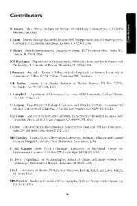
Fig. 6. Pathway for attachment of L-Ara4N to lipid A. The Ugd dehydrogenase converts UDP-glucose into UDP-glucuronic acid, which is a precursor for both colanic acid capsular polysaccharides and L-Ara4N. The first committed step of L-Ara4N biosynthesis is the AmA-catalyzed oxidative decarboxylation, which generates a novel UDP-4-keto-pyranose intennediate. Transamination catalyzed by AmB is followed by formylation due to a second catalytic domain in ArnA. Transfer of the fonnylated monosaccharide to undecaprenyl phosphate by Arne is presumably followed by translocation to the periplasmic side of the] M for deformylation. Undecaprenyl phosphate L-Ara4N is the substrate for ArnT, which transfers L-Ara4N to the lipid A acceptor.
a putative transporter may be specific for the formylated compound and that deformylation may then occur at the periplasmic surface [97]. These steps would ensure the vectorial translocation of the lipid across the 1M and avoid futile cycling. The necessity ofthe deformylation step is dictated by the fact that undecaprenyl phosphate-L-Ara4N is the substrate for PmrK (ArnT), which catalyzes the final transfer of L-Ara4N to lipid A at the periplasmic surface of the 1M [99, 100]. Roles for the remainingpmr genes in the transport and periplasmic deformylation reactions are suspected, but remain to be established.
EptA The putative pEtN adding enzyme EptA has recently been cloned from E. coli [101], and a homologous gene from Neisseria has been associated with
the addition of pEtN to Iipid A [102]. The EptA-encoding gene is the upstream
Endotoxin Structure and Function
15
OH
~"IO
o
0
o
eptA (pmrC)
/
Diacylglycerol
""
pagP PtdEtn
sn-1-lyso-PtdEln
Fig. 7. Modification of lipid A with pEtN and palmitate. EptA at the peri plasmic side of the 1M uses PtdEtn as the pEtN donor to generate diacylglycerol and pEtN-modified lipid A. PagP also uses PtdEtn (or another glycerophospholipid) as the palrnitoyl donor in the OM to generate sn-I-lyso-PtdEtn and lipid A modified by the addition of a palmitoyl group.
open reading frame that is part of the pmrAB operon, and is also known as pmrC (pagB) [103, 104]. PtdEtn is the reported pEtN donor (fig. 7) and several EptA homologues are likely responsible for pEtN addition to other cell envelope components including the inner core sugars of LPS. It is noteworthy that roughly one third of E. coli lipid A carries a diphosphate moiety instead of the monophosphate at position 1 [56], and that the putative phosphorylating enzyme shares with EptA the ability to generate a phosphodiester bond at the same position in lipid A.
PagP
PagP is encoded by a PhoP/PhoQ-activated gene and functions to transfer a palmitate chain from a phospholipid to the hydroxyl group of the N-linked 3-0H-l4:0 chain on the proximal glucosamine unit of lipid A [77, 83]. PagP was the first enzyme of lipid A biosynthesis shown to be localized in the OM
Bishop
16
[77]. Since thiolester-containing substrates are not available in the extracellular compartments, PagP uses a phospholipid as the palmitoyl donor instead (fig. 7). PagP appears to be responsible for the production of lipid Y as a side reaction in IpxB mutants. It was first identified in the salmonellae due to its role in providing resistance to CAMPs [83], and was subsequently purified from E. coli [77]. In addition to these enteric pathogens, PagP homologues are present in the respiratory pathogens Legionella pneumophila and Bordetella bronchiseptica, where PagP has been shown to be necessary for disease causation in animal models of infection [105, 106]. In B. bronchiseptica, PagP is controlled by a different two-component virulence signal transduction pathway known as BvgNBvgS, and palmitoylation occurs at the O-linked 3-0H-14:0 chain on the distal glucosamine sugar [106]. PagP homologues are also found in Yersinia, Photorhahdus and Erwinia species, which adopt pathogenic lifestyles in animals, insects, and plants, respectively. Current efforts to understand the structure and function of PagP are aimed at developing a treatment for infections caused by this important group of pathogens. The structure and dynamics of PagP in detergent micelles have been determined by both NMR spectroscopy [107] and X-ray crystallography [Bishop and Prive, unpub!. data]. PagP is an 8-stranded antiparallel f3-barrel preceded by an N-termjnal amphipathic a-helix. The f3-barrel is well defined in the structure while the extracellular loops are not. Unlike other f3-barrel membrane proteins, proline residues at two sites between f3-strands disrupt the continuity of hydrogen bonding in the outer leaflet half of the PagP f3-barre1. These non-hydrogenbonded regions are located between strands f3-1 and f3-2, generating a f3-bulge, and between strands f3-6 and f3-7. The f3-bulge is largely responsible for the highly dynamic nature of the extracellular loop L1 [107]. Additional features not seen in any other f3-barrel membrane protein include a tilting of the PagP barrel axis by 30° with respect to the membrane normal and the presence of an interior hydrophobic pocket in the upper half of the f3-barrel [Bishop and Prive, unpub!. data]. The hydrophobic pocket harbors a single detergent molecule and functions as a hydrocarbon ruler that allows the enzyme to distinguish palmitate from other acyl chains present in phospholipids. Mutation of Gly 88 lining the bottom of the hydrophobic pocket can modulate the acyl chain length specificity ofPagP [Bishop and Prive, unpubl. data]. Internalization of phospholipid palmitoyl groups within the barrel interior likely occurs by lateral diffusion through the non-hydrogen-bonded regions between the f3-strands in the upper half of the molecule. Three putative catalytic residues were identified by site-directed mutagenesis and mapped to the extracellular loops L 1 and L2, indicating that the active site is localized at the cell surface in the most dynamic region of the molecule [107]. The putative catalytic residues project their side chains toward the barrel
Endotoxin Structure and Function
17
interior and are positioned above the hydrocarbon ruler [Bishop and Prive, unpub1. data]. The requirement of invariant His 33, Asp 76, and Ser 77 for catalysis might suggest that PagP utilizes an acyl-enzyme mechanism characteristic of known serine esterases. However, the putative active site residues are not organized into a catalytic triad that could enhance the nucleophilic character of Ser 77 [107]. The presence of two non-hydrogen-bonded regions that could provide simultaneous access for both substrates to the f3-barrel interior raises the distinct possibility that PagP catalysis proceeds through the formation of a ternary complex. Such a mechanism could promote the direct transfer of the palmitoyl group from the phospholipid donor to the lipid A acceptor without the formation of an acyl-enzyme intermediate, but the detailed mechanism of PagP catalysis remains to be elucidated. The clear alignment of the PagP active site with the OM outer leaflet creates an important topological problem for the enzyme. How does PagP access phospholipids if OM lipid asymmetry is maintained? Chelating agents such as EDTA can strip a fraction ofLPS from the bacterial surface [108]. A large body of evidence indicates that EDTA promotes the migration of phospholipids into the OM outer leaflet [10]. Indeed, brief treatment of cells with EDTA rapidly induces lipid A palmitoylation through a process that is independent of both pagP gene regulation and de novo protein synthesis [Bishop, unpub1. data]. Lipid A palmitoylation induced by EDTA in vivo also requires functional MsbA [Bishop, unpubl. data], which is presumably needed to replenish phospholipids lost from the OM inner leaflet. These findings suggest that PagP may function to maintain the OM permeability barrier under Mg2+ -limited growth conditions, in addition to providing CAMP resistance and converting lipid A into an endotoxin antagonist.
LpxO An Fe2+la-ketoglutarate-dependent dioxygenase homologue in Salmonella has recently been shown to catalyze the hydroxylation of lipid A and is expressed in a PhoPlPhoQ-dependent manner [76]. Under aerobic conditions, LpxO uses molecular oxygen to hydroxylate the 3' secondary acyl chain to generate 2-0H-I4:0-modified lipid A (fig. 8). Homologues are found in other gram-negative bacteria that similarly incorporate S-2-0H groups into their lipid A. The function of S-2-hydroxylation is unknown, but the authors speculate that the action of leukocyte acyloxyacyl hydrolase, an enzyme that releases secondary acyl chains from the lipid A of phagocytosed bacteria, would release 2-0H-14:0, which is possibly converted into 2-0H-14:0-CoA, a known inhibitor of protein N-myristoylation needed for cell signaling functions.
Bishop
18
Fig. 8. S-2-hydroxylation and 3-0-deacylation of lipid A. LpxO is an 1M Fe2+!o:ketoglutarate-dependent dioxygenase homologue that uses molecular oxygen to incorporate a hydroxyl group into the secondary myristoyl group at position 3'. PagL is an OM lipase that removes the 3-0H-14:0 group at position 3.
S-2-hydroxylation may also function to provide an additional hydrogen-bond donor that could stabilize the lateral interactions between LPS molecules in the OM [67]. Given that S-2-hydroxylation is contingent upon lipid A acylation by LpxM, the LpxO reaction could occur on either side of the 1M without interfering with the sequential steps of the Raetz pathway. However, LpxO is predicted to be anchored on the periplasmic face of the 1M.
PagL and Rhizobium Lipid A
Lipid A 3-0-deacylase activity was observed in Salmonella during investigations of PagP in membranes from a PhoP-constitutive mutant [77]. The responsible enzyme was subsequently identified as the PagL gene product, which proved to be the second enzyme of lipid A metabolism that is located in the OM [78]. PagL functions to deacylate the O-linked 3-0H-14:0 chain at the proximal glucosamjne unit of lipid A (fig. 8). By exposing the 3-0H group in lipid A, PagL may provide a new hydrogen-bond donor to stabilize the lateral interactions between LPS molecules in the OM [67]. Although a similar
Endotoxin Structure and Function
19
reaction had been described in Rhizobium leguminosarum membranes [109], PagL homologues are only found in the various serovars of Salmonella. Lipid A recovered from Rhizobium species is structurally quite different from E. coli lipid A, a fact that may reflect the symbiotic relationship between nitrogenfixing rhizobia and leguminous plants, which normally mount an innate immune response to endotoxin. Rhizobium lipid A biosynthesis proceeds according to the Raetz pathway, but the molecule is subsequently remodeled by numerous modifYing enzymes. Besides the absence of phosphate groups at positions I and 4' [110], due to the presence of specific phosphatases [111, 112], the distal glucosamine sugar exhibits a 27-0H-28:0 acyl chain as part of a characteristic acyloxyacyl moiety at position 2' and a galacturonic acid residue at position 4' [113, 114]. LpxQ is the third OM enzyme found to be involved in lipid A modification [115, 116], and catalyzes the oxidation of the proximal I-dephospho sugar to generate an acylated 2-aminogluconate moiety. Rhizobium lipid A serves to illustrate a fundamental point that is supported by functional genomics; namely, that the essential enzymes ofthe Raetz pathway are highly conserved in gram-negative bacteria and that the observed variations in lipid A structure are a consequence of the presence of additional modifYing enzymes. Aside from variations in lipid A structure due to cytoplasmic ACPdependent acyltransferases [117-119] and Kdo transferases [120, 121] with distinct substrate specificities, it appears that most modifying enzymes act on the lipid A nucleus in the extracytoplasmic compartments. These observations may reflect a need to avoid futile cycling and to maintain a sequential order of Raetz pathway reactions. These principles should faithfully guide future discoveries of new enzymes that are employed to generate novel lipid A structures in diverse orgamsms.
Perspectives LPS structure and function are unique to gram-negative bacteria, but some intriguing parallels are seen with the cholesterol and glycosphingolipid-richl· lipid rafts, and N-linked protein glycosylation pathways of eukaryotic cells. Both lipid A and eukaryotic glycolipids differ from phospholipids by the presence of hydrogen-bonded lateral interactions that tend to exclude phospholipids leading to the formation of detergent resistant lipid domains [67, 122].1 Additionally, the undecaprenyl phosphate-dependent pathways for the synthesis and incorporation of O-antigens into the core-KdorJipid A molecule at the 1M mirrors the dolichol phosphate-dependent pathway in the endoplasmic reticulum, where Glc r Man 9 -GlcNAc2 is incorporated into targeted protein Asn residues [123]. Finally, it now appears that many of the Raetz pathway enzymes
Bishop
20
are conserved in the genomes of plants, perhaps reflecting the presence of lipid A-like molecules in plastids [5]. Lipid A and its regulated covalent modifications exhibit profound effects on bacterial and human physiology. Novel endotoxin antagonists and immune adjuvants have already been developed from modified lipid A structures [124, 125]. By revealing the biochemical details oflipid A structure and function we hope to understand its role in bacterial pathogenesis and to intervene with novel treatments for infection. However, we must remind ourselves that multiple molecular subtypes of lipid A are acting in concert in the bacterial cell. The need to unravel the interactions between individual lipid A modifications will provide fertile ground for future research.
Acknowledgments Work in the author's laboratory was supported by the Canadian Institutes of Health Research. Eileen I. Lo is acknowledged for her assistance with the initial drafts of this manuscript.
References
2 3 4 5 6 7 8 9
10 II 12 13
14
Beutler B, Rietschel ET: Innate immune sensing and its roots: The story of endotoxin. Nat Rev ImmunoI2003;3:169-I76. Janeway CA Jr: Approaching the asymptote? Evolution and revolution in immunology. Cold Spring Harb Symp Quant Bioi 1989;54: 1-13. Akira S: Toll-like receptor signaling. J Bioi Chern 2003;278:38105-38108. Inohara N, Nunez G: NODs: Intracellular proteins involved in inflammation and apoptosis. Nat Rev ImmunoI2003;3:371-382. Raetz CR, Whitfield C: Lipopolysaccharide endotoxins. Annu Rev Biochem 2002;71:635-700. Harald FM: Gleanings ofa chemiosmotic eye. Bioessays 2001;23:848-855. Duong F, Eichler J, Price A, Leonard MR, Wickner W: Biogenesis of the Gram-negative bacterial envelope. Cell 1997;91 :567-573. Holtje N: Growth of the stress-bearing and shape-maintaining murein sacculus of Escherichia coli. Microbiol Mol Bioi Rev 1998;62:181-203. Kamio Y, Nikaido H: Outer membrane of Salmonella typhimurium: Accessibility of phospholipid head groups to phospholipase C and cyanogen bromide activated dextran in the external medium. Biochemistry 1976; 15:2561-2570. Nikaido H, Vaara M: Molecular basis of bacterial outer membrane penneability. Microbiol Rev 1985;49: 1-32. Schulz GE: The structure of bacterial outer membrane proteins. Biochim Biophys Acta 2002; 1565:308-317. Vaara M: Antibiotic-supersusceptible mutants of Escherichia coli and Salmonella lyphimurium. Antimicrob Agents Chemother 1993;37:2255-2260. Poltorak A, He X, Smirnova I, Liu MY, Van Huffel C, Du X, Birdwell D, Alejos E, Silva M, Galanos C, Freudenberg M, Ricciardi-Castagnoli P, Layton B, Beutler B: Defective LPS signaling in C3H/HeJ and C57BL/IOScCr mice: Mutations in Tlr4 gene. Science 1998;282:2085-2088. Qureshi ST, Lariviere L, Leveque G, Clermont S, Moore KJ, Gros P, Malo D: Endotoxin-tolerant mice have mutations in Toll-like receptor 4 (Tlr4). J Exp Med 1999;189:615-625.
Endotoxin Structure and Function
21
15 16 17
18
19
20
21 22 23
24 25
26 27 28
29
30
31
32 33
34
Hoffmann JA, Kafatos FC, Janeway CA, Ezekowitz RA: Phylogenetic perspectives in innate immunity. Science 1999;284:1313-1318. Aderem A, Ulevitch RJ: Toll-like receptors in the induction of the innate immune response. Nature 2000;406:782-787. Nishijima M, Raetz CR: Membrane lipid biogenesis in Escherichia coli: Identification of genetic loci for phosphatidylgiycerophosphate synthetase and construction of mutants lacking phosphatidylglycerol. J Bioi Chern 1979;254:7837-7844. Takayama K, Qureshi N, Mascagni P, Nashed MA, Anderson L, Raetz CR: Fatty acyl derivatives of glucosamine I-phosphate in Escherichia coli and their relation to lipid A. Complete structure of A diacyl GlcN-I-P found in a phosphatidylglycerol-deficient mutant. J Bioi Chern 1983;258: 7379-7385. Takayama K, Qureshi N, Mascagni P, Anderson L, Raetz CR: Glucosamine-derived phospholipids in Escherichia coli. Structure and chemical modification of a triacyl glucosamine I-phosphate found in a phosphatidylglycerol-deficient mutant. J Bioi Chern 1983;258:14245-14252. Rietschel ET, Brade H, Brade L, Kaca W, Kawahara K, Lindner B, Luderitz T, Tomita T, Schade U, Seydel U, Ziihringer U: Newer aspects of the chemical structure and biological activity of bacterial endotoxins. Prog Clin Bioi Res 1985; 189:31-51. Nishijima M, Bulawa CE, Raetz CR: Two interacting mutations causing temperature-sensitive phosphatidylglycerol synthesis in Escherichia coli membranes. J Bacteriol 1981; 145: 113-121. Nishijima M, Raetz CR: Characterization of two membrane-associated glycolipids from an Escherichia coli mutant deficient in phosphatidylglycerol. J BioI Chern 1981 ;256: I0690-1 0696. Ray BL, Painter G, Raetz CR: The biosynthesis of Gram-negative endotoxin. Formation of lipid A disaccharides from monosaccharide precursors in extracts of Escherichia coli. J Bioi Chern 1984;259:4852-4859. Nishijima M, Amano F, AkamatsuY, Akagawa K, Tokunaga T, Raetz CR: Macrophage activation by monosaccharide precursors of Escherichia coli lipid A. Proc Natl Acad Sci USA 1985;82:282-286. Anderson MS, Bull HG, Galloway SM, Kelly TM, Mohan S, Radika K, Raetz CR: UDP-Nacetylglucosamine acyltransferase of Escherichia coli. The first step of endotoxin biosynthesis is thermodynamically unfavorable. J Bioi Chern 1993;268: 19858-19865. Raetz CR, Roderick SL: A left-handed parallel beta helix in the structure of UDP-N-acetylglucosamine acyltransferase. Science 1995;270:997-1000. Wyckoff TJ, Lin S, Cotter RJ, Dotson GO, Raetz CR: Hydrocarbon rulers in UDP-N-acetylglucosamine acyltransferases. J Bioi Chern 1998;273:32369-32372. Young K, Silver LL, Bramhill 0, Cameron P, Eveland SS, Raetz CR, Hyland SA, Anderson MS: The envA permeability/cell division gene of Escherichia coli encodes the second enzyme of lipid A biosynthesis. UDP-3-0-(R-3-hydroxymyristoyl)-N-acetylglucosamine deacetylase. J Bioi Chern 1995;270:30384-30391. Sorensen PG, Lutkenhaus J, Young K, Eveland SS, Anderson MS, Raetz CR: Regulation ofUDP3-0-[R-3-hydroxymyristoyl]-N-acetylglucosamine deacetylase in Escherichia coli. The second enzymatic step of lipid a biosynthesis. J BioI Chern 1996;271 :25898-25905. Onishi HR, Pelak BA, Gerckens LS, Sil ver LL, Kahan FM, Chen MH, Patchett AA, Galloway SM, Hyland SA, Anderson MS, Raetz CR: Antibacterial agents that inhibit lipid A biosynthesis. Science 1996;274:980-982. Jackman JE, Raetz CR, Fierke CA: Site-directed mutagenesis of the bacterial metalloamidase UDP-(3-0-acyl)-N-acetylglucosamine deacetylase (LpxC). Identification of the zinc binding site. Biochemistry 2001;40:514-523. Coggins BE, Li X, McClerren AL, Hindsgaul 0, Raetz CR, Zhou P: Structure of the LpxC deacetylase with a bound substrate-analog inhibitor. Nat Struct BioI 2003; 10:645-651. Whittington DA, Rusche KM, Shin H, Fierke CA, Christianson OW: Crystal structure of LpxC, a zinc-dependent deacetylase essential for endotoxin biosynthesis. Proc Nat! Acad Sci USA 2003; I00:8146-8150. Jackman JE, Fierke CA, Tumey LN, Pirrung M, Uchiyama T, Tahir SH, Hindsgaul 0, Raetz CR: Agents that target lipid A biosynthesis in gram-negative bacteria. Inhibition of diverse UDP-3-0(r-3-hydroxymyristoyl)-n-acetylglucosamine deacetylases by substrate analogs containing zinc binding motifs. J Bioi Chern 2000;275: 11002-11009.
Bishop
22
35
36
37 38
39
40 41
42
43
44 45
46
47
48
49 50
5\
52
53
Pirrung MC, Tumey LN, McClerren AL, Raetz CR: High-throughput catch-and-release synthesis of oxazoline hydroxamates. Structure-activity relationships in novel inhibitors of Escherichia coli LpxC: In vitro enzyme inhibition and antibacterial properties. J Am Chern Soc 2003; 125: 1575-1586. Kelly TM, Stachula SA, Raetz CR, Anderson MS: The firA gene of Escherichia coli encodes UDP-3-0-(R-3-hydroxymyristoyl)-glucosamine N-acyltransferase. The third step of endotoxin biosynthesis. J Bioi Chern 1993;268:19866-19874. Babinski KJ, Ribeiro AA, Raetz CR: The Escherichia coli gene encoding the UDP-2,3-diacylglucosamine pyrophosphatase of lipid A biosynthesis. J Bioi Chern 2002;277:25937-25946. Babinski KJ, Kanjilal SJ, Raetz CR: Accumulation of the lipid A precursor UDP-2,3-diacylglucosamine in an Escherichia coli mutant lacking the IpxH gene. J Bioi Chern 2002;277: 25947-25956. Garrett TA, Kadrmas JL, Raetz CR: Identification of the gene encoding the Escherichia coli lipid A 4' -kinase. Facile phosphorylation of endotoxin analogs with recombinant LpxK. J BioI Chern 1997;272:2 I855-2 I864. Garrett TA, Que NL, Raetz CR: Accumulation of a lipid A precursor lacking the 4' -phosphate following inactivation of the Escherichia coli IpxK gene. J Bioi Chern 1998;273:12457-12465. Lien E, Means TK, Heine H, Yoshimura A, Kusumoto S, Fukase K, Fenton MJ, Oikawa M, Qureshi N, Monks B, Finberg RW, Ingalls RR, Golenbock DT: Toll-like receptor 4 imparts ligandspecific recognition of bacterial lipopolysaccharide. J Clin Invest 2000; I05:497-504. Poltorak A, Ricciardi-Castagnoli P, Citterio S, Beutler B: Physical contact between lipopolysaccharide and toll-like receptor 4 revealed by genetic complementation. Proc Nat! Acad Sci USA 2000;97: 2163-2167. Clementz T, Raetz CR: A gene coding for 3-deoxy-D-manno-octulosonic-acid transferase in Escherichia coli. Identification, mapping, cloning, and sequencing. J Bioi Chern 1991 ;266: 9687-9696. Brozek KA, Raetz CR: Biosynthesis oflipid A in Escherichia coli. Acyl carrier protein-dependent incorporation oflaurate and myristate. J Bioi Chern 1990:265:15410-15417. Clementz T, Bednarski JJ, Raetz CR: Function of the htrB high temperature requirement gene of Escherchia coli in the acylation of lipid A: HtrB catalyzed incorporation of laurate. J BioI Chern 1996;271:12095-12102. Clementz T, Zhou Z, Raetz CR: Function of the Escherichia coli msbB gene, a multicopy suppressor of htrB knockouts, in the acylation of lipid A. Acylation by MsbB follows laurate incorporation by HtrB. J Bioi Chern 1997;272:10353-10360. Carty SM, Sreekumar KR, Raetz CR: Effect of cold shock on lipid A biosynthesis in Escherichia coli. Induction at 12 degrees C of an acyltransferase specific for palmitoleoyl-acyl carrier protein. J Bioi Chern 1999;274:9677-9685. Vorachek-Warren MK, Carty SM, Lin S, Cotter RJ, Raetz CR: An Escherichia coli mutant lacking the cold shock-induced palmitoieoyltransferase of lipid A biosynthesis: Absence of unsaturated acyl chains and antibiotic hypersensitivity at 12 degrees C. J Bioi Chern 2002;277: 14 I 86- 14 I93. Vorachek-Warren MK, Ramirez S, Cotter RJ, Raetz CR: A triple mutant of Escherichia coli lacking secondary acyl chains on lipid A. J Bioi Chern 2002;277: 14194-14205. Somerville JE Jr, Cassiano L, Bainbridge B, Cunningham MD, Darveau RP: A novel Escherichia coli lipid A mutant that produces an anti-inflammatory lipopolysaccharide. J Clin Invest 1996;97: 359-365. Heinrichs DE, Yethon JA, Whitfield C: Molecular basis for structural diversity in the core regions of the lipopolysaccharides of Escherichia coli and Salmonella enterica. Mol Microbiol \998;30: 221-232. Kanipes MI, Lin S, Cotter RJ, Raetz CR: Ca'+ -induced phosphoethanolamine transfer to the outer 3-deoxy-D-manno-octulosonic acid moiety of Escherichia coli lipopolysaccharide. A novel membrane enzyme dependent upon phosphatidylethanolamine. J BioI Chern 200 1;276: 1156-1163. Yethon JA, Heinrichs DE, Monteiro MA, Perry MB, Whitfield C: Involvement of waaY, waaQ, and waaP in the modification of Escherichia coli lipopolysaccharide and their role in the formation of a stable outer membrane. J Bioi Chern 1998;273:26310-26316.
Endotoxin Structure and Function
23
54
55 56
57 58 59 60 61
62 63 64 65 66 67 68 69 70 71 72
73
74
75
76
Heinrichs DE, Yethon JA, Amor PA, Whitfield C: The assembly system for the outer core portion of RI- and R4-type lipopolysaccharides of Escherichia coli. The RI core-specific betaglucosyltransferase provides a novel attachment site for O-polysaccharides. J BioI Chem 1998; 273:29497-29505. Valvano MA: Export ofO-specific lipopolysaccharide. Front Biosci 2003;8:s452-s47I. Zhou Z, White KA, Polissi A, Georgopoulos C, Raetz CR: Function of Escherichia coli MsbA, an essential ABC family transporter, in lipid A and phospholipid biosynthesis. J Bioi Chem 1998; 273:12466-12475. Doerrler WT, Reedy MC, Raetz CR: An Escherichia coli mutant defective in lipid export. J Bioi Chem 200 I ;276: I 1461-1 1464. Chang G, Roth CB: Structure of MsbA from E. coli: A homolog of the multidrug resistance ATP binding cassette (ABC) transporters. Science 200 1;293: 1793-1800. Doerrler WT, Raetz CR: ATPase activity of the MsbA lipid flippase of Escherichia coli. J BioI Chem 2002;277:36697-36705. Chang G: Structure of MsbA from Vibrio cholera: A multidrug resistance ABC transporter homolog in a closed conformation. J Mol Bioi 2003;330:419-430. Kol MA, van Dalen A, de Kroon AI, de KruijffB: Translocation of phospholipids is facilitated by a subset of membrane-spanning proteins of the bacterial cytoplasmic membrane. J Bioi Chem 2003; 278:24586-24593. Jones NC, Osborn MJ: Interaction of Salmonella typhimurium \\~th phospholipid vesicles. Incorporation of exogenous lipids into intact cells. J Bioi Chem 1977;252:7398-7404. Jones NC, Osborn MJ: Translocation of phospholipids bet\veen the outer and inner membranes of Salmonella typhimurium. J Bioi Chem 1977;252:7405-7412. Genevrois S, Steeghs L, Roholl P, Letesson JJ, van der Ley P: The Omp85 protein of Neisseria meningitidis is required for lipid export to the outer membrane. EMBO J 2003;22: 1780-1789. Voulhoux R, Bos Mp, Geurtsen J, Mols M, Tommassen J: Role of a highly conserved bacterial protein in outer membrane protein assembly. Science 2003;299:262-265. Coughlin RT, Tonsager S, McGroarty EJ: Quantitation of metal cations bound to membranes and extracted lipopolysaccharide of Escherichia coli. Biochemistry 1983;22:2002-2007. Niakido H: Molecular basis of bacterial outer membrane permeability revisited. Microbiol Mol Bioi Rev 2003;67:593-656. Hancock RE, Falla T, Brown M: Cationic bactericidal peptides. Adv Microb Physiol 1995;37: 135-175. Zasloff M: Antimicrobial peptides of multicellular organisms. Nature 2002;415:389-395. Groisman EA: The ins and outs of virulence gene expression: Mg'+ as a regulatory signal. Bioessays 1998;20:96-101. Forbes JR, Gros P: Divalent-metal transport by NRAMP proteins at the interface of host-pathogen interactions. Trends MicrobioI2001;9:397-403. Guo L, Lim KB, Gunn JS, Bainbridge B, Darveau RP, Hackett M, Miller SI: Regulation of lipid A modifications by Salmonella typhimurium virulence genes phoP-phoQ. Science 1997;276: 250-253. Zhou Z, Lin S, Cotter RJ, Raetz CR: Lipid A modifications characteristic of Salmonella typhimurium are induced by NH 4 VO] in Escherichia coli K12. Detection of 4-amino-4-deoxy-Larabinose, phosphoethanolamine and palmitate. J Bioi Chem 1999;274:18503-18514. Zhou Z, Ribeiro AA, Raetz CR: High-resolution NMR spectroscopy of lipid A molecules containing 4-amino-4-deoxy-L-arabinose and phosphoethanolamine substituents. Different attachment sites on lipid A molecules from NH 4 VO]-treated Escherichia coli versus kdsA mutants of Salmonella typhimuriwn. J Bioi Chem 2000;275:13542-13551. Zhou Z, Ribeiro AA, Lin S, Cotter RJ, Miller SI, Raetz CR: Lipid A modifications in polymyxinresistant Salmonella typhimurium: PMRA-dependent 4-amino-4-deoxy-L-arabinose, and phosphoethanolamine incorporation. J Bioi Chem 2001;276:43111-43121. Gibbons HS, Lin S, Cotter RJ, Raetz CR: Oxygen requirement for the biosynthesis of the S-2-hydroxymyristate moiety in Salmonella typhimurium lipid A. Function of LpxO, a new Fe2+/a lpha-ketoglutarate-dependent dioxygenase homologue. J Bioi Chem 2000;275: 32940-32949.
Bishop
24
77 78
79
80
81 82 83 84 85 86 87 88 89 90 91
92
93
94
95 96
97
98
Bishop RE, Gibbons HS, Guina T, Trent MS, Miller SI, Raetz CR: Transfer of palmitate from phospholipids to lipid A in outer membranes of Gram-negative bacteria. EMBO J 2000; 19:5071-5080. Trent MS, Pabich W, Raetz CR, Miller SI: A PhoP/PhoQ-induced lipase (PagL) that catalyzes 3-0-deacylation of lipid A precursors in membranes of Salmonella typhimurium. J Bioi Chern 2001;276:9083-9092. Helander 1M, Kilpelainen I, Vaara M: Increased substitution of phosphate groups in lipopolysaccharides and lipid A of the polymyxin-resistant pmrA mutants of Salmonella typhimurium: A 31p-NMR study. Mol Microbiol 1994; II :481-487. Gunn JS, Lim KB, Krueger J, Kim K, Guo L, Hackett M, Miller SI: PmrA-PmrB-regulated genes necessary for 4-aminoarabinose lipid A modification and polymyxin resistance. Mol Microbiol 1998;27:1171-1182. Bruch MD, Cajal Y, Koh IT, Jain MK: Higher-order structure of polymyxin B: The functional significant of topological specificity. J Am Chern Soc 1999;121: 11993-12004. Pristovsek P, Kidric J: Solution structure of polymyxins B and E and effect of binding to lipopolysaccharide: An NMR and molecular modeling study. .I Med Chem 1999;42: 4604-4613. Guo L, Lim K, Poduje C, Daniel M, Gunn.l, Hackett.l, Miller SJ: Lipid A acylation and bacterial resistance against vertebrate anti-microbial peptides. Cell 1998;95: 189-198. Hajjar AM, Ernst RK, Tsai JH, Wilson CB, Miller SI: Human Toll-like receptor 4 recognizes hostspecific LPS modifications. Nat Immunol 2002;3:354-359. Tanamoto K, Azumi S: Salmonella-type heptaacylated lipid A is inactive and acts as an antagonist of lipopolysaccharide action on human line cells. J Immunol 2000; 164:3149-3156. Muroi M, Ohnishi T, Tanamoto K: MD-2, a novel accessory molecule, is involved in species-specific actions of Salmonella lipid A. Infect Immun 2002;70:3546-3550. Groisman EA: The pleiotropic two-component regulatory system PhoP-PhoQ. .I Bacteriol 200 I; 183: 1835-1842. Garcia Vescovi E, Soncini FC, Groisman EA: Mg2+ as an extracellular signal: Environmental regulation of Salmonella virulence. Cell 1996;84: 165-174. Kox LF, Wosten MM, Groisman EA: A slTlJlll protein that mediates the activation of a twocomponent system by another two-component system. EMBO J 2000; 19: 1861-1872. Wosten MM, Kox LF, Chamnongpol S, Soncini FC, Groisman EA: A signal transduction system that responds to extracellular iron. Cell 2000;103: 113-125. Kato A, Latifi T, Groisman EA: Closing the loop: The PmrAlPmrB two-component system negatively controls expression of its posttranscriptional activator PmrD. Proc Natl Acad Sci USA 2003; 100:4706-4711. Bader MW, Navarre WW, Shiau W, Nikaido H, Frye JG, McClelland M, Fang FC, Miller SI: Regulation of Salmonella typhimurium virulence gene expression by cationic antimicrobial peptides. Mol Microbiol 2003;50:219-230. McPhee 18, Lewenza S, Hancock RE: Cationic antimicrobial peptides activate a two-component regulatory system, PmrA-PmrB, that regulates resistance to polymyxin B and cationic antimicrobial peptides in Pseudomonas aeruginosa. Mol MicrobioI2003;50:205-217. Vaara M, Vaara T, Jensen M, Helander I, Nurminen M, Rietschel ET, Makela PH: Characterization of the lipopolysaccharide from the polymyxin-resistant pmrA mutants of Salmonella typhimurium. FEBS Lett 1981;129:145-149. Mouslim C, Groisman EA: Control of the Salmonella ugd gene by three two-component regulatory systems. Mol Microbiol 2003;47:335-344. Breazeale SO, Ribeiro AA, Raetz CR: Oxidative decarboxylation of UDP-glucuronic acid in extracts of polymyxin-resistant Escherichia coli. Origin of lipid A species modified with 4-amino4-deoxy-L-arabinose. J Bioi Chern 2002;277:2886-2896. Breazeale SO, Ribeiro AA, Raetz CR: Origin of lipid A species modified with 4-amino-4-deoxyL-arabinose in polymyxin resistant mutants of Escherichia coli: An aminotransferase (ArnB) that generates UDP-4-amino-4-deoxy-L-arabinose. J Bioi Chern 2003;278: 24731-24739. Noland BW, Newman JM, I-Iendle J, Badger J, Christopher .lA, Tresser.l, Buchanan MD, WrightTA, Rutter ME, Sanderson WE, Muller-Dieckmann H.I, Gajiwala KS, Buchanan SG: Structural studies of Salmonella typhimurium ArnB (PmrH) aminotransferase: A 4-amino-4-deoxy-L-arabinose lipopolysaccharide-modifying enzyme. Structure 2002; I0: 1569-1580.
Endotoxin Structure and Function
25
99
100
101 102
103
104 105
106
107
108 109
I 10
III
112
I 13
114
115
116
I 17
Trent MS, Ribeiro AA, Lin S, Cotter RJ, Raetz CR: An inner membrane enzyme in Salmonella and Escherichia coli that transfers 4-amino-4-deoxy-L-arabinose to lipid A: Induction on polymyxin-resistant mutants and role of a novel lipid-linked donor. J Bioi Chern 200 1;276: 43122-43131. Trent MS, Ribeiro AA, Doerrler WT, Lin S, Cotter RJ, Raetz CR: Accumulation of a polyisoprenelinked amino sugar in polymyxin-resistant Salmonella typhimurium and Escherichia coli: Structural characterization and transfer to lipid A in the periplasm. J Bioi Chern 2001 ;276:43 I 32-43 144. Trent MS, Raetz CRH: Cloning ofEptA, the lipid A phosphoethanolamine transferase associated with polymyxin resistance. J Endotoxin Res 2002;8: 158. Cox AD, Wright JC, Li J, Hood OW, Moxon ER, Richards JC: Phosphorylation of the lipid a region of meningococcal lipopolysaccharide: Identification of a family of transferases that add phosphoethanolamine to lipopolysaccharide. J Bacteriol 2003; I 85:3270-3277. Gunn JS, Miller SI: PhoP-PhoQ activates transcription of pmrAB, encoding a two-component regulatory system involved in Salmonella typhimurium antimicrobial peptide resistance. J Bacteriol 1996; 178:6857-6864. Soncini FC, Groisman EA: Two-component regulatory systems can interact to process multiple environmental signals. J Bacteriol 1996; I78:6796-680 I. Robey M, O'Connell W, Cianciotto NP: Identification of Legionella pneumophila rcp, a pagP-like gene that confers resistance to cationic antimicrobial peptides and promotes intracellular infection. Infect lmmun 200 I ;69:4276-4286. Preston A, Maxim E, Toland E, Pishko EJ, Harvill ET, Caroff M, Maskell OJ: Bordetella bronchiseptica PagP is a Bvg-regulated lipid A palmitoyl transferase that is required for persistent colonization of the mouse respiratory tract. Mol Microbiol 2003;48:725-736. Hwang PM, Choy WY, Lo EI, Chen L, Forman-Kay JD, Raetz CR, Prive GG, Bishop RE, Kay LE: Solution structure and dynamics of the outer membrane enzyme PagP by NMR. Proc Natl Acad Sci USA 2002;99:13560-13565. Leive L: Release of lipopolysaccharide by EDTA treatment of E. coli. Biochem Biophys Res Commun 1965;21 :290-296. Basu SS, White KA, Que NL, Raetz CR: A deacylase in Rhizobium leguminosal1lm membranes that cleaves the 3-O-linked beta-hydroxymyristoyl moiety of lipid A precursors. J Bioi Chern 1999;274:1 I 150-1 I 158. Brozek KA, Kadrmas JL, Raetz CR: Lipopolysaccharide biosynthesis in Rhizobium leguminosal1lm. Novel enzymes that process precursors containing 3-deoxy-D-manno-octulosonic acid. J Bioi Chern 1996;271:32112-32118. Price Np, Jeyaretnam B, Carlson RW, Kadrmas JL, Raetz CR, Brozek KA: Lipid A biosynthesis in Rhizobium leguminosal1lm: Role of a 2-keto-3-deoxyoctulosonate-activated 4' phosphatase. Proc Natl Acad Sci USA 1995;92:7352-7356. Karbarz MJ, Kalb SR, Cotter RJ, Raetz CR: Expression cloning and biochemical characterization of a Rhizobium leguminosarum lipid A I-phosphatase. J Bioi Chern 2003;278: 39269-39279. Que NL, Lin S, Cotter RJ, Raetz CR: Purification and mass spectrometry of six lipid A species from the bacterial endosymbiont Rhizobium etli. Demonstration of a conserved distal unit and a variable proximal portion. J Bioi Chern 2000;275:28006-28016. Que NL, Ribeiro AA, Raetz CR: Two-dimensional NMR spectroscopy and structures of six lipid A species from Rhizobium etli CE3. Detection of an acyloxyacyl residue in each component and origin of the aminogluconate moiety. J Bioi Chern 2000;275:28017-28027. Que-Gewirth NL, Lin S, Cotter RJ, Raetz CR: An outer membrane enzyme that generates the 2-amino-2-deoxy-gluconate moiety of Rhizobium leguminosarum lipid A. J Bioi Chern 2003;278: 12109-12119. Que-Gewirth NL, Karbarz MJ, Kalb SR, Cotter RJ, Raetz CR: Origin of the 2-amino-2-deoxygluconate unit in Rhizobium leguminosarum lipid A. Expression cloning of the outer membrane oxidase LpxQ. J Bioi Chern 2003;278: 12120-12129. Sweet CR, Lin S, Cotter RJ, Raetz CR: A Chlamydia trachomatis UDP-N-acetylglucosamine acyltransferase selective for myristoyl-acyl carrier protein. Expression in Escherichia coli and formation of hybrid lipid A species. J Bioi Chern 2001;276: 19565-19574.
Bishop
26
118
119 120
121
122 123 124
125
Sweet CR, Preston A, Toland E, Ramirez SM, Cotter RJ, Maskell OJ, Raetz CR: Relaxed acyl chain specificity of Bordetella UDP-N-acetylglucosamine acyltransferases. J Bioi Chem 2002; 277:18281-18290. Basu SS, Karbarz MJ, Raetz CR: Expression cloning and characterization of the C28 acyltransferase of lipid A biosynthesis in Rhizobium leguminosarum. J Bioi Chem 2002;277:28959-28971. White KA, Kaltashov lA, Cotter RJ, Raetz CR: A mono-functionaI3-deoxy-D-manno-octulosonic acid (Kdo) transferase and a Kdo kinase in extracts of Haemophilus influenzae. J Bioi Chem 1997; 272: 16555-16563. Belunis CJ, Mdluli KE, Raetz CR, Nano FE: A novel 3-deoxy-D-manno-octulosonic acid transferase from Chlamydia trachomatis required for expression of the genus-specific epitope. J Bioi Chem 1992;267: 18702-18707. Munro S: Lipid rafts: Elusive or illusive? Cell 2003;115:377-388. Drickamer K, Taylor ME: Evolving views of protein glycosylation. Trends Biochem Sci 1998; 23:321-324. Christ WJ, Asano 0, Robidoux AL, Perez M, Wang YA, Dubuc GR, Gavin WE, Hawkins LD, McGuinness PO, Mullarkey MA, Lewis MD, Kishi Y, Kawata T, Bristol JR, Rose JR, Rossignol Dp, Kobayashi S, Hishinuma L, Kimura A, Asakawa N, Katayama K, Yamatsu I: E5531, a pure endotoxin antagonist of high potency. Science 1995;268:80-83. Ulrich JT, Myers KR: Monophosphoryl lipid A as an adjuvant; in Powell MF, Newman MJ (eds): Vaccine Design: The Subunit and Adjuvant Approach. New York, Plenum Press, 1995, pp 495-524.
Russell E. Bishop 6213 Medical Sciences Building, I King's College Circle Toronto, Ont. M5S 1A8 (Canada) Tel. + I 4169467103, Fax + 1 4169785959, E-Mail [email protected]
Endotoxin Structure and Function
27
Toxins Russell W, HelWald H (eds): Concepts in Bacterial Virulence. Contrib Microbiol. Basel, Karger, 2005, vol 12, pp 28-54
Bacterial Exotoxi ns Michel R. Popoff Unite des Baeteries anaerobies et Toxines, Tnstitut Pasteur, Paris, France
Amongst the various mechanisms developed by pathogenic bacteria to cause disease, toxins play an important role, since they are responsible for the majority of symptoms and lesions during infection. Exotoxins act at a distance from the infectious site and can diffuse through the organism. While some cytotoxins can cause disruption of cells permitting the pathogens access to nutrients, other toxins are only active on specific cells, for example intestinal cells, neuronal cells, or leukocytes. This is achieved by the recognition of specific cell surface receptors. When bound to the receptor, toxins can unleash their toxic program at the cell membrane by interfering with signal transduction pathways, pore formation, or enzymatic activities towards membrane compounds. In contrast, other toxins enter the cytosol, and recognize and modifY specific intracellular targets. According to the nature of the target and the type of modification, intracellular active toxins cause a dramatic alteration of cellular functions such as protein synthesis, cell homeostasis, cell cycle progression, vesicular traffic, and actin cytoskeletal rearrangements. Alternatively, invasive bacteria can directly inject toxins or virulence factors into target cells. This chapter is a comparative overview of the molecular mechanisms of the main bacterial exotoxins.
Toxins Active at the Cell Surface Toxins Modulating Signal Transduction Pathways Some enterotoxigenic Escherichia coli and other gram-negative enteropathogens (Yersinia enterocolitica, Vibrio cholerae) secrete heat-stable enterotoxins (STs) that can cause acute diarrhea in humans and animals. These toxins are small peptides which fall into two subgroups: methanol-soluble (STa or ST-I) and methanol-insoluble (STh or ST-II) toxins. Analysis ofSTs shows they possess a similar structure, containing 3 segments joined by 3 disulfide bridges. Ala13 in
Hormone-like toxins
Guanylate cyclase
if
~
/'
It \
CGMP
GTP
40dUlin Bacterial adenylcyclases
EF, Cya
ATP
8'1 "'~ /
CT
Gie<
l;
ADP-ribosylation
Pore-forming toxins
Fig. 1. Toxins that alter cell homeostasis. Some of the mechanisms used by bacteria to modify cell homeostasis are depicted. E. coli heat-stable enterotoxin (STa) binds to the extracellular domain of transmembrane guanylate cyclase, resulting in an increase in cyclic GMp, and secretion ofCI- and H 20. PFT inserted into the membrane cause leakage of ions and H 20. CT and E. coli heat-labile toxins enter the cell cytosol and ADP-ribosylate the GSLX subwlit ofheterotrimeric G proteins, leading to a permanent active molecule by inhibition of its GTPase activity and subsequent stimulation of adenylcyclase. The resulting increase in cyclic AMP induces the secretion ofCl- and H 20. PT inactivates the inhibitory heterotrimeric G protein Gio:, leading to a upregulation of adenylcyclase activity. Bacterial adenylcyclases, such as EF from anthrax toxin and Bordetella adenylcyclase (Cya), can also modulate cAMP levels in the cells.
the flexible central segment plays a key role in the toxin's activity. This residue is probably involved in the interaction of the toxin with its receptor. In the case of STa, the secreted protein encompasses 18-19 amino acids, including 6 cysteines, and is capable of forming 3 disulfide bridges to create a highly stable molecule. The carboxy-terminal segment of STs shares similarities with ionophores and is therefore expected to interact with metal ions. Enteroaggregative E. coli (EAggEC) strains also produce a heat-stable enterotoxin related to STa with simjlar pathological effects. STa induces watery diarrhea without causing obvious histological morphological damage. The toxin hinds to the extracellular domain of guanylate cyclase (GC-C) localized on the apical membrane of enterocytes. GC-C consists of 4 domains: an extracellular domain, a transmembrane segment, a kinase-like domain and an enzymatic domain, whjch catalyzes the formation of cyclic GMP (cGMP) (fig. I). The kinase-like domain has an inhibitory effect on the catalytic
Bacterial Exotoxins
29
activity. Binding of STa to the extracellular domain of GC-C has been suggested to induce a conformational change in the protein kinase-like domain resulting in an uncontrolled increase of GC-C activity. Elevation of intracellular cGMP activates protein kinase II (cGKII), which in turn stimulates the cystic fibrosis transmembrane conductance regulator (CFTR) CJ- channels. This results in a net fluid secretion through activation of apical CI- channels in pamllel with the inhibition of coupled NaCI tmnspOlters. These findings have been confirmed in GC-C knockout mice, which have a lower intestinal GC-C activity and do not exhibit a secretory response to STa treatment [reviewed in 1]. STa was the first ligand found to bind GC-C and later studies demonstrated that the hormones guanylin and uroguanylin are the natural ligands for this receptor. These hormones have been shown to be involved in the regulation of fluid and electrolyte transport in many tissues. Guanylin and uroguanylin consist of IS amino acids and are highly homologous to STa.
Toxins with Enzymatic Activity at the Cell Surface That Alters Cell Signaling Phospholipases The first toxin that was recognized to possess an enzymatic activity was the Clostridium pelfringens a-toxin. This protein is a zinc-dependent phospholipase C, which degrades phosphatidylcholine and sphingomyelin. Both in vitro and in vivo studies have shown that it has cytolytic, dermonecrotic, and hemolytic activities, and is lethal to animals at low doses. The toxin causes membrane damage to a variety of different human and animal cell types including platelets, leukocytes, and fibroblasts, as well as erythrocytes. It is the major toxin involved in gangrene, which is characterized by extensive local tissue destruction and necrosis progressing to profound shock and death. The secreted protein consists of 370 amino acids (43 kD), and contains 2 domains, an a-helical amino-terminal domain (residues 1-246) harboring the active site, and a l3-sandwich carboxy-terminal domain (residues 256-370), which mediates membrane binding. The carboxy-terminal domain is structurally similar to eukaryotic calcium-binding C2 domains, which are involved in Ca2 + -dependent phospholipid binding. a-Toxin preferentially binds to phospholipids in the intact membrane, opening the active site of the toxin and resulting in cleavage of phospholipids [2]. In the activated state, the active site contains two tightly bound zinc ions and one loosely bound zinc ion and is accessible for substrate binding, whereas in the closed or inactive conformation, the active site is occluded and one zinc ion binding site is lost [2--4]. In addition to its lytic activity, a-toxin is also involved in intracellular signaling and the activation of endogenous metabolism cascades. Diacylglycerol and ceramide generated from limited hydrolysis of phospholipids and sphingomyelin,
Popoff
30
respectively, activate endogenous phospholipases A2, C and D, and protein kinase C. This in turn stimulates membrane phospholipases and initiates the arachidonic acid pathway leading to the production of proinflammatory molecules (prostaglandins, thromboxanes, and leukotrienes responsible for vasodilatation, bronchostriction), and platelet aggregation [4]. Other bacterial phospholipases include phospholipase C from Pseudomonas, Listeria, and various Clostridium species, phospholipase A from Helicobacter pylori, phosphatidylinositol phospholipase C from Bacillus, Clostridium, and phospholipase D from Corynebacterium.
Bacteroides fragilis Enterotoxin B. fragilis enterotoxin (BFT) induces morphological changes in cultured intestinal and renal cells, including cell rounding, increase in volume, and effacement of microvilli and apical junctional complexes. BFT has zinc-dependent protease activity, which has been shown to cleave the extracellular domain of E-cadherin, the primary protein of the zonula adherens. Experimental studies have led to the proposed two-step hypothesis, whereby the extracellular domain of E-cadherin is cleaved by BFT, followed by intracellular degradation by as yet unidentified protease(s). As a consequence, nuclear signaling and actin rearrangement occm, which leads to the production of proinflammatory cytokines, diminished epithelial barrier function, and activation of apical membrane ion transporters. These cytotoxic effects are reversible, since 2-3 days after toxin treatment cells appear normal [reviewed in 5]. Pore-Forming Toxins So far more than 80 toxins have been identified that act by forming a transmembrane pore in the target cell. The general mechanism of pore-forming toxins (PFT) is to bind to cell surface receptors where they then oligomerize. The insertion of the oligomer into the cell membrane results in the formation of a channel, which impairs the osmotic balance of the cell and causes cytolysis. Most of the PFTs are cytolytic and/or hemolytic and they have been classified inlo several families [for review see 6-8]. RTX toxins (repeats in toxin) are synthesized by many gram-negative pathogens (Escherichia, Proteus, Pasteurella). Members of the RTX toxin family, including cytolytic toxins, metalloproteases and lipases, share a common gene organization and distinctive structural featmes. They are secreted by the type I secretion system which is mediated by the Sec machinery. At the carboxy-terminal end, RTX contains 10--40 repeats of glycine- and aspartate-rich nonapeptide domains. Most RTX toxins are posttranslationally activated by acylation. The prototype of this famjly is the a-hemolysin (110 kD) of E. coli and its target receptors on leukocytes have been identified as members of the ~2
Bacterial Exotoxins
31
integrin family. Insertion of a-hemolysin into the membrane, probably mediated by four predicted hydrophobic a-helices in the amino-terminal region, leads to the formation of a hydrophilic- and cation-selective pore of at least 1 nm in diameter [9]. A related family of hemolysins consists of streptolysin Sand streptolysin S-like cytolysins expressed in streptococci. Cholesterol-binding cytolysins are produced by a wide variety of bacterial species including Streptococcus, Bacillus, Clostridium, and Listeria. Perfringolysin 0 (PFO) is one of the best-studied toxins from this family. PFOs are secreted as water-soluble monomers, which contain 4 domains rich in f)-strands. A short hydrophobic loop in domain 4 is involved in the binding to cholesterol [10]. After cholesterol binding, PFO undergoes a conformational change resulting in the unfolding of domain 3 a-helices and the formation of two amphipathic f)-hairpins in each monomer. This leads to an association of neighboring monomers and the subsequent formation of a large f)-barrel, which then inserts into the membrane forming the pore. In general, cholesterol-binding cytolysins form large pores (300 A) containing about 50 monomers [11]. Staphylococcus aureus a-hemolysin, aerolysin and the binary staphylococcal leukocidins, such as LukF, are also synthesized as monomers consisting of a very hydrophilic sequence essentially arranged in f)-sheets. Binding of monomers to an as yet unidentified cell receptor triggers the heptamerization of the toxin, which adopts a mushroom shape with cap, rim and stem domains. The amino-terminus detaches from the core monomer unmasking a small hydrophobic surface and assembles with the corresponding domains of the neighboring monomers to form the cap. In contrast to PFO, only one antiparaHel f)-hairpin loop of each monomer unfolds and contributes to the stem formation, which consists of l4-stranded f)-barrels and results in pores with a small diameter (15--45 A) [II, 12]. Aerolysin is secreted as an inactive precursor, which binds to a glycosylphosphatidylinositol (GPI)-anchored protein. The toxin is activated by cleavage of a carboxy-terminal peptide (40 amino acids) by soluble proteases (trypsin or chymotrypsin) or furin. The localization of the aerolysin receptor on lipid rafts probably facilitates toxin oligomerization [13]. Clostridium septicum a-toxin, which is responsible for gangrene, shares a similar mode of activationl and pore formation with aerolysin [14]. The multi component leukocidins and )I-hemolysin from S. aureus also assemble in hexamers (l: 1 stoichiometry), which form transmembrane pores [7]. One component (class S) is involved in the recognition of a cell surface receptor and allows the binding of the other component (class F). The f)-toxid from C. per[ringens, which is involved in necrotic enteritis, is related to S. aureus a- and )I-hemolysin, and triggers pore formation [15]. C. perfringens enterotoxin is a toxin that causes food poisoning via the specific binding of the enterotoxin to receptor(s) from the c1audin family,
Popoff
32
present on enterocytes. This complex is then able to associate with additional membrane proteins, including occludin, to form larger complexes. It has been suggested that these complexes form pores in the plasma membrane, which alters the permeability of the plasma membrane for small molecules and ultimately causes cell death by lysis or metabolic shut-down [16].
Superantigens A particular class of bacterial toxins referred to as superantigens (enterotoxins, toxic shock syndrome toxins from Staphylococcus and Streptococcus) are characterized by their ability to bind both MHC class II molecules and T cell receptors. Unlike conventional antigens that are presented to the T cell receptor in complex with the MHC class II molecule, superantigens bind to the T cell receptors and MHC class 11 molecules outside the classical antigen-binding groove. This results in a massive antigen-independent proliferation of the targeted T lymphocytes, leading to the release of various cytokines and inflammatory factors [6].
Intracellularly Active Toxins
Inhibition ofProtein Synthesis Diphtheria Toxin - Inactivation of Elongation Factor 2 Corynebacterium diphtheriae is a human pathogen that normally colonizes the throat. The bacterium secretes a potent toxin, also known as diphtheria toxin (DT), which is one of the most extensively studied and well-understood bacterial toxins. Once DT has entered the bloodstream it can affect various organs, causing serious complications such as nephritis and cardiac dysfunction associated with high mortality rates. DT is a single-chain protein of 58 kD encompassing three structural and functional domains: a carboxy-terminal domain rich in ~-sheets (domain R), which binds to cell surface receptors, a central translocation domain containing 9 a-helices (domain T), and the amino-terminal catalytic domain consisting o[ a mixture o[ (X- and ~-structures with a cleft forming the active site (domain C). The toxin is activated by proteolysis at a furin cleavage site located in an exposed loop between Cys 186 and Cys201. The aminoterminal fragment corresponds to the catalytic domain and remains linked by a disulfide bridge to the rest of the molecule. The receptor for DT has been identified as heparin-binding epidermal growth factor-like growth factor precursor which forms complexes with other membrane components, including CD9, heparin sulfate proteoglycans and integrins. Epidermal growth factors are synthesized as transmembrane proteins, which are subsequently cleaved close to the transmembrane segment to release
Bacterial Exotoxins
33
DT ADP-ribosylation ExoA
YEF2
~-\~
ER
ST, VT
Golgi
Hydrolysis of N-glycosidic bond in ribosomal RNA
Leakage of nucleotides and amino acids
Fig. 2. Toxins that inhibit protein synthesis. DT enters the cytosol via the early endosomes (EE) and inactivates EF2 by ADP-ribosylation, which results in impaired protein synthesis. Pseudomonas exotoxin A (ExoA), Shiga toxin (ST) and E. coli verotoxin (VT) enter cells via the Golgi apparatus and ER. While ExoA inactivates EF2, ST and VT impair ribosomal RNA fimction by cleaving an N-glycosidic bond in the 60S subunit. PFT such as C. perfringens enterotoxin (ePE) inhibit protein synthesis by inducing leakage ofnucleotides, amino acids, and other small molecules.
the active growth factor. Once bound to the receptor complex, DT is proteolytically cleaved by furin and internalized into cells by receptor-mediated endocytosis via clathrin-coated vesicles. DT is then transported to late endosomes and lysozomes where further degradation occurs. An acidification of the early endocytic vesicles (pH less than 6) triggers a conformational change in the T domain, to form a molten globule structure exposing hydrophobic sites (in particular TH5-7 and TH8-9) that insert into the membrane forming cationselective channels. The amino-termjnal fragment of DT is then translocated in an unfolded state from the endosomes into the cytosol where it inhibits prolein synlhesis by ADP-ribosylalioll of elongalion fador 2 (EF2) [see delails inl· 17-21]. The DT catalytic domain belongs to a family of mono-ADP ribosyltransferases, whjch bind to NAD and transfer the ADP-ribose group to a specific residue on the target protein. The active site is conserved among the bacterial ADP-ribosylating toxins. It consists of an a-helix bent over a f3-strand, which forms the NAD-binding cavity that is flanked by two residues (His and Glu) that have a major role in catalytic activity. The ADP-ribosylation of diphtamide 715 by DT prevents the binding of EF2 to tRNA, resulting in the inhjbition of protein synthesis (fig. 2).
Popoff
34
Pseudomonas Exotoxin A - Inactivation of EF2 Pseudomonas exotoxin A (ExoA) is a 66-kD single-chain protein, which shares the same mechanism of action as DT. ExoA is the major virulence factor of the opportunistic pathogen Pseudomonas aeruginosa, which often infects immunocompromised patients. The toxin is synthesized as a precursor, containing an amino-terminal signal peptide that directs the polypeptide into the type II secretion pathway. The crystal structure reveals three distinct domains: an amino-terminal domain consisting of 17 antiparallel l3-strands that recognizes the cell surface receptor, a central domain composed of 6 a-helices forming the translocation domain, and a carboxy-terminal domain containing the catalytic site. ExoA binds to lipoprotein-receptor-related protein (LRP), which is a multifunctional scavenger receptor that is expressed by many cell types. Upon binding to LRP, ExoA is internalized into the cell by receptor-mediated endocytosis. Inside the endosome, the toxin is cleaved by furin, which results in two fragments. The enzymatic domain is transported from the Golgi to the endoplasmic reticulum (ER), where it is then translocated to the cytosol. In the cytosol the enzymatic domain of ExoA catalyzes the ADP-ribosylation ofEF2, resulting in an inhibition of protein synthesis and ultimately leading to cellular death [22, 23] (fig. 2). Shiga Toxin - Inactivation of Ribosomal RNA Another family of toxins consists of Shiga toxin, Shiga-like toxins, verotoxins, and verocytotoxins which are expressed by several enteric pathogens, including Shigella dysenteriae and enterohemorrhagic E. coli. This group of toxins plays an important role in the disease pathogenesis of a number of severe complications, such as hemorrhagic colitis and the hemolytic uremic syndrome. Shiga toxins are composed ofa catalytically active subunit (A subunit) and a receptor recognition subunit (B subunit). The B subunit that recognizes the cell surface receptor globotriosyl ceramide Gb3 consists of 5 B fragments that form a symmetrical ring-like structure in solution. The catalytic domain is located in the A subunit, which is activated by proteolytic cleavage leading to two fragments (AI and A2) that are linked together by a disulfide bridge. Several studies have previously shown that Shiga toxin enters the cell by the clathrin-dependent pathway and is then transported directly from early/recycling endosomes to the Golgi apparatus and then to the ER [24]. However, a clathrinindependent mechanism has also been described involving lipid rafts [25]. Activation of the catalytic domain probably occurs in the trans-Goigi network and/or in endosomes by the action of furin, and to a lesser extent by other cellular proteases. The Al fragment is released into the cytosol and inactivates the 60S subunit of host cell ribosomes by cleaving the N-glycosidic bond of adenosine 4324 of the 28S ribosomal RNA of the 60S subunit. This induces
Bacterial Exotoxins
35
a dramatic inhibition of cellular protein synthesis (fig. 2). It has been reported that Shiga toxin and verotoxins also cause apoptosis characterized by DNA degradation and subsequent cell lysis by an independent mitochondrial pathway [26].
Alteration a/Cell Homeostasis Alteration of Heterotrimeric G Protein Signaling Cholera Toxin. Cholera is a serious epidemic disease characterized by severe diarrhea and dehydratation, caused principally by the cholera toxin (CT). Other members of the CT family are the E. coli heat-labile enterotoxins LT-I and LT-II. The CT gene is localized to filamentous bacteriophage DNA and can be chromosomally integrated or replicated as a plasmjd [27]. Similarly, the heat-labile enterotoxin genes are located on plasmids (LT-I) or are integrated into the chromosome (LT-II) [28]. CT and lethal toxjn (LT) subunits are exported across the bacterial membrane by Sec proteins and assemble in the periplasm. In V cholerae, CT is actively secreted through the outer membrane, while the release of LT-I depends on cell lysis [for a revjew, see 29]. Like Shiga toxin, CT and LTs consist of an A subunit (28 kD) and 5 B suburuts (11 kD each) assembled in a pentamer (AB5 structure). The A subunit is proteolytically activated by a V cholerae endopeptidase into two components Al (approximately 22kD) and A2 (approximately 5.5 kD) which remain linked by a disulfide bridge. The carboxy-terminal part of A2 extends through the central pore of the B pentamer and is linked noncovalently to the B subunits. CT is internalized into noncoated vesicles after binding of the B subunits to ganglioside GMI, which is located at the epithelial cell surface. GMI directs the toxin into lipid rafts from where it enters the Golgi via early and late endosomes in a Rab9-dependent pathway [30]. In the perinuclear region of the Golgi, the A subunit dissociates from the B subunits and enters the ER via coatomer I-coated vesicles. The carboxy-terminal sequence of the A2 fragment contains an ER retention sequence (KDEL), whjch recognizes the receptor Erd2p and directs the Golgi-ER traffickjng ofCT [31]. B subunits lackjng an ER retention signal are also transported to the ER, via an unknown mechanism, and translocate into the cytosol via the Sec61 complex [32]. The Al fragment is responsible for the enzymatic activities of the toxin in the presence of the membrane factor Arf. This activity includes NAD hydrolysis ofADP-ribose and nicotinamide, and transfer of ADP-ribose to Argl87 of the rr-subunit of stimulatory protein (Gsrr), leailing to stimulation of adenylcyclase and elevated intracellular cAMP. The increased cAMP levels lead to an activation of protein kjnase A, which subsequently phophorylates numerous substrates in the cell [33]. This results in an increase of Cl- secretion by intestinal crypt cells (fig. I) and a decrease ofNaCI-coupled absorption by villus cells.
Popoff
36
Pertussis Toxin. Pertussis toxin (PT) is an important virulence factor of Bordetella pertussis, the causative agent of whooping cough in humans. PT is a hexameric protein consisting of an enzymatic A domain (subunit SI) and five binding B domains (subunits S2-S5). Whereas the 5 B subunits ofCT are identical and arranged in a regular pentamer, the corresponding PT subunits are distinct (11-26 kD) and organized as an oligomer (S5-S2-S4-S3-S4). This structure forms a disc-like base upon which the pyramid-shaped enzymatic A subunit (S I) rests. The different B subunits form a pentameric domain in the center of the B oligomer, consisting of a ring of30 antiparallel f3-strands, which is surrounded by a barrel of five a-helices. The pore of the barrel is partially penetrated by the carboxy-terminus of S I. Glycoproteins and glycolipids found on many types of eukaryotic cells have been shown to act as a receptor for the B oligomer of PT, seemingly via carbohydrate-recognizing domains on subunits S2 and S3. The interaction of the toxin with cells of the immune system leads to the induction of lymphocytosis, inhibition of macrophage migration, adjuvant activity, and T cell mitogenicity. The T cell mitogenic effect is mediated by the B oligomer and is thought to be independent of the S I subunit of the toxin. PT possibly undergoes a retrograde transport to the ER to deliver SI into the cytosol, although SI does not contain an ER retention sequence [34]. Internalization of PT is mediated by endocytosis through coated pits, and seems to be routed to the late endosome and to the Golgi apparatus. The SI subunit of PT, which shares high homology with the enzymatic domains of CT and LT, catalyzes the ADP-ribosylation of the inhibitory a-subunits of the heterotrimeric GTP-binding proteins (G proteins) involved in a variety of signaling pathways. This results in the prevention of the a-subunit coupling with the corresponding f3/-y-subunits, an increase of adenylcyclase activity, which is no longer negatively regulated, and the impairment of several second-messenger pathways including an increase in cAMP (fig. I). Adenylcyclase Activity Bordetella Adenylcyclase The adenylate cyclase toxin (Cya) of B. pertussis, the whooping cough agent, is a major virulence factor required for the early phases of lung colonization. Cya is a single-chain 177-kD protein consisting of two domains. The toxin is activated after posttranslational palmitoylation of the protein at Lys856 and Lys963. The enzymatic activity of Cya is located within the proximal 400 amino acids at the amino-terminus. The carboxy-terminal part, also referred to as the hemolysin domain, contains several glycine and aspartate-rich nonapeptide repeats that are related to those found in RTX toxins and represent the main Ca2 + -binding site of the protein. In addition to its intrinsic hemolytic
Bacterial Exotoxins
37
activity, this domain mediates the binding to and internalization of the toxin into eukaryotic cells. CyaA can penetrate a wide range of cell types, including erythrocytes and immune cells. In macrophages, neutrophils and dendritic cells, CyaA has been demonstrated to bind specifically aM~2 integrin (CD 11 b/CD 18) [35]. After internalization, possibly directly through the plasma membrane, Cya is cleaved and the catalytic domain is released into the cytosol, where it increases the cAMP levels in a calmodulin-dependent fashion (fig. 1). The toxin allows the pathogen to escape the host immune response by intoxicating neutrophils and macrophages, causing phagocyte impotence, and inducing apoptosis [36]. Anthrax Edema Toxin Anthrax toxin is a tripartite toxin consisting of the protective antigen (PA), edema factor (EF), and lethal factor (LF). PA is the binding component, which permits the entry of either EF or LF into the cell. The combination PA and EF is termed anthrax edema toxin, while PA and LF is termed lethal toxin (a further description of LT can be found in the section Apoptosis below). The genes of the three components are localized on a large plasmid (pXO 1) present in virulent Bacillus anthracis strains. The proteins are secreted by means of a signal peptide [reviewed in 37]. PA is secreted as an inactive protein (83 kD), which is activated after removal of a 20-kD amino-terminal peptide. The cleavage site contains the RKKR motif, which is sensitive to proteases such as trypsin or furin. The active protein (PA63) has four domains [38]: an amino-terminal domain (domain 1) that is relatively hydrophobic and which is involved in the binding ofEF or LF, a heptamerization domain (domain 2) containing a large amphipathic flexible loop implicated in membrane insertion, a small domain of unknown function (domain 3), and a carboxy-terminal receptor-binding domain which is rich in ~-strands (domain 4) [39,40]. The cell surface receptor for PA has been identified as a membrane protein containing a von Willebrand factor A domain that is located in lipid rafts [41, 42]. Receptor-bound PA is activated at the cell surface and clusters in lipid rafts, resulting in the formation of PA63 heptamers that bind EF or LF. The complex is endocytosed and transported to endosornall compartments where the low pH induces a conformational change in the PA63 heptamers, leading to its insertion into the membrane and the formation oft water-filled channels. The translocation of EF and LF into the cell occurs by different strategies. While LF is fully translocated into the cell cytoplasm, EFt remains membrane bound, exposing its catalytic domains to the cytosolic compartment [43]. EF is an adenylcyclase, which is only active when associated with calmodulin (fig. 1). Ca2+ -bound calmodulin is much more efficient at activating EF than the Ca2+ -free form. The catalytic domain of EF is homologous with B. pertussis adenylcyclase, and contains the consensus ATP binding motif
Popoff
38
(GxxxxGKS). The conversion of ATP by EF leads to an increase in intracellular cAMP levels. These effects are reversible and transient, since EF is instable in the cytosol. In human monocytes, EF enhances IL-6 production and inhibits LPS-dependent tumor necrosis factor (TNF) synthesis. It has been speculated that the main role of anthrax edema toxin is to impair the function of phagocytosing cells such as polymorphonuclear cells and macrophages, which may facilitate the early stages of bacterial infection [37].
Arrest of Cell Cycle Cytolethal distending toxins (CDTs) belong to a recently discovered family of toxins, which cause irreversible cell cycle arrest and ultimately death of the target cells. CDT was first described in 1987 when certain strains of E. coli were found to cause cytopathic effects that were distinct from those induced by E. coli toxins such as LT, ST, verotoxin, and hemolysin. Cells that are sensitive to CDT first increase in size (3- to 5-fold), followed by a slowly developing cell distention, that finally leads to cell death. Apart from E. coli, CDTs are produced by a wide variety of gram-negative bacteria including Shigella, Hemophilus ducreyi, Actinobacillus actinomycetemcomitans, H. pylori, and Campylobacter [44]. In E. coli, it has been shown that CDT is encoded by three adjacent or slightly overlapping genes, cdtA, cdtB, and cdtC, all of which are required for the activity of the toxin. While CdtB contains the enzymatic activity, CdtA and CdtC are required for the translocation of CdtB into the target cell. internalization of CDT from H. ducreyi occurs via endocytosis mediated by clathrincoated pits. The toxin has been shown to traffic through the Golgi apparatus into the cytosol and the nucleus. The proposed mechanisms of action of CDTs are not yet fully elucidated; however, it has been reported that the toxin blocks cells in the G2 phase of the cell cycle by preventing dephosphorylation of the inactive form of cdc2. In addition CdtBs possess DNase I activity that causes double-strand DNA breaks (fig. 3) [45]. Apoptosis Vacuolating Cytotoxin The vacuolating cytotoxin (VacA) is one of the most important virulence factors produced by H. pylori, a causative agent of severe gastric diseases such as ulcers and cancer. VacA has been shown to induce large cytoplasmic vacuoles in cultured cells and apoptosis in gastric epithelial and parietal cells. Cleavage ofthe secreted VacA protein (95 kD) results in an amino-terminal 34- to 37-kD (p37) and a carboxy-terminal 58-kD (p58) fragment that remain associated with each other. The p58 fragment mediates VacA monomer binding to the target cell via a GPI-anchored protein, which leads to VacA oligomerization in the membrane and the formation of anion-selective channels that release bicarbonate, chloride
Bacterial Exotoxins
39
ADPribosylation C3 EDIN
Glucosylation
"y
Deamidation Proteolysis Transglutamination
Large It'd' I cos,n la (tors
CNF DNT
Yo T p
/, /
Rho, Rae, Cdc42
ADP-ribosylation Clostridial binary toxins SpvB
GDP bound
•
Actin -----. Actin monomers filaments
+--
RhoK
GTF bound
Glucosylation Proteolysis LT
~
7 Ras....
LF ../.
Raj
.... MAPKK ...
Nuclear factors Proliferation, differentiation
CDT
DNase
,------------------.~
\
Phosphatase Cdc25C
----+
Cdc2-P
Cell cycle arrest
/
Fig. 3. Bacterial toxins that modify intracellular signaling, actin cytoskeleton rearrangement and cell cycle progression, Clostridial binary toxins and other toxins injected by the type III secretion system (SpvB) depolymerize actin filaments by ADP-ribosylation of actin monomers, While the large clostridial toxins and C3 inactivate Rho-GTPases and YopT impairs the translocation of Rho-GTPases to the membrane, CNF and DNT induce an activation of Rho-GTPases, In contrast, SopE and YopE activate Rho-GTPases via a GEF activity, or inactivate these molecules through a GAP activity, respectively, These factors are involved in the coordinated remodeling of the actin cytoskeleton permitting the bacterial invasion and the subsequent restitution of the normal cell architecture after bacterial entry. C. sardellii LT and anthrax LT (LF) downregulate the Ras signaling pathway by glucosylation of Ras molecules (LT) or proteolysis of MAPK kinase, whose subsequent molecular mechanisms and cell effects are still unclear. CDT interfere with the cell cycle through DNase activity, which induces DNA damage and subsequent cell cycle arrest. CDT probably also acts on the regulation of cyelin-dependent kinase (Cdc2) by converting this molecule to its phosphorylated inactive form,
Popoff
40
and urea from the cell cytosol [46, 47]. VacA toxin channels are then internalized and transported to the late endosomal compartments where they change the anion permeability, leading to an enhancement of the vacuolar ATPase proton pump activity [40, 41]. It has also been reported that the p34 fragment ofVacA targets mitochondria leading to the release of cytochrome c, activation of caspase 3 and cell apoptosis [48]. Anthrax Lethal Toxin B. anthracis LT is a zinc metalloprotease that causes hyperinflammatory conditions in macrophages, the release of reactive oxygen intermediates, and secretion of proinflammatory cytokines, such as TNF-a and interleukin-l r3 [49]. LF (90kD) is composed of 4 domains. As discussed for EF, domain 1 (amino acids 1-254) consisting of a 12-helix bundle, is involved in the interaction with PA. Interestingly, the structure of domain 2 is similar to that of the catalytic domain of Bacillus cereus VIP2 (vegetative insecticidal protein) and C. perfringens iota toxin (see below). However, LF is devoid ofADP-ribosylating activity. Domain 3 forms a small helical bundle, which is required for the substrate recognition and domain 4 (residues 552-776), consisting of a nine-helix bundle packed against a four-stranded r3-sheet, contains the metalloprotease active site (HExxH). Analysis of the crystal structme revealed that domains 2, 3 and 4 form a long deep groove that holds the 16-residue amino-terminal tail of mitogenactivated protein kinase kinase 2 (MAPKK-2) [50]. Subsequent studies have shown that MAPKK-2 is not the only target for LF, since MAPKK-l to 7 (except MAPKK-5) are also cleaved and inactivated by this enzyme [51, 52]. In macrophages, LF also inhibits the extracellular signalregulated kinase (ERK), c-Jun N-terminal kinase (JNK), and p38 MAPKs pathways (fig. 3). While high concentrations of LF cause cell necrosis, low concentrations (200 ng/mJ) induce apoptosis in macrophages. However, in order to trigger apoptosis, cells have to be activated, for instance by LPS or other inflammatory mediators. Apoptosis of activated macrophages was found to be dependent on p38 inactivation, however, the mechanism is not fully elucidated [53].
Alteration of Vesicular Traffic, Blockade ofNeuroexocytosis, Clostridial Neurotoxins The mode of action of botulinum (BoNT) and tetanus (TeTx) nemotoxins consists of four steps: binding, internalization, translocation and intracellular activation [see also reviews 54-58]. BoNT and TeTx recognize specific receptors on unmyelinated areas of the presynaptic membrane. The precise identity of nemotoxin receptors has still to be determined; however, gangliosides from the G 1b series and synaptic vesicle-associated proteins known as synaptotagmjns (a family of membrane-trafficking proteins) seem to be involved [59].
Bacterial Exotoxins
41
Proteolysis
c=:>
Inhibition of exocytosis
SNAP25
/
..
..
•••••• ~NT/C/'1~___
'
BoNTIE
BoNT/A Assembly of SNARE complex
Fig.4. Toxins that interfere with vesicular traffic. BoNTs and TeTx are zinc-dependent proteases, which cleave SNARE proteins (VAMP, SNAP25 and syntaxin) and result in SNARE complexes with a reduced stability. This prevents synaptic vesicles from fusing with the presynaptic membrane.
Neurotoxin bound to its receptor is internalized by receptor-mediated endocytosis. An essential difference between BoNTs and TeTxs is that the BoNTs are directly endocytosed in clathrin-coated vesicles, resulting in a translocation of the light chain into the cytosol. In the peripheral nervous system, the BoNT light chain blocks the release of acetylcholine at the neuromuscular junctions, leading to a flaccid paralysis. In contrast, TeTx is sorted to the fast axonal retrograde transport route, and delivered to the motoneurons, which are located in the spinal cord. TeTx enters inhibitory interneurons probably via coated vesicles, pennitting the delivery of light chain into the cytosol where it inhibits the release of glycine and GABA. The light chains of clostridial neurotoxins contain a conserved zincdependent proteolytic site (His-Glu-x-x-His) with endopeptidase activity [60, 61]. It has been shown that the different neurotoxins preferentially target proteins belonging to the SNARE (soluble N-ethylmaleimide-sensitive fusion protein attachment protein receptors) family, comprising the three membrane-associated proteins VAMP/synaptobrevin, SNAP-25, and syntaxin. While TeTx, BoNTIE, D, F and G cleave VAMP/synaptobrevin and BoNTIA and E cleave SNAP25, BoNTle1 utilizes both SNAP25 and syntaxin as substrates (fig. 4). Each neurotoxin recognizes its substrate at specific binding sites termed SNARE motifs (two in VAMP and syntaxin, and four in SNAP25), resulting in a cleavage pattern which is characteristic for each toxin. It should be noted that TeTx and BoNTIE cleave VAMP at the same site. While SNARE proteins are unstructured in solution, when they lie parallel to the membrane surface, they assemble in a ternary complex (SNARE complex) consisting of four tightly packed a-helices.
Popoff
42
The SNARE complex is able to recruit a number of soluble cytosolic proteins such as NSF (N-ethymaleimide-sensitive factor) and SNAPs (soluble NSF accessory proteins). The resulting 20S SNARE complex has been recognized as essential in vesicle targeting and fusion. It has been shown that this complex is rapidly disassembled by NSF-dependent hydrolysis of ATP. Assembly and disassembly of SNARE proteins within the complex are thought to be essential in the exocytosis process. Importantly, clostridial neurotoxins can only cleave SNARE proteins when they are disassembled. The cleavage of SNARE proteins by clostridial neurotoxins results in a reduction of SNARE complex stability and impaired neurotransmitter release. Even though VAMP, SNAP25 and syntaxin have different physiological properties at neuromuscular junctions, all clostridial neurotoxins cause similar symptoms. However, the intensity and duration of neurotransmission inhibition vary depending on the neurotoxin [56, 57]. Alteration ofActin Cytoskeleton and Small G Protein Signaling Toxins Active on Actin Actin ADP-Ribosylating Toxins. Actin ADP-ribosylating toxins are binary toxins which share a common structure, composed of two individual proteins, a binding/translocation component and an enzyme component, which are nonlinked and assemble on the target cell. So far three families have been identified. The iota family, which encompasses iota toxin, produced by C. perfiingens type E, Clostridium spiroforme toxin and an ADP-ribosyltransferase synthesized by some strains of Clostridium dijJicile. The second family (C2 family) contains the C2 toxins expressed by Clostridium botulinum type C and D, which have been shown to cause necrotizing enteritis and diarrhea. The third family concerns the insecticidal binary toxins or VIP produced by B. cereus and Bacillus thuringiensis [62]. The binding component binds to the surface ofthe target cell and is essential for the import of the toxin into the cell. For this, the binding component has to be activated by protease cleavage. In solution, the binding components of iota and C2 toxins (Ib and C2-1I, respectively) can be processed by trypsin or a-chymotrypsin. However, unprocessed Ib and C2-II can also bind to the cell surface receptor, but do not mediate the entry of the enzymatic component. The processed binding component recognizes specific cell membrane receptors, heptamerizes and forms small ion-permeable channels that trap the enzymatic component into endocytic vesicles. The enzymatic component is subsequently translocated into the cytosol [63-66]. The enzymatic component catalyzes the ADP-ribosylation of actin monomers at Arg 177 but not of polymerized F-actin, since Arg 177 is located in the actin-actin binding site. The cumbersome ADP-ribose at the actin-binding site prevents the nucleation and polymerization of ADP-ribosylated actin
Bacterial Exotoxins
43
monomers. Moreover, ADP-ribosylated actin acts as a capping protein, it binds to the barbed end of the actin filament and inhibits the further addition of unmodified actin monomers. Actin filaments depolymerize at the pointed end and the released actin monomers are immediately ADP-ribosylated (fig. 3). In addition, ADP-ribosylation inhibits the intrinsic ATPase activity of actin. Cell microinjection ofADP-ribosylated actin monomers induces the same effect as C2 or iota toxin. This results in a complete disassembly of the actin filament and accumulation of actin monomers [67, 68]. While the microtubules are unaffected, the intermediate filaments are disorganized. As a consequence cells become rounded, detach from the surface, and die [reviewed in 68, 69]. Studies with epithelial and endothelial cells have shown that clostridial ADP-ribosylating toxins alter the tight and adherens junctions resulting in a loss of cell barrier function [70, 71]. While toxins of the iota family modify all actin isoforms, including cellular and muscular isoforms, C2 toxins only interact with cytoplasmic and smooth muscle ')'-actin. Substrates for VIP have not yet been reported. TYpe III Secretion System-DependentADP-Ribosylating Toxins. Nontyphoid Salmonella strains that are commonly associated with severe systemic infections carry a large plasmid harboring spv genes, which are required for bacterial growth in macrophages and monocytes. Among the four-gene operon (spvABCD), it has been demonstrated that the spvB gene, encoding a 65.6-kD protein, is essential for the virulence phenotype. Based on database searches it has been proposed that SpvB has two functional domains, an amino-terminal domain related to the insecticidal toxin Tcal from Photorhabdus luminescens with an as yet unknown mechanism of action, and a carboxy-terminal domain that shares homology with the ADP-ribosylating part of iota, C2 and VIP. Recombinant SpvB ADP-ribosylates nonmuscle actin and microinjection of SpvB into CHO cells causes a breakdown of actin filaments (fig. 3). In vivo studies have shown that SpvB is crucial for the virulence in mice while a mutant strain lacking the spvB gene shows marked attenuation of virulence [72]. Evidence has been provided demonstrating that SpvB is injected into host cells by a type III secretion system. Once bacteria have entered epithelial cells and macrophages, SpvB is expressed after 6 h, and in infected macrophages SpvB-dependent cytotoxicity is evident after 10-12h. Like Sptp, SpvB reverses the actin cytoskeleton reorganization mediating bacterial entry, and permits the infected cells to regain their normal architecture after invasion. Another ADP-ribosyltransferase toxin that also targets actin and which is secreted into the target cell by a type III system has been found in Aeromonas salmonicida (AexT) [73]. Toxins Activating Small G Proteins Enzymatic Modification o/the GTPase Site. Some E. coli strains have been shown to produce cytotoxic necrotizing factors (CNFs). To date, two variants
Popoff
44
tenned CNF I and CNF2 have been characterized. CNF I is synthesized by strains mainly isolated from human urinary infections and neonatal meningitis, whereas CNF2 is produced by strains that infect animals [74]. Both factors are highly homologous at the amino acid level (86% identity) and are produced as single-chain proteins with a molecular weight of about 110 kD. CNFs are related to the dennonecrotic factor (DNT) from Bordetella, and homologous sequences to the cnfl gene have been found in the genomes of Yersinia pestis and Yersinia pseudotuberculosis. CNF toxins consist of three functional domains: an amino-terminal domain (amino acids 1-299), which is involved in the recognition of a cell surface receptor, a central domain (amino acids 299-720) containing two hydrophobic regions which have been proposed to translocate the toxin across the cell membrane, and a carboxy-tenninal (720-1,014) catalytic domain. The carboxy-terminal domain of CNF1 has a novel protein fold as detennined by crystal structure analysis. This unusual compact domain is fonned by a central l3-sandwich, that is composed of two mixed l3-sheets, and surrounded by helices and extensive loop regions [75]. CNF1 catalyzes the deamidation of Gln63 in Rho and Gln61 in Rac and Cdc42 to glutamic acid. Gln63/Gln61 are located in the switch II region of the Rho protein. This region has an important function in the turn-off mechanism of RhoGTPases and is essential for GTP hydrolysis by this family of proteins [76, 77]. Thereby, CNFI blocks the RhoGTPases in their active form linked to GTP. Studies with fibroblasts (Vero cells) have shown that CNF1 causes dense actin stress fibers and focal contact point formations, whereas in epithelial cells (Hep2) the formation of lamellipodia and filopodia predominates. In both cell types, CNF1 leads to cell spreading resulting from the increase in actin filament fonnation at the leading edge and anchorage of actomyosin filaments to focal contact points. This is followed by contraction of these filaments in a similar way to that seen in actin-based motility. These findings suggest that in epithelial cells CNF1 first activates Cdc42 and Rac followed by the activation of Rho, whereas in fibroblasts activation of Rho is predominant [78]. Activation ofRhoGTPases by CNFs is only transient and it has been shown that deactivation of Rac correlates with an increase in the susceptibility 0 its deamidated fonn to ubiquitin/proteasome-mediated degradation. During the first phase of CNF intoxication, which corresponds to the activation of RhoGTPases, uroepithelial cells begin spreading followed by intense membrane ruffling. In the next phase of intoxication, lamellipodia are replaced by filopodia, cells become highly motile, and there is an alteration in cellular junction dynamics. This probably favors bacterial internalization, which requires coordinated RhoGTPase activation and inactivation for a maximal efficiency [79]. Type 11/ Toxin-Activating RhoGTPases by Guanine Nucleotide Exchange Factor Activity. Salmonella enters the cell by a trigger mechanism that induces the
Bacterial Exotoxins
45
formation oflarge membrane ruffles, which engulf the bacteria. The subsequent rearrangements of the actin cytoskeleton and the plasma membrane are reminiscent of lamellipodia and filopodia responses stimulated by various agonists such as growth factors, hormones, or activated oncogenes. It has been demonstrated that Cdc42 and to a lesser extent Rac are involved in the Salmonella-dependent cytoskeletal rearrangements. These effects are mediated by SopE, which is delivered into the cell by a type 1II secretion system. Like guanine nucleotide exchange factors (GEFs), SopE activates Racl, Rac2, Cdc42, RhoG, and also to a lesser extent RhoA by catalyzing the exchange of GDP for GTP [80]. Interestingly, SopE2, an isoform of SopE, interacts with Cdc42 but not with Rac1 [81]. SopE binds to the switch I and switch II regions of Cdc42 and promotes guanine nucleotide release. This mechanism is similar to that used by the eukaryotic Dbl-like exchange factor Tiaml in complex with Rac1 (fig. 3). However, the catalytic domain of SopE has a different structure to that ofTiam I and interacts with the switch regions via a GAGA motif [82]. SopE also acts as a GEF for Rab5 and mediates the recruitment ofRab5 in its GTP form to phagosomes containing Salmonella. This promotes the fusion of these phagosomes with early endosomes, preventing their transport to Iysozomes and subsequent destruction [83]. In addition, activation of Cdc42 and Rac by SopE leads to stimulation of p21-activated kinase (PAK) and subsequent activation of JNK, the MAP kinase pathway and a number of transcriptional factors [80]. Toxins Inactivating Small G Proteins ADP-Ribosylating C3 Exoenzyme. C. botulinum C3 exoenzyme belongs to the family of Rho-ADP-ribosylating toxins. Other C3-like ADPribosyltransferases have been identified in S. aureus and B. cereus and are termed EDIN (epithelial differentiation inhibitor) and B. cereus exoenzyme, respectively. It should be noted that genes encoding EDlN have a higher prevalence in S. aureus strains isolated from infection sites than in strains isolated from nasal carriers [84]. The C3-like exoenzymes ADP-ribosylale Asn41, which is localed wilhin the ~-strancl, align next to the switch I region of the Rho-GTPases [85]. However, the Asn41 residue is not accessible when Rho is associated with GDl (guanine nucleotide dissociation inhibitor), resulting in a protein that is resistant to C3 exoenzyme ADP-ribosylation. Studies have shown that ADP-ribosylation of Rho-Asn41 by C3 exoenzyme does not affect the activity of the protein [86, 87], but prevents Rho translocation to the membrane that is required for its activation and subsequent interaction with effector molecules [83]. While C3 exoenzyme recognizes RhoA, Band C, but not RhoE, EDIN ribosylates all four proteins. This results in the disassembly of actin filaments (fig. 3).
Popoff
46
Glucosylating Toxins. Glucosylating toxins, also referred to as large clostridial toxins, are proteins with a molecular weight of approximately 250-300 lcD. The family consists of C. difficile toxin A and B (ToxA, ToxB), Clostridium sordellii LT and hemorrhagic toxin, and Clostridium novyi a toxin (a-novyi). In Clostridium isolates that cause intestinal disease and myonecrosis, the toxins are considered to be the main virulence factors. The glucosylating toxins are single chain proteins containing three functional domains. In ToxA and ToxB, the carboxy-terminal domains contain multiple repeated sequences and are involved in cell surface receptor recognition. A trisaccharide (Gal-al-3Gal-l31-4GlcNac) has been found to be the motif recognized by ToxA. The central domain contains hydrophobic sequences that are thought to mediate the translocation of the toxin across the membrane and the enzymatic and cytotoxic activity (DxD motif) of the toxins is found at the amino-terminus. Sequence analysis has revealed that ToxB and LT are highly homologous (76% amino acid sequence identity) and are more distantly related to ToxA and a-novyi (48--60% identity) [88]. The large clostridial toxins enter cells by receptor-mediated endocytosis. The cytotoxic effects are blocked by endosomal and lysosomal acidification inhibitors (monensin, bafilomycin AI, ammonium chloride) and the inhibiting effects can be bypassed by an extracellular acidic pulse. This indicates that the large clostridial toxins translocate from early endosomes upon an acidification step. At low pH, ToxB and LT induce channel formation in cell membranes and artificial lipid bilayers, and show an increase in hydrophobicity [89, 90]. This is thought to involve a conformational change and insertion of the toxin into the membrane mediated by the hydrophobic segment of the central domain. Large clostridial toxins catalyze the glucosylation of 21-kD G proteins using UDP-glucose as the sugar donor (with the exception of a-novyi that preferentially uses UDP-N-acetylglucosamine) (fig. 3). The toxins transfer the glucose or N-acetylglucosamine moiety to the acceptor amino acid Thr37 of Rho or Thr35 ofRac, Cdc42 and Ras proteins [91,92]. Rho complexed to GDI is not a substrate for glucosylation, and modified Rho does not bind to GDI [93]. The conserved glucosyJated TIll" (Thr37/35) is located in switch I of RholRas GTPases. Thr3 7/35 is involved in the coordination of Mg2+ and subsequently to the binding of the f3 and 'Y phosphates ofGTP. The hydroxyl group of Thr3 7/35 is exposed at the surface of the molecule in its GDP-bound form, which is the only accessible substrate for glucosylating toxins. Crystal structure analysis of Ras modified by LT shows that glucosylation prevents the formation of the GTP conformation in the effector loop of Ras, which is required for the interaction with the effector Raf [94]. Similar results were found when RhoA glucosylation by ToxB was studied [91]. It has been shown that glucosylation of GTPase by the toxins reduces the intrinsic GTPase activity, completely
Bacterial Exotoxins
47
inhibits GTPase-activating protein (GAP)-stimulated GTP hydrolysis, and leads to accumulation of the GTP-bound fonn of Rho at the membrane [93, 95]. The modification of Rho proteins by the large clostridial toxins induces cell rounding, the loss of actin stress fibers, reorganization of cortical actin, and disruption of the intercellular junctions. ToxB and ToxA have been reported to trigger apoptosis as a consequence of Rho glucosylation. In addition to the effects on the cytoskeleton, the inactivation of Rho proteins impairs other cellular functions such as endocytosis, exocytosis, NADPH oxidase regulation, and transcriptional activation mediated by JNK and/or p38 [88]. Proteolytic Toxins. YopT is one of the six Yop effector proteins which are injected into host cells by the Yersinia type III secretion system. This protein inactivates Rho-GTPases leading to the disruption of actin filaments and the accumulation of inactive RhoA in the cytosol (fig. 3). Recently, it has been reported that YopT is a cysteine protease that cleaves prenylated Rho-GTPases near their carboxy-termini and results in the release of these proteins from the membrane [96]. Rho-GTPases Inactivating Toxins by GAP Activity. As discussed earlier, Salmonella enters nonphagocytic cells by delivering effector proteins, such as SopE, into the host cell cytosol by the type III secretion system that directly modulates host actin dynamics to facilitate bacterial uptake. Importantly, the infected cells quickly recover from the above-mentioned cytoskeletal rearrangements. It has been shown that the reversal of actin cytoskeleton rearrangements is promoted by SptP, another type III-secreted protein, which acts as a GAP for Cdc42 and Rac. SptP is a modular molecule that consists of an amino-tenninal domain that shares sequence similarity with YopE of Yersinia spp. and ExoS of P aeruginosa and binds to Rac and Cdc42 but not Rho in the GTP-bound form. The carboxy-terminal domain is related to YopH and several eukaryotic tyrosine phosphatases [97]. Crystal structure analysis revealed that SptP binds Racl exclusively through an amino-terminal four-helix bundle domain that targets the nucleotide and both the switch I and switch II regions of the GTPase. Interestingly, eukaryotic GAPs show a larger surface of interaction with Rho-GTPases than SptP. This outlines the minimal structure involved in the GAP activity and argues for a convergent evolution of eukaryotic and bacterial GAPs. SptP binding to Rac does not change the confonnation of the carboxytenninal domain (tyrosine phosphatase domain). It is possible that the GAP domain targets the tyrosine phosphatase to its relevant substrate(s) [98,99] and it has been speculated that the tyrosine phosphatase activity of SptP is involved in the downregulation of the subsequent nuclear response to Cdc42 and Rac stimulation [100]. While SopE is rapidly degraded by the proteasome pathway, the
Popoff
48
degradation kinetics of SptP is much slower, permitting the transient reorganization of the actin cytoskeleton involved during bacterial invasion [101]. YopE from Yersinia, ExoS and ExoT from P. aeruginosa are secreted into macrophages by the type III secretion system and display a GAP activity towards Rho-GTPases. Despite the fact that the amino acid sequences are not highly conserved among these proteins, the GAP domains ofYopE and ExoS show a similar structure to that of SptP with a conserved Arg finger that is essential for activity [102]. These factors induce actin cytoskeleton disorganization and cell rounding, and support the antiphagocytic activity permjtting the survival of the bacterium [103, 104]. In addition, ExoS exerts an ADP-ribosyltransferase activity towards several proteins including Ras [105].
Concluding Remarks
Whilst most of the bacterial toxjns form pores that act on cell membranes, many of them have the ability to enter host cells and enzymatically modify intracellular targets. As discussed in the present revjew, while some toxins contain specific translocation domains that attach to the cell membrane forming small pores, others lack such domains and are directly injected into cells by a type III secretion system. Over the last years, evidence has accumulated showing that many bacterial toxjns interfere with physiological processes by modulating host effector systems. In contrast to the host, bacteria, however, are not able to regulate these cascades, since toxins often act in an uncontrolled manner. This may trigger a noxious amplification of the signal and lead to severe systemic complications from the infection. Among the numerous potential cellular targets, bacterial toxins have only selected some key physiological pathways, such as the inactivation of EF and ribosomal RNA, leading to the inhibition of protein synthesis, as well as interfering with cell homeostasis by stimulating the overproduction of secondary messengers. It is also interesting to note that even though the regulation of actin polymerization requires a large number of proteins, bacterial toxins only act on two essential targets, namely monomeric actin and RhoGTPases. Many toxins target the same host effector systems; however, the physiological effects can differ from species to species. For instance, Clostridium secretes actin-modifying exotoxins, which act at a distance from the bacterium and disrupt cell barriers and tissues, permitting massive bacterial colonization of necrotic tissues. In contrast, some bacteria use specific toxins at the site of infection which interfere with the cytoskeleton, facilitating the invasion into target cells or preventing phagocytosis.
Bacterial Exotoxins
49
Finally, the specificity of bacterial toxins makes these molecules highly attractive as potential therapeutic agents (for instance, botulinum neurotoxins and immunotoxins), valuable tools in cell biology and the vectorization of molecules into cells.
References
2
3 4 5 6 7
8
9
10 11 12 13 14
15 16 17
18
19
Hirayama T, Wada A: Heat-stable enterotoxin of Escherichia coli; in Aktories K, Just I (eds): Bacterial Protein Toxins. Berlin, Springer, 2000, pp 577-593. Eaton JT, Naylor CE, Howells AM, Moss OS, Titball RW, Basak AK: Crystal structure of the C. perfi'ingens alpha-toxin with the active site closed by a flexible loop region. J Mol BioI 2002;319:275-281. Naylor CE, Eaton n, Howells A, Justin N, Moss OS, Titball RW, Basak AK: Structure of the key toxin in gas gangrene. Nat Struct Bioi 1998;5:738-746. Titball RW, Naylor CE, Basak AK: The Clostridium perfringens a-toxin. Anaerobe 1999;5:51-64. Sears CL: The toxins of Bacteroides fragilis. Toxicon 2001 ;39: 1737-1746. Alouf JE, Mulier-AloufH: Staphylococcal and streptococcal superantigens: Molecular, biological and clinical aspects. Int J Med Microbiol 2003;292:429-440. A10uf JE: Molecular features of the cytolytic pore forming bacterial protein toxins. Folia Microbiol 2003;48:5-16. Menestrina G, Semjen BV: Biophysical methods and model membranes for the study of bacterial pore-forming toxins; in Alouf JE, Freer JE (cds): The Comprehensive Sourcebook of Bacterial Protein Toxins. London, Academic Press, 1999, pp 287-309. Ludwig A, Goebel W: The family of the multigenic encoded RTX toxins; in A10uf JE, Freer JE (eds): The Comprehensive Sourcebook of Bacterial Protein Toxins. London, Academic Press, 1999, pp 330-348. Ramachandran R, Heuck Ap, Tweten RK, Johnson AE: Structural insights into the membraneanchoring mechanism ofa cholesterol-dependent cytolysin. Nat Struct Bioi 2002;9:823-827. Heuck Ap, Tweten RK, Johnson AE: S-Barrel pore-forming toxins: Intriguing dimorphic proteins. Biochemistry 2001 ;40:9065-9073. Bhakdi S, Walev NI, Palmer M, Valeva A: Staphylococcal a-toxin; in Aktories K, Just I (eds): Bacterial Protein Toxins. Berlin, Springer, 2000, pp 509-527. Fivaz M, Abrami L, Tsitrin Y, van der Goot FG: Not as simple as just punching a hole. Toxicon 2001 ;39: 1637-1645. Tweten RK, Sellman BR: Clostridium septicum pore-forming and lethal a-toxin; in Alouf JE, Freer JE (eds): The Comprehensive Sourcebook of Bacterial Protein Toxins. London, Academic Press, 1999, pp 435-442. Tweten RK: Clostridium perfi'ingens beta toxin and Clostridium septicum alpha toxin: Their mechanisms and possible role in pathogenesis. Vet Microbiol 2001 ;82: 1-'1. McClane BA, Rood J1: Clostridial toxins involved in human enteric and histotoxic infections; in Bahl H, Durre P (eds): Clostridia. Weinheim, Willey-VCH, 2001, pp 169-209. Murphy JR, vanderSpek J, Lemichez E, Boquet P: Diphtheria toxin and related fusion proteins: Autonomous systems for the delivery of proteins and peptides to the cytosol of eukaryotic cells; in Moss J, Iglewski B, Vaughan M, Tu AT (eds): Bacterial Toxins and Virulence Factors in Disease. New York, Marcel Dekker, 1995, vol 8, pp 23-45. Ren J, Kachel K, Kim H, Malenbaum SE, Collier JR, London E: Interaction of diphtheria toxin T domain with molten globule-like proteins and its implications for translocation. Science 1999; 284:955-957. Hal11l11ond K, Caputo GA, London E: Interaction of the membrane-inserted diphtheria toxin T domain with peptides and its possible implications for chaperone-like T domain behavior. Biochemistry 2002;41 :3243-3253.
Popoff
50
20
21
22
23
24
25 26 27 28
29
30 31
32 33 34 35
36
37 38 39 40 41
42
Lemichez E, Bomsel M, Devilliers G, vanderSpek J, Murphy JR, Lukianov EY, Olsnes S, Boquet P: Membrane translocation of diphtheria toxin fragment A exploits early to late endosome trafficking machinery. Mol Microbiol 1997;23:445-457. Ratts R, Zeng H, Berg EA, Blue C, McCom ME, Costello CE, vanderSpek JC, Murphy JR: The cytosolic entry of diphtheria toxin catalytic domain requires a host cell cytosolic translocation factor complex. J Cell Bioi 2003;160: 1139-1150. Pizza M, Masignani Y, Rappuoli R: Molecular, functional and evolutionary aspects of ADPribosylating toxins; in Aiouf JE, Freer JH (eds): The Comprehensive Sourcebook of Bacterial Protein Toxins. London, Academic Press, 1999, pp 45-72. West SEH: Pseudomonas aeruginosa Exotoxin A: Structure/function, production, and intoxication ofeukaryotic cells; in Aktories K, Just I (eds): Bacterial Protein Toxins. Berlin, Springer, 2000, pp 67-89. Mallard F, Antony C, Tenza D, Salamero J, Goud B, Johannes L: Direct pathway from early/recycling endosomes to the Golgi apparatus revealed through the study of Shiga toxin B-fragment transport. J Cell Bioi 1998; 143:973-990. Johannes L, Lamaze C: Clathrin-dependent or not: Is it still the question? Traffic 2002;3:443-451. Fujii J, Matsui T, Heatherly Dp, Schlegel KH, Lobo PI, Yutsudo T, Ciraolo GM, Morris RE, Obrig T: Rapid apoptosis induced by Shiga toxin in Hela cells. Infect Immun 2003;71 :2724-2735. Waldor MK, Mekalanos JJ: Lysogenic conversion by a filamentous phage encoding cholera toxin. Science 1996;272:1910-1914. Holmes RK, Jobling MG, Conell TD: Cholera toxin and related enterotoxins of gram-negative bacteria; in Moss J, Iglewski B, Vaughan M, Tu AT (eds): Bacterial Toxins and Virulence Factors in Disease. New York, Marcel Dekker, 1995, pp 225-255. Hirst TR: Cholera toxin and Escherichia coli heat-labile enterotoxin; in Alouf JE, Freer JH (eds): The Comprehensive Sourcebook of Bacterial Protein Toxins. London, Academic Press, 1999, pp 104-129. Orlandi PA, Fishman PH: Filipin-dependent inhibition of cholera toxin: Evidence for toxin internalization and activation through caveolae-like domains. J Cell Bioi 1998; 141 :905-915. Majoul I, Sohn K, Wieland FT, Pepperkok R, Pizza M, Hillemann J: KDEL receptor (Erd2p)mediated retrograde transport of the cholera toxin A subunit from the Golgi involves COPI, p23, and the COOH terminus of Erd2p. J Cell BioI 1998;143:601-612. Schmitz A, Herrgen H, Winkeler A, Herzog V: Cholera toxin is exported from microsomes by the Sec61 p complex. J cell Bioi 2000; 148: 1203-1212. Kaper JB, Morris JG, Levine MM: Cholera. Clin Microbiol Rev 1995;8:48-86. Locht C, Antoine R, Veithen A, Raze 0: Pertussis toxin: Structure-function relationship; in Aktories K, Just I (eds): Bacterial Protein Toxins. Berlin, Springer, 2000, pp 167-185. Guermonprez p, KhelefN, Blouin E, Rieu P, Ricciardi-Castagnoli P, Guiso N, Ladant 0, Leclerc C: The adenylate cyclase toxin of Bordetella pertussis binds to target cells via the aM132 integrin (CDllb/CDI8). J Exp Med 2001;193:1035-1044. Gueirard P, Druilhe A, Pretolani M, Guiso N: Role of adenylate cyclase-hemolysin in alveolar macrophage apoptosis during Bordetella pertussis infection in vivo. Infect Immun 1998;66: 1718-1725. Leppla SH: Anthrax toxin; in Aktories K, Just I (eds): Bacterial Protein Toxins. Berlin, Springer, 2000, pp 445-472. Petosa C, Collier JR, Klimpel KR, Leppla SH, Liddington RC: Crystal structure of the anthrax toxin protective antigen. Nature 1997;385:833-838. Bradley KA, Mogridge J, Mourez M, Collier RJ, Young JAT: Identification of the cellular receptor for anthrax toxin. Nature 200 I ;414:225-229. Cunningham K, Lacy DB, Mogridge J, Collier RJ: Mapping the lethal factor and edema factor binding sites on oligomeric anthrax protective antigen. Proc Natl Acad Sci USA 2002;99:7049-7053. Mogridge J, Cuningham K, Lacy DB, Mourez M, Collier RJ: The lethal and edema factors of anthrax toxin bind only to oligomeric forms of the protective antigen. Proc Natl Acad Sci USA 2002;99:7045-7048. Abrami L, Liu S, Cosson P, Leppla SH, van der Goot FG: Anthrax toxin triggers endocytosis of its receptor via a lipid raft-mediated clathrin-dependent process. J Cell Bioi 2003;160:321-328.
Bacterial Exotoxins
51
43 44 45
46
47
48
49 50
51
52
53 54 55 56 57
58 59
60 6\
62
63
Guidi-Rontani C, Weber-Levy M, Mock M, Cabiaux V: Translocation of Bacillus anthracis lethal and edema factors across endosome membranes. Cell Microbiol 2000;2:259-264. Cortes-Bratti X, Frisan T, Thelestam M: The cytolethal distending toxins induce DNA damage and cell cycle arrest. Toxicon 2001 ;39: 1729-1736. Cortes-Bratti X, Karlsson C, Lagergard T, Thelestarn M: The Haemophilus ducreyi cytolethal distending toxin induces cell cycle arrest and apoptosis via the DNBA damage check point pathways. J Bioi Chern 2001;276:5296-5302. Ricci V, Galmiche A, Doye A, Necchi V, Solcia E, Boquet P: High cell sensitivity to Helicobacter pylori VacA toxin depends on a GPI-anchored protein and is not blocked by inhibition of the clathrin-mediated pathway of endocytosis. Mol Bioi Cell 2000; II :3897-3909. Montecucco C, Papini E, de Bernard M, Telford JL, Rappuoli R: Helicobacter pylori vacuolating cytotoxin and associated pathogenic factors; in Alouf JE, Freer JH (eds): The Comprehensive Sourcebook of Bacterial Protein Toxins. London, Academic Press, 1999, pp 264-283. Galmiche A, Rassow J, Doye A, Cagnol S, Chambard JC, Contamin S, de Thillot V, Just I, Ricci V, Sol cia E, van Obberghen E, Boquet P: The N-tenninal 34 kDa fragment of f-Ielicobaeter pylori cytotoxin targets mitochondria and induces cytochromec release. EMBO J 2000; 19:6361--6370. Hanna P: Anthrax pathogenesis and host response. Curr Top Microbiol Immunol 1998;225: 13-35. Pannifer AD, Wong TY, Scharzenbacher R, Renatus M, Petosa C, Bienkowska J, Lacy DB, Collier RJ, Park S, Leppla SH, Hanna P, Uddington RC: Crystal structure of the anthrax lethal factor. Nature 200 1;414:229-232. Duesbery NS, Webb CP, Leppla SH, Gordon VM, Klimpel KR, Copeland TD, Ahn NG, Oskarsson MK, Fukasawa K, Paul KD, Vande Woude GF: Proteolytic inactivation of MAPkinase-kinase by anthrax lethal factor. Science 1998;280:734-737. Vitale G, Bernardi L, Napolitani G, Mock M, Montecucco C: Susceptibility of mitogen-activated protein kinase kinase family members to proteolysis by anthrax lethal factor. Biochem J 2000;352: 739-745. Park 1M, Greten FR, Li ZW, Karon M: Macrophage apoptosis by lethal factor through p38 MAP kinase inhibition. Science 2002;297:2048-2051. Meunier FA, Schiavo G, Molgo J: Botulinum neurotoxins: From paralysis to recovery of functional neuromuscular transmission. J Physiol 2002;96: I05-113. Schiavo G, Matteoli M, Montecucco C: Neurotoxins affecting neuroexocytosis. Physiol Rev 2000;80: 717-766. Humeau Y, Doussau F, Grant NJ, Poulain B: How botulinum and tetanus neurotoxins block neurotransmitter release. Biochimie 2000;82:427-446. Meunier FA, Herreros J, Schiavo G, Poulain B, Molgo J: Molecular mechanism of action of botulinal neurotoxins and the synaptic remodeling they induce in vivo at the skeletal neuromuscular junction; in Massaro J (ed): Handbook of Neurotoxicology. Totowa, Humana Press, 2002, vol I, pp 305-347. Bigalke H, Shoer LF: Clostridial neurotoxins; in Aktories K, Just I (eds): Bacterial Protein Toxins. Berlin, Springer, 2000, pp 407-443. Nishiki T, Tokuyama Y, Kamata Y, Nemoto Y, Yoshida A, Sato K, Sekigichi M, Taakahashi M, Kozaki S: The high-afTinity of Clostridium botulinum type B neurotoxin to synaptotagmin II associated with gangliosides GTIB/GDla. FEBS Lett 1996;378:253-257. Niemann H: Molecular biology of Clostridial neurotoxins; in Alouf JE, Freer J (eds): Sourcebook of Bacterial Protein Toxins. New York, Academic Press, \99\, pp 299-344. Popoff MR, Marvaud JC: Structural and genomic features of clostridial neurotoxins; in Alouf JE, Freer JH (eds): The Comprehensive Sourcebook of Bacterial Porte in Toxins. London, Academic Press, 1999, pp 174-20 I. Warren G, Koziel M, Mullins MA, Nye G, Carr B, Desai N, Kostichka K, Duck N, Estruch JJ: Novel pesticidal proteins and strains. World Intellectual Property Organization Patent Application, WO 96/10083,1996. Barth H, Blocker D, Behlke J, Bergsma-Schutter W, Brisson A, Benz R, Aktories K: Cellular uptake of Clostridium botulinum C2 toxin requires oligomerization and acidification. J Bioi Chern 2000;275: 18704-18711.
Popoff
52
64 65
66 67
68 69 70 71
72
73 74 75 76 77 78 79
80
81
82
83
84
85
Knapp 0, Benz R, Gibert M, Marvaud JC, Popoff MR: Interaction of Clostridium perfringens iota-toxin with lipid bilayer membranes. J Bioi Chern 2002;277:6143-6152. Bachmeyer C, Benz R, Barth H, Aktories K, Gibert M, Popoff MR: Interaction of Clostridiwn botulinum C2 toxin with lipid bilayer membranes and Vero cells: Inhibition of channel function in chloroquine and related compounds in vitro and toxin action in vivo. FASEB J 2001;15: 1658-1660. Stiles BG, Hale ML, Marvaud JC, PopoffMR: Clostridiwn perfringens iota toxin: Characterization of the cell-associated iota b complex. Biochem J 2002;367:801-808. Aktories K, Koch G: Modification of actin and Rho proteins by clostridial ADP-ribosylating toxins; in Moss J, Iglewski B, Vaughan M, Tu AT (eds): Bacterial Toxins and Virulence Factors in Disease. New York, Marcel Dekker, 1995, vol 8, pp 491-520. Aktories K: Bacterial protein toxins as tools in cell biology and pharmacology; in Cossart P, Boquet P, Normark S, Rappuoli R (eds): Cellular Microbiology. Washington, ASM Press, 2000, pp 221-237. Ohishi I: Structure and function of actin-adenosine-diphosphate-ribosylating toxins; in Aktories K, Just I (eds): Bacterial Protein Toxins. Berlin, Springer, 2000, vol 145, pp 253-273. Richard .IF, Mainguy G, Gibert M, Marvaud .IC, Stiles B, Popoff MR: Transcytosis of iota toxin across polarized CaCo-2 cell monoJayers. Mol Microbiol 2002;43:907-917. Ermert L, Duncker HR, Briickner H, Grimminger F, Hansen T, R6ssig R, Aktories K, Seeger W: Ultrastructural changes of lung capillary endothelium in response to botulinum C2 toxin. J Appl Physiol 1997;82:382-388. Lesnick ML, Reiner NE, Fierer J, Guiney DG: The Salmonella spvB virulence gene encodes an enzyme that ADP-ribosylates actin and destabilizes the cytoskeleton of eukaryotic cells. Mol MicrobioI2001;39:1464-1470. Burr SE, Stuber K, Wahli T, Frey J: Evidence for a type III secretion system in Aeromonas salmonicida subsp. salmonicida . .I BacterioI2002;184:5966-5970. Boquet P: The cytotoxic necrotizing factor I (CNF I) from Escherichia coli. Toxicon 200 I;39: 1673-1680. Bluetow L, Flatau G, Chiu K, Boquet P, Ghosh P: Structure of the Rho-activating domain of Escherichia coli cytotoxic necrotizing factor I. Nat Stmct Bioi 200 I ;8:584-588. Flatau G, Lemichez E, Gauthier M, Chardin P, Paris S, Fiorentini C, Boquet P: Toxin-induced activation of the G protein p21 Rho by deamidation of glutamine. Nature 1997;387:729-733. Schmidt G, Sehr P, Wilm M, Selzer J, Mann M, Aktories K: Gln63 of rho is deaminated by Escherichia coli cytotoxic necrotizing factor-I. Nature 1997;387:725-729. Boquet P, Fiorentini C: The cytotoxic necrotizing factor I from Escherichia coli; in Aktories K, Just I (eds): Bacterial Protein Toxins. Berlin, Springer, 2000, pp 361-384. Doye A, Mettouchi A, Bossis G, Clement R, Buisson-Touati C, Flatau G, Gagnoux L, Piechaczyk M, Boquet P, Lemichez E: CNFI exploits the ubiquitin-proteasome machinery to restrict Rho GTPase activation for bacterial host cell invasion. Cell 2002; III :553-564. Hardt WD, Chen LM, Schuebel KE, Bustelo XR, Galan .IE: Salmonella typhimurium encodes an activator of Rho GTPases that induces membrane ruffling and nuclear response in host cells. Cell 1998;93:815-826. Friebel A, Ilchmann H, Aepfelbancher M, Ehrbar K, Machleidt W, Hardt WD: SopE and SopE2 from Salmonella typhimurium activate ditTerent sets of RhoGTPases of the host cell. J Bioi Chern 2001 ;276:34035-34040. Buchwald G, Friebel A, Galan JE, Hardt WD, Wittinghofer A, Scheffzek K: Structural basis for the reversible activation of a Rho protein by the bacterial toxin SopE. EMBO J 2002;21: 3286-3295. Fujihara H, Walker LA, Gong MC, Lemichez E, Boquet P, Somlyo AV, Somlyo AP: Inhibition of RhoA translocation and calcium sensitization by in vivo ADP-ribosylation with the chimeric toxin DC3B. Mol Bioi Cell 1997;8:2437-2447. Czech A, Yamaguchi T, Bader L, Linder S, Kamisnki K, Sugai M, Aepfelbacher M: Prevalence of Rho-inactivating epidermal cell differentiation inhibitor toxins in clinical Staphylococcus aureus isolates. J Infect Dis 200 I; 184:785-788. Wei Y, Zhang Y, Derewenda U, Liu X, Minor W, Nakamoto RK, Somlyo AY, Somlyo AP, Derewenda ZS: Crystal structure of RhoA-GDP and its functional implications. Nat Struct Bioi 1997;4:699-703.
Bacterial Exotoxins
53
86
87
88 89 90 91
92
93 94 95 96
97 98 99 100 101 102 103 104 105
Ren XD, Bokoch GM, Traynor-Kaplan A, Jenkins GH, Anderson RA, Schwartz MA: Physical association of the small GTPase Rho with a 68-kDa phosphatidylinositol 4-phosphate 5-kinase in Swiss 3T3 cells. Mol Bioi Cell 1996;7:435-442. Sehr P, Gili J, Genth 1-1, Just I, Pick E, Aktories K: Glucosylation and ADP ribosylation of Rho proteins: Effects on nucleotide binding, GTPase activity, and effector coupling. Biochemistry 1998;37:5296-5304. Just I, Hofmann F, Aktories K: Molecular mechanism of action of the large clostridial cytotoxins; in Aktories K, Just I (eds): Bacterial Protein Toxins. Berlin, Springer, 2000, pp 307-331. Barth H, Pfeifer G, Hofmann F, Maier E, Benz R, Aktories K: Low pH-induced formation of ion channels by Clostridium difficile toxin B in target cells. J Bioi Chern 200 I;276: I0670-1 0676. Qa'dan M, Spyres LM, Ballard JD: pH-induced cytopathic effects of Clostridium sordellii lethal toxin. Infect Immun 200 1;69:5487-5493. Just I, Wilm M, Selzer J, Rex G, von Eichel-Streiber C, Mann M, Aktories K: The enterotoxin from Clostridium difflcile (ToxA) monoglucosylates the Rho proteins. J BioI Chern 1995;270: 13932-13936. Popoff MR, Chaves-O!arte E, Lemichez E, Von Eichel-Streiber C, The!estam M, Chardin P, Cussac D, Antonny B, Chavrier P, Flatau G, Giry M, de Gunzburg J, Boquet P: Ras, Rap, and Rac small GTP-binding proteins are targets for Clostridium sordellii lethal toxin g!ucosylation. J Bio! Chern 1996;271: 10217-10224. Genth H, Aktories K, Just I: Monoglucosylation of RhoA at threonine 37 blocks cytosol membrane recycling. J Bioi Chern 1999;274:29050-29056. Vetter IR, Hofmann F, Wohlgemuth S, Hermann C, Just I: Structural consequences of monoglucosylation of Ha-Ras by Clostridium sordellii lethal toxin. J Mol Bioi 2000;30 I: 1091-1095. Hermann C, Ahmadian MR, Hofmann F, Just I: Functional consequences of monoglucosylation of Ha-Ras at effector domain amino acid threonine 35. J Bioi Chern 1998;273: 16134-16139. Shao F, Merritt PM, Bao Z, Innes RW, Dixon JE: A Yersinia effector and a Pseudomonas avirulence protein define a family of cysteine proteases functioning in bacterial pathogenesis. Cell 2002;109:575-588 Fu Y, Gallan JE: A Salmonella protein antagonizes Rac-I and Cdc42 to mediate host-cell recovery after bacterial invasion. Nature 1999;40 1:293-297. Stebbins CE, Galan JE: Modulation of host signaling by a bacterial mimic: Structure of the Salmonella effector SptP bound to Rac!. Mol Cell 2000;6:1449-1460. Stebbins CE, Galan JE: Structural mimicry in bacterial virulence. Nature 200 1;412:701-705. Galan JE: Salmonella interactions with host cells: Type III secretion at work. Annu Rev Cell Dev Bioi 2001;17:53-86. Kubori T, Galan E: Temporal regulation of Salmonella virulence etTector function by proteasomedependent protein degradation. Cell 2003; 115:333-342. Evdokimov A, Tropea JE, Routzahn KM, Waugh OS: Crystal structure of the Yersinia pestis GTPase activator YopE. Protein Sci 2002; II :40 1-408. Wiirtele M, Wolf E, Pederson KJ, Bucwald G, Ahmadian MR, Barbieri JT, Wittinghofer A: How the Pseudomonas aeruginosa ExoS toxin downregulates Rac. Nat Struct Bioi 2001;8:23-26. Kazmierczak BI, Engel IN: Pseudomonas aeruginosa ExoT acts in vivo as a GTPase-activating protein for RhoA, Racl and Cdc42. Infect Immun 2002;70:2198-2205. Ganesan AK, Frank OW, Misra Rp, Schmidt G, Barbieri JT: Pseudomonas aeruginosa exoenzyme S ADP-ribosylates Ras at multiple sites. J Bioi Chem 1998;273:7332-7337.
Michel R. Popoff Unite des Bacteries anaerobies et Toxines, Institut Pasteur 28 rue du Dr Roux, FR-75724 Paris Cedex 15 (France) Tel. +33 I 456838307, Fax +33 I 40613123, E-Mail [email protected]
Popoff
54
Toxins Russell W, Herwald H (eds): Concepts in Bacterial Virulence! I Contrib Microbiol. Basel, Karger, 2005, vol 12, pp 55-66
Capsular Polysaccharides and Their Role in Virulence Clare M. Taylor, Ian S. Roberts School of Biological Sciences, University of Manchester, Manchester, UK
Bacterial pathogens exhibit a number of virulence factors that enable them to invade and colonize the tissues of host organisms. A number of these virulence factors are displayed on the cell surface and include adhesins that mediate attachment to host cells, toxins that may be secreted resulting in host tissue damage, and the possession of molecules that render them resistant to host antimicrobial defences. Capsular polysaccharide (CPS) has long been recognized as an important virulence determinant in isolates capable of causing infection in humans and animals [1]. CPS is found on the outermost surface of a wide range of the bacteria [2] and may be linked to the cell surface via covalent attachments to phospholipid or lipid A molecules [3]. In contrast, extracellular polysaccharide (EPS) molecules appear to be released onto the cell surface with no visible means of attachment. Such EPS can be loosely associated with the cell surface and easily sloughed off as slime. CPS molecules are highly hydrated and typically constitute more than 95% water [4]. They are composed of repeating single monosaccharide units that are joined by glycosidic linkages. CPS may be homo- or heteropolymers and can be substituted with both organic molecules such as acetyl groups, and inorganic molecules such as phosphate. In addition, two monosaccharides may be joined in a number of configurations due to the presence of multiple hydroxyl groups within each monosaccharide that may be involved in the glycosidic linkage. Thus, CPS are a diverse range of molecules that can differ not only in their constituent monosaccharides but also in the manner in which they are joined. This diversity is illustrated in bacterial species such as Escherichia coli where over 80 distinct capsular serotypes have been described while in Streptococcus pneumoniae, there are over 90 capsular serotypes. The introduction of branches and substitution with organic or inorganic molecules to polysaccharide chains adds a further layer of structural complexity. However, chemically identical CPS may also be synthesised by
different bacterial species. The group B capsule of Neisseria meningitidis, a homopolymer of a2,8-linked N-acetylneuraminic acid (NeuNAc), is identical to the K I antigen of E. coli [5], while the CPS of Pasteurella multocida type D is identical to the E. coli K5 capsule which comprises repeating disaccharides of glucuronic acid linked to N-acetylglucosamine [6]. The apparent conservation of particular CPS structures between taxonomically diverse genera of bacterial species raises intriguing questions regarding the evolution of capsule diversity and the acquisition of capsule biosynthesis genes.
Functions of Bacterial Capsules
As the polysaccharide capsule represents the outermost layer of the bacterial cell, it is not surprising that the capsule mediates interactions between the bacterium and its immediate environment. Accordingly, a number of functions has been ascribed to bacterial capsules. Each of these functions (resistance to desiccation, adherence, resistance to nonspecific host immunity, resistance to specific host immunity) is directly relevant to pathogenicity and as such contributes to the role of CPS as a virulence factor.
Resistance to Desiccation As CPS are highly hydrated molecules that surround the cell surface, they may protect bacteria from the harmful effects of desiccation [7]. This property is probably most relevant in the transmission and survival of encapsulated bacteria in the environment demonstrated in the cases of isolates of E. coli, Acinetobacter calcoaceticus and Erwinia stewartii, which have been shown to be more resistant to desiccation than their isogenic acapsular mutants [8]. Furthermore, the capsule probably provides protection during transmission from host to host. In the case of E. coli, genes encoding enzymes for the biosynthesis of capsular colanic acid have been shown to be upregulated in response to desiccation [8]. While the mechanism of regulation is unclear, it is thought that external osmolarity is altered during desiccation, and it has been shown that expression of alginate EPS of Pseudomonas aenlginosa as well as expression of the Vi CPS of Salmonella typhi, which is essential for virulence, are increased in response to high osmolarity [9, 10]. Adherence CPS may mediate adhesion of bacteria to surfaces (both biotic and abiotic) and to each other. Adhesion to abiotic surfaces may result in the establishment of biofilms and EPS-mediated interspecies co-aggregation within biofilms can enhance colonization of various ecological niches [11]. In addition, growth of
Taylor/Roberts
56
bacteria as a biofilm may offer some protection from phagocytic protozoa and present nutritional advantages, while it is thought that the presence of EPS acts as a permeability barrier against antimicrobial agents [12]. While adhesion to host tissues is undoubtedly a multifactorial process involving an array of bacterial surface components, CPS has been implicated in the adhesion of a number of human pathogens to host tissues. Streptococcus pyogenes or group A Streptococcus (GAS) is responsible for a range of clinical infections including skin infections, acute rheumatic fever, streptococcal pharyngitis, streptococcal toxic shock syndrome and necrotizing fasciitis [13, 14]. In the development of pharyngitis, colonization of the pharynx by streptococci not only represents a vital stage in the life cycle of GAS, but it is also likely that that the pharynx serves as a reservoir for infection from which GAS may be disseminated to other hosts as well as causing invasive infections such as necrotizing fasciitis. It has been demonstrated that the hyaluronic acid capsule of GAS binds to CD44 molecules on the surface of human keratinocytes, the predominant cell type in skin and the pharyngeal epithelium [15]. Once bound, bacterial contact with the epithelial surface induces lamellipodia formation on the surface of keratinocytes, which is not observed in an isogenic acapsular mutant [16]. Gram-negative pathogens such as Salmonella and Shigella spp. also induce lamellipodia formation following binding to host epithelial cells; however subsequent fusion of the lamellipodia entraps the bacteria, resulting in their internalization. GAS are inefficiently internalized as a consequence of the possession of their hyaluronic acid capsule. Furthermore, the binding of GAS to CD44 induces marked cytoskeletal rearrangements and cell signalling events leading to the opening of intercellular junctions, which is thought to promote tissue penetration by GAS [16]. Clearly this is not the case for all encapsulated pathogens, as the case of GAS involves molecular mimicry, with the CPS being identical to host hyaluronic acid. In other pathogens, initial attachment to host cells has been shown to be inhibited by encapsulation, as is the case for binding of Klebsiella pneumoniae to epithelial cell lines in vitro [17]. Paradoxically, encapsulated isolates of the same strain adhered better to a mucus-producing cell line than an acapsular mutant. These data suggest that in some cases the CPS may promote initial colonization of the mucus layer, while subsequent interaction with the underlying epithelial layer is reduced by the presence of a capsule, presumably due to the masking of bacterial components required for specific interaction with the epithelial surface. These observations support the notion that there is some form of co-ordinate regulation of capsule expression during the early stages of infection.
Resistance to Non-Specific Host Immunity During invasive infections of humans and animals by encapsulated pathogens, interactions between the bacterial CPS and immune system of the
Capsules and Virulence
57
host playa critical role in determining the fate of the infection [18]. During an innate host response, the bacterial capsule may confer some resistance to complement-mediated killing. The main function of the complement system is the binding of host peptides to foreign organisms. Once bound, these are recognized by specific complement receptors on host phagocytes that facilitate opsonization and subsequent destruction. Thus, activation of the complement cascade involves an array of serum and cell surface proteins and three pathways of activation are recognized. In the classical pathway, an antibody response is generated, while the alternative pathway can be activated in the absence of specific antigen-antibody recognition. The mannan-binding lectin pathway recognizes surface polysaccharides and then activates the complement cascade [19]. These pathways generate C3 convertases that cleave C3 (the major complement component) to C3b, which can then bind to the cell surface. Factor C3b and its degradation product iC3b are the primary complement opsonins [20]. In the absence of specific antibody, CPS is thought to activate the alternative pathway in which C3b binds non-specifically to the bacterial surface. Bound C3b is then activated by interaction with factor B and forms the C3 convertase C3bBb, which binds to the bacterial surface along with further C3b. This complex termed C3b 2 Bb acts as the C5 convertase and promotes formation of the membrane attack complex (MAC), which can form pores in certain bacteria, causing their destruction. CPS that contain NeuNAc are known to be poor activators of the alternative pathway [21, 22] and it is thought that this is because NeuNAc binds directly to factor H [21]. Bound factor H promotes the binding of factor I to C3b, forming iC3b, which breaks the amplification loop of the cascade, which in turn prevents formation of the MAC [23]. In such cases, the bacterial capsule usually acts in concert with other surface structures such as the a-antigen oflipopolysaccharide to confer resistance to complement-mediated killing [24]. Thus, a particular combination of surface structures can confer a high degree of resistance to the innate immune response. In the case of other encapsulated pathogens, it is thought that the presence of a CPS may actually provide a barrier to complement components by physically masking underlying surface structures that would normally be potent activators of the alternative pathway [24]. Finally, CPS may confer resistance to complement-mediated opsonophagocytosis. In the case of Staphylococcus aureus, the presence of a thick capsule has been shown to be antiphagocytic, as it interfered with recognition of cell-bound C3b and iC3b by phagocytic receptors [25]. Similar observations have been made in the case of S. pneumoniae where CPS also appears to block cell-bound C3b [26]. Furthermore, many CPS are highly negatively charged molecules and may also confer resistance to phagocytosis [1, 27, 28]. In addition to these direct interactions between CPS and components of the complement system, certain
Taylor/Roberts
58
CPS may modulate the host's immune system by stimulating the release of certain cytokines resulting in the disruption of the cell-mediated immune response [29]. One such example is the CPS of K. pneumoniae, which was shown to induce high levels of interleukin-l 0 (lL-IO) in experimentally infected mice, in contrast to an acapsular mutant [30]. High levels of lL-IO inhibit gamma interferon-induced activation of macrophages, and therefore cell-mediated reactions such as delayed-type hypersensitivity, which are normally visible 24-48 h after infection.
Resistance to Specific Host Immunity Although many CPS elicit a specific (antibody-mediated) immune response in the host, a certain small set of CPS are able to confer some resistance. Capsules such as those that contain NeuNAc, e.g. E. coli K I and N. meningitidis serogroup B [31] in addition to the E. coli K5 polysaccharide which is identical to N-acetyl heparosan (precursor in heparin/heparan sulfate biosynthesis) [32], are poorly immunogenic. Infected individuals only mount a poor immune response to these antigens as a consequence of the structural similarities of these capsules to host polysaccharides encountered abundantly in the extracellular matrices [18, 33]. As a result, the expression of these capsules that mimic host structures provides protection against the specific arm of the host's immune response.
Polysaccharide Capsules of Pathogenic E. coli A large number of capsule gene clusters, representing various capsular serotypes, have been identified and cloned from a number of gram-negative pathogens. In all cases, the capsule genes are clustered at a single locus allowing for the co-ordinate regulation of capsule gene expression. Each of the capsule serotypes appears to be represented within E. coli, and to date, E. coli capsules are amongst those most intensively studied. Thus capsule gene clusters of E. coli are regarded as a paradigm for capsule gene clusters in gramnegative bacteria. As previously mentionecl, over 80 different serologically and chemically distinct types of polysaccharide capsule have been described in E. coli [34]. Termed K antigens, these have been classified into four functional groups (table 1) based on a number of biochemical and genetic criteria [35]. Most pathogenic extra-intestinal E. coli express group 2 K antigens [2]. Group 2 CPS represent a heterogeneous group concerning composition, while in terms of structure and cell surface assembly they resemble the capsules of other gram-negative pathogens, N. meningitidis and Haemophilus influenzae.
Capsules and Virulence
59
Table 1. Classification of E. coli capsules [adapted from 34] Characteristics
Group 2
3
4
Former K antigen group
IA
II
VII or III
IE (O-antigen capsules)
Co-expressed with 0 serogroups
Limited range (08,09,020,OlDI)
Many
Many
Often 08, 09 but sometimes none
Co-expressed with colanic acid
No
Yes
Yes
Yes
Thennostability
Yes
No
No
Yes
Terminal lipid moiety
Lipid A core in K LPS ; unknown for K antigen
et-
a-
Glycerophosphate
Glycerophosphate
Lipid A core in K LPS ; unknown for K antigen
Reducing terminus
Non-reducing terminus
Non-reducing terminus?
Reducing tenninus
Direction of chain growth Polymerization system
Wzy-dependent
Processive
Processive?
Wzy-dependent
Transplasma membrane export
Wzx (PST)
ABC-2 exporter
ABC-2 exporter?
Wzx (PST)
Elevated levels of CMP-Kdo synthetase
No
Yes
No
No
Genetic locus
cps near his and rfb
kps near serA
kps near serA
rfb near his
Thermoregulated (not expressed below 20°C)
No
Yes
No
No
Positively regulated by Rcs system
Yes
No
No
No
Model system
K30
KI,KS
KIO, K54
K40,0111
Similar to
Klebsiella, Erwinia
Neisseria, Haemophilus
Neisseria, Haemophilus
Many genera
A model for assembly and attachment of group 2 capsules to the cell surface is shown in figure I.
Genetic Organization and Regulation of E. coli Group 2 Capsule Gene Clusters
A number of group 2 capsule gene clusters have been cloned, and analysis has revealed that they have a conserved modular genetic organization, consisting
Taylor/Roberts
60
Biosynthesis of Phosphatidyl-Kdo (PA-Kdo)
\.
U
III
'to
:::J
n
Ul
~q
n
:5
B
Polymerization \t::::CA.::~---l c 0
~
u 0 "iii c [1l
1M
C)
o
f0-
aM
Phosphatidic acid 2-Keto-deoxymanno-octonic acid (Kdo) Repeat monosaccharide
Fig. 1. A model for the assembly of group 2 capsules. Polysaccharide is polymerized at the non-reducing terminus and subsequently ligated to phosphatidyl-Kdo (PA-Kdo) at the reducing end prior to export across the cytoplasmic membrane. The presence of PA-Kdo may act as a motif for the export proteins as structurally diverse group 2 polysaccharides are all exported via the same conserved export proteins. IM = Inner membrane; OM = outer membrane.
of three functional regions (fig. 2). Furthermore it appears that this modular organization is applicable to capsule gene clusters of other bacteria [2]. Gene expression is achieved following transcription from two convergent promoters P I and P3 which flank regions I and 3, respectively. Regions I and 3 are conserved amongst group 2 gene clusters and encode proteins necessary for the transport of the polysaccharide from its site of synthesis to the cell surface. Region 2 is serotype specific and encodes the enzymes responsible for biosynthesis (where necessary) and polymerization of the individual monosaccharides that comprise the particular polysaccharide. The size of this region is variable; however, size is thought to reflect the complexity of the polysaccharide, as region 2 in isolates that produce CPS with complex structures often encodes a larger number of open reading frames [36]. Region I comprises 6 genes kpsFEDUCS organized in a single transcriptional unit (fig. 2) that encode proteins involved in transport of the polysaccharide. A single E. coli (J"70 promoter (PI) has been mapped 225 bp upstream of kpsF and transcription from P I generates an 8.0-kb polycistronic transcript that is subsequently processed to generate a stable 1.3-kb kpsS-specific transcript [37]. This may facilitate the differential expression of KpsS, which may influence the attachment of phosphatidyl-Kdo (2-keto-3-deoxymanno-octonic acid) to nascent
Capsules and Virulence
61
3
IHF
RfaH
P~" ,- - - -U_ _ ------; -~-~~~ _L-----'---_�~I'______'_I
_ ___'_I_______'U = I
kpsFEDUC
S kfi
D
C
B
A
Fig. 2. Genetic organization and regulation of E. coli group 2 capsule gene clusters. In this example, the gene cluster of E. coli K5 is shown. The numbers at the top refer to the three functional regions; the serotype-specific region, region 2, is shaded. P I and P3 represent the region I and 3 promoters, respectively, and the straight arrows denote the major transcripts.
polysaccharide and regulate its entry into the export machinery. An intragenic Rho-dependent transcriptional terminator has also been identified within kpsF. This may play a role in regulating transcription by preventing synthesis of untranslated region 1 transcripts under conditions of physiological stress [38]. Region 3 of the gene cluster contains two genes kpsM and kpsT organized in a single transcriptional unit [2, 39]. The promoter (P3), which has a typical E. coli rr70 -10 consensus sequence but no -35 motif, has been mapped 741 bp upstream of the initiation codon of kpsM. No consensus binding sites for other alternative rr-factors or other DNA-binding proteins have been identified [40]. However, region 3 is subject to control by an antitermination process, conferred by RfaH and ops elements. A cis-acting regulatory sequence termed ops, which is essential for the function of RfaH has been identified 33 bp upstream of the initiation codon of kpsM [40]. The ops element, with the sequence GGCGGTAC, is contained within a larger regulatory element of 39 bp termed JUMPstart (just upstream from many polysaccharide-associated gene starts) [41]. RfaH is known to regulate a number of gene operons in E. coli including the hemolysin operon and the gene clusters for LPS core and O-antigen biosynthesis [42,43]. In addition, RfaH is a homolog of NusG, an essential transcription elongation factor that is necessary for Rho-dependent transcription termination and bacteriophage A-N-mediated antitermination. RfaH is thought to act as a transcriptional elongation factor that allows transcription to proceed over long distances. As such, mutations in liaR give rise to increased transcription polarity throughout RfaHregulated operons without disrupting initiation from operon promoters [42].
Taylor/Roberts
62
To act, ops elements must be located on the nascent mRNA transcript, where they recruit RfaH, and perhaps other proteins, promoting transcription elongation. It is thought that the JUMPstart sequence on the mRNA molecule may permit the formation of stem-loop structures at the 5' end, which mediate the interaction with RfaH [4 I]. A mutation in lfaH or deletion of the JL1MPstart sequence has been shown to abolish capsule production in E. coli K 1 and K5 [40] and serves to confirm the role of RfaH in the regulation of group 2 capsule gene clusters. The genetic organization of region 2 is serotype specific and differs among group 2 K antigens. In the case of K5, region 2 comprises 4 genes /ifiABCD [44] while KI comprises 6 genes neuDBACES [39]. In each case, transcription of region 2 proceeds in the same direction as that of region 3, which is important in the RfaH-mediated regulation of region 2 expression [40]. A feature of group 2 capsule gene expression pertinent to pathogenicity is that transcription from both PI and P3 is temperature regulated, enabling capsule expression at 37°C but not at 18°C [37, 45]. Temperature regulation is in part achieved via the action of the global regulatory protein H-NS (histone-like nucleoid-associated protein), since hns mutants show comparable levels of transcription from P I at both 18 and 37°C, albeit lower than those usually seen in a wild-type strain at 37°C, indicating that H-NS is required for maximal transcription at 37°C as well as repression at 18°C [46]. This situation is analogous to the H-NS-mediated thermoregulation of the virB promoter in Shigella jlexneri. In this system however, activation of the virB promoter has an absolute requirement for the AraC-like protein VirF [47]. It is not yet clear whether an AraC-like transcriptional activator is involved in modulating transcription from PI and P3. Mutations in bipA also result in increased transcription at 18°C and reduced transcription at 37°C [44]. Although this phenomenon mirrors the effect ofmutations in hns, the phenotype of a bipA mutant cannot be explained by a loss of H-NS function as this is unaffected in a bipA mutant. BipA was first described as a tyrosine-phosphorylating protein in enteropathogenic E. coli (EPEC) [48]. EPEC bipA mutants are unable to trigger cytoskeletal rearrangements in host cells, are hypersensitive to BPI (bactericidal/permeability-increasing protein) protein and demonstrate increased flagella expression and motility [48]. Futihermore, BipA is a GTPase with similarity to the TetO resistance protein and elongation factor G (EF-G), both of which interact with ribosomes. These data have led to the suggestion that BipA may represent a novel class of regulators that interact directly with the ribosome by regulating translation elongation [48]. It is therefore likely that BipA does not regulate PI and P3 directly, but that regulation is achieved via interaction with other proteins that do modulate transcription of P I and P3. This hypothesis is currently under investigation in our laboratory. At 3 the mechanism of temperature regulation is further complicated by the interaction of integration host factor (IHF) with PI. IHF is required for
rc,
Capsules and Virulence
63
optimal capsule gene expression and IHF binding sites have been identified that flank PI [40]. IHF usually acts to facilitate the activity of other regulatory proteins [49] and as such it is likely that lHF also acts in concert with an as yet unidentified regulatory protein or proteins that act to control transcription from regions I and 3 at 37°C. However, the lack of IHF binding sites in the region 3 promoter [40] demonstrates that the requirement for IHF is not absolute. In summary, the regulation of E. coli group 2 capsules is complex, involving a number of overlapping regulatory circuits. However, of relevance to pathogenicity and virulence, there are still many unanswered questions. How are changes in temperature, such as those concomitant with entry into a susceptible host, sensed and transduced to induce changes in gene expression? How is capsule gene expression regulated in response to attachment and interaction with host cells? While it is known that encapsulation is an important virulence factor, understanding the regulation of capsule expression during the stages of infection still represents an interesting challenge. One further important area for future research is the understanding of the export of CPS onto the bacterial cell surface. Such understanding will lend itself to the design of chemotherapeutic agents targeted to selectively disrupt capsule export and therefore combat infections caused by encapsulated bacteria.
References
2 3 4 5 6 7 8 9 10
II
Moxon ER, Kroll JS: The role of bacterial polysaccharide capsules as virulence factors. Curr Microbiol ImmW1011990;21:221-231. Roberts IS: The biochemistry and genetics of capsular polysaccharide production in bacteria. Annu Rev MicrobioI1996;50:285-315. Whitfield C, Valvano M: Biosynthesis and expression of cell-surface polysaccharides in gramnegative bacteria. Adv Microbiol Phys 1993;35: 135-146. Costerton JW, Irvin RT, Cheng K-J: The bacterial glycocalyx in nature and disease. Annu Rev Microbiol 1981 ;35:299-324. Grados 0, Ewing VM: Antigenic relationships between Escherichia coli and Neisseria meningitidis group B. J Infect Dis 1970;122:100-103. DeAngelis PL, White CL: Identification and molecular cloning of a heparosan synthase from Pasteurella multocida type D. J BioI Chern 2002;277:7209-7213. Roberson E, Firestone M: Relationship between desiccation and exopolysaccharide production in soil Pseudomonas sp. Appl Environ Microbiol 1992;58: 1284-1291. Ophir T, Gutnick D: A role for exopolysaccharides in the protection of micro-organisms from desiccation. Appl Environ Microbiol 1994;60:740-745. Berry A, DeVault J, Chakrabarty A: High osmolarity is a signal for enhanced algD transcription in mucoid and nonmucoid Pseudomonas aeruginosa strains. J BacterioI1989;171:2312-2317. Pickard D, Roberts M, Maskell D, Hone D, Levine M: Characterization of defined ompR mutants of Salmonella typhi: OmpR is involved in the regulation of Vi polysaccharide. Infect Immun 1994;62:3984-3993. Costerton JW, Cheng K-J, Geesey GG, Ladd n, Nickel JC, Dasgupta M, Marrie T: Bacterial biofilms in nature and disease. Annu Rev Microbiol 1987;41:435-464.
Taylor/Roberts
64
12 13
14
15 16 17 18 19 20 21 22 23 24
25 26
27
28 29 30
31
32
33 34
Costerton JW, Stewart PS, Greenberg EP: Bacterial biofilms: A common cause of persistent infections. Science 1999;284:1318-1322. Hoge CW, Scwartz B, Talkington OF, Breiman RF, MacNeill EM, Englender SJ: The changing epidemiology of invasive group A streptococcal infections and the emergence of streptococcal toxic shock-like syndrome. A retrospective population-based study. JAMA 1993;269:384-389. Kaul R, McGeer A, Low DE, Green K, Scwartz B: Population based surveillance for necrotizing fasciitis: Clinical features, prognostic indicators, and microbiologic analysis of seventy-seven cases. Ontario Group A Streptococcal Study. Am J Med 1997; I03: 18-24. Cywes C, Stamenkovic I, Wessels M: CD44 as a receptor for colonization of the pharynx by group A Streptococcus. J Clin Invest 2000; I06:995-1 002. Cywes C, Wessels M: Group A Streptococcus tissue invasion by CD44-mediated cell signalling. Nature 2001;414:648-652. Favre-Bonte S, Joly B, Forestier C: Consequences of reduction of Klebsiella pneumoniae capsule expression on interactions of this bacterium with epithelial cells. Infect Immun 1999;67:554-561. Roberts IS, Saunders FK, Boulnois GJ: Bacterial capsules and interactions with complement and phagocytes. Biochem Soc Trans 1989; 17:462-464. Frank MM, Fries LF: The role of complement in the defense against bacterial disease; in Bailliere's Clinical Immunology and Allergy. Philadelphia, Bailliere Tindall, 1998, vol 2, pp 335-361. Morgan BP: Physiology and pathophysiology of complement: Progress and trends. Crit Rev Clin Lab Sci 1995;32:265-298. Michalek M, Mold C, Bremer E: Inhibition of the alternative pathway of human complement by structural analogues of sialic acid. J Immunol 1988; 140: 1588-1594. Stevens P, Huang SNH, Welch WD, Young LS: Restricted complement activation by Escherichia coli with the K I capsuJar serotype: A possible role in pathogenicity. J Immunol 1978; 121 :2171-2180. Frank M, Joiner K, Hammer C: The function of antibody and complement in the lysis of bacteria. Rev Infect Dis 1987;9:S527-S545. Howard CJ, Glynn AA: The virulence for mice of strains of Escherichia coli related to the effects of K antigens on their resistance to phagocytosis and killing by complement. Immunology 1971 ;20:767-777. Cunnion M, Lee JC, Frank MM: Capsule production and growth phase influence binding of complement to Staphylococcus aureus. Infect Immun 200 1;69:6796-6803. Abeyta M, Hardy GG, Yother J: Genetic alteration of capsule type but not PspA type affects accessibility of surface-bound complement and surface antigens of Streptococcus pneumoniae. Infect Immun 2003;71 :218-225. Brown EJ, Joiner KA, Garther TA, Hammer CH, Frank MM: The interaction of C3b bound to pneumococci with factor H (beta IH globulin), factor I (C3b/C4b inactivator), and properdin factor B of the human complement system. J Immunol 1983; 131 :409-415. Horwitz MA, Silverstein SC: Influence of the Escherichia coli capsule on complement fixation and on phagocytosis. J Clin Invest 1990;65:82-94. Cross A: The biological significance of bacterial encapsulation. Curr Top Microbiol Immunol 1990; 150:87-95. Yoshida K, Matsumoto T, Tateda K, Uchida K, Tsujimoto S, Yamaguchi K: Induction ofinterleukin10 and down-regulation of cytokine production by Klebsiella pneumoniae capsule in mice with pulmonary infection. J Med MicrobioI2001;50:456-461. Bhattacharjee A, Jennings H, Kenny C, Martin A, Smith I: Structural determination of the sialic acid polysaccharide antigens of Neisseria meningitidis serogroups Band C with carbon 13 nuclear magnetic resonance. J Bioi Chern 1975;250: 1926-1932. Vann WF, Schmidt M, Jann B, Jann K: The structure of the capsular polysaccharide (K5 antigen) of urinary tract infective Escherichia coli 0 I0:K5:H4. A polymer similar to desulfo-heparin. Eur J Biochem 1981;116:359-364. Wyle F, Artenstein M, Brandt BL, Tramont EC, Kasper DL: Immunological response of man to group B meningococcal polysaccharide vaccines. J Infect Dis 1972; 126:514-521. Jann K, Jann B: Capsules of Escherichia coli, expression and biological significance. Can J Microbiol 1992;38:705-710.
Capsules and Virulence
65
35 36 37 38 39 40 41 42 43
44
45 46
47 48
49
Whitfield C, Roberts IS: Structure, assembly and regulation of expression of capsules in Escherichia coli. Mol Microbiol 1999;31:1307-1319. Boulnois G, Drake R, Pearce R, Roberts I: Genome diversity at the serA-linked capsule locus in Escherichia coli. FEMS Microbiol Lett 1992; I00: 121-124. Simpson DA, Hammarton TC, Roberts IS: Transcriptional organization and regulation of expression of region I of the Escherichia coli K5 capsule gene cluster. J Bacteriol 1996; 178:6466-6474. Richardson JP: Preventing the synthesis of unused transcripts by Rho factor. Cell 1991;64: 1047-1049. Bliss JM, Silver RP: Coating the surface: A model for expression of capsular polysialic acid in Escherichia coli K I. Mol Microbiol 1996;2\ :221-23\. Stevens MP, Clarke BR, Roberts IS: Regulation of the Escherichia coli K5 capsule gene cluster by transcription antitermination. Mol Microbiol 1997;24:1001-1012. Hobbs M, Reeves PR: The JUMPstart sequence: A 39 bp element common to several polysaccharide gene clusters. Mol Microbiol 1994; 12:855-856. Bailey MJ, Hughes C, Koronakis V: RfaH and the ops element, components of a novel system controlling bacterial transcription elongation. Mol Microbiol 1997;26:845-851. Marolda CL, Valvano MA: Promoter region of the Escherichia coli 07-specific lipopolysaccharide gene cluster: Structural and functional characterization of an upstream untranslated mRNA sequence. J Bacteriol 1998; 180:3070-3079. Petit C, Rigg GP, Pazzani C, Smith A, Sieberth V, Boulnois G, Jann K, Roberts IS: Region 2 of the Escherichia coli K5 capsule gene cluster encoding proteins for the biosynthesis of the K5 polysaccharide. Mol Microbiol 1995; 17:611-620. Cieslewicz M, Vimr E: Thermoregulation of kpsF, the first region I gene in the kps locus for polysialic acid biosynthesis in Escherichia coli Kl. J BacterioI1996;178:3212-3220. Rowe S, Hodson N, Griffiths G, Roberts IS: Regulation of the Escherichia coli K5 capsule gene cluster; evidence for the role of H-NS, BipA and IHF in the regulation of group 1I capsule gene clusters in pathogenic E. coli. J BacterioI2000;182:2741-2745. Dorman CJ, Porter ME: The Shigella virulence gene regulatory cascade: A paradigm of bacterial gene control mechanisms. Mol Microbiol 1998;29:677-684. Farris M, Grant A, Richardson TB, O'Connor CD: BipA: A tyrosine-phosphorylated GTPase that mediates interactions between enteropathogenic Escherichia coli (EPEC) and epithelial cells. Mol Microbiol 1998;28:265-279. Freundlich M, Ramani N, Mathew E, Sikiro A, Tsui P: The role of integration host factor in gene expression in Escherichia coli. Mol Microbiol 1992;6:2557-2563.
Clare M. Taylor School of Biological Sciences, 1.800 Stopford Building, University of Manchester Oxford Road, Manchester, MI3 9PT (UK) Tel. +44 161 2755601, Fax +44 161 2755656, E-Mail [email protected]
Taylor/Roberts
66
Adhesins Russell W, Herwald H (eds): Concepts in Bacterial Virulence! I Contrib Microbiol. Basel, Karger, 2005, vol 12, pp 67-89
Fimbriae, Pili, Flagella and Bacterial Virulence Ann-Beth Jonson, StafJan Normark, Mikael Rhen Microbiology and Tumor Biology Center, Karolinska Institute, Stockholm, Sweden
Filamentous surface structures have been detailed on gram-negative enterobacteria since the introduction of electron microscopy [1]. The bacteria appeared to express two types of extruding appendages: wavy flagella exceeding the length of the bacterium itself, and more rigid but somewhat thinner 'fimbriae' [2] or 'pili' [3]. Soon after the description offimbriae and pili, it was realized that their expression correlated with the ability of the bacteria to bind to cells from potential host organisms. Fimbriated and piliated bacteria agglutinated erythrocytes in a fashion resembling classical hemagglutination and adhered to host epithelial cells [2, 4-6]. Moreover, for some strains bacteriainduced hemagglutination was inhibited by the addition of the monosaccharide mannose. This suggested that mannose is used as a receptor for adherence and that the free mannose functions as a hapten. For other bacteria-erythrocyte reactions hemagglutination was not inhibited by mannose implying another receptor selectivity in the binding reaction [7-9]. Since the initial notion that fimbriae or pili function as specific adhesive organelles that aid bacterial colonization of mucosal surfaces, a myriad of bacterial adhesive factors have been described, many of which have turned out to act as virulence factors and to have a fimbria! morphology. Thematically, therefore, the unique but often separate binding specificity expressed by the various fimbriae participates in determining host and mucosal tropism (fig. 1) [10, 11]. While such notions remain rather unchallenged, many recent observations imply additional functions for fimbriae. Distinct fimbriae are known to bind plasma proteins and to initiate intrinsic proteolytic cascades [12], whereas others are capable of activating Ca2+ influx and signal transduction cascades in host target cells [13]. In addition, fimbriae have been shown to act as invasion and motility factors, whereas bacterial flagella that typically mediate bacterial
p+
p-
a
b Fig. 1. a Transmission electron microscopy of type TV piliated (P+) and nonpiliated (P-) N. gonorrhoeae. b The adherence of fluorescently labelled P+ and P- bacteria to human cervical tissue sections is shown. The presence of type IV pili enables the bacteria to attach.
motility have also been ascribed functions in tenns of bacterial adherence [14] and in the initiation of proinflammatory responses [IS]. One purpose of this review is to highlight the more recently defined virulence-associated functions of fimbriae, pili (see The Role of Fimbriae in Pathogenesis of Mammalian Hosts as Illustrated through a Few Examples) and flagella (see Flagella as Virulence Factors) using a few illustrating examples, and to argue that these organelles have a role in the infection pathogenesis beyond the first step of smface colonization.
Classification and Biosynthesis of Fimbrial Adhesive Factors
The early notion of variation among fimbrial adhesive virulence factors brought the need for classification schemes [16]. However, the multitude by
Jonson/Normark/Rhen
68
which various bacterial organelles with different binding specificities started to emerge soon implied that typing approaches could become difficult based on a single characteristic, such as antigenicity or receptor specificity. Even for a single fimbrial type, the antigenic variation could be significant [17], and for many fimbriae no defined receptor structure was identified. Furthermore, not all adhesive factors appeared typically 'fimbrial' in morphology although they showed receptor-specific binding abilities [18, 19]. Finally, in selected cases even flagella are known to function as adhesive organelles [14]. While fimbriae and flagella can be defined as distinct structures, they share the need to create a polymer architecturally outside the ordinary bacterial anabolic machinery. This is also reflected in the complex organization of genes that are needed for either fimbrial or flagellar biosynthesis. The elucidation of the precise events involved in fimbrial biosynthesis by several laboratories has clearly formulated distinct fimbrial 'families' and assembly pathways that actually can define groups of fimbrial types. Thus, a given assembly pathway can be used as a gross classification criterion [20], within which fimbriae can be defined based on receptor specificity or antigenic variation.
Fimbriae Produced through the 'Chaperone/Usher ' Pathway The classical 'common' type-I fimbriae that mediate mannose-sensitive hemagglutination, and the P-blood-group-antigen-binding P-fimbriae, or Pap pili, are produced through the so-called 'chaperone/usher' pathway [21]. The biogenesis and the basis for receptor recognition have been extensively studied for these two types of fimbriae. Therefore, the defined biogenesis and function oftype I and Pap pili have functioned as guidelines when dissecting the molecular biology of fimbriae not only belonging to this class, and as a 'reference system' when dissecting other types of bacterial adhesins. Yet, fimbriae that belong to this 'chaperone-usher' family come in several different variants, and are not only defined to Escherichia coli. Gene clusters that provide the fimbrial subunits, protein chaperones and outer membrane anchors for the fimbrial shaft, as well as specific fimbrial regulatory genes code for these fimbriae. Altogether nine 'biosynthetic' genes and two intrinsic fimbria I regulatory genes are included in the E. coli pap gene cluster responsible for the expression of P-fimbriae [21, 22]. The fimbrial constituents are translocated to the periplasm through the housekeeping Sec system, and are met by the fimbrial chaperones once translocated. Principally, the chaperones translocate fimbrial subunits to the usher which then initiates translocation and polymerization of the fimbrial subunits across the outer membrane. Thus, the fimbria elongates from the proximal end of its shaft. Initially, these fimbriae were considered genuine homopolymers of the fimbrial protein subunit, the fimbrillin [23]. Furthermore, isolated fimbriae that
Fimbriae, Pili, Flagella and Bacterial Virulence
69
appeared as one major protein species in Coomassie-blue-stained SDS polyacrylamide gels could agglutinate cells, implying that receptor recognition was closely associated with fimbrial subunits [24]. However, during the dissection of P-fimbrial biosynthesis it became evident that the formation of fimbria and the ability of fimbriae to mediate adhesion or hemagglutination (receptor recognition) could be separated [25,26]. This showed that the P-fimbriae actually were composite fibers. The fimbrial fibers include at least two distinct functions: the constitution of a filament and the recognition of the receptor; these functions were separable [27]. Indeed, in addition to the major fimbrial subunit PapA, P-fimbrial filaments were found to contain minor subunits, including the PapE, PapF, PapK and PapG proteins located at the distal end of the fiber [21, 27]. The ability to bind the receptor resided in the PapG subunit, whereas other tip-located Pap proteins functioned as initiators of fimbrial polymerization and for adapting PapG to the fimbrial shaft. However, PapA alone forms the micrometer long shaft and hence substantially dominates preparations of isolated fimbriae [26]. This may explain why isolated fimbriae perform receptor recognition but require sensitive staining techniques to reveal minor components in gel analyses. Not surprisingly, type I fimbriae are also composite fibers [22, 28-30], and may even include minor components scattered throughout the filament [31]. This could be due to a need to enhance fimbrial polymerization, and/or due to a need to include receptor-binding entities along the fimbrial shaft [20, 31]. That is to say, the addition of minor nucleator or lectin components along the shaft could increase the efficiency of polymerization, or the avidity of the receptor-recognizing potential of the fimbrial filament. Other fimbriae belonging to this class include the E. coli S-fimbriae recognizing sialyl galactosides and type 1C fimbriae [21, 22, 32]. Crystallographic studies have demonstrated that the periplasmic chaperone not only fulfils a transporting function for the respective pilus subunit proteins in the periplasmic space as initially thought [33]. The pilus subunit proteins have an incomplete immunoglobulin fold, due to the lack of the seventh l3-strand creating a large hydrophobic groove in the pilus subunit protein. In pilus biogenesis this groove is transiently occupied by the G I strand of the chaperone [34]. At the site of the usher, the chaperone G 1 strand is replaced by the amino-terminal extension of the next subunit protein to become incorporated via a donor strand exchange mechanism [35]. During donor strand exchange, the subunit undergoes a topological transition that triggers the closure of the groove and seals the amino-terminal extension of the incoming subunit in place [36]. These findings help explain the ordered assembly of pili heteropolymers. A contributing factor to the ordered assembly is the different affinities that chaperone-subunit complexes have for the outer membrane usher
Jonson/Normark/Rhen
70
protein [37]. Outer-membrane PapC molecular ushers discriminately recognize periplasmic chaperone-pilus subunit complexes. That the initiating step in pilus assembly is an interaction between the adhesin, in complex with the chaperone and the outer membrane usher, explains why the adhesin ends up at the pilus tip [38]. Evidently, comparable complex strategies of assembly are also applied by other fimbria and fimbrial types, and reflected in the multitude of participating gene functions [20, 39]. The atomic structures of three minor fimbrial lectin subunits or lectin domains associated with the cognate receptor have been determined [34, 40, 41]. While these three lectin proteins do not share obvious sequence identity, they share a remarkably similar structural outline. The three proteins in question that, respectively, recognize mannosides (FimH of the type I fimbriae), Galal ~ 4Gal13 (PapG of the P-fimbriae) and terminal N-acetyl-D-glucosamine (GafD or F17G of the F17 fimbriae) share an immunoglobulin-like folding pattern forming an ellipsoid structure [41]. The receptor-binding pockets, however, seem to be somewhat differently positioned in relation to the superimposed imaginary core structure [41]. Thus, although all these fimbria I lectin proteins share the ability to bind a small carbohydrate epitope and to become integrated into the fimbrial filament, the lectin proteins apparently have not evolved just through modifications in one existing carbohydrate-binding pocket. Fimbriallectins are interesting candidate antigens for vaccine development. Due to the incomplete structural nature of the adhesin, vaccine trials have been conducted with adhesin-chaperone dimeric complexes. The FimHJFimC complex provided protection against uropathogenic E. coli in both a murine and a primate cystitis model [42, 43].
The CSI Fimbria! Family Fimbriae belonging to the class of the CS I fimbrial family are assembled in a manner that phenotypically resembles the 'chaperone-usher' pathway [20]. The CS I fimbria forms the prototype ofthis class that includes several antigenic variants, including the classical CFAII fimbriae of enterotoxigenic E. coli (ETEC), and the type 11 pili of Burkho!deria cepacia [44,45]. The CSI fimbrial, subunit CooA is translocated to the periplasm through a Sec-dependent path J way, and then assisted by a protein CooB with chaperone-like function [20, 46,1 47]. CooA is then fed to a larger transmembrane protein CooC concomitan~ with fimbrial polymerization. However, polymerization needs the presence of minor fimbriaI subunit protein CooD, which functions both as an initiator and the lectin subunit [45, 48]. The constituents of the transport and assembly machinery do not show apparent amino acid sequence homology to the P-fimbrial chaperone or usher components. In addition, the number of CS I-specific genes that participate in
J
Fimbriae, Pili, Flagella and Bacterial Virulence
71
fimbrial biogenesis as well as the number of specific fimbrial components tend to be more restricted within the CS I family. Still, the number of genes involved may not be a definitive characteristic of a fimbrial class; N-acetyl-D-glucosamine-binding FI7 fimbriae also need only four genes for their expression in E. coli KI2 and yet show many characteristics of the P-fimbrial family [48,49].
TYpe IV Pili Type IV pili are multifunctional adhesive structures expressed by a number of diverse microorganisms, including Neisseria meningitidis, Neisseria gonorrhoeae, Pseudomonas aeruginosa, Dichelobacter nodosus and Mora-cella bovis [50]. Related structures have also been identified in Vibrio cholerae (toxin-coregulated pili, Tcp) and enteropathogenic E. coli (bundle-forming pili, Bfp) [51, 52]. Type IV pili are typically 5-7nm in diameter and can extend several micrometers in length (fig. 1). They share an unusual (amino-terminal) N-methyl phenylalanine, a high conservation of the amino-terminal 32 amino acids, and a proposed immunogenic carboxy-terminal disulfide-bound region. As with other types offimbriae, type IV pili are composed primarily of a single protein subunit, termed pilin, which are arranged in a helical conformation with 5 subunits per turn. In addition, and somewhat unorthodoxal for prokaryotic structural proteins, type IV pili can be glycosylated and/or phosphorylated depending on the bacterial species [53-57]. Type IV pilus assembly is hypothesized to occur within the cytoplasmic membrane or periplasm. The assembly of pili requires a nucleotide-binding protein, a polytopic inner membrane protein, the prepilin peptidase, and a multimeric outer membrane protein that forms a pore in the outer membrane for pilus protrusion [58]. One most astonishing aspect of type IV pili is their ability to intimate their initial contact through pilus retraction. A core set of mechanisms, fiber assembly and extension, fiber adhesion, fiber disassembly and retraction, account for these functions. Genetic analysis has revealed multiple clusters of genes, scattered through the microbial genome coding for type IV pilus biogenesis genes, as well as [or major pilin and minor pil in-I ike proleins. The [acllhal almosl40 genes have been identified in P aeruginosa as essential for biogenesis and functionality of type IV pili evidently reflects the complexity both in pilus assembly and function [59, 60]. While bacterial fimbriae belonging to chaperone/usher or CS I family may have evolved through a divergent evolutionary need to produce sticky, surface-located adhesive organelles [20], type IV pili may share evolutionary origins with filamentous bacteriophages [61], and with genes required for bacterial type II protein export and DNA uptake systems [62]. Type IV pili bind to a variety of surfaces, including both 'inert' nonbiological surfaces, to other bacteria, as well as to eukaryotic cells. In the case of type IV
Jonson/Normark/Rhen
72
pili, the tip of the pilus binds to specific receptors on mammalian epithelial cells as an initial engaging event. Pili attached to cells are always observed anchored to surfaces at their distal end, and broken pi Ii also only attach via an end [63]. In P aenlginosa, the above-mentioned carboxy-terminal disulfidebonded region is exposed at the tip of the pilus and binds the carbohydrate moiety of the asialo-GMI and asialo-GM2 glycosphingolipids on epithelial cells [64, 65]. Consequently, type IV pili of Neisseria are composed of a major pilus subunit PilE and several other pilus-associated proteins, which have different functions in pilus assembly and adhesion [66, 67]. One of these proteins is PiIC, which is associated with the tip and the shaft of the pili [68] and the basal part in the outer membrane [69]. Adhesion of Neisseria to cells requires PiIC, which appears to function as a tip adhesin, although it is also found in the cell membrane. The pili of Neisseria recognize and interact with the cell surface receptor complement regulator CD46 [70].
Fimbriae Produced through the Extracellular Nucleator Pathway: Curli Organelles Many enterobacteria are capable of expressing elongated surface organelles, called AgfA fimbriae, with an 'aggregative' and chemically robust character [71, 72]. AgfA fibers appear not as straight but rather as twisted, curly structures and hence are referred to as 'curli' fimbriae [73]. Curli fibers of E. coli and Salmonella enterica sv Tyhpimurium are coded for by the cfg gene cluster. The cluster consists of two divergently transcribed units that include the csgABC and csgDEFG genes, respectively. Although curli fibers are coded for genetic elements comparable in size to the P-fimbrial pap operon [74, 75], the curli fiber polymerization process is apparently different. Interestingly, curli fibers show all the typical characteristics of amyloid fibers, such as the binding to the dye Congo red. However, unlike amyloid formation in human neurodegenerative disorders such as Alzheimer's disease, curli amyloids require a specific assembly machinery [76]. Thus, the CsgA and CsgB fimbrial subunits appear to be secreted out from the bacteria [72, 74], where after an interaction between the subunits in the extracellular compartment then leads to polymerization. The CsgA subunit occurs in excess in the isolated filament, whereas in vitro both the CsgB subunit [72] and the isolated CsgA subunit [76, 77] are capable of self-polymerization. Thus, as in analogy with type 1, P- and CS 1 fimbriae the assembly of curli organelles also involves a nucleator component (CsgB), proteins with apparent chaperone functions (CsgE) [76], or a nucleator center (CsgG) [78]. As with type IV pili, curli fibers have a rather diverse spectrum of receptor targets. Curli fibers are reported to mediate binding to mouse small intestinal epithelial cells [73], in addition to various plasma and
Fimbriae, Pili, Flagella and Bacterial Virulence
73
extracellular matrix proteins [12, 71, 79, 80]. One reason for this promiscuity might reside in the participation of curli in the formation of biofilms [81, 82]. A more flexible binding specificity might be more efficient in collecting various organic molecules into the biofilm as compared to an organelle with a highly specific, but concomitantly more narrow receptor repertoire. Since the CsgD transcriptional regulator also affects bacterial production of cellulose an important role of curli mjght be to interact with cellulose fibrils in an extracellular matrix [83].
The Role of Fimbriae in Pathogenesis of Mammalian Hosts as Illustrated through a Few Examples
Chaperone/Usher Fimbriae and Urinary Tract Inftction Adhesion E. coli is by far the most common causative agent of urinary tract infections (UTI) [84]. Consequently, the role of E. coli fimbriae in the infection pathogenesis of UTI has been given much attention, and has been used as a template for the analysis of other fimbrial structures [21, 85]. The ability to express certain types and sets of fimbriae seems overrepresented among urinary tract isolates of E. coli. The expression of type 1 fimbriae appears to be both an important colonization factor and a factor contributing to the persistence in the bladder epithelium [85]. However, the pattern of mannose binding by the protein FimH is somewhat different among commensal and UTI E. coli; UTI isolates seem capable of binding D-mannose whereas commensals seem to prefer trimannoside structures [86]. This difference in specificity resides in minute differences in the FimH fimbrial lectin molecule as coded for by separate alleles of fimH. Uroplakins, or rather mannosides contained on uroplakin, are believed to be the actual epithelial receptor in the urinary tract [84, 87]. Thus, it appears that the type I fimbria can be equipped with different variants of FimH, and that the receptor preferences expressed by FimH in turn steer the mucosal tropism of the bacterial even within a single host orgallism. The P-fimbriae is another group of bacterial adhesins often expressed by UTI isolates of E. coli, in particular among strains causing upper UTI and urosepsis [84, 88]. P-fimbriae recognize the core Galcd ----7 4Galf3 entity contained in blood group antigen-carrying globoseries glycolipids [78]. Thereby, as the receptor is present on cells lining the human urinary tract, it provides an adhesion target for P-fimbriated bacteria ascending from the bladder to the ureter and further up into the kidney [84]. As with the FimH protein of type I pili, PapG possesses allelic polymorphism: the class I, II and III adhesins. Of these,
Jonson/Normark/Rhen
74
the class II G adhesin recognizes most members of Gala I ---7 4Gal~-containing globoseries glycolipids and has been considered important for kidney infection in persons with a nonobstructed urinary tract [84,89-91]. Beyond Adherence Besides mediating adherence to the urinary tract epithelium, type 1 and P-fimbriae have been implicated in the later phases of infection, and in the generation of innate proinflammatory responses in the infected urinary tract epithelium. First, although cystitis-associated E. coli have generally been regarded as noninvasive bacteria, type I fimbriated E. coli have been observed to enter human bladder epithelial cell lines in vitro in a FimH-dependent manner [87]. Invasion could be mimicked by applying FimH-coated beads, and invasion was associated with host protein tyrosine phoshorylation and host actin cytoskeleton rearrangement [92]. This suggests that FimH alone, in analogy with Yersinia invasion factor Inv [93], can activate host signal transduction events that subsequently trigger actin cytoskeletal rearrangements in the host leading to bacterial uptake. Later it was observed in a mouse cystitis model that the bacteria were internalized into bladder epithelial cells and subsequently formed a biofilm-like mass [94]. Apart from type 1 fimbriated bacteria, uroplakin was also found in the biofilm. Thus, type I fimbriae appear multifunctional in the pathogenesis of UTI; they mediate initial adherence, invasion and seem to participate in the formation of an intracellular biofilm. Many types of fimbriae, including type 1, type I C and P-fimbriae have all been associated with the induction of proinflammatory responses in epithelial cells [95-97]. Type 1 fimbriated E. coli induce cytokine expression from both A498 kidney epithelial cells as well as in bladder cell lines [96, 98]. However, in bladder epithelial cells the majority of the IL-6 response seems to derive from lipopolysaccharide (LPS) signalling through the CD14-TLR4 pathway [98]. Still, type l/fimH+ fimbriae appear to be somewhat more potent inducers of IL-6 as compared to type l/fimH- bacteria in LPS-hyporesponsive A498 cells. Likewise, type 1C fimbriae, also associated with cystitis, augment bacterial IL-8 release [rom A498 cells [95]. Il is thus possible that bacterial allachI ment, the prerequisite for the infection in the first place, is also one cause fori the symptoms of cystitis. The mechanism by which P-fimbriae induces signal transduction casacades in kidney A498 cells appears complex, and differs from those mecha-I nisms used by type I fimbriae [96,99]. Binding ofP-fimbriated bacteria causes a release of ceramide in the target cells concomitant with an activation of cytokine release [96]. Cytokines, such as TNF-a, also cause the release of ceramide from sphingomyelin, which eventually results in the activation of transcription factor NF-KB [100]. It has thus been suggested that ceramide
Fimbriae, Pili, Flagella and Bacterial Virulence
75
Initial adherence
Tight adherence
Cellular responses CD46
Pilus retraction
CD44
ICAM-1
2
Ca + signalling Trigger lysosome exocytosis/LAMP-1 to cell surface
Fig. 2. Initial adherence of type IV piliated Neisseria involves initial contact with cell surface receptors followed by sophisticated cell signalling leading to tight adherence and invasion of host cells. Failure in the pilus retraction events and/or host cell signalling leads to lost or changed adherence patterns, and a loss of ability to enter and invade host cells.
release caused through attachment of P-fimbriated E. coli could induce nuclear responses as a result of ceramide release. Furthermore, the LPS-recognizing Toll-like receptorTLR4 has been implicated in P-fimbria-induced host responses [99]. Possibly, P-fimbriae can adapt both cermjde- and TLR4-mediated signals to induce NF-KB nuclear translocation. Bindillg ofP-fimbriated bacteria to A498 cells also caused an upregulation in the expression of TLR4 mRNA suggesting that one function of P-fimbria-meruated host cell responses might be to modify the surface of the host cell to better accommodate or promote the infection.
Type IV Pili in Sequential Attachment and Invasion ofPathogenic Neisseria Adhesion The important initial interaction between pili of Neisseria and its host cell occurs through the receptor molecule CD46, a human cell surface protein involved in the regulation of complement activation. In cultured epithelial cells, binding of pili to CD46 is followed by release of Ca2+ from intracellular stores [13, 101]. This Ca2+ transient is sufficient to mediate exocytosis of a pool of the lysosomal/late endosomal vacuoles resulting in the increase of surface lysosomal components such as h-Lamp-I, and possibly other factors that could contribute to a tighter adherence of bacteria. During initial contact between bacteria and cells, pilus retraction exerts tensile forces upon the plasma membrane (fig. 2) [102]. The mechanical forces applied to the plasma membrane trigger actin polymerization accompanied by accumulation of phosphotyrosine-containing
Jonson/Normark/Rhen
76
proteins, which leads to the formation of compact microcolonies and so-called pilus-associated cortical plaques on the host cell [103, 104]. The cortical plaque structures are characterized by the accumulation of actin and actin-associated proteins, and trigger recruitment of transmembrane proteins such as CD44, ICAM-I, EGFR, and components of the cortical cytoskeleton, i.e. ezrin and cortactin, and contain tyrosine-phosphorylated host cell proteins beneath the microcolony [104, 105]. At later times after infection, bacteria disperse from the microcolonies, pili disappear, and individual diplococci become intimately associated with the host plasma membrane. Pilus loss, bacterial dispersal, and intimate adhesion are all blocked in a pitT mutant [106, 107]. The full set of rearrangements requires the expression of both type IV pili and PilT. Obviously, pilus retraction could account for elongation of microvilli towards the bacterial microcolony and bring the host cell and bacterial membrane into close contact [108]. For example pitT mutants of N gonorrhoeae are unable to make intimate contact with or form attaching effacing lesions on epithelial cells [95]. P aeruginosa pitT mutants are not infective in corneal tissue and exhibit reduced cytotoxicity to epithelial cells in culture [109-111]. To summarize, type IV pili do not only simply anchor the bacteria at the cell surface, they initiate a multistep adhesion cascade, which starts with a loose adherence and ends with the intimate attachment of bacteria [112]. Establishment of intimate attachment appears to require an intensive host-pathogen cross talk, and a complex sequence of bacteria-host cell interactions. Type IV pili also assist in the formation of biofilms [112] that may support further tissue colonization and protect the bacteria against antibodies and antibiotics. Beyond Adhesion In an experimental model system of Neisseria infection, using transgenic mice expressing human CD46, the crossing of the blood-brain barrier by bacteria occurred in CD46 mice but not in nontransgenic mice, indicating an important role for CD46 in meningococcal meningitis [113]. Intranasal infection of CD46 mice required piliated bacteria for the development of disease, supporting that CD46 facilitates pilus-dependent interactions at the epithelial mucosa.
Binding ofFimbrial Structures to Extracellular Components Although a primary role offimbriae indeed might be to mediate adhesion and subsequent events through binding to specific structures on host (epithelial) cells, it has recently become evident that fimbriae can also bind various connective tissue proteins, as well as plasma and serum proteins. Moreover,
Fimbriae, Pili, Flagella and Bacterial Virulence
77
binding to selected plasma components can induce subsequent intrinsic cascades leading to the activation of zymogen proteases and the release of biologically active host peptides [12, 80]. Such observations illustrate that fimbriae may contribute to the infection pathogenesis even after they have assisted adhesion and invasion. The FI7 fimbriae occur characteristically in E. coli isolates causing diarrhea and septicemia in newborn calves. F 17 fimbriae mediate binding to the calf intestinal epithelium, which suggests a role for F 17 fimbria in the intestinal colonization. In addition, the F17 fimbria is capable of binding plasminogen [114] and the extracellular matrix protein laminin [115]. Binding to laminin is inhibited by the receptor analogue N-acetyl-D-glucosamine, indicating that carbohydrate receptors on the extracellular matrix protein are recognized by the minor fimbrial lectin protein GafD [115]. The binding to plasminogen is not inhibited by the receptor analogue, but instead the binding leads to conversion of plasminogen to proteolytically active plasmin. Binding and activation of plasmin is not unique to F17 fimbriae. For instance, it has been shown that meningitis-associated S-fimbriae and S. enterica sv Typhimurium type 1 fimbriae as well as curli fibers are both plasminogen binders and activators [79, 114, 116, 117]. Such observations suggest that fimbriae may assist bacteria during tissue dissemination by directing them to extracellular matrix proteins, and by coating them with proteolytically active proteins that enable the bacteria to penetrate through the tissue. Indeed, enterobacteria capable of binding and activating plasminogen have been shown to degrade extracellular matrix proteins, and to penetrate reconstituted basement membranes in vitro [117]. Yet another aspect of binding to plasma proteins is illustrated by the ability of bacterial curli fimbriae to activate the contact phase pathway of the coagulation system, and thereby to induce proinflammatory reactions [12, 80]. Facto XI, factor XII, prokallikrein and H-kininogen are absorbed to curilated E. coli and S. enterica sv Typhimurium, but not to isogenic noncurliated mutants. Binding of the contact phase proteins by the curliated bacteria lead to a rapid release of vasodilatory bradykinin from kininogens and to prolonged clottin times of the infected plasma. While it is difficult to ascertain the biological function of such reactions, possibly they reflect an aspect of the innate line of defenses, such observations imply that a more massive encounter with curJiated bacteria may contribute to the symptoms of septic shock [80]. Pili and Motility The term twitching motility was used by Lautrop [118] in 1961 to describe flagella-independent spasmodic movements of bacteria. Twitching motility occurs in a wide range of bacteria, and has been well studied in N gonorrhoeae and P aeruginosa. It occurs on solid, wet surfaces and is mediated by type IV
Jonson/Normark/Rhen
78
pili. Twitching motility occurs by extension, tethering, and then retraction of type IV pili, which operate in a manner similar to a grapping hook, which has been shown by elegant studies in N. gonorrhoeae [102], Myxococcus xanthus [119], and P aeruginosa [III]. Type IV pili serve as an initial bridge between bacteria and cells, and twitching motility allows bacteria to spread in the infected tissue. P aeruginosa is an important pathogen, being the major cause of lung damage in patients suffering from cystic fibrosis as well as of opportunistic infections in immunocompromised individuals, such as burn victims or patients undergoing chemotherapy. Twitching motility has been shown to be important for infection by P aeruginosa as well as for biofilm formation, which appears to be involved in chronic infection [110, 112]. Type IV pili generate considerable force by retraction [102, 120]. For some pilus-dependent functions, the amount of force is critical, e.g. in host-cell responses and movement of bacteria through viscous mucous layers. Although pilT mutants adhere and colonize surfaces, pilT mutants are avirulent in many experimental model systems. PilT mutants are unable to retract their pili, leading to hyperpiliation and loss of twitching motility. It could be speculated that a signal could pass from the tip of the fiber to its base, by the propagation of a helix dislocation or a mechanical force such as tension, compression or flexion. The dislocation signal that reaches the base of the pilus could induce a beneficial movement response to the cell. PiIT, an ATPase associated with various cellular activities (AAA), seems to act as a molecular motor [121, 122]. Pilus retraction is thought to occur by filament disassembly mediated by PiIT, a process that has been estimated to occur at around 1,000 pilin subunits per second. Genetic studies and structural data support the following molecular model. The cytoplasmjc membrane has reservoir of the prepilin subunits that are cleaved by PilD, the prepilin pept~1 dase, and then polymerized into pili. In the model PilT is actively involved i' the dissociation of a pilus. PilT is a member of the GspE family of hexameriaJ AAAs, and one PilT unjt could hydrolyze several (up to six) ATP molecules i~ the process of dissociating one pilin subwlit. It is possible that epithelial cells sense the amount of force generated by pilus retraction and respond in a similar manner.
1
Phase Variation ofPilus Structures As mentioned above, fimbriae and pili of the same type can be expressed as antigeillc variants. For example, separate strains of UTI E. coli can express separate antigenic variants of the major fimbrial subunit protein [123], and a single strain can contain more than one P-fimbrial gene cluster [124]. Furthermore, as different P-fimbrial gene clusters may contain separate papG alleles [89], and
Fimbriae, Pili, Flagella and Bacterial Virulence
79
as P-fimbriae are subject to phase variation [125], the set-up provides E. coli with flexibility in terms of varying antigenicity and function of P-fimbriae. Sti 11, for a given strain the repertoire is restricted to the number offimbrial gene clusters contained, and hence usually narrow. One extraordinary characteristic of the pathogenic Neisseria species is their enormous capability to vary their surface pili [17]. In this context, the changing in the antigenic structures of surface proteins is certainly an important immune escape mechanism [126]. Furthermore, the variation also modifies the function of these adhesions [127-130]. Small alterations on the primary structures of neisserial pilins cause changes in immunoreactivity, posttranslational modification, adhesive function, and ability to form bundles of pili. As a consequence, the pathogens can selectively interact with certain cell types and thus occupy special niches in their host. Many pilin variants that promote strong adhesion to host cells also aggregate into laminar bundles, whereas variants that promote weaker adhesion tend to exist as single filaments [131, 132]. It is unclear how bundling promotes adhesion. Bundles could promote bacterial aggregation, increase receptor avidity by oligomerizing binding sites, or increase pilus stiffness. Bundles might also promote twitching motility by promoting coordinated fiber extensions and retraction processes that would be unfeasible with less-ordered structures.
Flagella as Virulence Factors
Like fimbriae, flagella are protein polymers, each flagellum consisting of thousands of fl age11 in monomers [14]. These filaments are connected to the cell surface through the 'hook' structure, and the basal structure that forms the rotation device and that traverses the bacterial cell wall. Consequently, flagella are complex structures and coded for by a large set of genes. While the primordial role of flagella is to ensure motility, either as swimming movement in liquid medium or as swarming on solid surfaces, these traits are also applied in bacterial virulence [14]. For example, Jlagella-rnediated motility acts as a virulence function for V cholerae [133], Helicobacter pylori [134] and for Proteus mirabilis [135]. The former two pathogens are noninvasive colonizers of the digestive tract. Evidently, these bacteria apply motility to gain contact with the intestinal or gastric mucosal cells, respectively, and thus to establish the infection. p. mirabilis, on the other hand, is believed to apply motility for ascending from the ureter to the bladder, and further up to kidney structures. For V cholerae and H. pylori, the role of flagella as virulence factors is also supported through transcriptome analyses, which show an upregulation of motility genes in de facto infecting bacteria [136-138]. For P. mirabilis, the swarming state involves a
Jonson/Normark/Rhen
80
transItIOn to a hyperflagellated state and an upregulation, the expression of selected virulence functions [135]. Besides mediating motility, flagella are in many instances known to adapt functions typically ascribed to fimbriae. The flagellar FliC and FliD proteins ofthe gram-positive anaerobe Clostridium difflcile, a causative agent of pseudomembranous colitis, have been shown to bind both to mouse catcall mucous and cultured cells [139]. Similarly, a nonflagellated P aeruginosa mutant was shown to be attenuated in a mouse pneumonia infection model. In parallel flagellin was been shown to bind GMI, asialoGMl and GDI glycolipids in vitro [140]. For S. enterica sv Enteritidis, nonflagellated mutants are abrogated for their ability to adhere to gut epithelium and epithelial cells, and for their ability to invade host cells [141]. Whereas some bacteria, like S. enterica sv Typhimurium, can phase-variate between the expression of two alternative flagellar subunit proteins, others, like Vibrio parahaemolyticus and Aeromonas spp., apply two separate sets of flagella: polar and lateral sets [14]. The different flagellar sets expressed by Aeromonas primarily associate with a shift in motility, the lateral set being used for swarming. However, there is also evidence for different adhesive characters disposed by polar and lateral Aeromonas flagella [142]. Therefore, as for P mirabilis, the switch to a swarming phenotype reflects a more fundamental alteration in the expression of the bacterial virulence potential. As with fimbriae, flagella also activate host cell signal transduction cascades and inflammatory responses. At least in part, this response originates from the fact that flagella, like bacterial LPS, act as pattern molecules that are recognized by the host innate responses. While LPS is recognized by TLR4, flagellin from both gram-positive and gram-negative bacteria is recognized by TLR5 [15]. The interaction between flagellin and TLR5 signals via Myd88 to cause activation of inflammatory responses [143]. Both LPS and flagellin can cause tolerance in host cells, the cells becoming non- or hypo responsive after prior exposure to the ligand. What is interesting in this context is that LPS and flagellin can cause cross-tolerance, at least in cell lines [144]. The flagellar assembly pathway is related to the contact-dependent, so-called type III protein secretion pathway that is applied by many pathogens, like S. enterica sv Typhimurium and Yersiniae, for the translocation of bacterial virulence protein into host cells [145]. In selected cases it has been observed that the flagellar basal body and hook structures in S. enterica sv Typhimurium can substitute for the transport function of virulence proteins [146, 147]. While this was observed against a background with the ordinary secretion machinery inactivated, it suggests that the flagellar protein secretion potential, normally reserved for flagellar components, also could be applied for more sinister purposes.
Fimbriae, Pili, Flagella and Bacterial Virulence
81
Concluding Remarks
The ability to express surface structures related to adhesiveness and motility appears to be a widespread ability among prokaryotes, reflecting the necessity of corresponding traits for microorganisms. In many cases these organelles mediate colonization and adhesion of the bacteria to their growth niche: the plant root or a vertebrate epithelium. However, the further investigations proceed from describing adhesion to resolving the biogenesis and host responses, the more complex the functions of the ad priori adhesive and motility organelles appear. Indeed, type IV pili are known not merely to function as passive adhesive fibers, but in addition as dynamic machines that participate in a surprising number of functions: adhesion to host cell surfaces, modulation target cell specificity, twitching motility, DNA transformation, and bacterial autoagglutination. Furthermore, fimbrial receptor recognition can actually represent the prelude to a much more elaborated host-parasite cross talk. This is illustrated by type 1 fimbria-mediated activation of host signal transduction cascades that result in concomitant bacterial internalization, or by P-fimbria that activates TLR4mediated proinflammatory and causes increase in TLR4 expression to further amplify the process. Considering the impact of adhesion and motility in virulence, it is interesting to note that, at least in selected cases, the same proteinaceous extensions are being applied both for adhesion and motility. Perhaps it is the extendedness of the structure that makes it suitable for such purposes. It is important to mention that the host has evolved systems that recognize bacterial flagellae. It remains to be evaluated whether there are specific innate specific recognition systems for fimbriae or whether the ability of fimbriae to initiate proinflammatory responses in fact reflects attempts to eradicate bacterial colonization.
References
2 3
4 5 6
Houwink AL, Iterson W: Electron microscopical observations on bacterial cytology. II. A study on tlagellation. Biochim Biophys Acta 1950;5: 10-44. Duguid Jp, Smith IW, Dempster G, Edmunds PN: Non-tlagellar filamentous appendages (fimbriae) and haemagglutination activity in Bacterium coli. J Pathol Bacteriol 1955;70:335-348. Brinton CC Jr: The structure, function, synthesis and genetic control of bacterial pili and a molecular model for DNA and RNA transport in gram negative bacteria. Trans NY Acad Sci 1965;27: 1003-1054. Jones GW, Rutter JM: The association 0[K88 antigen with haemagglutination activity in porcine strains of Escherichia coli. J Gen Microbiol 1974;84: 135-144. Burrows MR, Sellwood R, Gibbons RA: Haemagglutination and adhesive properties associated with K99 antigen of bovine strains of Escherichia coli. J Gen Microbiol 1976;96:269-275. Isaacson RE, Fusco PC, Brinton CC, Moon HW: In vitro adhesion of Escherichia coli to porcine small intestinal epithelial cells: Pili as adhesive factors. Infect lmmun 1978;21 :392-397.
Jonson/Normark/Rhen
82
7
8
9
10
II 12
13 14 15
16
17 18
19
20 21 22 23
24 25
26 27 28
Evans OJ, Evans DG, DuPont HL: Hemagglutination patterns of enterotoxigenic and enteropathogenic Escherichia coli determined within human, bovine, chicken, and guinea pig erythrocytes in the presence and absence of mannose. Infect Immun 1979;23:336-346. Kallenius G, MOllby R, Svenson SB, Winberg J, Lundblad A, Svensson S, Cedergren B: The pk antigen as a receptor for the haemagglutinin of pyelonephritogenic Escherichia coli. FEMS Microbiol Lett 1980;7:297-302. Leffler H, Svanborg-Eden C: Chemical identification ofa glycosphingolipid receptor for Escherichia coli attaching to human urinary tract epithelial cells and agglutinating human erythrocytes. FEMS Microbiol Lett 1980;8: 127-134. Nowicki B, Holthofer H, Saraneva T, Rhen M, Vaisanen-Rhen V, Korhonen TK: Location of adhesion sites for P-fimbriated and for 075X-positive Escherichia coli in the human kidney. Microb Pathog 1986; I: 169-180. Korhonen TK, Parkkinen J, Haker J, Finne J, Pere A, Rhen M, HolthOfer H: Binding of Escherichia coli S fimbriae to human kidney epithelium. Infect Immun 1986;54:322-327. Herwald H, Morgelin M, Olsen A, Rhen M, Dahlback B, Muller-Esterl W, Bjorck L: Activation of contact-phase system on bacterial surfaces - A clue to serious complications in infectious diseases. Nat Med 1998;4:298-302. Kallstrom H, Islam MS, Berggren P-O, Jonsson A-B: Cell signa11ing by the type IV pilus ofpathogenic Neisseria. J Bioi Chern 1998;273:21777-21782. Kirov SM: Bacteria that express lateral flagella enable dissection of the multifunctional roles of flagella in pathogenesis. FEMS Microbiol Lett 2003;224:151-159. Hayashi F, Smith KD, Ozinsky A, Hawn TR, Yi EC, Goodlett DR, Eng JK, Akira S, Underhill OM, Aderem A: The innate immune responses to bacterial flagellin is mediated by Toll-like receptor 5. Nature 2001;410:1099-1103. Nimmich W, Zingler G, 0rskov I: Fimbrial antigens of Escherichia coli 0 I:K I :H7 and 0 I:K I: H-strains isolated from patients with urinary tract infections. Zentralbl Bakteriol Mikrobiol Hyg 1984;258: 104-111. Hagblom P, Segal E, Billyard E, So M: Intragenic recombination leads to pilus antigenic variation in Neisseria gonorrhoeae. Nature 1985;315: 156--158. Labigne-Roussel AF, Lark 0, Schoolnik G, Falkow S: Cloning and expression of an afimbrial adhesin (AFA-I) responsible for P-blood-group-independent mannose-resistant hemagglutination from a pyelonephritogenic Escherichia coli strain. Infect Immun 1984;46:251-259. Rhen M, Klemm p, Korhonen TK: Identification of two new hemagglutinins of Escherichia coli: N-acetyl-D-glucosamine-specific fimbriae and a blood group M-specific agglutinin, by cloning the corresponding genes in Escherichia coli K-12. J BacterioI1986;168:1234-1242. Smyth CJ, Marron MB, Twohig JMGJ, Smith GJS: Fimbrial adhesins: Similarities and variations in structure and biogenesis. FEMS Immunol Med MicrobioI1996;16:127-139. Hung DL, Hultgren S: Pilus biogenesis via the chaperon/usher pathway: An integration of structure and function. J Struct Bioi 1998; 124:201-220. Krogfelt KA: Bacterial adhesion: Genetics, biogenesis, and role in pathogenesis of fimbrial adhesins in Escherichia coli. Rev Infect Dis 1991;13:721-735. Brinton CC Jr: The structure, function, synthesis and genetic control of bacterial pili and a molecular mechanism for DNA and RNA transport in gram negative bacteria. Trans NY Acad Sci 1965;27: 1003-1054. Korhonen TK: Yeast cell agglutination by purified enterobacterial fimbriae. FEMS Microbiol Lett 1979;6:421-425. Norgren M, Normark S, Lark 0, O'Hanley p, Schoolnik G, Falkow S, Svanborg-Eden C, Baga M, Uhlin BE: Mutations in E. coli cistrons affecting adhesion to human cells do not abolish Pap fiber formation. EMBO J 1984;3:1159-1165. Lindberg Fp, Lund B, Normark S: Genes ofpyelonephritogenic E. coli required for digalactosidespecific agglutination of human cells. EMBO J 1984;3: 1167-1173. Kuehn MJ, Heuser J, Normark S, Hultgren S: P pili in uropathogenic E. coli are composite fibers with distinct fibrillar adhesive tips. Nature 1992;356:252-255. Klemm P, Christiansen G: Threefim genes required for the regulation of length and mediation of adhesion of Escherichia coli type I fimbriae. Mol Gen Genet 1987;208:439-445.
Fimbriae, Pili, Flagella and Bacterial Virulence
83
29
30
31 32
33 34
35 36 37 38 39 40
41
42
43
44
45 46
47
48
49
Jones CH, Pinkner JS, Nicholes AY, Slonim LN, Abraham SN, Hultgren SJ: FimC is a periplasmic PapD-like chaperone that directs assembly of type I pili in bacteria. Proc Natl Acad Sci USA 1993;92:2081-2085. Jones CH, Pinkner JS, Roth R, Heuser J, Nicholes AY, Abraham S, Hultgren SJ: FimH adhesin of type I pili is assembled into a fibrillar tip structure in Enterobacteriaceae. Proc Nat! Acad Sci USA 1995;90:8397-840 I. Klemm P, Krofelt KA: Type I fimbria of Escherichia coli; in Klemm P (ed): Fimbriae: Aspects of Adhesion, Genetics, Biogenesis and Vaccines. Boca Raton, CRC Press, 1994, pp 9-26. Schmoll T, Morschhauser J, Ott M, Ludwig B, van Die I, Hacker J: Complete genetic organization and functional aspects of the Escherichia coli S fimbrial adhesion determinant: Nucleotide sequence of the genes sfa S, C, D, E, F. Microb Pathog 1990;9:331-343. Lindberg F, Tennet JM, Hultgren SJ, Lund B, Normark S: PapD, aperiplasmic transport protein in P pilus biogenesis. J Bacteriol 1989; 171:6052-6058. Chodhury D, Thompson A, StojanoffY, Langermann S, Pinkner J, Hultgren SJ, Knight SD: X-ray structure of the FimC-FimH chaperon-adhesin complex from uropathogenic Escherichia coli. Science 1999;285: I061-1 066. Sauer FG, Futter K, Pinkner JS, Dodson KW, Hultgren SJ, Waksman G: Structural basis of chaperone function and pilus biogenesis. Science 1999;285: I058-1 061. Sauer FG, Pinkner JS, Waksman G, Hultgren SJ: Chaperone priming of pilus subunits facilitates a topological transition that drives fiber formation. Cell 2002; I II :543-551. Dodson KW, Jacod-Dubuisson F, Striker RT, Hultgren SJ: Outer-membrane Pape molecular usher protein. Proc Natl Acad Sci USA 1993;90:3670-3674. Bamart MM, Sauer FG, Pinkner JS, Hultgren SJ: Chaperon-subunit-usher interactions required for donor strand exchange during bacterial pilus assembly. J Bacteriol 2003; 185:2723-2730. Mooi FR, Wouters C, Wijfjes A, de Graaf FK: Construction and characterization of mutants impaired in the biosynthesis of the K88ab antigen. J Bacteriol 1982; 150:512-521. Dodson KW, Pinkner JS, Rose T, Magnusson G, Hultgren SJ, Waksman G: Structural basis of the interaction of the pyelonephritogenic Escherichia coli adhesin to human kidney receptor. Cell 200 I; 105:733-743. Buts L, Bouckaert J, De Genst E, Loris R, Oscarson S, Lahman M, Messens J, Brosens E, Wyns L, De Greve H: The fimbrial adhesin F17-G of enterotoxigenic Escherichia coli has an immunoglobulinlike lectin domain that binds N-acetylglucosamine. Mol MicrobioI2003;49:705-715. Langermann S, Palaszynski S, Barnhart M, Auguste G, Pinkner JS, Burlein J, Barren P, Koenig S, Leath S, Jones CH, Hultgren SJ: Prevention of mucosal Escherichia coli infection by FimHadhesin-based systemic vaccination. Science 1997;276:607-611. Langermann S, Mollby R, Burlein JE, Palaszynski SR, Auguste CG, DeFusco A, Strouse R, Schenerman MA, Hultgren SJ, Pinkner JS, Winberg J, Guldevall L, Soderhall M, Ishikawa K, Normark S, Koenig S: Vaccination with FimH adhesin protects cynomolgus monkeys from colonization and infection by uropathogenic Escherichia coli. J Infect Dis 2000; 181 :774-778. Sajjan US, Sun L, Goldstein R, Forstner JF: Cable (Cbl) type II pili of cystic fibrosis-associated Surholderia (Pseudomonas) cepacia: Nucleotide sequence of the cblA major pilin subunit pilin gene and novel morphology of the assembled appendage fibers. J Bacteriol 1995;30: I030-1 038. Sakellaris H, Scott JR: Novel tools in an old trade: CSI pilus morphogenesis. Mol Microbiol 1998;30:681-688. Voegele K, Sakellaris H, Scott JR: CooB plays a chaperone-like role for the proteins involved in formation of CS I pili of enterotoxigenic Escherichia coli. Proc Natl Acad Sci USA 1997;94: 13257-13261. Sakellaris H, Munson GP, Scott JR: A conserved residue in the tip proteins ofCSI and CFA/I pili of enterotoxigenic Escherichia coli that is essential for adherence. Proc Natl Acad Sci USA 1997; 96: 12828-12832. Saarela S, Taira S, Nurmiaho-Lassila E-L, Makkonen A, Rhen M: The Escherichia coli G-fimbrial lectin protein participates both in fimbrial biogenesis and in recognition of the receptor N-acetyl-D-glucosamine. J Bacteriol 1995;177: 1477-1484. Lintermanns P: Karakterizatie van de F 17 en F III fimbriae van Escherichia coli en genetische analyse van de F 17 genkluster; Academi Dissertation Rijksuniversitet Gent, 1990.
Jonson/Normark/Rhen
84
50 51 52
53 54
55
56
57
58 59 60
61 62 63 64
65
66 67
68 69 70 71 72
Strom MS, Lory S: Structure-function and biogenesis of the type IV pili. Annu Rev Microbiol 1993;47:565-596. Faast R, Ogierman MA, Stroeher UH, Manning PA: Nucleotide sequence of the structural gene, tcpA, for a major pilin subunit of Vibrio cholerae. Gene 1989;85:227-231. Donnenberg MS, Giron JA, Nataro Jp, Kaper JB: A plasmid-encoded type IV fimbrial gene of enteropathogenic Escherichia coli associated with localized adherence. Mol Microbiol 1992;22: 3427-3437. Castric P, Cassels FJ, Carlson RW: Structural characterization of the Pseudomonas aeruginosa 1244 pilin glycan. J Bioi Chern 2001;276:26479-26485. Forest KT, Dunham SA, Koomey M, Tainer JA: Crystallographic structure reveals phosphorylated pilin from Neisseria: Phosphoserine sites modify type IV pilus surface chemistry and fibre morphology. Mol Microbiol 1999;31:743-752. Marceau M, Forest K, Beretti JL, Tainer J, Nassif X: Consequences of the loss of O-linked glycosylation of meningococcal type IV pilin on piliation and pilus-mediated adhesion. Mol Microbiol 1998;27:705-715. Stimson E, ViIji M, Makepeace K, Dell A, Morris HR, Payne G, Saunders JR, Jennings MP, Barker S, Panico M, et a1: Meningococcal pilin: A glycoprotein substituted with digalactosyl 2,4diacetamido-2,4,6-trideoxyhexose. Mol Microbiol 1995; I 7: 120 1-1214. Stimson E, Virji M, Barker S, Panico M, Blench I, Saunders J, Payne G, Moxon ER, Dell A, Morris HR: Discovery ofa novel protein modification: Alpha-glycerophosphate is a substituent of meningococcal pilin. Biochem J 1996;316:29-33. Tonjum T, Koomey M: The pilus colonization factor of pathogenic neisserial species: Organelle biogenesis and structure/function relationships - A review. Gene 1997; 192/ I: 155-163. Aim RA, Mattick JS: Genes involved in the biogenesis and function of type-4 fimbriae in Pseudomonas aeruginosa. Gene 1997; 192:89-98. Beatson SA, Whitchurch CB, Sargent JL, Levesque RC, Mattick JS: Differential regulation of twitching motility and elastase production by Vfr in Pseudomonas aeruginosa. J Bacteriol 2002; 184:3605-36 I3. Karaolis OK, Somara S, Maneval DR Jr, Johnson .lA, Kaper .IE: A bacteriophage encoding a pathogenicity island, a type-IV pilus and a phage receptor in cholera bacteria. Nature 1999;399:375-379. Fussenegger M, Rudel T, Barten R, Ryll R, Meyer TF: Transformation competence and type-4 pilus biogenesis in Neisseria gonorrhoeae - A review. Gene 1997;192: 125-134. Skerker JM, Berg HC: Direct observation of extension and retraction of type IV pili. Proc Natl Acad Sci USA 2001;98:6901-6904. Lee KK, Sheth HE, Wong WY, Sherburne R, Paranchych W, Hodges RS, Lingwood CA, Krivan H, Irvin RT: The binding of Pseudomonas aeruginosa pili to glycosphingolipids is a tip-associated event involving the C-tenninal region of the structural pilin subunit. Mol MicrobioI1994;11:705-713. Hazes B, Sastry PA, Hayakawa K, Read RJ, Irvin RT: Crystal structure of Pseudomonas aeruginosa PAK pilin suggests a main-chain-dominated mode of receptor binding. J Mol Bioi 2000;299: 1005-1017. Wolfgang M, van Putten JP, Hayes SF, Dorward D, Koomey M: Components and dynamics offiber formation define a ubiquitous biogenesis pathway for bacterial pili. EMBO J 2000; 19:6408-6418. Scheuerpflug I, Rudel T, Ryll R, Pandit J, Meyer TF: Roles of PilC and PilE proteins in pilusmediated adherence of Neisseria gonorrhoeae and Neisseria meningitidis to human erythrocytes and endothelial and epithelial cells. Infect Immun 1999;67:834-843. Rudel T, Scheurerpflug I, Meyer TF: Neisseria PilC protein identified as type-4 pilus tip-located adhesin. Nature 1995;373:357-359. Rahman M, Kallstrom H, Normark S, Jonsson AB: PilC of pathogenic Neisseria is associated with the bacterial cell surface. Mol Microbiol 1997;25: 11-25. Kallstrom H, Liszewski MK, Atkinson Jp, Jonsson AS: Membrane cofactor protein (MCP or CD46) is a cellular pilus receptor for pathogenic Neisseria. Mol Microbiol 1997;25:639-647. Olsen A, Jonsson A, Normark S: Fibronectin binding mediated by a novel class of surface organelles on Escherichia coli. Nature 1989;338:652-655. Sian Z, Nonnark S: Nucleator function of CsgS for the assembly of adhesive surface organelles in Escherichia coli. EMBO J 1997; 16:5827-5836.
Fimbriae, Pili, Flagella and Bacterial Virulence
85
73
74
75
76
77 78
79
80
81
82
83
84
85 86
87
88
89 90
91
Sukupolvi S, Lorenz RG, Gordon ]I, Bian Z, Pfeifer JD, Normark SJ, Rhen M: Expression of thin aggregative fimbriae promotes interaction of Salmonella typhimurium SR-II with mouse intestinal epithelial cells. Infect Immun 1997;65:5320-5325. Hammar M, Amqvist A, Bian Z, Olsen A, Normark S: Expression of two csg operon is required for production of fibronectin- and Congo red-binding curli polymers in Escherichia coli K-12. Mol MicrobioI1995;18:661-670. Romling U, Bian Z, Hammar M, Sierralta WD, Nonnark S: Curli fibers are highly conserved between Salmonella typhimurium and Escherichia coli with respect to operon structure and regulation. J Bactriol 1998; 180:722-731. Chapman MR, Robinson LS, Pinkner JS, Roth R, Heuser J, Hammar M, Nonnark S, Hultgren SJ: Role of Escherichia coli curli operons in directing amyloid fiber formation. Science 2002;295: 851-855. Hammar M, Bian Z, Normark S: Nucleator-dependent intercellular assembly of adhesive curli organelles in Escherichia coli. Proc Natl Acad Sci USA 1996;93:6562-6566. LOferer H, Hammar M, Normark S: Availability of the fibre subunit CsgA and the nucleator protein CsgB during assembJy of fibronectin-binding curJi is limited by the intraceJJular concentration of the novel lipoprotein CsgG. Mol Microbiol 1997;26: J 1-23. Sjobring U, Pohl G, Olsen A: Plasminogen, absorbed by Escherichia coli expressing curli or by Salmonella enteritidis expressing thin aggregative fimbriae, can be activated by simultaneously captured tissue-type plasminogen activator (t-PA). Mol Microbiol 1994; 14:443-452. Ben Nasr A, Olsen A, Sjobring U, Muller-Esterl W, Bjorck L: Assembly of human contact phase proteins and release of bradykinin at the surface of curl i-expressing Escherichia coli. Mol Microbiol 1996;20:927-935. Prigent-Combaret C, Presnier G, Le Thi TT, Vidal 0, Lejeune P, Dorel C: Developmental pathway for biofilm formation in curl i-producing Escherichia coli strains: Role of flagella, curli and colanic acid. Environ Microbiol 2000;2:450-464. Zogaj X, Bokranz W, Nirnz MM, Romling U: Production of cellulose and curli fimbriae by members of the family Enterobacteriaceae isolated from the human gastrointestinal tract. Infect Immun 2003;71 :4151-4158. Zogaj X, Nimz M, Rohde M, Bokranz W, Romling U: The multicellular morphotypes of Salmonella typhimurium and Escherichia coli produce cellulose as a second component of the extracellular matrix. Mol MicrobioI2001;39:1452-1463. Wullt B, Bergsten G, Samuelsson M, Svanborg C: The role of P fimbriae for Escherichia coli establishment and mucosal inflammation in the human urinary tract. Int J Antimicrob Agents 2002; 19:522-538. Schilling JD, Mulvey MA, Hultgren SJ: Structure and function of Escherichia coli type I pili: New insight into the pathogenesis of urinary tract infection. J Infect Dis 2001; 183:S36-S40. Hung C-S, Bouckaert J, Hung D, Pinkner J, Widberg C, DeFusco A, Auguste CG, Strouse R, Langermann S, Waksman G, Hultgren SJ: Structural basis of trophism of Escherichia coli to the bladder during urinary tract infection. Mol Microbiol 2002;44:903-915. Mulvey MA, Lopez-Boado YS, Wilson CL, Roth R, Parks WC, Heuser J, Hultgren SJ: Induction and evasion of host defenses by type I-piliated uropathogenic Escherichia coli. Science 1998; 282: 1494-1497. Soderhiill M, Bergerheim US, Jacobson SH, Lundahl J, Mollby R, Normark S, Winberg J: Molecular evidence for papG-specific adhesion of Escherichia coli to human renal cells. J Urol 1997;157: 346-350. Haslam DB, Boren T, Falk P, liver D, Chou A, Xu Z, Normark S: The amino-terminal domain of the P-pilus adhesin detennines receptor specificity. Mol Microbiol 1994; 14:399-409. Jantunen ME, Siitonen A, Koskimies 0, Wikstrom S, Karkkainen S, Salo E, Saxen H: Predominance of class II papG allele of Escherichia coli in pyelonephritis in infants with normal urinary tract anatomy. J Infect Dis 2000; 181: 1822-1824. Tseng CC, Huang JJ, Ko WC, Yan JJ, Wu JJ: Decreased predominance of papG class \I allele in Escherichia coli strains isolated from adults with acute pyelonephritis and urinary tract abnormalities. J UroI2001;166:1643-1646.
Jonson/Normark/Rhen
86
92 93
94 95
96 97
98
99
100
101
102 103
104 105 106 107
108 109
110
III 112
Martinez JJ, Mulvey MA, Schilling JD, Pinkner JS, Hultgren SJ: Type I pilus-mediated bacterial invasion of bladder epithelial cells. EMBO J 2000; 19:2803-2812. Marra A, Isberg RR: Invasin-dependent and invasin-independent pathways for translocation of Yersinia pseudotuberculosis across the Peyer's patch intestinal epithelium. Infect Immun 1997;65: 3412-3421. Anderson GG, Palermo JJ, Schilling JD, Roth R, Heuser J, Hultgren SJ: Intracellular bacterial biofilm-like pods in urinary tract infection. Science 2003;301: I05-1 07. Backhead F, Alsen B, Roche N, Angstrom J, von Euler A, Breimer ME, Westerlund-Wickstrom B, Teneberg S, Richter-Dahlfors A: Identification of target tissue glycosphingolipids for uropathogenic, FI C-fimbriated Escherichia coli and its role in mucosal inflammation. J Bioi Chern 2002; 277: 18198-18205. Hedlund M, Svensson M, Nilsspn A, Duan R-D, Svanborg C: Role of ceramide-binding in cytokine responses to P-fimbriated Escherichia coli. J Exp Med 1996; 183: I037-1 044. Khlgatian M, Nassar H, Chou HH, Gibson FC 3rd, Genco CA: Fimbria-dependent activation of cell adhesion molecule expression in Porphyromonas gingivalis-infected endothelial cells. Infect lmmun 2002;70:257-267. Schilling JD, Martin SM, Hunstad DA, Patel KP, Mulvey MA, Justice SS, Lorenz RG, Hultgren SJ: CDI4- and Toll-like receptor dependent activation of bladder epithelial cells by lipopolysaccharide and type I piliated Escherichia coli. Infect lrnmun 2003;71: 1470-1480. Frendeus B, Wachtler C, Hedlund M, Fischer H, Samuelsson P, Svensson M, Svanborg C: Escherichia coli P fimbriae utilize the Toll-like receptor 4 pathway for cell activation. Mol Microbiol 2001; 40:37-51. Schutze S, PotthoffK, Machleidt T, Berkovic C, Weigman K, Kronke M: TNF activated NF-KB by phosphatidylcholine-specific phospholipase C-induced 'acidic' sphingomyelin breakdown. Cell 1992;71 :765-776. Ayala BP, Vasquez B, Clary S, Tainer JA, Rodland K, So M: The pilus-induced Ca 2+ flux triggers lysosome exocytosis and increases the amount of Lamp I accessible to Neisseria 19A I protease. Cell MicrobioI2001;3:265-275. Merz AJ, So M, Sheetz MP: Pilus retraction powers bacterial twitching motility. Nature 2000; 407:98-102. Merz AJ, So M: Attachment ofpiliated, Opa- and Opc- gonococci and meningococci to epithelial cells elicits cortical actin rearrangements and clustering of tyrosine-phosphorylated proteins. Infect Immun 1997;65:4341-4349. Merz AJ, Enns CA, So M: Type IV pili of pathogenic Neisseriae elicit cortical plaque formation in epithelial cells. Mol Microbiol 1999;32: 1316-1332. Popp A, Billker 0, Rudel T: Signal transduction pathways induced by virulence factors of Neisseria gonorrhoeae. Int J Med Microbiol 2001 ;291 :307-314. Pujol C, Eugene E, de Saint Martin L, Nassif X: Interaction of Neisseria meningitidis with a polarized monolayer of epithelial cells. Infect Immun 1997;65:4836-4842. Pujol C, Eugene E, Marceau M, Nassif X: The meningococcal PilT protein is required for induction of intimate attachment to epithelial cells following pilus-mediated adhesion. Proc Natl Acad Sci USA 1999;96:4017-4022. Merz AJ, So M: Interactions of pathogenic neisseriae with epithelial cell membranes. Annu Rev Cell Dev Bioi 2000;16:423-457. Hazlett LD, Moon MM, Singh A, Berk RS, Rudner XL: Analysis of adhesion, piliation, protease production and ocular infectivity of several P aeruginosa strains. Curr Eye Res 1991; I0: 351-362. Comolli JC, Hauser AR, Waite L, Whitchurch CB, Mattick JS, Engel IN: Pseudomonas aeruginosa gene products Pi IT and PilU are required for cytotoxicity in vitro and virulence in a mouse model of acute pneumonia. Infect lmmun 1999;67:3625-3630. Zolfaghar I, Evans OJ, Fleiszig SM: Twitching motility contributes to the role of pili in corneal infection caused by Pseudomonas aeruginosa. Infect Immun 2003;71 :5389-5393. O'Toole GA, Kolter R: Flagellar and twitching motility are necessary for Pseudomonas aeruginosa biofilm development. Mol Microbiol 1998;30:295-304.
Fimbriae, Pili, Flagella and Bacterial Virulence
87
113 114
115
I 16
117 I 18 119 120 121 122 123 124 125 126
Johansson L, Rytkonen A, Bergman P, Albiger B, Kallstrom H, Hokfelt T, Agerberth B, Cattaneo R, Jonsson AB: CD46 in meningococcal disease. Science 2003;30 1:373-375. Kukkonen M, Saarela S, Lahteenmaki K, Hyvonen U, Westerlund-Wickstrom B, Rhen M, Korhonen TK: Identification of two laminin-binding fimbriae, the type I fimbria of Salmonella enterica serovar typhimurium and the G fimbria of Escherichia coli, as plasminogen receptors. Infect Imrnun 1998;66:4965--4970. Saarela S, Westerlund-Wickstrom B, Rhen M, Korhonen TK: The GatD protein of the G (FI7) fimbrial complex confers adhesiveness of Escherichia coli to laminin. Infect Imrnun 1996;64: 2857-2860. Parkkinen J, Hacker J, Korhonen TK: Enhancement of tissue plasminogen activator-catalyzed plasminogen activation by Escherichia coli S fimbriae associated with neonatal septicaemia and meningitis. Thromb Haemost 1991 ;65 :483--486. Liihteenmaki K, Westerlund B, Kuusela P, Korhonen TK: Immobilization of plasminogen on Escherichia coli flagella. FEMS Microbiol Lett 1993; I06:309-314. Lautrop H: Bacterium anitmtum transferred to the genus cytophaga. Int Bull Bacteriol Nomencl 1961;1:107-108. Sun H, Zusman DR, Shi W: Type IV pilus of Myxococcus xanthus is a motility apparatus controlled by the frz chemosensory system. Curr BioI 2000;10:1 143-1146. Maier B, Potter L, So M, Long CD, Seifert HS, Sheetz MP: Single pilus motor forces exceed 100pN. Proc Natl Acad Sci USA 2002;99:16012-16017. Kaiser D: Bacterial motility: How do pili pull? Curr BioI2000;10:R777-R780. Mattick JS: Type IV pili and twitching motility. Annu Rev MicrobioI2002;56:289-314. Rhen M, Wahlstrom E, Korhonen TK: P-fimbriae of Escherichia coli: Fractionation by immune precipitation. FEMS Microbiol Lett 1983; 18:227-232. Rhen M, Knowles J, Penttila ME, Sarvas M, Korhonen TK: P-fimbriae of Escherichia coli: Molecular cloning of DNA fragments containing the structural genes. FEMS Microbiol Lett 1983; 19:119-123. Rhen M, Makela PH, Korhonen TK: P-fimbriae of Escherichia coli are subject to phase variation. FEMS Microbiol Lett 1983;19:267-271 Seifert HS: Questions about gonococcal pilus phase- and antigenic variation. Mol Microbiol
1996;21 :433--440. 127
128
129 130 131 132
133 134 135 136
Rudel T, van Putten Jp, Gibbs Cp' Haas R, Meyer TF: Interaction of two variable proteins (PilE and PiIC) required for pilus-mediated adherence of Neisseria gonorrhoeae to human epithelial cells. Mol Microbiol 1992;6:3439-3450. Virji M, Saunders JR, Sims G, Makepeace K, Maskell 0, Ferguson OJ: Pilus-facilitated adherence of Neisseria meningitidis to human epithelial and endothelial cells: Modulation of adherence phenotype occurs concurrently with changes in primary amino acid sequence and the glycosylation status of pi lin. Mol MicrobioI1993;10:1013-1028. Nassif X, Lowy J, Stenberg P, O'Gaora P, Ganji A, So M: Antigenic variation of pilin regulates adhesion of Neisseria meningitidis to human epithelial cells. Mol Microbiol 1993;8:719-725. Jonsson AB, liver 0, Falk P, Pepose J, Normark S: Sequence changes in the pilus subunit lead to tropism variation of Neisseria gonorrhoeae to human tissue. Mol Microbiol 1994; 13:403--416. Marceau M, Beretti JL, Nassif X: High adhesiveness of encapsulated Neisseria meningitidis to epithelial cells is associated with the formation of bundles of pili. Mol Microbiol 1995; 17:855-863. Park HS, Wolfgang M, van Putten Jp, Dorward 0, Hayes SF, Koomey M: Structural alterations in a type IV pilus subunit protein result in concurrent defects in multicellular behaviour and adherence to host tissue. Mol Microbiol 2001 ;42:293-307. Richardson K: Roles of motility and flagellar structure in pathogenicity of Vibrio choleme: Analysis of motility mutants in three animal models. Infect Immun 1991 ;59:2727-2736. Ottemann KM, Lowenthal AC: Helicobacter pylori uses motility for initial colonization and to attain robust infection. Infect Immun 2002;70: 1984-1990. Liaw SJ, Lai HC, Ho SW, Luh KT, Wang WE: Role of RsmA in the regulation of swarming motility and virulence factor expression in Proteus mimbi/is. J Med Microbiol 2003;52: 19-28. XU Q, Dziejman M, Mekalanos JJ: Determination of the transcriptome of Vibrio cholerae during intraintestinal growth and midexponential phase in vitro. Proc Natl Acad Sci USA 2003; I00: 1286--1291.
Jonson/Normark/Rhen
88
137 138
139 140 141 142
143
144
145 146
147
Merrell OS, Butler SM, Qadri F, Oolganov NA, Alam A, Cohen MB, Calderwood SB, Schoolnik GK, Camilli A: Host-induced epidemic spread of the cholera bacterium. Nature 2002;417:642-645. Kavermann H, Bums BP, Angermuller K, Odenbreit S, Fischer W, Melchers K, Haas R: Identification and characterization of Helicobacter pylori genes essential for gastric colonization. J Exp Med 2003; 197:813-822. Tasteyre A, Barc MC, Collignon A, Boureau H, Karjalainen T: Role ofFliC and FliO flagellar proteins of Clostridium difficile in adherence and gut colonization. Infect Immun 200 I;69:7937-7940. Feldman M, Bryan R, Scheffler L, Brunnert S, Tang H, Prince A: Role oftlagella in pathogenesis of Pseudomonas aeruginosa pulomary infection. Infect Immun 1998;66:43-51. Allen-Vercoe E, Woodward MJ: The role of flagella, but not fimbriae, in the adherence of Salmonella enterica serotype enteritidis to chick gut explant. J Med Microbiol 1999;48:771-780. Gavin R, Merino S, Altarriba M, Canals R, Shaw JG, Tomas JM: Lateral flagella for increased cell adherence, invasion and biofilm formation by Aeromonas spp. FEMS Microbiol Lett 2003; 224:77-83. Gewirtz AT, Navas TA, Lyons S, Godowski PJ, Madara JL: Cutting edge: Bacterial flagellin activates basolaterally expressed TLR5 to induce epithelial proinflammatory gene expression. J Immunol 200 I; 167: 1882-1885. Mizel SB, Snipes JA: Gram-negative flagellin-induced self-tolerance is associated with a block in interleukin-I receptor-associated kinase release from toll-like receptor 5. J Bioi Chern 2002;277: 22414-22420. Hueck CJ: Type III secretion systems in bacterial pathogens of animals and plants. Microbiol Mol Bioi Rev 1998;62:379-433. Young GM, Schmiel OH, Miller VL: A new pathway for the secretion of virulence factors by bacteria: The flagellar export apparatus functions as a protein-secretion system. Proc Natl Acad Sci USA 1999;96:6456-6461. Murray RA, Oilva M, McCormick BA, Lee CA: Salmonella typhimurium induced enteritis: Identification of alternative pathways for SipA secretion; in Microbial Pathogenesis and Host Response. Cold Spring Harbor, Cold Spring Harbor Laboratory, 2001, P 140.
Mikael Rhen Microbiology and Tumor Biology Center, Karolinska Institute Nobels vag 16, SE-171 77 Stockholm (Sweden) Fax +468301797, E-Mail [email protected]
Fimbriae, Pili, Flagella and Bacterial Virulence
89
Adhesins Russell W, HelWald H (eds): Concepts in Bacterial Virulence. Contrib Microbiol. Basel, Karger, 2005, vol 12, pp 90-113
Gram-Positive Adhesins Susanne R. Talay GBF-Gerrnan Research Centre for Biotechnology, Braunschweig, Germany
In the process of bacterial infection, adhesion to host tissues represents an initial and essential step. Adhesion allows the pathogen to attach to and colonize specific sites of the body, thereby withstanding eradication through cleansing mechanisms such as excretion and peristalsis. Once attached to the target tissue, bacteria may either remain extracellular, multiply, and eventually spread into deeper tissue, or trigger their own uptake by host cells, resulting in an intracellular location that may allow the pathogen to persist or further spread within the cellular or subcellular compartment. Bacterial surface components that mediate adherence are called adhesins. Among gram-positive pathogens, surface proteins represent the largest group of adhesins, although other factors such as polysaccharides and lipids may also display adhesive functions. Targets for these microbial adhesins are host molecules found on mucosal surfaces, skin, and wounds. Depending on the strength of this interaction, adhesins allow the pathogen to loosely associate with or intimately bind to specific cells or tissues. Most gram-positive pathogens express multiple adhesins that may bind to either the same or distinct target molecules. Multiple adhesins of one pathogen are likely to be involved in different stages of an infection, expressed under different environmentally determined conditions, and may display a redundant function. In the present article, adhesins of pathogenic gram-positive bacteria belonging to the genus Streptococcus, Staphylococcus and Listeria, as well as the most important host molecules targeted by these adhesins are reviewed.
The Extracellular Matrix -A MajorTarget for Pathogens
Many adhesins function by specifically recognizing and binding to various components found in the extracellular matrix (ECM) of the host. The ECM
fonns the major structural support for cells and tissues and is responsible for maintaining the strength and elasticity of the body. Thus, it is ubiquitously present and frequently exposed in cases such as trauma and injury, a situation that renders its constituents ideal targets for many adhesins. The following section gives a short overview on the major ECM components, their structure and their basic function. Collagens Collagens are the most abundant proteins in the mammalian body and it is well recognized that collagens fulfill an important structural role in the ECM in a number of tissues. More than 25 distinct collagen types have been identified, in which identical or distinct a chains fonn a triple helix. Collagens can be divided into fibril-forming interstitial collagens (e.g. types I, II, III, V, and XI) and non-fibril-forming collagens such as type IV, VI, and X [I]. Type I collagen is found in tendons and muscle, while type II collagen is the major constituent of cartilage. The nonfibrillar type IV collagen is the major constituent of basement membranes, forming a network with laminins, nidogen, and sulfated proteoglycans. Collagen IV is composed of six chains (a I-a6) that form three basic sets of triple helical molecules. Collagens may interact with a variety of factors, including other matrix components such as fibronectin and laminin, as well as matrix metalloproteinases. The binding of collagen to cells is mediated by integrins, which constitute another group of receptors for collagens. Currently four collagen-binding integrins are known, al~I' a2~" alO~1 and all~l' that mediate cellular binding and signalling. Bacterial binding to collagens, such as cartilage collagen and basement membrane collagen, represent important adhesion mechanisms among pathogens. Fibronectin Fibronectin, which exists both as a soluble protein in plasma and as a fibrillar polymer in the ECM, is a large glycoprotein involved in cell adhesion, migration, and differentiation. Fibronectin exists as a dimer composed of two 250-kD subunits which are carboxy-terminally linked via a pair of disulfide bonds [2]. Each subunit contains three distinct types of modules, the type I, II and III modules (fig. I). Fibronectin efficiently binds to cell surfaces via numerous integrins, including the classic fibronectin-binding integrin, a5~1 integrin. Integrin binding is mediated by an RGD sequence and also involves secondary sites on the fibronectin molecule. In addition to the interaction with integrins, fibronectin associates with heparin, collagen/gelatin, and fibrin. Heparin binding is governed by three domains that interact with heparan sulfate proteoglycans. Binding to collagen is mediated by type I repeats 6-9 and the two type II repeats. The two fibrin-binding sites are located at the carboxy- and
Gram-Positive Adhesins
91
a
1-5 F1
-------c
NH 2
1F1
~
----~ues
A
I
COOH LI
Spacer
-----'/
FnBR
LPATG
Fig. 1. a Modular structure offibronectin (one subunit). Module types 1,2 and 3 are symbolized by pentagons, hexagons, and circles, respectively. The amino-terminal domain (I-sFI), the collagen-binding domain (CBD), alternatively spliced sites (curved labels) and the major integrin-binding site (RGD) are labelled. b The extended tandem l3-zipper model of SfbI binding to I-sF I. A model of the amino-terminal domain is shown on top. Short consecutive segments of a fibronectin-binding repeat form anti parallel l3-strands on triplestranded l3-sheets of all five homologous F I modules. c Molecular organization ofSfbI from S. pyogenes. S = Signal peptide; A = nonhomologous region; PRR = proline-rich repeats; spacer = upstream fibronectin-binding region; 1-5 = Fn-binding repeats; W = cell wall spanning sequence; LPATG = cell wall anchor; M = membrane spanning region. (The tandem zipper model was kindly provided by Dr. Ulrich Schwarz-Linek, University of Oxford, UK.)
amino-tem1inal part of the molecule, the major site being fom1ed by type I modules 4 and 5 located at the amino-tem1inal domain. Fibrin-binding and cross-linking to fibronectin via factor XlIIa is important in the generation of fibrin clots that form a provisional ECM network in the wound healing process. Fibronectin is an ideal target for many pathogens due to its wide presence in exudates, blood, wounds, as well as on the surface of cells. Laminin Laminin is a 900-kD glycoprotein and is a major component of the basement membrane. Its macromolecular structure is fOm1ed by assembly of three distinct
Talay
92
polypeptide chains, a, [3, and 'Y [3]. Laminin functionally interacts with other components of basement membranes such as collagen IV and a variety of proteoglycans and ECM molecules. More than 10 different isoforms of laminin are known to be involved in cell proliferation and attachment, as well as in chemotaxis and angiogenesis. In the case of epithelial and endothelial injury, basement membrane components such as laminin are likely to be exposed and may serve as target structures for bacterial colonization of damaged tissue. Elastin Elastin is the major ECM protein of lung, skin and large arteries such as the aorta, imparting characteristics of extensibility and elastic recoil [4]. Elastin is formed by polymerization and cross-linking of its precursor tropoelastin. This process of ordered self-aggregation is called coacervation. Once deposited in tissues, polymeric elastin is not subject to turnover, but is able to sustain its mechanical resilience through millions of cycles of extension and recoil. Elastin consists of approximately 36 domains with alternating hydrophobic and cross-linking characteristics. The rubber-like mechanical properties result from the repetitive hydrophobic domains of tropoelastin that display an unstructured organization with higher entropy in the relaxed state, and a structured organization with lower entropy in the extended state. The major binding partners for tropoelastin are fibrillins, the main components of microfibrils which themselves may be attached to cells. Elastin serves as a target for pathogenic staphylococci, which use this molecule for adhesion to host tissue. Vi/raneetin Vitronectin is a multifunctional 75-kD glycoprotein present in blood and the ECM [5]. It binds collagen, plasminogen and the urokinase receptor, and stabilizes the inhibitory conformation of plasminogen activation inhibitor-I, thereby regulating the proteolytic degradation of the ECM. It further interacts with glycosaminoglycans via its carboxy-terminal part, and integrins of the av family via an RGD sequence located at the most amino-terminal part. Binding uf the RGD se4uence tu integrins induces signalling cascades, and mediates attachment and spreading of cells on the matrix. Through its localization in the ECM and its binding to plasminogen activation inhibitor-I, vitronectin can potentially regulate the proteolytic degradation of this matrix. In addition, vitronectin binds to complement factors, heparin and thrombin-antithrombin III complexes, and therefore participates in the regulation of clot formation. The biological functions of vitronectin can be modulated by proteolytic enzymes, and exo- and ecto-protein kinases which are present in blood. Like fibronectin, vitronectin is an ideal target for adhesins of pathogens due to its presence in the ECM, in blood, and at sites of tissue injury.
Gram-Positive Adhesins
93
Fibrinogen Fibrinogen is a 340-kD plasma glycoprotein composed of six polypeptide chains, two Aa, two Bf3, and two 'Y chains that form a dimer. In the vascular system, fibrinogen mediates platelet adherence and aggregation at sites of trauma and injury, thereby acting as an important clotting factor [6]. Upon interaction with thrombin, subsequent stabilization of the fibrin clot is achieved by transglutaminase/factor XlIIa-mediated cross-linkage of the 'Y and a chains of fibrinogen. Binding to platelets is mediated through the interaction of fibrinogen with integrin aUbf33 on the platelet surface. In addition to its function in the coagulation system, fibrinogen also participates in inflammatory responses. Fibrinogen mediates leukocyte attachment to the vessel wall and transmigration through the endothelium. Fibrinogen binds to aMf32 integrin on leukocytes and to aMf33 integrin on macro phages, thereby regulating phagocytic clearance of fibrin clots during wound healing. Interaction with integrins is governed by two RGD sequences and other defined epitopes on the fibrinogen molecule. In addition, fibrinogen has the ability to bind a variety offactors such as fibronectin, collagen, and components of the fibrinolytic system, implicating this protein as a key factor in matrix organization, remodelling and wound repair. Many gram-positive pathogens have evolved distinct factors that specifically bind fibrinogen, evoking bacterial adhesion, aggregation, and evasion of phagocytosis. Glycosaminoglycans Glycosaminoglycans are polysaccharide chains covalently linked to a protein core to form proteoglycans. Being composed of distinct repeating disaccharide units, these molecules can be divided into different classes such as heparan sulfate, dermatan sulfate, and chondroitin sulfate. Glycosaminoglycans are present in the ECM of connective tissue but are also expressed on the surface of eukaryotic cells. Heparan sulfate [7] and dermatan sulfate [8] are ubiquitously found on the surface of cells and in the ECM and skin. Glycosaminoglycans function as stabilizers, cofactors and coreceptors of cytokines and chemokines, regulators of enzymatic activity, and signalling molecules in response to inj ury or infection. Glycosaminoglycans may mediate adherence and entry of pathogens including bacteria, viruses and parasites [8].
Streptococcal Adhesins Streptococcus pyogenes
S. pyogenes, the group A Streptococcus, is an important human pathogen that causes localized infections of the respiratory tract and the skin, but also in
Talay
94
severe invasive diseases, such as sepsis and toxic shock-like syndrome. Severe nonsuppurative sequelae such as acute rheumatic fever and glomerulonephritis may follow primary group A streptococcal infection. S. pyogenes initiates infection by interacting specifically with host molecules present on mucosal surfaces or skin. A variety of different adhesins that either bind to identical or distinct target molecules are expressed by S. pyogenes (table 1). Among the large number of bacterial factors that bind to host molecules, only those for which adhesive properties were clearly demonstrated are herein termed adhesins. S. pyogenes possesses at least nine distinct fibronectin-binding adhesins. Some of these occur in a large number of serotypes, such as SfbI protein or FBP54, whereas others such as Ml or M3 protein are exclusively expressed by Ml or M3 serotypes, respectively. Among all fibronectin-binding adhesins of S. pyogenes, SfbI protein and its allelic variant Fl are the most extensively studied. Identified in 1992, SfbllFl was shown to act as an adhesin on epithelial cells [9, 10]. SfbI protein has a modular architecture [11], and binds to fibronectin via two distinct domains [12, 13]. The carboxy-terminal repeat region and the adjacent nonrepetitive domain termed spacer 2 or UR synergistically bind to two distinct regions on the fibronectin molecule: the amino-terminal fibrin-binding fragment (harboring fibronectin Fl modules 1-5) and the gelatine/collagenbinding fragment (harboring Fl modules 6-9 and the two F2 modules) [14]. The carboxy-terminal repeat region of SfbI was demonstrated to be sufficient to mediate adherence to epithelial cells [14]. However, besides this activity, SfbI acts as a potent invasin that triggers internalization into eukaryotic cells [14-18]. SfbI mediates attachment to epithelial cells of the oral mucosa and the lung, but also to endothelial cells [18]. Binding to human cells was shown to be dependent on the presence of fibronectin-binding integrins [17], leading to the concept that fibronectin acts as a bridging molecule between bacteria and host cell integrins. Besides its potential to bind to cell surfaces, SfbI has the ability to recruit collagen via prebound fibronectin, a mechanism that enables the bacteria to form aggregates and renders the organism capable of colonizing collagen matrix [19]. The overall pathogenic potential of this protein is underlined by vaccination studies using recombinant SfbI that protected mice from lethal S. pyogenes infection [20]. Recently, the first three-dimensional structure for a bacterial fibronectin-binding peptide, the B3T peptide derived from the Streptococcus dysgalactiae FnBp, in complex with the 'FPFI module of fibronectin was obtained. Based on this structural information, a compelling model for the interaction of the fibronectin-binding repeats of SfbI with the amino-terminal domain of fibronectin was developed, termed the tandem ~-zipper model [21]. Short motifs within each of the carboxy-terminal repeats of SfbI were predicted to form antiparallel ~ strands along the five Fl modules in the amino-terminal domain of fibronectin (fig. 1), leading to binding affinities in the nanomolar
Gram-Positive Adhesins
95
Table 1. Streptococcal aclhesins
Adhesin
Ligand molecule
Target cells, tissue
Reference no.
Spyogenes SfbUFI
fibronectin, collagen
pharynx and lung epithelial cells, endothelial cells, collagen matrix n.d. buccal epithelial cells epithelial cells epithelial cells epithelial cells epithelial cells epithelial cells, macrophages collagen matrix
9-19,21
F2/PFBP FBP54 Fba FbaB Protein H MI protein LTA
Lbp SpeB R28 Sc1AJSc11 ScIB/ScI2
fibronectin fibronectin fibronectin fibronectin fibronectin, M proteins fibronectin fibronectin, macrophage scavenger receptor type I and IV collagen, fibronectin type I collagen type I and IV collagen, C044 C046 glucosaminoglycans, M proteins laminin laminin, glycoproteins n.d. n.d. n.d.
S agalactiae ScpB
fibronectin
Lmb Alpha C protein
laminin n.d.
S pneumoniae SpsA/CbpAJPspC Phosphorylcholine
SC, sIgA, factor H PAF receptor
PavA
fibronectin
M3 protein Cpa HA capsule M6 protein M proteins
n.d.
=
n.d. collagen matrix, keratinocytes keratinocytes epithelial cells, fibroblast cells epithelial cells n.d. cervical epithelial cells pharyngeal cells fibroblast cells pharynx and lung epithelial cells n.d. cervical epithelial cells plgR-expressing cells endothelial and epithelial cells n.d.
22,23 24 26 27 28,29 30-32 33,34 35, 36 37 35,38 39,40 29,41,42 43 44 45 46,47 48, 49 50,511 52 53
I
56-61 62 63,64
Not determined.
Talay
96
range. This is extremely important since high affinity binding is a prerequisite for bacterial attachment, a mechanism that has to withstand shear forces occurring on the mucosal surfaces or during the internalization process. Protein F2 or PFBP are homologous but distinct fibronectin-binding proteins, found in most isolates of S. pyogenes lacking the sjbJ/prtFJ gene [22,23]. Like SfbI, protein F2 possesses two binding domains that interact with fibronectin. Among the genes encoding fibronectin-binding proteins, the gene for FBP54 is the most abundant and found in all S. pyogenes isolates [24]. Although lacking the classical membrane anchor motif of gram-positive surface proteins, it appears to be localized on the streptococcal surface by a distinct mechanism [25], thereby acting as an adhesin for buccal epithelial cells but not for HEp2 cells [24]. These data also indicate that distinct fibronectin-binding factors may target different cell types and have a substantial effect on cell tropism. Two other recently discovered fibronectin-binding proteins are Fba and FbaB [26, 27]. The jba gene was found in 5 serotypes of S. pyogenes including M types 1 and 49. An Fba mutant showed diminished adhesion to HEp2 cells, suggesting that this protein has adhesive properties [26]. However, it should be noted that the FbaB protein was only found in serotype M3/M 18 S. pyogenes isolates and appears to be genetically most closely related to protein F2 [27]. Protein H, a member of the M protein family, binds to fibronectin in a unique manner [28]. Unlike the proteins described so far that mainly interact with the type I or type II module containing domains offibronectin, protein H binds to the type III modules. In addition, protein H was shown to mediate streptococcal aggregation through a so-called AHP sequence that also promoted adhesion to epithelial cells [29]. Ml protein, another member of the M protein famjly, was demonstrated to bind fibronectin [30], and Ml-specific antibodies efficiently blocked adherence to HeLa cells. Moreover, an Ml-deficient mutant showed reduced adherence and invasion, indicating that Ml protein acts as an adhesin and invasin in serotype MIS. pyogenes strains [31]. Importantly, as in the case of SfbI protein, a5~1 integrins are the terminal receptor proteins on the cellular swface [32]. Lipoteichoic acid (LTA) was suggested to interact with fibronectin or hydrophobic residues on the cellular surface. It was defined as a first step adhesin, mediating low affinity and reversible binding to the ligand, whereas protein adhesins with high affinity binding to the ligand were termed second step adhesins [reviewed in 33]. At least one other cellular receptor exists for LTA: the type I macrophage scavenger receptor which exhjbits a broad range of binding specificity [34]. Recent findings identified M3 protein as an important adhesin that binds to soluble type I and type IV collagen as well as to the native collagen matrix
Gram-Positive Adhesins
97
Fig. 2. Serotype M3 S. pyogenes adhering to collagen type I fibers via the M3 protein. (The scanning electron-microscopic image was kindly provided by Dr. Manfred Rohde, GBF, BraWlschweig, Germany.)
[35] (fig. 2). The amino-termjnal variable but M3-specific region ofM3 protein is essential for collagen binding, explaining why other M proteins lack this function. Besides attaching bacteria directly to collagen matrix, aggregation of soluble collagen on the bacterial surface leads to formation of large bacterial aggregates that facilitate the colonization process [35]. The only other collagen-binding protein of S. pyogenes described so far is Cpa, which was identified in the M49 serotype and was suggested to mediate attachment to immobilized type I collagen [37]. In highly encapsulated M 18 streptococci, collagen-binding activity and adhesive properties are mediated by the hyaluronic acid (HA) capsule. The assumption that M3 protein and streptococcal HA could indeed mediate adherence to the collagenous matrix was demonstrated ex vivo on native collagen fibers and in vivo by using a skin infection mouse model [35]. Apart from binding to collagen, HA interacts with human CD44 on the surface ofkeratinocytes, acting as an adhesin for the major cell type of the human pharyngeal epithelium and external skin [38]. This fmding was of particular importance since former studies suggested an inhibitory role ofHA in streptococcal cell attachment. The current concept, however, is that HA may act as an adhesin itself but may also mask binding interactions of other streptococcal surface molecules, depending on the type of the M serotype or tissue [38]. Another target receptor present on the surface of keratinocytes is CD46, the membrane cofactor protein which is bound by M6 protein [39]. The carboxy-terminal region of M6 protein as well as the short consensus domains 3 and 4 ofCD46 were shown to be crucial for M6/CD46-mediated keratinocyte
Talay
98
attachment [40]. Although structurally closely related, M proteins represent a heterogeneous group of adhesins with respect to their ligands or target cells [41]. In contrast to the binding properties displayed by individual M proteins, such as the fibronectin-binding activity of M I protein or the collagen-binding of M3 protein, homophilic interactions of M protein [29] and interactions with glycosaminoglycans [42] represent common adherence mechanisms among several types of M proteins. This is underlined by the finding that interactions with several types of glycosaminoglycans such as dermatan sulfate and heparan sulfate are predominantly, although not exclusively, mediated via the conserved carboxy-terminal part of the M proteins [42]. Laminin, another constituent of the ECM, also represents a target for S. pyogenes. Two laminin-binding proteins are known, Lbp that has adhesive properties for epithelial cells [43], and SpeB, the secreted cysteine protease which also displays glycoprotein-binding activity [44]. Whether SpeB indeed mediates adherence to host cells remains to be determined. Within S. pyogenes, three adhesins have been identified of which the ligand molecules are still unknown. R28, a higWy repetitive surface protein related to the Streptococcus agalactiae surface proteins Rib and a, binds to cervical epithelial cells [45], and two distinct collagen-like proteins termed SclNScl1 and SclB/Scl2 were shown to bind to pharyngeal and fibroblast cells, respectively [46-49]. Since the sci genes appear to be prevalent in all S. pyogenes serotypes, are differentially regulated, and display adhesive function, precise functional analysis of these potentially important factors will be helpful to understand their role in the infection process. During recent years a large number of S. pyogenes adhesins have been identified and considerable progress has been made by analyzing the molecular mechanisms underlying the process of bacterial attachment to host cells and tissue. Future challenges will be to elucidate the three-dimensional structure of receptor! ligand complexes that will lead to a better understanding of the molecular nature of these interactions, and the development and use of appropriate in vivo and ex vivo models for studying the role of the adhesins in the infection process. The emerging nwnber of available knockout cell lines and mice will serve as helpful tools, defining a promising interdisciplinary cutting edge between mouse genomics and infection biology. S. agalactiae S. agalactiae, the group B streptococcus, is a gram-positive commensal of the human vagina, but also the major cause of neonatal sepsis and meningitis. S. agalactiae may also cause serious infections in immunocompromised adults. Compared to S. pyogenes, the number of adhesins identified so far is relatively small (table I). The host molecules known to be targeted by S. agalactiae are
Gram-Positive Adhesins
99
fibronectin [50, 51], lamjnin [52, 53], and cytokeratin 8 [54]. The only known fibronectin-binding factor of group B streptococci is C5a peptidase (ScpB), a large serine protease that is secreted but also attached to the streptococcal surface. Purified recombinant ScpB was demonstrated to bind to immobilized fibronectin [51], as well as to HEp2 and A549 cells [50]. Lmb, a surface-associated lipoprotein belonging to the LraI family of proteins, was shown to meiliate attachment of group B streptococci to laminin [52]. Whether Lmb indeed acts as an adhesin remains to be determined. Other data suggest a direct role for the alpha C protein in adherence to cervical epithelial cell [53]. The alpha C protein is the prototype for a family oflong tandem repeatcontaining surface proteins that also include R28 of S. pyogenes and Esp of Enterococcus faecalis. The cellular receptor for alpha C protein is, as in the case of R28, still unknown. The molecular nature of another streptococcal adhesin that binds to cytokeratin 8 [54], a molecule potentially important for colonjzation of keratinized epithelium or damaged cells, also remains to be identified. Streptococcus pneumonjae S. pneumoniae, the pneumococcus, is a natural coloruzer of the nasopharyngeal epithelium and has the ability to penetrate the epithelial barrier, to translocate into deeper tissue, where it can cause severe infections such as pneumonia, menjngitis and sepsis. Although binding to laminin, type IV collagen, and vitronectin was described over a decade ago [55], only three adhesins that bind to other target molecules have been identified in this streptococcal species. To date, the best-studied adhesin of S. pneumoniae is SpsA, also named CbpA or PspC [56-58]. SpsA binds to human secretory IgA [56, 59], mediates adherence to activated human cells [57], and uses the human polymeric immunoglobulin receptor as a terminal receptor on the surface of host cells for adherence and translocation [60]. In addition to these properties, SpsA is a protective antigen that also binds to factor H [58, 61], suggesting a multifunctional role for this adhesin. Attachment of pneumococci to activated cells was also shown to be mediated through phosphorylcholine on the bacterial surface, employing the plateletactivating factor (PAF) receptor as a target molecule on the cellular surface [62]. PAF receptor-mediated adherence was found to be coupled to invasion of epithelial and endothelial cells, suggesting a direct role for this interaction in subcellular spreading of the pathogen [62]. Among the various ECM molecules, fibronectin is one of the target molecules used by pneumococci for attachment [63]. The binding site of pneumococci was suggested to be located within the carboxy-terminal portion of fibronectin. Immobilized rather than soluble fibronec6n was shown to be bound by this bacterial species, discriminating this binding factor from most of the
Talay
100
fibronectin-binding proteins found in S. pyogenes or Staphylococcus aureus, which efficiently bind to soluble fibronectin as well. PavA, a surface-associated pneumococcal protein, was identified as receptor for immobilized fibronectin [64]. It displays high similarity to FBP54, its orthologue found in S. pyogenes. Evident data demonstrate that PavA is essential for virulence [64]; however, its precise role in mediating cell or tissue adherence remains to be defined.
Staphylococcal Adhesins
S. aureus is an important opportunistic pathogen of humans and animals. The spectrum of diseases ranges from superficial skin infection to serious infections such as endocarditis, septic arthritis, and community-acquired and nosocomial sepsis. Besides this, S. aureus is a major cause of infections originating from catheters and implanted synthetic medical devices. Many S. aureus isolates have the ability to bind fibronectin. Most strains express FnbpA and FnbpB (table 2), two related fibronectin-binding proteins encoded by closely linked genes [65--67]. These two proteins were shown to bind soluble and immobilized fibronectin via their carboxy-terminal repeat region, whereas FnbpA was also shown to bind fibrinogen via its amino-terminal A domain [68]. In vitro infection experiments employing distinct cell types as well as isogenic S. aureus strains either expressing or lacking one or both Fnbps revealed that fibronectin-coated devices, human epithelial cells, endothelial cells, and T lymphocytes are targets for Fnbp-mediated adhesion [69-73]. As in the case for the S. pyogenes fibronectin-binding proteins SfbIlF I and M1, the underlying mechanism for this interaction was shown to be the use offibronectin as a bridging molecule between the bacteria and host cell integrins such as CXS131 integrin [71, 74, 75]. Consequently, S. aureus Fnbps may act as invasins governing the uptake of staphylococci by human epithelial and endothelial cells [74-78]. Analogous to the fibronectin-binding repeat region ofSfbl from S. pyogenes, the domain in fibronectin which is recognized by the Fnbp repeat region is located at the amino-terminus of the molecule, being composed of five Fl modules [79-82]. The interacting Fnbp repeat region was suggested to be unfolded, undergoing a conformational shift upon interaction with the F I modules of fibronectin [83, 84]. Based on recent NMR-based structural data, FnbpA contains II fibronectin-binding repeat segments, each of which can potentially bind sequential F 1 modules, most likely through the tandem l3-zipper mechanism that has also been suggested for Sfbl protein [21] (fig. 1). Altogether, these findings provide substantial insight into the molecular mechanisms offibronectinmediated adherence of pathogenic cocci. Whether Fnbps of S. aureus are also able to recruit collagen via prebound fibronectin remains to be determined.
Gram-Positive Adhesins
10J
Table 2. S. aureus adhesins Adhesin
Ligand molecule
Target cells, tissue
Reference no.
FnbpA
fibronectin, fibrinogen
65-75
FnbpB
fibronectin
Ebh Cna ClfA
fibronectin collagen fibrinogen
ClfB
fibrinogen, cytokeratin
SasG PIs Bbp Spa vWbp Map/Eap
EbpS PIA
? ? bone sialoprotein vWF vWF fibronectin, fibrinogen, vitronectin, bone sialoprotein, thrombospondin, collagen, osteopontin TCAM-I fibronectin, fibrinogen, vitronectin, collagen elastin ?
epithelial cells, endothelial cells, mammary glands, T lymphocytes epithelial cells, endothelial cells, mammary glands ? cartilage thrombi, implanted biomaterial thrombi, implanted biomaterial, keratinocytes, nasal epithelial cells nasal epithelial cells nasal epithelial cells bone tissue damaged endothelium ? epithelial cells, fibroblast cells
Capsule
?
Emp
67, 69-75 85 86-94 95-100 101-102
103,105 104,105 106 108 109 110-116
?
117
?
118-120 121
biofilm formation, cell-cell adhesion epithelial cells, endothelial cells
122
Another fibronectin-binding protein of S. aureus is Ebh, a large 1.1megadalton surface-associated protein that has been shown to bind soluble and immobilized fibronectin [85]. The role of Ebh in cell adherence is, however, still undefined. Cna, the collagen-binding factor ofS. aureus is an important adhesin which mediates attachment to collagen suhstrates and collagenous tissues [R6, R7]. In addition to this, Cna is able to mediate adherence to cartilage, a potentially important mechanism during septic arthritis [88, 89] and/or osteomyelitis [90]. The ligand-binding domain of Cna was identified to be located on a I68-amino-acid-long segment within the amino-terminal A domain ofthe protein
Talay
102
[91]. A synthetic peptide mimicking a subdomain of this segment inhibited collagen binding to the bacteria and identified the critical residues for collagen binding [92]. Structural resolution of the binding domain revealed a trenchshaped organization of the binding module that was predicted to accommodate the collagen triple helix [93]. Interestingly, collagen binding to S. aureus cells is inhibited by capsule expression, suggesting a masking role for the surface polysaccharide [94]. This is in contrast to the collagen-binding characteristics observed in S. pyogenes where HA capsule expression does not inhibit but enhances collagen binding of S. pyogenes by directly binding to collagen [35]. S. aureus expresses two adhesins that mediate binding to fibrinogen, ClfA and CItB. ClfA enables S. aureus to adhere to fibrinogen-containing substrates such as plasma clots and to clump in the presence of fibrinogen, giving this protein its name: clumping factor [95]. ClfA is a potentially important virulence factor since ClfA negative mutant staphylococci showed reduced virulence in a rat endocarditis model [96]. The ligand binding domain of ClfA was mapped to a 329-amino acid segment within the amino-terminal A domain [97]. ClfA recognizes the carboxy-terminus of the 'Y chain of fibrinogen, a region also recognized by the allb~3 integrin on platelets, and thus inhibits platelet aggregation [98]. Analogous to the integrin/fibrinogen interaction, ClfA-mediated fibrinogen binding is affected by Ca 2 + [99]. The structural basis for this interaction was found by analyzing the crystal stnlcture of the fibrinogen-binding domain. A variant of the immunoglobulin (IgG) fold, a structure found in IgG, was defined to mediate adhesion, placing ClfA into the IgG fold group of adhesins [100]. CItB, the second fibrinogen-binding clumping factor and adhesin of S. aureus, has an overall organization similar to ClfA [I OJ]. However, in contrast to ClfA, ClfB binds to the a and ~ chains of fibrinogen. Another characteristic ofClfB is its ability to bind cytokeratin 10 via the amino-terminal A domain [102]. It was shown to promote adherence to human keratinocytes and desquamated nasal epithelial cells, suggesting that this adhesin plays an important role in nasal colonization [102]. SasG, a recently identified surface protein of S. aureus [103], and PIs, a surface protein of methicillin-resistant S. aureus [104], also promote adherence to desquamated nasal epithelial cells [105]; their receptor on the cellular surface is, however, still unknown. Bone sialoprotein (BSP) is bound by Bbp, a surface protein of S. aureus [106]. BSP is present in high concentrations in newly formed bone tissue, the osteoid, and thus suspected to be of relevance in osteomyelitis, an infection mostly affecting the osteoid. Bbp, like ClfA and ClfB, belongs to the Sdr family of surface proteins, characterized by the presence of carboxy-terminal serineapartic acid dipeptide repeats [107].
Gram-Positive Adhesins
103
S. aureus has the ability to adhere to von Willebrand factor (vWF), a multimeric glycoprotein present at damaged endothelial sites. Two proteins have been identified that mediate binding of S. aureus to human vWF: staphylococcal protein A (Spa) and vWbp [108, 109]. Binding to soluble or immobilized vWF may not only be responsible for S. aureus endovascular adherence but also increase the risk of disturbed hemostasis and vascular thrombosis, both symptoms observed during severe S. aureus infection. A surface-associated protein with broad matrix protein binding specificity was identified in 1993 [110], and subsequently characterized as Map or Eap protein [Ill, 112]. MaplEap was shown to bind fibrinogen, fibronectin, thrombospondin, vitronectin, bone sialoprotein, osteopontin and collagen, and occurs as a secreted but also surface-associated protein [110-112]. Map/Eap was demonstrated to mediate adherence to cultured epithelial and endothelial cells [113, 114], and appears to enhance staphylococcal internalization into eukaryotic cells [115]. Furthermore, due to its binding ability towards ICAM-l and the resulting impairment of leukocyte recruitment, MaplEap plays a role as anti-inflammatory immune modulator [116]. Emp, another surface-associated protein of S. aureus, binds to fibronectin, fibrinogen, vitronectin and collagen [117]. Like Map/Eap, Emp lacks the carboxyterminally located LPXTG membrane anchor motif present in several grampositive adhesins, but is found on the surface of S. aureus cells where it may display adhesive function. The ECM component elastin is a target for EbpS, an elastin-binding protein [118]. The elastin-binding domain was localized within the amino-terminal portion of the transmembrane molecule, encompassing 21 amino acid residues shown to be exposed on the surface of intact S. aureus cells [119, 120]. As for Ebh, vWbp, and Emp, its role in mediating cell adherence remains to be investigated. In addition to the various protein adhesins, S. aUl-eus expresses polysaccharides that display an adhesive function: PIA, the polysaccharide intercellular adhesin, is required for biofilm formation and cell-to-cell adhesion [121]. Capsular polysaccharide of serotype 5 or 8, most frequently found to be expressed by S. aureus isolated from human infections, binds to monocytes as well as to epithelial and endothelial cells, demonstrating adhesive properties for the S. aureus capsule [122].
Other Gram-Positive Adhesins Listeria monocytogenes is a gram-positive food-borne human pathogen that causes listeriosis, a severe invasive infection during which bacteria are disseminated to the fetoplacental unit and the central nervous system. Although
Talay
104
the overall number of cases of listeriosis is low, the severity of infection is high and the factors responsible for host cell interaction and spreading are well studied. L. monocytogenes expresses two important invasins, internalin A and B (lnlA, InlB), that also mediate adhesion to host cells. The cellular receptor for InJA was shown to be human E-cadherin [123], a cell surface adhesion molecule contributing to cell cohesion via homophilic dimerization and formation of adherens junctions. Interestingly, the species specificity of listeriosis arises from a single amino acid variation in E-cadherins of distinct species: the presence of a proline residue at position 16 in human E-cadherin was demonstrated to be crucial for cell interaction, explaining the finding that mouse and rat E-cadherin harboring a glutamic acid residue at that position was not susceptible for listeriosis [124, 125]. Different mammalian cell lines have varying susceptibilities to InlA and InLB. The human intestinal epithelial cell line Caco-2 and the hepatocyte HepG2 cells are targets for InlA and InlB. Interaction with monkey kidney Vero cells, mouse hepatocytes, and human endothelial cells is mediated via InlB [125]. Three receptor molecules have been identified for InlB [reviewed in 126]. InlB binds to HGF-R or Met, a receptor tyrosine kinase that acts as a receptor for hepatocyte growth factor [127], to gC Iq-R or p32, a receptor of the complement component Clq [128], and to proteoglycans [129]. Recent work has demonstrated that autolysins of gram-positive pathogens may also display adhesive properties. The first autolysin shown to act as an adhesin was AtlE of Staphylococcus epidermidis, a commensal of the skin and an opportunistic pathogen [130]. AtlE was suggested to playa role in the attachment to polystyrene surfaces and to vitronectin, thereby contributing to biofilm formation of S. epidermidis on implanted polymers. Aas, an orthologous autolysin of Staphylococcus saprophyticus, mediates adhesion and binds to fibronectin [131]. The third autolysin found to mediate bacterial attachment was Ami of L. monocytogenes [132]. Adhesive properties were localized within the noncatalytic carboxy-terminal cell wall-anchoring domain, composed of so-called GW modules, short dipeptide repeats containing the amino acid residues glycine and tryptophane [133]. Linkage of GW modules to LTA, as well as to glycosaminoglycans, anchor GW module-containing proteins to the surface of gram-positive bacteria [134]. GW modules are found within all adhesive autolysins described herein, but also in eight other listerial proteins including InlB [134]. Thus, to define the adhesive properties of the yet uncharacterized GW module-containing proteins will be a future goal. Interestingly, Cwp66 of Clostridium difficile, the first identified adhesin of this gram-positive spore-forming pathogen belonging to the genus clostridia, exhibits homology to the catalytic domain ofCwLB, the autolysin of Bacillus subtilis [135]. In contrast to the above-described adhesive autolysins, Cwp66 lacks repetitive GW modules
Gram-Positive Adhesins
105
but may be linked to the gram-positive cell wall via an alternative mechanism, explaining its surface localization. It is important to mention that a variety of adhesins, colonization and cross-linking factors have been identified and characterized in commensal gram-positive bacteria such as oral streptococci, enterococi, and staphylococci. Since these adhesins were not the subject of this chapter, the reader should be referred to these reviews [136-140] summarizing the adhesive mechanisms of commensal organisms that may also play an important role as opportunistic human pathogens in the susceptible host. Concluding Remarks
Among bacterial virulence factors adhesins represent an important group. Many gram-positive pathogens express adhesins with a broad specificity, as well as adhesins that recognize particular target molecules such as collagen or fibronectin. These proteins have evolved in distinct gram-positive and gramnegative pathogens via convergent mechanisms. Adhesins very often function synergistically and are highly specific factors that are a prerequisite for infection which subsequently governs the interplay between the microbe and the host. In particular cases, they may even have a direct impact on the phenotype of a disease such as septic arthritis in case of the S. aureus collagen-binding adhesin ena [88, 89] or in autoimmune reaction based on the M3 protein of S. pyogenes [35]. Further characterization of the concerted function of multiple adhesins is a hallmark in understanding the initiation and progress of infection caused by a particular pathogen. Defining the target molecules in the adhesion process will help to understand individual host susceptibilities, and will link recent data on molecular interactions with epidemiological data collected over a whole century. The growing knowledge in the field of molecular mechanisms of pathogen adhesion will open up new perspectives in prevention and treatment strategies. Rational drug design based on the availability of structural data on receptor/ligand complexes, fine-tuned vaccination approaches based on minimal functional domains, and identification of new vaccine candidates are the challenging perspectives of future research in this field. References
2
Cremer MA, Rosloniec EF, Kang AH: The cartilage collagens: A review of their structure, organization, and role in the pathogenesis of experimental arthritis in animals and in human rheumatic disease. J Mol Med 1998;76:275-288. Pankov R, Yamada KM: Fibronectin at a glance. J Cell Sci 2002;115:3861-3863.
Talay
106
3 4 5 6 7 8 9
10
II
12
13
14
15
16 17 18 19
20
21
22
23 24
25
Colognato H, Yurchenco PO: Form and function: The laminin family of heterotrimers. Dev Dyn 2000;218:213-234. Debelle L, Tamburro AM: Elastin: Molecular description and function. Int J Biochem Cell BioI 1999;31 :261-272. Schvartz I, Seger 0, Shaltiel S: Vitronectin. Int J Biochem Cell BioI 1999;31:539-544. Dempfle CE, Mosesson MW : Theme issue: Fibrinogen and fibrin - Structure, function, interactions and clinical applications. Thromb Haemost 2003;89:599-600. Capila I, Linhardt RJ: Heparin-protein interactions. Angew Chem 1m Ed Engl 2002;41 :391-412. Trowbridge JM, Gallo RL: Dermatan sulfate: New functions from an old glycosaminoglycan. Glycobiology 2002;12:117R-125R. Talay SR, Valentin-Weigand P, Jerlstrom PG, Timmis KN, Chhatwal GS: Fibronectin-binding protein of Streptococcus pyogenes: Sequence of the binding domain involved in adherence of streptococci to epithelial cells. Infect Immun 1992;60:3837-3844. Hanski E, Caparon M: Protein F, a fibronectin-binding protein, is an adhesin of the group A streptococcus. Proc Natl Acad Sci USA 1992;89:6172-6176. Talay SR, Valentin-Weigand P, Timmis KN, Chhatwal GS: Domain structure and conserved epitopes of Sfb protein, the fibronectin-binding adhesin of StreptocOCG1lS pyogenes. Mol Microbiol 1994; 13:531-539. Sela S, Aviv A, Tovi A, Burstein I, Caparon MG, Hanski E: Protein F: An adhesin of Streptococcus pyogenes binds fibronectin via two distinct domains. Mol Microbiol 1993;10: 1049-1055. Ozeri V, Tovi A, Burstein I, Natanson-Yaron S, Caparon MG, Yamada KM, Akiyama SK, Vlodavsky I, Hanski E: A two-domain mechanism for group A streptococcal adherence through protein F to the extracellular matrix. EMBO J 1996;15:989-998. Talay SR, Zock A, Rohde M, Molinari G, Oggioni M, Pozzi G, Guzman CA, Chhatwal GS: Co-operative binding of human fibronectin to Sfbl protein triggers streptococcal invasion into respiratory epithelial cells. Cell MicrobioI2000;2:521-535. Molinari G, Talay SR, Valentin-Weigand P, Rohde M, Chhatwal GS: The fibronectin-binding protein of Streptococcus pyogenes, Sfbl, is involved in the internalization of group A streptococci by epithelial cells. Infect Immun 1997;65: 1357-1363. Jadoun J, Ozeri V, Burstein E, Skutelsky E, Hanski E, Sela S: Protein FI is required for efficient entry of Streptococcus pyogenes into epithelial cells. J Infect Dis 1998; 178: 147-158. Ozeri V, Rosenshine I, Mosher OF, Fassler R, Hanski E: Roles of integrins and fibronectin in the entry of Streptococcus pyogenes into cells via protein F I. Mol Microbiol 1998;30:625--637. Rohde M, Muller E, Chhatwal GS, Talay SR: Host cell caveolae act as an entry-port for group A streptococci. Cell Microbiol 2003;5:323-342. Dinkla K, Rohde M, Jansen WTM, Carapetis JR, Chhatwal GS, Talay SR: Streptococcus pyogenes recruits collagen via surface-bound fibronectin: A novel colonization and immune evasion mechanism. Mol MicrobioI2003;47:861-869. Guzman CA, Talay SR, Molinari G, Medina E, Chhatwal GS: Protective immune response against Streptococcus pyogenes in mice after intranasal vaccination with the fibronectin-binding protein Sfbi. J Infect Dis 1999; 179:90 1-906. Schwarz-Linek U, Werner JM, Pickford AR, Gurusiddappa S, Kim JH, Pilka ES, Briggs JA, Gough TS, Hook M, Campbell !D, Potts JR: Pathogenic bacteria attach to human fibronectin through a tandem beta-zipper. Nature 2003;423: 177-181. Jaffe J, Natanson-Varon S, Caparon MG, Hanski E: Protein F2, a novel fibronectin-binding protein from Streptococcus pyogenes, possesses two binding domains. Mol Microbiol 1996;21: 373-384. Rocha CL, Fischetti VA: Identification and characterization of a novel fibronectin-binding protein on the surface of group A streptococci. Infect Immun 1999;67:2720-2728. Courtney HS, Dale 18, Hasty DI: Differential effects of the streptococcal fibronectin-binding protein, FBP54, on adhesion of group A streptococci to human buccal cells and HEp-2 tissue culture cells. Infect Immun 1996;64:2415-2419. Chhatwal GS: Anchorless adhesins and invasins of gram-positive bacteria: A new class of virulence factors. Trends Microbiol 2002; I0:205-208.
Gram-Positive Adhesins
107
26
27
28 29 30 31
32
33 34
35 36
37 38
39
40
41
42
43
44
45
Terao Y, Kawabata S, Kunitomo E, Murakami J, Nakagawa I, Hamada S: Fba, a novel fibronectinbinding protein from Streptococcus pyogenes, promotes bacterial entry into epithelial cells, and thejba gene is positively transcribed under the Mga regulator. Mol Microbiol 2001 ;42:75-86. Terao Y, Kawabata S, Nakata M, Nakagawa I, Hamada S: Molecular characterization of a novel fibronectin-binding protein of Streptococcus pyogenes strains isolated from toxic shock-like syndrome patients. J Bioi Chern 2002;277:47428-47435. Frick 1M, Crossin KL, Edelman GM, Bj6rck L: Protein H - A bacterial surface protein with afTinity for both immunoglobulin and fibronectin type III domains. EMBO J 1995; 14: 1674-1679. Frick 1M, M6rgelin M, Bj6rck L: Virulent aggregates of Streptococcus pyogenes are generated by homophilic protein-protein interactions. Mol Microbiol 2000;37: 1232-1247. Cue D, Dombek PE, Lam H, Cleary PP: Streptococcus pyogenes serotype MI encodes multiple pathways for entry into human epithelial cells. Infect Immun 1998;66:4593-460 I. Dombek PE, Cue D, Sedgewick J, Lam H, Ruschkowski S, Finlay BB, Cleary PP: High-frequency intracellular invasion of epithelial cells by serotype M I group A streptococci: M I protein-mediated invasion and cytoskeletal rearrangements. Mol Microbiol 1999;31 :859-870. Cue D, Southern SO, Southern PJ, Prabhakar J, Lorelli W, Smallheer JM, Mousa SA, Cleary PP: A nonpeptide integrin antagonist can inhibit epithelial cell ingestion of Streptococcus pyogenes by blocking formation of integrin alpha 5beta I-fibronectin-M I protein complexes. Proc Natl Acad Sci USA 2000;97:2858-2863. Courtney HS, von Hunolstein C, Dale JB, Bronze MS, Beachey EH, Hasty DL: Lipoteichoic acid and M protein: Dual adhesins of group A streptococci. Microb Pat hog 1992; 12: 199-208. Dunne Ow, Resnick 0, Greenberg J, Krieger M, Joiner KA: The type I macrophage scavenger receptor binds to gram-positive bacteria and recognizes lipoteichoic acid. Proc Natl Acad Sci USA 1994;91: 1863-1867. Dinkla K, Rohde M, Jansen WT, Kaplan EL, Chhatwal GS, Talay SR: Rheumatic fever-associated Streptococcus pyogenes isolates aggregate collagen. J Clin Invest 2003; III: 1905-1912. Schmidt KH, Mann K, Cooney J, Kohler W: Multiple binding of type 3 streptococcal M protein to human fibrinogen, albumin and fibronectin. FEMS Immunol Med Microbiol 1993;7: 135-143. Podbielski A, Woischnik M, Leonard BA, Schmidt KH: Characterization ofnra, a global negative regulator gene in group A streptococci. Mol Microbiol 1999;31: I051-1 064. Schrager HM, Alberti S, Cywes C, Dougherty GJ, Wessels MR: Hyaluronic acid capsule modulates M protein-mediated adherence and acts as a ligand for attachment of group A Streptococcus to CD44 on human keratinocytes. J Clin Invest 1998; 101: 1708-1716. Okada N, Liszewski MK, Atkinson JP, Caparon M: Membrane cofactor protein (CD46) is a keratinocyte receptor for the M protein of the group A streptococcus. Proc Natl Acad Sci USA 1995;92: 2489-2493. Giannakis E, Jokiranta TS, Ormsby RJ, Duthy TG, Male DA, Christiansen 0, Fischetti VA, Bagley C, Loveland BE, Gordon DL: Identification of the streptococcal M protein binding site on membrane cofactor protein (CD46). J Immunol 2002; 168:4585-4592. Berkower C, Ravins M, Moses AE, Hanski E: Expression of different group A streptococcal M proteins in an isogenic backgrow1d demonstrates diversity in adherence to and invasion of eukaryotic cells. Mol Microbiol 1999;31:1463-1475. Frick 1M, Schmidtchen A, Sjobring U: Interactions between M proteins of Streptococcus pyogenes and glycosaminoglycans promote bacterial adhesion to host cells. Eur J Biochem 2003;270:2303-2311. Terao Y, Kawabata S, Kunitomo E, Nakagawa I, Hamada S: Novel laminin-binding protein of Streptococcus pyogenes, Lbp, is involved in adhesion to epithelial cells. Infect Immun 2002;70: 993-997. Hytonen J, Haataja S, Gerlach D, Podbielski A, Finne J: The SpeS virulence factor of Streptococcus pyogenes, a multifunctional secreted and cell surface molecule with strepadhesin, laminin-binding and cysteine protease activity. Mol Microbiol 200 I;39:512-519. Stalhammar-Carlemalm M, Areschoug T, Larsson C, Lindahl G: The R28 protein of Streptococcus pyogenes is related to several group B streptococcal surface proteins, confers protective immunity and promotes binding to human epithelial cells. Mol Microbiol 1999;33:208-219.
Talay
108
46 47
48
49 50 51
52
53
54 55 56
57
58
59
60
61 62
63 64
65
Rasmussen M, Eden A, Bjorck L: SclA, a novel collagen-like surface protein of Streptococcus pyogenes. Infect Immun 2000;68:6370-6377. Lukomski S, Nakashima K, Abdi I, Cipriano VJ, Ireland RM, Reid SO, Adams GG, Musser JM: Identification and characterization of the sci gene encoding a group A Streptococcus extracellular protein virulence factor with similarity to human collagen. Infect Immun 2000;68: 6542-6553. Lukomski S, Nakashima K, Abdi I, Cipriano VJ, Shelvin BJ, Graviss EA, Musser JM: Identification and characterization of a second extracellular collagen-like protein made by group A Streptococcus: Control of production at the level of translation. Infect Immun 200 1;69: 1729-1738. Rasmussen M, Bjorck L: Unique regulation of SclB - A novel collagen-like surface protein of Streptococcus pyogenes. Mol MicrobioI2001;40:1427-1438. Cheng Q, Stafslien D, Purushothaman SS, Cleary P: The group B streptococcal C5a peptidase is both a specific protease and an invasin. Infect Immun 2002;70:2408-2413. Beckmann C, Waggoner JD, Harris TO, Tamura GS, Rubens CE: Identification of novel adhesins from group B streptococci by use of phage display reveals that C5a peptidase mediates fibronectin binding. Infect Immun 2002;70:2869-2876. Spellerberg B, Rozdzinski E, Martin S, Weber-Heynemann J, Schnitzler N, Liitticken R, Podbielski A: Lmb, a protein with similarities to the Lral adhesin family, mediates attachment of Streptococcus agalactiae to human laminin. Infect Immun 1999;67:871-878. Bolduc GR, Baron MJ, Gravekamp C, Lachenauer CS, MadoffLC: The alpha C protein mediates internalization of group B Streptococcus within human cervical epithelial cells. Cell Microbiol 2002;4:751-758. Tamura GS, Nittayajarn A: Group B streptococci and other gram-positive cocci bind to cytokeratin 8. Infect Immun 2000;68:2129-2134. Kostrzynska M, Wadstrom T: Binding oflaminin, type N collagen, and vitronectin by Streptococcus pnel.lmoniae. Zentralbl Bakteriol 1992;277:80-83. Hammerschmidt S, Talay SR, Brandtzaeg P, Chhatwal GS: SpsA, a novel pneumococcal surface protein with specific binding to secretory immunoglobulin A and secretory component. Mol Microbiol 1997;25: 1113-1124. Rosenow C, Ryan P, Weiser IN, Johnson S, Fontan P, Ortqvist A, Masure HR: Contribution of novel choline-binding proteins to adherence, colonization and immunogenicity of Streptococcus pneumoniae. Mol Microbiol 1997;25:819-829. Brooks-Walter A, Briles DE, Hollingshead SK: The pspC gene of Streptococcus pneumoniae encodes a polymorphic protein, PspC, which elicits cross-reactive antibodies to PspA and provides immunity to pneumococcal bacteremia. Infect Immun 1999;67:6533-6542. Hammerschmidt S, Tillig MP, Wolff S, Vaerman Jp, Chhatwal GS: Species-specific binding of human secretory component to SpsA protein of Streptococcus pneumoniae via a hexapeptide motif. Mol Microbiol 2000;36:726-736. Zhang JR, Mostov KE, Lamm ME, Nanno M, Shimida S, Ohwaki M, Tuomanen E: The polymeric immunoglobulin receptor translocates pneumococci across human nasopharyngeal epithelial cells. Cell 2000;102:827-837. Dave S, Brooks-Walter A, Pangburn MK, McDaniel LS: PspC, a pneumococcal surface protein, binds human factor H. Infect Immun 2001 ;69:3435-3437. Cundell DR, Gerard Np, Gerard C, Idanpaan-Heikkila I, Tuomanen EI: Streptococcus pneumoniae anchor to activated human cells by the receptor for platelet-activating factor. Nature 1995;377: 435--438. van der Flier M, Chhun N, Wizemann TM, Min J, McCarthy JB, Tuomanen EI: Adherence of Streptococcus pneumoniae to immobilized fibroneetin. Infect Immun 1995;63:4317--4322. Holmes AR, McNab R, Millsap KW, Rohde M, Hammerschmidt S, Mawdsley JL, Jenkinson HF: The pavA gene of Streptococcus pnel.lmoniae encodes a fibronectin-binding protein that is essential for virulence. Mol Microbiol 200 I;41: 1395-1408. Flock JI, Froman G, Jonsson K, Guss B, Signiis C, Nilsson B, Raucci G, Hook M, Wadstrom T, Lindberg M: Cloning and expression of the gene for a fibronectin-binding protein from Staphylococcus aureus. EMBO J 1987;6:2351-2357.
Gram-Positive Adhesins
109
66
67
68
69
70
71
72
73
74
75
76
77
78
79
80
81
82
Signas C, Raucci G, Jonsson K, Lindgren PE, Anantharamaiah GM, Hook M, Lindberg M: Nucleotide sequence of the gene for a fibronectin-binding protein from Staphylococcus aureus: Use of this peptide sequence in the synthesis of biologically active peptides. Proc Natl Acad Sci USA 1989;86:699-703. Jonsson K, Signas C, Muller HP, Lindberg M: Two different genes encode fibronectin binding proteins in Staphylococcus aureus. The complete nucleotide sequence and characterization of the second gene. Eur J Biochem 1991;202:1041-1048. Wann ER, Gurusiddappa S, Hook M: The fibronectin-binding MSCRAMM FnbpA of Staphylococcus aureus is a bifunctional protein that also binds to fibrinogen. J Bioi Chern 2000; 275: 13863-13871. Greene C, McDevitt 0, Francois P, Vaudaux PE, Lew Dp, FosterTJ: Adhesion properties of mutants of Staphylococcus aureus defective in fibronectin-binding proteins and studies on the expression ofjiJb genes. Mol Microbiol 1995;17: 1143-1152. Peacock SJ, Foster TJ, Cameron BJ, Berendt AR: Bacterial fibronectin-binding proteins and endothelial cell surface fibronectin mediate adherence of Staphylococcus aureus to resting human endothelial cells. Microbiology 1999; 145:3477-3486. Miyamoto YJ, Wann ER, Fowler T, Duffield E, Hook M, McIntyre BW: Fibronectin binding protein A of Staphylococcus aureus can mediate human T lymphocyte adhesion and coactivation. J lmmunoI2001;166:5129-5138. Mongodin E, Bajolet 0, Cutrona J, Bonnet N, Dupuit F, Puchelle E, de Bentzmann S: Fibronectinbinding proteins of Staphylococcus aureus are involved in adherence to human airway epithelium. Infect Immun 2002;70:620-630. Brouillette E, Talbot BG, Malouin F: The fibronectin-binding proteins of Staphylococcus aureus may promote mammary gland colonization in a lactating mouse model of mastitis. Infect Immun 2003;71 :2292-2295. Sinha B, Francois Pp, Nusse 0, Foti M, Hartford OM, Vaudaux P, FosterTJ, Lew DP, Herrmann M, Krause KH: Fibronectin-binding protein acts as Staphylococcus aureus invasin via fibronectin bridging to integru1 alpha5beta I. Cell Microbiol 1999;1·101-117. Fowler T, Wann ER, Joh D, Johansson S, Foster TJ, Hook M: Cellular invasion by Staphylococcus aureus involves a fibronectin bridge between the bacterial fibronectin-binding MSCRAMMs and host cell betal integrins. Eur J Cell BioI 2000;79:672-679. Sinha B, Francois P, Que YA, Hussain M, Heilmann C, Moreillon P, Lew 0, Krause KH, Peters G, Herrmann M: Heterologously expressed Staphylococcus aureus fibronectin-binding proteins are sufficient for invasion of host cells. Infect Immun 2000;68:6871--6878. Massey RC, Kantzanou MN, Fowler T, Day NP, Schofield K, Wann ER, Berendt AR, Hook M, Peacock SJ: Fibronectin-binding protein A of Staphylococcus aureus has multiple, substituting, binding regions that mediate adherence to fibronectin and u1Vasion of endothelial cells. Cell Microbiol 2000;3:839-851. Dziewanowska K, Patti lM, Deobald CF, Bayles KW, Trumble WR, Bohach GA: Fibronectin binding protein and host cell tyrosine kinase are required for internalization of Staphylococcus aureus by epithelial cells. Infect Immun 1999;67:4673-4678. McGavin MJ, Gurusiddappa S, Lindgren PE, Lindberg M, Raucci G, Hook M: Fibronectin receptors from Streptococcus dysgalactiae and Staphylococcus aureus. Involvement of conserved residues in ligand binding. J BioI Chern 1993;268:23946-23953. Huff S, Matusuka YY, McGavin MJ, Ingham KC: Interaction of N-terminal fragments of fibronectin with synthetic and recombinant D motifs from the binding protein of Staphylococcus aureus studied using fluorescence anisotropy. J BioI Chern 1994;269: 15563-15570. Joh 0, Speziale P, Gurusiddappa S, Manor J, Hook M: Multiple specificities of the staphylococcal and streptococcal fibronectin-binding MSCRAMMs. Eur J Biochem 1998;258: 897-905. Penkett CJ, Dobson CM, Smith LJ, Bright JR, Pickford AR, CampbelllD, Potts JR: Identification of residues involved in the interaction of Staphylococcus aureus fibronectin-binding protein with the (4)FI(5)FI module pair of human fibronectin using heteronuclear NMR spectroscopy. Biochemistry 2000;39:2887-2893.
Talay
110
83 84
85
86 87
88
89
90
91
92
93
94 95 96
97
98
99
100
101
House-Pompeo K, Xu J, Joh 0, Speziale P, Hook M: Conformational changes in the fibronectinbinding MSCRAMMs are induced by ligand binding. J Bioi Chern 1996;271: 1379-1384. Penkett CJ, Redfield C, Jones JA, Dodd I, Hubbard J, Smith RAG, Smit LJ, Dobson CM: Structural and dynamical characterization of a biologically active unfolded fibronectin-binding protein from Staphylococcus aureus. Biochemistry 1998;37: 17054-17067. Clarke SR, Harris LG, Richards RG, Foster SJ: Analysis of Ebh, a I.I-megadalton cell wallassociated fibronectin-binding protein of Staphylococcus aureus. Infect Immun 2002;70: 6680-6687. Switalski LM, Speziale p, Hook M: Isolation and characterization of a putative collagen receptor from Staphylococcus aureus strain Cowan I. J Bioi Chern 1989;264:21080-21086. Patti lM, Jonsson H, Guss B, Switalski LM, Wiberg K, Lindberg M, Hook M: Molecular characterization and expression of a gene encoding a Staphylococcus aureus collagen adhesin. J BioI Chern 1992;267:4766-4772. Switalski LM, Patti JM, Butcher W, Gristina AG, Speziale P, Hook M: A collagen receptor on Staphylococcus aureus strains isolated from patients with septic arthritis mediates adhesion to cartilage. Mol Microbiol 1993;7:99-107. Patti lM, Bremell T, Krajewska-Pietrasik 0, Abdelnour A, Tarkowski A, Ryden C, Hook M: The Staphylococcus aureus collagen adhesin is a virulence determinant in experimental septic arthritis. Infect lmmun 1994;62:152-161. Elasri MO, Thomas JR, Skinner RA, Blevins JS, Beenken KE, Nelson CL, Smelter MS: Staphylococcus aureus collagen adhesin contributes to the pathogenesis of osteomyelitis. Bone 2002;30:275-280. Patti lM, Boles JO, Hook M: Identification and biochemical characterization of the ligand binding domain of the collagen adhesin from Staphylococcus aureus. Biochemistry 1993;32: 11428-11435. Patti JM, House-Pompeo K, Boles 10, Garza N, Gurusiddappa S, Hook M: Critical residues in the ligand-binding site of the Staphylococcus aureus collagen-binding adhesin (MSCRAMM). J BioI Chern 1995;270: 12005-120 II. Symersky J, Patti JM, Carson M, House-Pompeo K, Teale M, Moore 0, Jin L, Schneider A, DeLucas LJ, Hook M, Narayana SV: Structure of the collagen-binding domain from aStaplrylococcus aureus adhesin. Nat Struct Bioi 1997;4:833-838. Gillaspy AF, Lee CY, Sau S, Cheung AL, Smeltzer MS: Factors affecting the collagen binding capacity of Staphylococcus aureus. Infect Immunol 1998;66:3170-3178. McDevitt 0, Francois P, Vaudaux P, Foster TJ: Molecular characterization of the clumping factor (fibrinogen receptor) of Staphylococcus aureus. Mol Microbiol 1994; II :237-248. Moreillon P, Entenza lM, Francioli P, McDevitt 0, Foster TJ, Francois P, Vaudaux P: Role of Staphylococcus aureus coagulase and clumping factor in pathogenesis of experimenta I endocarditis. Infect lmmun 1995;63:4738-4743. McDevitt 0, Francois P, Vaudaux P, Foster TJ: Identification of the ligand-binding domain of the surface-located fibrinogen receptor (clumping factor) of Staphylococcus aureus. Mol Microbiol 1995; 16:895-907. McDevitt 0, Nanavaty T, House-Pompeo K, Bell E, Turner N, McIntire L, Foster T, Hook M: Characterization of the interaction between the Staphylococcus aureus clumping factor (ClfA) and fibrinogen. Eur J Biochem 1997;247:416-424. O'Connell Dp, Nanavaty T, McDevitt 0, Gurusiddappa S, Hook M, Foster TJ: The fibrinogenbinding MSCRAMM (clumping factor) of Staphylococcus aureus has a Ca 2 + -dependent inhibitory site. J Bioi Chern 1998;273:6821--6829. Deivanayagam CC, Wann ER, Chen W, Carson M, Rajashankar KR, Hook M, Narayana SV: A novel variant of the immunoglobulin fold in surface adhesins of Staphylococcus aureus: Crystal structure of the fibrinogen-binding MSCRAMM, clumping factor A. EMBO J 2002;21: 6660--6672. Ni Eidhin 0, Perkins S, Francois P, Vaudaux P, Hook M, FosterTJ: Clumping factor B (ClfB), a new surface-located fibrinogen-binding adhesin of Staphylococcus aureus. Mol Microbiol 1998;30: 245-257.
Gram-Positive Adhesins
III
102
103
104
105
106 107
108
109 110 III 112 113
114 115
116
117
118
I 19
120
121
O'Brien LM, Walsh EJ, Massey RC, Peacock SJ, Foster TJ: Staphylococcus aureus clumping factor B (CltB) promotes adherence to human type I cytokeratin 10: Implications for nasal colonization. Cell Microbiol 2002;4:759-770. Roche FM, Massey R, Peacock SJ, Day NPJ, Visai L, Speziale P, Lam A, Pallen M, Foster TJ: Characterization of novel LPXTG-containing proteins of Staphylococcus aureus identified from genome sequences. Microbiology 2003;149:643-654. Savolainen K, Paulin L, Westerlund-Wikstrom B, FosterTJ, Korhonen TK, Kuusela P: Expression of pis, a gene closely associated with the mecA gene of methicillin-resistant Staphylococcus aureus, prevents bacterial adhesion in vitro. Infect Immun 200 I;69:30 13-3020. Roche FM, Meehan M, FosterTJ: The Staphylococcus aureus surface protein SasG and its homologues promote bacterial adherence to human desquamated nasal epithelial cells. Microbiology 2003;149:2759-2767. Tung H, Guss B, Hellman U, Persson L, Rubin K, Ryden C: A bone sialoprotein-binding protein from Staphylococcus aureus: A member of the staphylococcal Sdr family. Biochem J 2000;345:611-619. Josefsson E, McCrea KW, Ni Eidhin D, O'Connell D, Cox J, Hook M, FosterTJ: Three new members of the serine-aspartate repeat protein multigene family of Staphylococcus aureus. Microbiology 1998; 144:3387-3395. Hartleib J, Kohler N, Dickinson RB, Chhatwal GS, Sixma JJ, Hartford OM, Foster TJ, Peters G, Kehrel BE, Herrmann M: Protein A is the von Willebrand factor binding protein on Staphylococcus aureus. Blood 2000;96:2149-2156. Bjerketorp J, Nilsson M, Ljungh A, Flock JI, Jacobsson K, Frykberg L: A novel von Wi\\ebrand factor binding protein expressed by Staphylococcus aureus. Microbiology 2002; 148:2037-2044. McGavin MH, Krajewska-Pietrasik D, Ryden C, Hook M: Identification of a Staphylococcus aureus extracellular matrix-binding protein with broad specificity. Infect Immun 1993;61:2479-2485. Jonsson K, McDevitt D, McGavin MH, Patti 1M, Hook M: Staphylococcus aureus expresses a major histocompatibility complex class II analog. J BioI Chern 1995;270:21457-21460. Palma M, Haggar A, Flock JI: Adherence of Staphylococcus aureus is enhanced by an endogenous secreted protein with broad binding activity. J Bacteriol 1999; 181 :2840-2845. Hussain M, Haggar A, Heilmann C, Peters G, Flock JI, Herrmann M: Insertional inactivation of Eap in Staphylococcus aureus strain Newman confers reduced staphylococcal binding to fibroblasts. Infect Immun 2002;70:2933-2940. Kreikemeyer B, McDevitt D, Podbielski A: The role of the Map protein in Staphylococcus aureus matrix protein and eukaryotic cell adherence. Int J Med Microbiol 2002;292:283-295. Haggar A, Hussain M, Lonnies H, Herrmann M, Norrby-Teglund A, Flock JI: Extracellular adherence protein from Staphylococcus aureus enhances internalization into eukaryotic cells. Infect lmmun 2003;71:2310-2317. Chavakis T, Hussain M, Kanse SM, Peters G, Bretzel RG, Flock JI, Herrmann M, Preissner KT: Staphylococcus aureus extracellular adherence protein serves as anti-inflammatory factor by inhibiting the recruitment of host leukocytes. Nat Med 2002;8:687-693. Hussain M, Becker K, von Eiff C, Schrenzel J, Peters G, Herrmann M: Identification and characterization of a novel 38.5-kilodalton cell surface protein of Staphylococcus aureus with extended-spectrum binding activity for extracellular matrix and plasma proteins. J Bacteriol 200 I; 183:6778-6786. Park PW, Rosenbloom J, Abrams WR, Rosenbloom J, Mecham RP: Molecular cloning and expression of the gene for elastin-binding protein (ebpS) in Staphylococcus aureus. J Bioi Chern 1996; 271: 15803-15809. Park PW, Broekelmann TJ, Mecham BR, Mecham RP: Characterization of the elastin binding domain in the cell-surface 25-kDa elastin-binding protein of Staphylococcus aureus (EbpS). J Bioi Chern 1999;274:2845-2850. Downer R, Roche F, Park PW, Mecham Rp, Foster TJ: The elastin-binding protein of Staphylococcus aureus (EbpS) is expressed at the cell surface as an integral membrane protein and not as a cell wall-associated protein. J Bioi Chern 2002;277:243-250 Cramton SE, Gerke C, Schnell NF, Nichols WW, Gotz F: The intercellular adhesion (ica) locus is present in Staphylococcus aureus and is required for biofilm formation. Infect Immun 1999; 67:5427-5433.
Talay
112
122
123 124
125
126 127 128 129
130 131 132
133
134 135
136 137 138 139 140
Soell M, Diab M, Haan-Archipoff G, Beretz A, Herbelin C, Poutre I B, Klein JP: Capsular polysaccharide types 5 and 8 of Staphylococcus aureus bind specifically to human epithelial (KB) cells, endothelial cells, and monocytes and induce release of cytokines. Infect Immun 1995;63: 1380-1386. Mengaud J, Ohayon H, Gounon P, Mege R-M, Cossart P: E-cadherin is the receptor for intemalin, a surface protein required for entry of L. monocytogenes into epithelial cells. Cell 1996;84:923-932. Lecuit M, Dramsi S, Gottardi C, Fedor-Chaiken M, Gumbiner B, Cossart P: A single amino acid in E-cadherin responsible for host specificity towards the human pathogen Listeria monocytogenes. EMBO J 1999;18:3956-3963. Lecuit M, Vandormael-Pournin S, Lefort J, Huerre M, Gounon p, Dupuy C, Babinet C, Cossart P: A transgenic model for listeriosis: Role of internal in in crossing the intestinal barrier. Science 200 1;292: 1722-1725. Bierne H, Cossart P: InIB, a surface protein of Listeria monocytogenes that behaves as an invasin and a growth factor. J Cell Sci 2002;115:3357-3367. Shen Y, Naujokas M, Park M, Ireton K: InIB-dependent internalization of Listeria is mediated by the Met receptor tyrosine kinase. Cell 2000; I03:50 I-51 O. Braun L, Ghebrehiwet B, Cossart P: gC Iq-Rlp32, a C Iq-binding protein, is a receptor for the InlB invasion protein of Listeria monocytogenes. EMBO J 2000;19:1458-1466. Jonquieres R, Pizarro-Cerda J, Cossart P: Synergy between the N- and C-terminaJ domains ofInIB for efficient invasion of non-phagocytic cells by Listeria monocytogenes. Mol MicrobioI2001;42: 955-965. Heilmann C, Hussain M, Peters G, Giitz F: Evidence for autolysin-mediated primary attachment of Staphylococcus epidermidis to a polystyrene surface. Mol Microbiol 1997;24: 1013-1024. Hell W, Meyer HG, Gatermann SG: Cloning ofaas, a gene encoding a Staphylococcus saprophyticus swface protein with adhesive and autolytic properties. Mol Microbiol 1998;29:871-881. Milohanic E, Pron B, Berche p, Gaillard JL: Identification of new loci involved in adhesion of Listeria monocytogenes to eukaryotic cells. European Listeria Genome Consortium. Microbiology 2000; 146:731-739. Milohanic E, Jonquieres R, Cossart P, Berche P, Gaillard JL: The autolysin Ami contributes to the adhesion of Listeria monocytogenes to eukaryotic cells via its cell wall anchor. Mol Microbiol 200 1;39: 1212-1224. Cabanes 0, Dehoux P, Dussurget 0, Frangeul L, Cossart P: Surface proteins and the pathogenic potential of Listeria monocytogenes. Trends MicrobioI2002;10:238-245. Waligora AJ, Hennequin C, Mullany P, Bourlioux P, Collignon A, Karjalainen T: Characterization of a cell surface protein of Clostridium difJicile with adhesive properties. Infect Immun 200 I;69: 2144-2153. Banas JA, Vickerman MM: Glucan-binding proteins of the oral streptococci. Crit Rev Oral Bioi Med 2003;14:89-99. Love RM, Jenkinson HF: Invasion of dentinal tubules by oral bacteria. Crit Rev Oral Bioi Med 2002; 13:171-183. Jenkinson HF, Lamont RJ: Streptococcal adhesion and colonization. Crit Rev Oral Bioi Med 1997; 8:175-200. O'Gara Jp, Humphreys H: Staphylococcus epidermidis biofilms: Importance and implications. J Med MicrobioI2001;50:582-587. Mack 0: Molecular mechanisms of Staphylococcus epidelmidis biofilm formation. J Hosp Infect 1999;43:S113-S125.
Susanne R. Talay Department of Microbial Pathogenesis and Vaccine Research uBF-uerman Research Centre for Biotechnology Mascheroder Weg I, DE-38124 Braunschweig (Germany) Tel. + 49 5346 912110, Fax + 49 531 6181 708, E-Mail [email protected]
Gram-Positive Adhesins
113
Adhesins Russell W, HelWald H (eds): Concepts in Bacterial Virulence. Contrib Microbiol. Basel, Karger, 2005, vol 12, pp 114-131
Microbial Pathogenesis and Biofilm Development Andreas Reisner a, Niels H@ibyb, Tim Tolker-Nielsen a, S@ren Molina aBioCentrum-DTU, Technical University of Denmark, Lyngby, and bDepartment of Clinical Microbiology, Rigshospitalet, Copenhagen, Denmark
Microbial infections constitute a major cause of premature death in large parts of the world, and for several years we have seen an alarming tendency towards increasing problems of controlling such infections by antibiotic treatments. It is hoped that an improved understanding of the infectious cycles of different microorganisms will eventually lead to improved treatments. Several bacteria have evolved specific strategies for virulent colonization of humans in addition to their otherwise harmless establishment as environmental inhabitants. In many such cases biofilm development seems to playa highly significant role in connection with chronic infections [I]. Bacterial growth on surfaces depends on several factors [2]. In nature, surfaces are probably often conditioned with a thin film of organic molecules, which may serve as attractants for bacterial chemotactic systems and which subsequently permit bacterial growth to occur. In laboratory model systems the growth of the surface-associated bacteria is supported by the nutrient supply in the moving or standing liquid. A benchmark ufbiufilm furmatiun by several organisms in vitru is the development of three-dimensional structures that have been termed 'maturation', which is thought to be mediated by a differentiation process. Maturation into late stages of biofilm development resulting in stable and robust structures may require the formation ofa matrix of extracellular polymeric substances (EPS), which are most often assumed to consist of polysaccharides. A recent striking finding is that DNA released from biofilm cells may be important as an initial matrix former [3]. At later times other EPS molecules may add to the shape and quality of the mature biofilm structure. Figure I summarizes the principle steps involved in the development of microbial biofilms.
1) Reversible attachment
2) Irreversible attachment
~
~
=
~ ~ ==:.>
0\.--1 o coS?
'-= ":)
~=-
4) Biofilm maturation
3) Cell proliferation <;:,
e::::::::::t>
0
c:::::;)
.::::::::::;:>
bb_~~ 5) Dissolution
Fig. 1. The biofilm development cycle. Biofilm development is depicted as a general scheme involving attachment to the surface, fonnation of a tight association between bacterial cells and a surface, growth and intercellular adhesion allowing microcolony fonnation, maturation including EPS matrix development, and local dissolution leading to release of bacteria, which may eventually restart the cycle.
How do bacteria know that they are located in a biofilm? There is no doubt that cell density is an important factor that distinguishes the usually dilute suspensions of planktonic cells in water from the very cell-dense surface communities found where organic matter is abundant. One answer to the question therefore is: very high cell density. Another characteristic ofbiofilms and other types of surface-associated communities is the prevalence of internally heterogeneous environments and microenvironments, often generated and maintained by the presence of EPS. For the biofilm-associated bacteria this scenario is recognized as gradients of nutrients and stress factors. For planktonic cells such gradients rarely playa role. It is often argued that attachment to surfaces is the most important feature, and that surface-induced gene expression is therefore one of the key determinants ofbiofilm development. It should be remembered, however, that cellular contact with the substratum in a biofilm is a transient phenomenon (bul mosl likely imporlanl [or early gene aClivalion), which is quickly converted to a state where essentially all bacterial cells are located far above the surface in microcolonies or in EPS-embedded 'mushrooms'. In these entities it is difficult to imagine any bacterial sensing of the surface association as a physical signal. Thus, it seems that biofilm-associated bacteria must respond to the (1) very high cell density and (2) to the various positive and negative gradients. If it is assumed that bacterial evolution is mainly connected to the dominant life form of these organisms, and that bacteria in natural environments almost exclusively live an active proliferating life associated with surfaces (in biofilms), it is to be
Biofilms and Pathogenesis
115
expected that evolution has provided bacteria with properties that allow adaptation to life under high cell density conditions in environments with nutrient and antagonist gradients. This leaves the following issues as the major common themes for biofilm investigations related to the microbial capacity to develop mature, heterogeneously structured surface-associated communities: How are the specific structural features in a biofiLm created and maintained? Which functions are involved in the adaptation to high cell densities and nutrient gradients? How do biofilm bacteria evolve, and what are the major selective forces? In the following we will present an overview of the current understanding of microbial biofilm development and its clinical relevance in relation to two examples of gram-negative pathogens, Escherichia coli and Pseudomonas aeruginosa, for which the biofiLm lifestyle seems to be relevant dming the comse of infection.
E. coli
As the dominant facultative anaerobe of the normal human intestinal flora, E. coli remains harmlessly confined to the intestinal lumen. However, highly adapted clones have evolved the ability to cause a broad spectrum of diseases ranging from minary tract infection (UTI) and diarrhea to sepsis and meningitis [4]. Many of these infections are initiated by bacterial colonization of mucosal surfaces of the genitourinary, gastrointestinal or respiratory tracts. Successful establishment in the host depends on the ability to overcome host defenses and shear forces present at most of these surfaces. Since biofilm formation has also been suggested to be an ancient bacterial smvival strategy [5], it seems possible that at least a fraction of pathogenic E. coli clones have conserved or evolved the ability to enter a sessile lifestyle in multicellular biofilm communities in the host environment. Through investigations in recent years we now begin to realize that bacterial cell-cell interactions among E. coli cells on biotic and abiotic smfaces playa more significant role in pathogenicity than previously anticipated. It has therefore been of significant interest to clarify the mechanism(s) by which this organism colonizes surfaces and develops into substantial and robust biofilms.
In vitro Biofilm Development Since E. coli K-12 has been the workhorse bacterium for molecular biologists for nearly 50 years, standard laboratory strains became model organisms used in an approach to assign a developmental program to E. coli biofilms formed in vitro. A simple genetic screen was implemented utilizing 96-well
Reisner/Hoiby/Tolker-NielseniMolin
116
microtiter dishes as abiotic substrates for biofilm development in vitro, allowing large-scale isolation of mutants attenuated in biofilm formation under static conditions. Underlined by microscopic observations, the results ofthese initial studies were integrated in a developmental model for E. coli biofilm formation [6]. According to this model, E. coli K-12 utilizes flagella-mediated motility and type I pili to initiate early attachment processes. The major phase-variable outer membrane protein Ag43 was implicated in further development of microcolonies, and in agreement with the classical role ascribed to exopolysaccharides in stabilization of mature biofilms, the production of colanic acid was found to be required for the development of normal biofilm architecture in vitro. In subsequent similar approaches, additional factors have been found to affect biofilm formation of E. coli on abiotic surfaces in conventional growth media; however, only the effects of a few of them have been studied in detail [7]. The intracellular localization of most of the proposed effector proteins such as the disulfide bond formation catalyzing DsbA or the acetate kinase AckA suggests an indirect influence, possibly by altering expression, assembly or function of already implicated surface appendages and outer membrane proteins. The importance of others such as the stress-response sigma factor RpoS or the stringent response proteins RelA and SpoT might simply indicate the requirement for metabolic pathways and stress responses within the heterogeneous biofilms that are less important during exponential growth in suspension. Interestingly, the growth of E. coli K-12 biofilms in continuous hydrodynamic culture leads to the identification of biofilm-promoting factors, reflecting the reduced biofilm-forming capability ofK-12 lab strains under these conditions. An E. coli ompR234 mutant was isolated from the glass surface of a long-term continuous culture that was found to constitutively overexpress curli fimbriae [8]. The significantly improved biofilm formation phenotype was independent of flagella [9]. In 2001, Ghigo [10] discovered that conjugative plasm ids enhance biofilm formation on submerged Pyrex slides under continuous flow when the expression of conjugative pili is derepressed. Mutant analysis demonstrated that at least for plasmid F, functional conjugative pili are indeed necessary to obtain the observed induction. In a subsequent study, evidence was provided that the promotion of biofilm formation in the presence of the conjugative transfer genes of plasmid F is independent of flagella, type I pili or Ag43 synthesis [11]. As the biofilm lifestyle is thought to be fundamentally different from bacterial life in mixed suspension, major differences in gene expression were expected to be encountered upon switching from planktonic to biofilm growth. This view was confirmed by an experimental approach that used random chromosomal insertions of a promoterless lacZ reporter gene [12]. A large fraction
Biofilms and Pathogenesis
117
(38%) of 885 fusions was differentially expressed in a curli-promoted static E. coli K-12 biofilm when compared to planktonic cells. However, a recent microarray analysis of a biofilm formed by a wild-type K-12 strain under continuous flow indicated a more modest impact on global gene expression [13]. The transcript level of only 5.4 and 13.6% of the 4,290 protein-encoding genes was found to be significantly different as compared to expression in either exponential or stationary planktonic culture, respectively. It is unclear whether these drastically different results in terms of changes in global gene expression can be ascribed to the different strain background and/or the experimental setup. Due to the exclusive focus on K-12 strains in the vast majority of genetic studies, the relevance of the implicated factors for biofilm formation of nondomesticated E. coli isolates remains uncertain. For example, whereas the role of type I and curli fimbriae in the adherence of Shiga toxin-producing E. coli has been confirmed [14], a recent study suggests that the expression of colanic acid blocks adhesion of uropathogenic E. coli (UPEC) to inert abiotic surfaces [15]. Given the significantly elevated genome size of pathogenic E. coli as compared to K-12, determination of the diversity of molecular mechanisms used by the species E. coli in bacterial cell-cell interactions will necessitate the application of the already established molecular approaches at least to prototypic clinical E. coli isolates. Gastrointestinal Biofilms As a minority member of the normal flora of the large intestine in vertebrates, E. coli has to compete for nutrients with approximately 500 other indigenous species. In principle, successful coexistence can only be achieved by a growth rate that is at least equivalent to the washout rate from the intestine or by adherence to the intestinal epithelial cells [16]. Indeed, E. coli is capable of growing rapidly in intestinal mucus both in vivo and in vitro, whereas growth in luminal contents seems to be poor [17]. In addition, in situ hybridization experiments detected only separated single cells of commensal E. coli strains within the mucus layer but no bacterial cells associated with the epitheliuIIl [17, 18]. Thus, benign E. coli cells do not seem to be able to overcome the innate barriers that impede colonization in a healthy host and the natural lifestyle of these strains appears to be to reside and grow within the mucus layer almost exclusively as single cells. In contrast, each highly adapted E. coli clone causing diarrheal disease has evolved efficient ways to penetrate the mucus layer and stably adhere to the underlying epithelial cells even at intestinal sites normally not colonized by E. coli, such as the small bowel mucosa [19]. As for other mucosal pathogens, surface colonization by diarrheagenic E. coli is a prerequisite to initiate disease.
Reisner/Hoiby/Tolker-Nielsen/Molin
118
Not surprisingly therefore, the most useful phenotypic assay for the diagnosis and differentiation of diarrheagenic E. coli pathotypes is an adherence assay using monolayers of epithelial HEp-2 cells. Strikingly, the adherence pattern of members of two major pathotypes of diarrheagenic E. coli, enteropathogenic (EPEC) and enteroaggregative (EAEC) E. coli involves - in addition to binding to eukaryotic cells - apparent strong interactions between bacterial cells leading to three-dimensional structures typically observed in bacterial biofilms. EPEC develop a characteristic localized adherence pattern appearing as microcolonies on the surface, whereas EAEC appear to aggregate both on the surface as well as more distantly from the epithelium in a characteristic stacked-brick configuration [19]. Most importantly, similar biofilm-like adherence patterns have also been observed for both EPEC and EAEC in vivo. While the adherence to epithelial cells has been extensively studied, little information is currently available about the factors that trigger bacterial cell-cell adherence or the relevance of the size ofthese cell aggregates for pathogenicity [20]. Although the plasmid-encoded bundle-forming pili (BFP) of EPEC have been suggested to mediate interbacteria I interactions allowing formation of three-dimensional microcolonies on the surface ofepithelia [21], BFP-expressing EPECs were found to bind to epithelial cells rather than to already formed microcolonies. Interestingly, BFP are subject to morphological changes from thin to thick pili as infection proceeds, resulting in loosening and dispersal of the aggregates [20]. A bfpF mutant that was found unable to undergo this morphological change was significantly attenuated in virulence, indicating that formation and dispersal of microcolonies are both important for virulence. Likewise, plasmid-encoded thin aggregative adherence fimbriae were found to mediate the adherence and aggregation pattern of EAEC strains in vivo and in vitro [19]. Interestingly, the aggregative adherence pattern also requires expression of a secreted coat protein designated Aap (antiaggregation protein), which appears to promote dispersal ofEAEC on the intestinal mucosa by forming a protein capsule on the bacterial surface. Mutations in aap lead to increased aggregation and significantly reduced mucus penetration in vitro, indicating that bacterial cell-cell adherence has to be tightly controlled in order to be advantageous in the intestinal environment [22]. Nevertheless, a large fraction of EPECs and EAECs lack BFP and aggregative adherence fimbriae, respectively [19, 23]. Thus, E. coli clones seem to have evolved various divergent pathways to solve the same problem. Intracellular Biofilm-Like Pods in UTI The human urinary tract is usually a sterile system protected from the intestinal microflora by nonspecific resistance mechanisms that include phagocytosis, endotoxin-induced shedding of bladder epithelial cells, and the flushing
Biofilms and Pathogenesis
119
effect of urine flow. However, UTIs are considered to be the most common bacterial infections [24], with UPEC remaining the predominantly isolated species [25]. Generally, UPECs are thought to migrate from the gastrointestinal tract to the periurethral area where they eventually enter the bladder via the urethra [26]. Further transport into the kidneys may even enable an invasion into the bloodstream. Since intestinal E. coli clones are not equally able to survive within and colonize the urinary tract, UPECs are thought to be equipped with a variety of virulence factors including various adhesins offimbrial nature such as curli, type I pili, P, S, and FIC fimbriae [27]. These surface appendages bind to specific host cell receptor molecules and facilitate attachment of bacteria to specific epithelial cells they encounter during their transit [28]. However, despite the clear importance of cell-surface interactions during the course of infection, bacterial cell aggregates typical for biofilm formation have not been demonstrated on epithelial cells in vivo. Recent evidence suggests a novel role for biofilm-like cell-cell interactions during recurrent UTI. After artificial UTI infection of mice, Anderson et a1. [29] observed large pod-like bacterial cell aggregates within superficial cells of dissected bladders whereas un infected bladders appeared smooth. Bacteria within the pods had a uniform coccoid morphology, were interconnected by fibers and encased in a polysaccharide matrix. Although the presence of persistent E. coli in the bladder following acute UTI has been shown before, these large biofilm-like pods are observed after only 24 h of infection and represent a previously unrecognized intracellular microbial community and might playa role in the frequent recurrence of uncomplicated UTI (cystitis). However, the occurrence of these bacterial cell communities in human UTI has not yet been demonstrated.
Colonization ofIndwelling Devices For every artificial appliance placed in humans there is a corresponding microbial infection [30]. The crucial importance of biofilms associated with contamination of medical implant devices has been well established. Although E. coli has been found to adhere to implanted endotracheal tubes and contact lenses [6, 31], it is predominantly isolated from the surface of urinary catheters. Catheter-associated UTIs are indeed the most common among nosocomial infections. For example, 10-50% of patients experiencing short-term «7 days) urinary catheterization [32], and virtually all patients undergoing long-term (> 1 month) catheterization became infected [33]. During early stages of infection, E. coli is assumed to be present as a single species, whereas longer catheterization periods commonly lead to the formation of mixed communities of mainly gram-negative opportunistic pathogens,
Reisner/Hoiby/Tolker-NielseniMolin
120
including P aeruginosa, Proteus mirabilis, and Klebsiella pneumoniae [34]. Such E. coli-dominated biofilms formed on the luminal surfaces can reach more than 400 j.1m in height, are usually embedded in a polysaccharide matrix [35], and can contain minerals such as hydroxyapatite and struvite that crystallize at the biofilm-urine interface as a result of the elevated pH achieved by bacterial urease activity. Although symptoms are seldom associated with the infection initially, ultimate blockage of the inner lumen of the catheter and/or ascent of bacteria to the bladder and kidney manifest severe consequences for the patient if left untreated. Further support for a biofilm mode of growth after catheter colonization is derived from studies indicating that bacteria in these biofilms survive the urinary concentrations of antibiotics generated by standard treatment [36]. As a consequence, removal of the colonized device is the only efficient way to clear the infection. Given these complications generated by biofilms, several attempts have been made to prevent infection and bacterial colonization of catheters by incorporating conventional antibiotics or biocides such as silver oxide into the catheter material [34, 36]. Unfortunately, although the onset of bacteriuria could be delayed for several days with some catheter materials and treatments, most of these strategies were ineffective in preventing colonization [31]. A better insight into biofilm formation and ecology on catheters therefore appears to be required in order to identify more suitable and specific drug targets or to design more resistant catheters. It needs to be addressed whether initial colonization by E. coli supports a later establishment of other pathogens. Subsequent colonizers could attach to initial E. coli biofilms or benefit from provision of more suitable conditions in the local microenvironment such as changes of pH and nutrient supply. Interactions between different species during biofilm formation such as coaggregation might play an important role, as such phenomena have already been observed between lactobacilli and UPEC [37]. However, since standardized in vitro and in vivo models are crucial for obtaining any relevant information about virulence mechanisms, the lack of a nondestructive, longitudinal monitoring system is a major problem faced in indwelling-device-related biofilm research. A recently described mouse model of chronic biofilm infection that relies on biophotonic imaging ofbioluminescent reporter bacteria constitutes an appealing approach to overcome this bottleneck [38].
P. aeruginosa
P aeruginosa is an environmental microorganism found especially in freshwater and soil. In humans, P aeruginosa may cause a wide range of infections.
Biofilms and Pathogenesis
121
The most prevalent and severe chronic lung infection in cystic fibrosis (CF) patients is caused by mucoid, biofilm-forming P. aeruginosa, which has become endemic in CF patients [1]. CF is the most common congenital, inherited disease among Caucasian populations with an incidence rate of 1:2,500-1:4,500. The pathology of the lung infection, however, is similar in severe chronic obstructive pulmonary disease, where the number of patients is much higher.
In vitro Biofllm Development In contrast to the biofilm development for E. coli, which appears to be a case of relatively simple self-assembly processes in concert with surface association, P. aeruginosa is considered an example of a more elaborate biofilm developmental pathway involving several distinct steps of early and late maturation. Most of the work clarifYing this developmental cycle has been performed with reference strains - PAO I, P. aeruginosa 14 and PAK - and so far it appears that at least these strains share the major features of the biofilm developmental cycle. In particular, the highly structured P. aeruginosa biofilms (comprising 'mushrooms', 'towers', voids and water channels) observed under some conditions have been a challenge to molecular geneticists, and below we will briefly summarize the current understanding of how the development progresses and is controlled. It is first of all important to stress that structural biofilm development by P. aeruginosa appears to be conditional. The immediate environment is a key determinant of the eventual biofilm structure, illustrated by the finding that in flow chambers supplied with a citrate minimal medium P. aeruginosa fonns a flat biofilm, while in flow chambers supplied with glucose mjnimal medium it forms a heterogeneous biofilm with mushroom-shaped multicellular structures [39]. In a series of investigations, it was shown that the formation of the flat P. aenlginosa biofilm occurs via initial growth of sessile bacteria forming microcolonjes at the substratum, followed by expansive migration of the bacteria on the substratum, resulting in the fonnation of a flat biofilm [39]. Since biofilm formation by a P. aeruginosa pilA mutant (which is deficient in biogenesis of type IV pili) occurred without the expansive phase that results in discrete protruding microcolonies, it was suggested that the expansive mjgration of the bacteria on the substratum is type IV pili-driven, and that the shift may be induced by some sort of lirilltation arising in the initial microcolonies. The formation of the mushroom-shaped structures in the heterogeneous glucose-grown P. aeruginosa biofilm was shown to occur in a sequential process involving a nonmotile bacterial subpopulation, which formed the initial microcolonies by growth in certain foci of the bioItlm, and a migrating bacterial subpopulation, which initially fonned a monolayer on the substratum, and subsequently fonned the mushroom caps by climbing the microcolonies [40].
Reisner/Hoiby/Tolker-NielseniMolin
122
The nature of bacterial cell agglutinating factor(s) in very dynamic P aenlginosa biofilms is not lrnown at present. A role of alginate as a cell-to-cell interconnecting substance has been proposed previously [41], but recently it was concluded that alginate is not expressed at any significant level in such in vitro biofilms and therefore cannot be a key structural determinant under the defined conditions [42]. As we will see later, this situation is completely reversed in biofilms developing in some clinical cases, where alginate production appears to be essential for robust biofilm development. Some bacterial cell populations are apparently kept in the biofilm by substances that allow type IV pili-driven migration. Since twitching motility is powered by a mechanism involving extension, grip, and retraction of type IV pili [43], it is possible that type IV pili can playa role as cell-to-cell and cell-to-substratum interconnecting compounds. It has been reported that extracellular DNA may playa role as a cell-to-cell interconnecting substance in P aenlginosa biofilms [3, 44], and interestingly there is evidence that type IV pili bind to DNA [45]. Yet, other bacterial cell-to-substratum and cell-to-cell connections keep the pi/A mutant bacteria substratum-associated and agglutinated in the biofilms. Evidence is emerging that a novel type offimbriae may function as adhesin in P aeruginosa biofilms [46], and that certain exopolysaccharides may function as cell-to-cell interconnecting substances [Friedmann and Kolter, pers. commun.]. Such compounds could likely interconnect nonmigrating P aeruginosa populations. The apparent complexity of the biofilm developmental cycle of P aeruginosa has stimulated the search for genetic regulatory activities, and the findings of Davies et al. [47] that quorum-sensing control seems to be essential for normal biofilm formation was in accord with the characteristics of the process. In light of the current lrnowledge about the above-described steps ofbiofilm development for this organism it is, however, important to emphasize that so far no specific target for quorum-sensing control has been identified as relevant for these particular processes. It therefore remains to be seen whether quorum sensing is regulating any of the described process features such as bacterial cell-cell adherence, colony climbing or population differentiation. Chronic Lung Infections in CF CF patients are intermittently colonized with nonmucoid P aeruginosa strains for an average of 12 months before the infections become chronic, and the presence of mucoid strains and an antibody response is a sign of chronicity [48, 49]. The chronic P aeruginosa lung infections in CF patients is responsible for most of the morbidity and mortality of these patients [50], and this state of the infection constitutes a lung-associated biofilm [51, 52]. The biofilm is characterized by the mucoid phenotype of P aeruginosa producing an abundance of alginate [53]. In the conductive zone of the lungs the majority of the bacteria
Biofilms and Pathogenesis
123
stay inside the mucus and grow under anaerobic conditions using nitrate as electron acceptor [54]. Most of the bacteria are not located on the epithelial cells, but they induce an endobronchitis and endobronchiolitis without spreading to the blood or to other organs [54, 55]. In the respiratory zone of the airways, however, the environment is aerobic [56]. Foci of pneumonia in the alveolar tissue with extensive infiltration of polymorphonuclear leukocytes (PMNs) surround localized biofilms of P aeruginosa which are situated within the alveoles and alveolar ducts [55, 57]. The location and organization of the bacteria in these biofilms are similar to those observed in mucoid colonies and in sputum from CF patients with microcolonies of mucoid P aeruginosa [58]. High levels of antibodies are produced against alginate and other P aeruginosa antigens, but elimination ofthe infections is not accomplished [59], and the resulting persistent immune-complex-mediated inflammation is the major cause of the lung tissue damage [59]. The biofilm mode of growth is resistant to the patients' defense mechanisms and to antibiotic treatment [59] and is the major reason for the persistence of the infection lasting for more than 30 years in some patients.
Adaptation ofP. aeruginosa to CF Lungs The CF lung is a stressful environment for P aeruginosa, and, therefore, they have developed a range of survival strategies. When particles of >5 j.Lm containing bacteria are inhaled, they are deposited in connection with the gel phase of the mucus on the airway surfaces in the relatively small conducting zone of the central airways, which are covered by ciliated epithelial cells and coordinated movements of these cilia beating in the sol phase (=epithelial lining fluid) remove the gel phase of the mucus towards the trachea [56]. The gel phase of the mucus is produced by submucosal glands and goblet cells. In normal persons the effect of the cilia's beating (also named the mucociliary escalator) removes the mucus towards the trachea in this way rapidly (60 j.Lm/s) clearing the bacteria within 6 h [54, 60]. This clearance mechanism is the most important part ofthe noninflammatory defense mechanism of the respiratory tract. In CF patients, however, the basic defect of the CFTR protein leads to a reduced volume of the epithelial lining fiuid [60], and the mucociliary clearance of the bacteria is therefore greatly reduced, leading to robust bacterial growth [54] and recruitment ofthe inflammatory defense mechanisms (PMNs) [59]. When particles of2-5 j.Lm containing bacteria are inhaled, they are deposited in the much larger peripheral respiratory zone of the lungs without mucus or cilia, and the major defense mechanism are the alveolar macrophages, which belong to the inflammatory defense mechanisms [56]. In accordance, bronchoalveolar lavage studies on CF infants have shown that recruitment of the inflammatory defense mechanisms (dominated by the phagocytic cells, PMNs and macrophages) takes place when aspirated microorganisms are colonizing the lower respiratory
Reisner/Hoiby/Tolker-NielseniMolin
124
tract [61]. When PMNs and macrophages engulf bacteria there is a metabolic burst in the phagosomes leading to a release of reactive oxygen species, some of which are leaked to the environment [62]. These oxygen radicals induce killing, DNA damage and mutations in the bacteria [62, 63]. Oxygen radicals produced by the inflammatory response (PMNs) induce mutations in e.g. the mucA gene leading to the alginate production, which is characteristic for P aeruginosa biofilm infections in CF [64]. Alginate, on the other hand, is an oxygen radical scavenger [65] and provides mucoid P aeruginosa with protection against further DNA damage compared to nonmucoid strains [66]. Alginate can also make the bacteria resistant to phagocytosis by PMNs and macrophages [67]. Alginate production of P. aeruginosa biofilms in CF lungs, therefore, seems to be the major mechanism of adaptation permitting mucoid strains to persist in the hostile environment of oxygen radicals originating from the phagocytic cells of the inflammatory defense mechanisms. The lungs consist of the central conducting zone and the peripheral respiratory zone. When P aeruginosa grow in the peripheral respiratory zone (niche), the growth condition is comparable to growth in an aerobic or microaerophilic incubation chamber (5-20% oxygen). The respiratory zone is the area of the lungs where the venous blood becomes oxygenated in the dense capillary network of the alveoles, thus providing continuous culture conditions with nutrient and oxygen from the blood [56]. The central conductive zone of the respiratory tract (the bronchi), on the other hand, where P aeruginosa is located in sputum, is a completely different niche, since no oxygen is present in sputum [54]. Sputum consists mainly of dead PMNs and an abundance of released DNA [68] and leukocyte proteases [69] originating from PMNs in addition to mucus. In sputum the environment is anaerobic and the growth condition for P aeruginosa is comparable to a batch culture in the stationary phase. There is not so much blood supply of the conducting zone compared with the respiratory zone [56] and the bacteria are located inside sputum and not at the epithelial surface [54]. Under these conditions P aeruginosa may rely on anaerobic growth with N0 3 - as the electron acceptor [54]. In cases of infection with mucoid P aeruginosa cells, which dominates chronic infections, a pronounced antibody response against the bacteria is observed in connection with deteriorating lung function and poor prognosis. In contrast, the few CF patients colonized only with nonmucoid P aeruginosa have a low antibody response, and they maintain their lung function at the same nearly normal level similar to that ofCF patients without chronic infection [70]. The persistent PMN inflammation around P aeruginosa infection areas in the respiratory zone destroys the lung tissue of the infected foci of the lungs of the CF patients [71]. The alveolar macrophages in this zone [61], which migrate to the lymph nodes [56], are antigen-presenting cells, which are important for
Biofilms and Pathogenesis
125
initiating the antibody production of the B lymphocytes. Colonization of the conducting zone of the lungs, on the other hand, primarily leads to obstruction due to the abundance of mucus, and antibody production and lung tissue damage of the respiratory zone are normally not severe [54]. These observations suggest that severe respiratory failure in CF patients is caused by infection of the respiratory zone with mucoid P aeruginosa located in biofilms [55, 57]. Pieces of these biofilms are visible in gram-stained smears of sputum from CF patients [58]. Although the mucoid phenotype of P aeruginosa is characteristic for colonization of the respiratory zones in CF patients, nonmucoid variants of the same genotype are regularly present simultaneously in sputum [66]. The reason for this diversity has so far been obscure [58], but indications from in vitro investigations of stratified bacterial populations may be relevant for a better understanding of the phenotypical diversity of infectious P aeruginosa populations in CF lungs [57, 72-74]. In a population of lung-associated mucoid P aeruginosa, isogenic nonmucoid variants could represent a subpopulation of the original infecting cells (most likely not mucoid) occupying a niche in which mucoidy is not selectively favorable. Alternatively, the nonmucoid variants may be phenotypic revertants arising either as 'cheaters', benefiting from the alginate production of other bacteria within the biofilm, or as niche specialists in the anaerobic conductance zone. The fact that these variants seem to appear as individual bacteria outside the mucoid biofilm areas in sputum may indicate that they predominantly derive from the anaerobic zone.
Antibiotic Therapy Bacteria growing in biofilms are often much more resistant to antibiotics than planktonic cells of the same isolate. Minimal inhibitory concentration and minimal bactericidal concentration may be increased 100- to I,OOO-fold in old biofilms, whereas young biofilms are less resistant [75]. In contrast, planktonic bacteria released from such resistant biofilms are most often found to be as sensitive to antibiotics as the original planktonic cells [75]. Biofilm-induced resistance to antibiotics can be caused by several factors, such as slow growth, reduced oxygen concentrations at the base of the biofilm, penetration barriers e.g. binding of positive charges on the antibiotic molecules to the negatively charged alginate polymers, the presence ofl3-lactamase from the bacteria which cleaves and/or traps f3-lactam antibiotics and overexpression of efflux pumps [53, 76]. The increased resistance of biofilm bacteria usually results in the failure of antibacterial therapy with respect to eradication of the bacteria, but the antibiotic treatment regularly leads to temporary clinical improvement of the patient [53]. The development of traditional mechanisms of resistance to antibiotics occurs frequently in CF due to the intensive selective pressure provided by the
Reisner/Hoiby/Tolker-Nielsen/Molin
126
large amount of antibiotics used in these patients [53]. Mucoid and nonmucoid variants of the same strain are frequently simultaneously present in sputum but the nonmucoid variants are more resistant to antibiotics, possibly reflecting a higher antibiotic selection pressure outside the alginate biofilm [66]. The number of P aeruginosa in sputum may be as high as 108-10 1O CFU/ml. The high number of bacteria implies that mutations do occur in sputum. In addition, high frequencies (> 30%) of hypermutable P aeruginosa variants have been found in CF lung infection [77, 78], and the mutator strains (hypermutable strains) showing >20-fold higher mutation frequency than control strains [78] were also multiply resistant. The observations from P aeruginosa strains from CF patients showed the occurrence of a high frequency of hypermutable P aeruginosa, a high level of resistance to many antibiotics and, in the case of ciprofloxacin, several different mutations which increased over time [79]. In addition, mutations can be induced by means of oxygen radicals from PMNs, which in vitro leads to alginate production due to mutations in the mucA gene [64]. Furthennore, there is an antioxidant imbalance in the CF lung, which leads to oxygen radical damage [80]. Taken together, all these observations have led us to suggest that it is the chronic inflammation dominated by PMNs which induces a high level of mutations in P aeruginosa in the CF lungs and that the resistant mutants are then selected by the heavy use of antibiotics. These conventional resistance mechanisms are then added to the physiological resistance caused by the biofilm mode of growth in the CF lung.
Perspectives
There is an increasing documentation concerning the importance ofbiofilms in connection with microbial infections - in particular in relation to persistent infections of opportunistic pathogens. The detailed investigation of several microbial biofilms has produced interesting infonnation indicating that the multicellular life of bacteria may have its own genetic background that is controlled by baclerial inleraclions, which in some cases may resemble complex eukaryolic tissue development. One important question in relation to pathogenic bacteria is whether it is possible to extrapolate from these detailed in vitro observations and mechanisms to the conditions in the infected host. A word of caution is probably warranted: it is important to keep in mind that there is no indication of a consensus developmental program, and we therefore must resolve the individual biofilm pathways case by case. We also have strong indications that the in vitro biofilm conditions applied in the laboratory cannot be compared to those prevailing in the host, and it is therefore important to develop better model systems, if not perfonning the investigations in vivo. The genomic diversity of
Biofilms and Pathogenesis
127
bacteria is an additional complication; different isolates of the same species often behave quite differently from each other or when compared with reference strains or laboratory strains. We also have to keep in mind that simple molecular identification and characterization of various bacterial cell-cell interaction mechanisms only constitute the first step in an approach to interfere with cellcell interactions necessary for virulence. Since the overall physical strength and resistance of biofilms to shear force presumably playa critical role in vivo, a better understanding of the binding forces exhibited by the individual implied molecular factors is required to identify realistic drug targets. We now have some fundamental knowledge about the principles of bacterial life forms which seem to be important for a range of pathogens causing severe therapeutic problems in the clinic, and the technological and conceptual advances that have been made during the last 10 years ofbiofilm research should be applied with increased intensity in the investigations of infectious diseases. In particular, it will be important to establish the boundaries for our extrapolations from in vitro biofilm studies to the conditions prevailing in clinical cases, just as we must expand our investigation scenarios to encompass conditions which much better reflect what goes on in cases of suspected biofilm infections.
References
2 3 4 5 6 7 8
9
10 II 12
Costerton JW, Stewart PS, Greenberg EP: Bacterial biofilms: A common cause of persistent infections. Science 1999;284: 1318-1322. Costerton JW, Lewandowski Z, Caldwell DE, Korber DR, Lappin-Scott l-IM: Microbial biofilms. Annu Rev Microbiol 1995;49:711-745. Whitchurch CB, Tolker-Nielsen T, Ragas PC, Mattick JS: Extracellular DNA is required for bacterial biofilm formation. Science 2002;295:1487-1489. Donnenberg MS: Escherichia coli: Virulence Mechanisms of a Versatile Pathogen. London, Academic Press, 2002. Stood ley P, Sauer K, Davies DG, Costerton JW: Biofilms as complex differentiated communities. Annu Rev Microbiol 2002;56: 187-209. Davey ME, a'Toole GA: Microbial biofilms: From ecology to molecular genetics. Microbiol Mol Bioi Rev 2000;64:847-867. Ghigo JM: Are there biofilm-specific physiological pathways beyond a reasonable doubt? Res MicrobioI2003;154:1-8. Vidal a, Longin R, Prigent-Combaret C, Dorel C, l-Iooreman M, Lejeune P: Isolation of an Escherichia coli K-12 mutant strain able to form biofilms on inert surfaces: Involvement of a new ompR allele that increases curli expression. J Bacteriol 1998; 180:2442-2449. Prigent-Combaret C, Prensier G, Le Thi TT, Vidal a, Lejeune P, Dorel C: Developmental pathway for biofilm formation in curl i-producing Escherichia coli strains: Role of flagella, curli and colanic acid. Environ Microbiol 2000;2:450-464. Ghigo 1M: Natural conjugative plasmids induce bacterial biofilm development. Nature 2001; 412:442-445. Reisner A, Haagensen JAJ, Schembri MA, Zechner EL, Molin S: Development and maturation of Escherichia coli K-12 biofilms. Mol Microbial 2003;48:933-946. Prigent-Combaret C, Vidal a, Dorel C, Lejeune P: Abiotic surface sensing and biofilm-dependent regulation of gene expression in Escherichia coli. J Bacterial 1999; 181 :5993-6002.
Reisner/Hoiby/Tolker-Nielsen/Molin
128
13 14 15 16 17
18
19 20 21 22
23 24 25 26 27 28 29 30 31 32 33 34 35 36 37 38
Schembri MA, Kjaergaard K, Klemm P: Global gene expression in Escherichia coli biofilms. Mol Microbiol 2003;48:253-267. Cookson AL, Cooley WA, Woodward MJ: The role of type I and curli fimbriae of Shiga toxinproducing Escherichia coli in adherence to abiotic surfaces. IntJ Med Microbiol 2002;292: 195-205. Hanna A, Berg M, Stout V, Razatos A: Role of capsular colanic acid in adhesion of uropathogenic Escherichia coli. Appl Environ Microbiol 2003;69:4474--4481. Freter R, Brickner H, Fekete .I, Vickerman MM, Carey KE: Survival and implantation of Escherichia coli in the intestinal tract. Infect Immun 1983;39:686-703. Moller AK, Leatham Mp, Conway T, Nuijten PJ, de Haan LA, Krogfelt KA, Cohen PS: An Escherichia coli MG 1655 lipopolysaccharide deep-rough core mutant grows and survives in mouse cecal mucus but fails to colonize the mouse large intestine. Infect Immun 2003;71:2142-2152. Poulsen LK, Lan F, Kristensen CS, Hobolth P, Molin S, Krogfelt KA: SpatiaJ distribution of Escherichia coli in the mouse large intestine inferred from rRNA in situ hybridization. Infect Immun 1994;62:5191-5194. Nataro .IP: Diarrhoeagenic Escherichia coli; in Sussman M (ed): Molecular Medical Microbiology. London, Academic Press, 200 I, vol 2, pp 1463-1504. Nougayrede JP, Fernandes PJ, Donnenberg MS: Adhesion of enteropathogenic Escherichia coli to host cells. Cell MicrobioI2003;5:359-372. Giron .lA, Ho AS, Schoolnik GK: An inducible bundle-forming pilus of enteropathogenic Escherichia coli. Science 1991 ;254:71 0-713. Nishi .I, Sheikh .I, Mizuguchi K, Luisi B, Burland V, Boutin A, Rose DJ, Blattner FR, Nataro JP: The export of coat protein from enteroaggregative Escherichia coli by a specific ATP-binding cassette transporter system. .I Bioi Chern 2003;278:45680-45689. Trabulsi LR, Keller R, Tardelli-Gomes TA: Typical and atypical enteropathogenic Escherichia coli. Emerg Infect Dis 2002;8:508-513. Foxman B: Epidemiology of urinary tract infections: Incidence, morbidity, and economic costs. Am .I Med 2002;113:5S-13S. Ronald A: The etiology of urinary tract infection: Traditional and emerging pathogens. Am .I Med 2002; 113: 14S-19S. Sussman M: Urinary tract infections: A clinical overview; in Sussman M (ed): Molecular Medical Microbiology. London, Academic Press, 2001, vol 2, pp 1507-1514. Oelschlaeger TA, Dobrindt U, Hacker .I: Virulence factors ofuropathogens. CUfT Opin Uro12002; 12:33-38. Miihldorfer I, Ziebuhr W, Hacker .I: Escherichia coli in urinary tract infections; in Sussman M (ed): Molecular Medical Microbiology. London, Academic Press, 2001, vol 2, pp 1515-1540. Anderson GG, Palermo JJ, Schilling JD, Roth R, Heuser .I, Hultgren SJ: Intracellular bacterial biofilm-like pods in urinary tract infections. Science 2003;30 I: 105-107. Bisno AL, Waldvogel FA: Infections Associated with Indwelling Medical Devices. Washington, ASM Press, 1989. Donlan RM, Costerton JW: Biofilms: Survival mechanisms of clinically relevant microorganisms. Clin Microbiol Rev 2002;15:167-193. Mulhall AB, Chapman RG, Crow RA: Bacteriuria during indwelling urethral catheterization. .I Hosp Infect 1988;11 :253-262. Kunin CM: Care of the urinary catheter; in Detection, Prevention and Management of Urinary Tract Infections, ed 4. Philadelphia, Lea & Febiger, 1987, pp 245-298. Donlan RM: Biofilms and device-associated infections. Emerg Infect Dis 200 1;7:277-281. Ganderton L, Chawla .I, Winters C, Wimpenny .I, Stickler D: Scanning electron microscopy ofbacterial biofilms on indwelling bladder catheters. Eur .I C1in Microbiollnfect Dis 1992; II :789-796. Stickler DJ: Susceptibility of antibiotic-resistant gram-negative bacteria to biocides: A perspective from the study of catheter biofilms. .I Appl Microbiol 2002;92: 163S-170S. Rickard AH, Gilbert P, High NJ, Kolenbrander PE, Handley PS: Bacterial coaggregation: An integral process in the development of multi-species biofilms. Trends Microbiol 2003; II :94-100. Kadurugarnuwa JL, Sin LV, Albert E, Yu .I, Francis KP, DeBoer M, Rubin M, Bellinger-Kawahara C, Parr TR Jr, Contag PR: Direct continuous method for monitoring biofilm infection in a mouse model. Infect Immun 2003;71 :882-890.
Biofilms and Pathogenesis
129
39
40
41 42
43 44
45 46
47 48
49 50 51
52 53 54
55
56 57
58
59 60
Klausen M, Heydorn A, Ragas P, Lambertsen L, Aaes-J0rgensen A, Molin S, Tolker-Nielsen T: Biofilm formation by Pseudomonas aeruginosa wild type, flagella, and type IV pili mutants. Mol MicrobioI2003;48:1511-1524. Klausen M, Aaes-J0rgensen A, Molin S, Tolker-Nielsen T: Involvement of bacterial migration in the development of complex multicellular structures in Pseudomonas aeruginosa biofilms. Mol Microbiol 2003;50:61-68. Boyd A, Chakrabarty AM: Role of alginate lyase in cell detachment of Pseudomonas aeruginosa. Appl Environ Microbiol 1994;60:2355-2359. Wozniac OJ, Wyckoff no, Starkey M, Keyser R, Azadi P, O'Toole GA, Parsek MR: Alginate is not a significant component of the extracellular polysaccharide matrix of PA 14 and PAO 1 Pseudomonas aeruginosa biofilms. Proc Natl Acad Sci USA 2003; 100:7907-7912. Skerker lM, Berg HC: Direct observation of extension and retraction of type IV pili. Proc Natl Acad Sci USA 2001;98:6901-6904. Nemoto K, Hirota K, Murakami I(, Taniguti I(, Murata H, Viducic 0, Miyake Y: Effect ofVaridase (streptodornase) on biofilm formed by Pseudomonas aeruginosa. Chemotherapy 2003;49: 121-125. Schaik EJ, Irvin RT: DNA uptake in Pseudomonas aeruginosa: An additional type IV pilus function (poster). ASM Conference, Victoria, 2003. Vallet I, Olson JW, Lory S, Lazdunski A, Filloux A: The chaperone/usher pathways of Pseudomonas aeruginosa: Identification of fimbrial gene clusters (cup) and their involvement in biofilm formation. Proc Natl Acad Sci USA 200 I ;98:69 11-6916. Davies DG, Parsek MR, Pearson JP, Iglewski BH, Costerton JW, Greenberg EP: The involvement of cell-to-cell signals in the development of a bacterial biofilm. Science 1998;280:295-298. Hoiby N: Pseudomonas aeruginosa infection in cystic fibrosis. Relationship between mucoid strains of Pseudomonas aenlginosa and the humoral immune response. Acta Pathol Microbiol Scand Microbiol Immunol 1974;82:551-558. Johansen HK, Hoiby N: Seasonal onset of initial colonisation and chronic infection with Pseudomonas aenlginosa in patients with cystic fibrosis in Denmark. Thorax 1992;4712: 109-111. Hoiby N, Frederiksen: Microbiology of cystic fibrosis; in Hodson ME, Geddes 0 (eds): Cystic Fibrosis, ed 2. London, Arnold, 2000, pp 83-107. Singh PK, Schaefer AL, Parsek MR, Moninger TO, Welsh MJ, Greenberg EP: Quorum-sensing signals indicate that cystic fibrosis lungs are infected with bacterial biofihns. Nature 2000;407: 762-764. Middleton B, Rodgers HC, Camara M, Knox AJ, Williams P, Hardman A: Direct detection of N-acylhomoserine lactones in cystic fibrosis sputum. FEMS Microbiol Lett 2002;207/1:1-7. Hoiby N: Understanding bacterial biofilms in patients with cystic fibrosis: Current and innovative approaches to potential therapies. J Cystic Fibrosis 2002; I :249-254. Worlitzsch 0, Tarran R, Ulrich M, Schwab U, Cekici A, Meyer KC: Effects of reduced mucus oxygen concentration in airway Pseudonwnas aeruginosa infections of cystic fibrosis patients. J Clin Invest 2002;109:317-325. Baltimore RS, Christie CDC, Smith GJW: Immunohistopathologic localization of Pseudomonas aeruginosa in lungs from patients with cystic fibrosis - Implications for the pathogenesis of progressive lung deterioration. Am Rev Respir Dis 1989; 140: 1650-1661. Westh JB: Pulmonary Physiology and Pathophysiology. Philadelphia, Lippincott Williams & Wilkins, 200 I. Hoffmann N, Rasmussen TB, Jensen P0, Stub C, Hentzer M, Ciofu 0, Givskov M, Molin S, Johansen HI(, Hoiby N: A novel mouse model of chronic Pseudomonas aeruginosa lung infection mimicking cystic fibrosis. Infect Immun, submitted. Hoiby N: Pseudomonas aeruginosa infection in cystic fibrosis. Diagnostic and prognostic significance of Pseudomonas aeruginosa precipitins determined by means of crossed immunoelectrophoresis. A survey. Acta Pathol Microbial Scand 1977;262(suppl):3-96. Hoiby N, Johansen HK, Moser C, Song ZJ, Ciofu 0, Kharazmi A: Pseudonwnas aentginosa and the biofilm mode of growth. Microbes Infect 2001 ;3: 1-13. Boucher RC: An overview of the pathogenesis of cystic fibrosis lung disease. Adv Drug Deliv Rev 2002;54: 1359-1371.
Reisner/Hoiby/Tolker-Nielsen/Molin
130
61
62 63 64
65 66
67 68 69 70 71 72 73
74 75 76 77 78
79
80
Armstrong OS, Grimwood K, Carzino R, Carlin JB, Olinsky A, Phelan PO: Lower respiratory infection and inflammation in infants with newly diagnosed cystic fibrosis. Br Med J 1995;310: 1571-1572. Miller RA, Britigan BE: Role of oxidants in microbial pathophysiology. Clin Microbiol Rev 1997;10:1-18. Hull J, Vervaart p, Grimwood K, Phelan P: Pulmonary oxidative stress response in young children with cystic fibrosis. Thorax 1997;52:557-560. Mathee K, Ciofu 0, Sternberg C, Lindum PW, Campbell JIA, Jensen p, Johnsen AH, Givskov M, Ohman DE, Molin S, Hoiby N, Kharazmi A: Mucoid conversion of Pseudomonas aeruginosa by hydrogen peroxide: A mechanism for virulence activation in the cystic fibrosis lung. Microbiology 1999; 145: 1349-1357. Simpson J, Smith SE, Dean RT: Scavenging by alginate of free radicals released by macrophages. Free Radic Bioi Med 1989;6:347-353. Ciofu 0, Fussing V, Bagge N, Koch C, Hoiby N: Characterization of paired mucoid/non-mucoid Pseudomonas aeruginosa isolates from Danish cystic fibrosis patients: Antibiotic resistance, betalactamase activity and RiboPrinting. J Antimicrob Chemother 200 I;48:391-396. Simpson JA, Smith SE, Dean RT: Alginate inhibition of the uptake of Pseudomonas aernginosa by macrophages. J Gen Microbiol 1988;134:29-36. Shah PL, Scott SF, Knight RA, Marriott C, Ranasinha C, Hodson ME: In vivo effects of recombinant human DNase I on sputum in patients with cystic fibrosis. Thorax 1996;51/2: I 19-125. Goldstein W, Doring G: Lysosomal enzymes from polymorphonuclear leukocytes and proteinase inhibitors in patients with cystic fibrosis. Am Rev Respir Dis 1986;134:49-56. Pedersen SS, Hoiby N, Espersen F, Koch C: Role of alginate in infection with mucoid Pseudomonas aeruginosa in cystic fibrosis. Thorax 1992;47:6-13. Tiddens HAWM: Detecting early structural lung damage in cystic fibrosis. Pediatr Pulmonol 2002;34:228-231. Spiers AJ, Buckling A, Rainey PB: The causes of Pseudomonas diversity. Microbiology 2000; 146: 2345-2350. Wyckoff TJO, Thomas B, Hassett OJ, Wozniak OJ: Static growth of mucoid Pseudomonas aerugillOsa selects for non-mucoid variants that have acquired flagellum-dependent motility. Microbiology 2002; 148:3423-3430. Rainey PB, Travisano M: Adaptive radiation in a heterogenous environment. Nature 1998;394: 69-72. Anwar H, Strap JL, Costerton JW: Establishment of aging biofilms: Possible mechanism ofbacterial resistance to antimicrobial therapy. Antimicrob Agents Chemother 1992;36: 1347-1351. Stewart PS, Costerton JW: Antibiotic resistance of bacteria in biofilms. Lancet 200 1;358: 135-138. Oliver A, Canton R Campo P, Baquero F, Blazquez J: High frequency ofhypermutable Pseudomonas aeruginosa in cystic fibrosis lung infection. Science 2000;288:1251-1253. Oliver A, Baquero F, Blazquez J: The mismatch repair system (mutS, mutL and uvrD genes) in Pseudomonas aerngillOsa: Molecular characterization of naturally occurring mutants. Mol Microbiol 2002;43: 1641-1650. Jalal S, Ciofu 0, Hoiby N, Gotoh N, Wretlind B: Molecular mechanisms offluoroquinolone resistance in Pseudomonas aeruginosa isolates from cystic fibrosis patients. Antimicrob Agents Chemother 2000;44:710-712. Wood LG, Fitzgerald DA, Gibson PG, Cooper OM, Collins CE, Garg ML: Oxidative stress in cystic fibrosis: Dietary and metabolic factors. J Am Coli Nutr 200 I;20/2: 157-165.
Soren Molin Molecular Microbial Ecology Group, BioCentrum-DTU Building 30 I, DTU DK-2800 Lyngby (Denmark) Tel. +4545252513, Fax +4545887328, E-Mail [email protected]
Biofilms and Pathogenesis
131
Enzymes Russell W, HelWald H (eds): Concepts in Bacterial Virulence. Contrib Microbial. Basel, Karger, 2005, vol 12, pp 132-180
Bacterial Peptidases Jan Potempaa, Robert N. Pike b Department of Microbiology, Faculty of Biotechnology, Jagicllonian University, Krakow, Poland, and Department of Biochemistry and Molecular Biology, University of Georgia, Athens, Ga., USA; b Department of Biochemistry and Molecular Biology, Victorian Center for Oral Health Sciences and CRC for Oral Health Sciences, Monash University, Clayton, Australia a
Enzymes that catalyze the hydrolysis of peptide bonds are referred to as proteases or peptidases. They are widely distributed in nature, where a variety of biological functions and processes depend on their activity. Regardless of the complexity of the organism, peptidases in general are essential at every stage in the life of every individual cell, since all protein molecules produced must be proteolytically processed and eventually degraded. Therefore, it is not surprising that throughout cellular life forms, genes encoding proteases occur at a relatively high frequency, ranging from 1.15% (Pirellula sp.) to 6.06% (Buchnera aphidicola) of the total gene count, with the average being about 3%. Among bacterial species which are pathogenic for humans, the number of peptidases known and putatively functional ranges from 9-15 in small genomes, such as those of the Mycoplasma spp. (1.45-2.07% of the total gene count) to 98 (2.64%) and 121 (2.85%) in genornes such as Pseudomonas aeruginosa and Escherichia coli, respectively. Fortunately, only a small fraction of the expressed peptidases in any pathogen impose a direct or indirect deleterious effect on their human host and may therefore be considered a virulence factor. With respect to the number of protease genes, the record in the microbial world goes to Bacillus cereus [179 potentially functional peptidase genes out of a total of 5,243 genes (3.99%)]. In comparison, only three times more functional protease genes have been identified in Homo sapiens (489 + 143 out of 23,531, 2.7% of the total gene count).
Classification of Peptidases Three major criteria are currently used to classify peptidases: (I) the reaction catalyzed, (2) the chemjcal nature of the catalytic site, and (3) the evolutionary relationship to other proteases, as revealed by the primary and/or tertiary structure of the protein. Based on the reaction they catalyze, peptidases are divided into two classes, comprising the exopeptidases and endopeptidases. The exopeptidases act only near the ends of polypeptide chains. Those acting at a free amino-terminus to liberate a single amino acid residue, a dipeptide or a tripeptide are referred to as aminopeptidases, dipeptidyl-peptidases, and tripeptidyl-peptidases, respectively. On the other hand, exopeptidases that cleave a single residue or dipeptide from a free carboxy-termmus are called carboxypeptidases and dipeptidyl-dipeptidases, respectively. Other exopeptidases are specific for dipeptides (dipeptidases), or the removal of termmal residues, either carboxy- or amino-terrrunal, that are substituted, cyclized, or linked by isopeptide bonds. Isopeptide bonds are peptide linkages other than those joining an a-carboxyl to an a-amino group. This last group is collectively referred to as the omega peptidases and is of particular importance for prokaryotic organisms producing nascent proteins that start with N-formylrnethionine at the beginning of their sequence, which needs to be removed. In contrast to the exopeptidases, endopeptidases preferentially hydrolyze peptide bonds in the inner regions of peptide chains, away from the termini. Typically, the presence of free a-amino or a-carboxyl groups has a negative effect on the activity of these enzymes, but it must be kept in mind that it is not unusual for an endopeptidase to have both exo- and endopeptidase activity. A subset of the endopeptidases, with activity limited to oligopeptides or fairly short polypeptide chains, are referred to oligopeptidases. According to the nature of their catalytic site, peptidases are divided into 6 types differing in their catalytic mechanism. The aspartic peptidases, sometimes incorrectly referred to as carboxypeptidases, have two aspartic acid residues invulved in the catalytic process. The cysteine-type peptidases (incorrectly called thiol peptidases) have a cysteine residue in their active center. The metallopeptidases use a metal ion (commonly zinc) in their catalytic mechanism. The activity of the serine-type peptidases depends on an active serine residue, while threonine-type peptidases utilize a catalytic threorune. The last group constitutes a numher ofpeptidases that cannot yet he assigned to any particular catalytic type. Among prokaryotic organisms, including pathogenjc bacteria, peptidases of all 6 catalytic types are common, although the frequency of their appearance is often strongly disproportionate (see following sections).
Bacterial Peptidases
133
A third way to classify peptidases is based on the evolutionary and structural relationship among enzymes, inferred from the comparison of amino acid sequences and/or tertiary structures. This method, introduced by Barrett et al. [2003], and currently implemented in the MEROPS database server (www. merops.ac.uk) [Rawlings et aI., 2004], is a powerful tool, allowing the logical classification of all peptidases, since the structural similarities within a family of peptidases commonly reflect important similarities in catalytic mechanism and other properties. However, in some cases, the classification is not fully consistent with three-dimensional structural data, as observed for the structurally distinct astacins and adamolysins, englobed in the same family M 12, or serralysins and matrixins, grouped into family MlO. This classification may even extend to assigning the biological function of an enzyme for which only the encoding DNA sequence is known. Therefore, the classification system briefly described below will be used here to discuss bacterial peptidases. The term 'family' is used to describe a group of peptidases in which each member shows an evolutionary relationship to at least one other, either throughout the whole sequence or at least in the part of the sequence responsible for catalytic activity. Each family is identified by an upper-case letter representing the catalytic type (A for aspartic type, C for cysteine type, M for metallo-type, S for serine type, T for threonine type, and U for unknown type), followed by a unique number. A family that contains deeply divergent groups is sometimes divided into subfamilies, identified by upper-case letters. Families are further clustered into clans. A clan contains all the present peptidases that have evolved from a single origin. It represents one or more families that show evidence of their evolutionary relationship, judged by similar tertiary structures, or when structures are not available, by the order of catalytic-site residues in the polypeptide chain and often by common sequence motifs around the catalytic residues. Each clan is identified by two letters, the first representing the catalytic type of the families included in the clan (with the letter 'P' being used for a clan containing families of more than one of the catalytic types: serine, threonine or cysteine). For the purpose of this review it is worth introducing a fourth classification of bacterial peptidases according to their role in pathogenicity. Pathogenicity, which is a term synonymous with virulence, is generally delineated as the ability of a bacterium to cause infection. Virulence factors represent either bacterial products or a strategy that contributes to virulence, which entails the pathogen to colonize the host, evade host defense mechanisms, facilitate dissemination, and cause host damage [Isenberg, 1988; Mekalanos, 1992]. In many respects, proteolytic enzymes produced by several pathogenic bacterial species fit into the category of virulence factors since they are directly involved in one or more of the processes listed above. Taking into account the numbers of peptidases produced by bacteria, relatively few can be considered sensu stricto as virulence factors. In
Potempa/Pike
134
this chapter we refer to peptidases, which preferentially target host proteins as 'primary virulence factors'. Many other peptidases are indirectly involved in pathogenicity, since they are indispensable for the expression of virulence factors per se. Such proteinases we call 'auxiliary virulence factors'. Finally, many other peptidases have well defined housekeeping functions. They do not harm the host either directly or indirectly, but are needed to withstand the stress of living in a hostile environment. We name them 'bystander virulence factors'.
Aspartic Peptidases
The MEROPS database currently (March 24, 2004) contains a total of 19,682 peptidase-related sequences and aspartic peptidases represent 6.3% of all peptidases, compared with 19.8% for cysteine, 30.2% for metallo-, 35.0% for serine, and 4.1 % for threonine peptidases. The aspartic peptidases are subdivided into six clans. Two clans (clans AC and AF) contain enzymes present only in the major domain of living organisms made up by bacteria. Bacterial peptidases also constitute a separate family within clan AD. They are represented by three archetypal enzymes: lipoprotein signal peptidase (LspA) often referred to as signal peptidase II (SPase II), a type IV prepilin peptidase and omptin. SPase II participates in prolipoprotein translocation through the cytoplasmic membrane of both gram-negative and gram-positive bacteria. With the exception of only three bacterial species, including Mycoplasma penetrans, Mycoplasma gallisepticum and onion yellows phytoplasma, the gene encoding a potentially functional protein has been found in all other species for which there is a completely sequenced genome (total 94). SPase II is a good example of a nonessential housekeeping enzyme, which, in the case of some pathogens, can contribute to their virulence. Apparently in Listeria monocytogenes, a gram-positive facultative intracellular human pathogen, temporally regulated expression of surface lipoproteins is critical for efficient phagosomal escape of L. monocytogenes. Mutants deficient in SPase II activity stayed entrapped inside the phagosomes of infected rnacrophages and have severely allenuated virulence [Reglier-Poupet et aI., 2003]. The gene encoding a potentially functional homologue of the type IV prepilin peptidase is strongly conserved amongst bacteria (clan AD, subfamily 24A), although not to the same degree as SPase II. The enzyme cleaves, among other substrates, the leader sequence from type 4 prepilins or prepilin-like proteins secreted by a wide range of bacterial species. Its activity is required for a variety of functions, including type 4 pilus formation, secretion of toxins and other enzymes through the type II protein secretion system in gramnegative bacteria, gene transfer and biofilm formation. In many regards,
Bacterial Peptidases
135
prepilin peptidase can be considered a housekeeping enzyme, but it contributes to the expression of well-defined virulence factors in several pathogenic species. In enteropathogenic E. coli, assembly of the type IV fimbriae known as the bundle-forming pilus (BFP) is dependent on the activity of the prepilin peptidase encoded by the bjjJP gene [Anantha et aL, 2000]. Biogenesis of BFP is required for autoaggregation and localized adherence to host cells and enteropathogenic E. coli mutants deficient in these surface appendages are nonvirulent in orally challenged human volunteers. Similarly, a knockout of the prepilin peptidase gene (pi/D) in Legionella pneumophi/a greatly impaired the ability of the bacterium to grow within amoebae and human macrophage-like U937 cells [Liles et al., 1999]. The mutant showed strongly attenuated virulence in animal models due to the malfunction of the prepilin peptidase-dependent type II secretion system operating inside the phagocytes [Rossier et al., 2004]. In the case of Vibrio cholerae, functioning of the extracellular protein secretion apparatus encoded by the eps gene is strongly dependent on prepilin peptidase activity. Deletion of the peptidase gene resulted in a dramatic decrease in cholera toxin secretion and abolished surface expression of the type 4 pilus responsible for mannose-sensitive hemagglutination [Marsh and Taylor, 1998]. In contrast to SPase II and the prepilin peptidase, which are good examples of auxiliary virulence factors, the plasminogen activating surface peptidase, Pia, of the plague bacterium Yersinia pestis is a paradigm for the primary virulence factor. The Pia surface peptidase resembles mammalian plasminogen activators in function and converts plasminogen to plasmin by limited proteolysis. At the same time, the Pia peptidase inactivates arantiplasmjn, a potent inhibitor of plasmin [Kukkonen et al., 2001], facilitating unrestrained activity of this broad-spectrum peptidase that in tum degrades fibrin and noncollagenous proteins of the extracellular matrix and activates latent procollagenases. This causes local damage of the connective tissue and enables the highly efficient spread of Y pestis from a subcutaneous site, where the pathogen is introduced by a vector bite, into the circulation [Sodeinde et al., 1992]. In addition, independent of proteolytic activity, the Pia peptidase mediates Y pestis adhesion to basement membrane and invasion into human endothelial cells, which may also contribute to dissemination of the bacterium in the host [Lahteenmakj et aI., 2001]. The Pia peptidase shares signjficant ammo acid sequence identity (about 50%) with the E. coli integral outer membrane peptidases, OmpT and OmpP, referred to as omptins. Since some serine protease inhibitors weakly affect OmpT activity and site-directed mutagenesis studies appeared to implicate Ser99 and His212 as the active site residues [Kramer et al., 2000], the omptins have been classified as novel serine proteases (family S18) [Rawlings and Barrett, 1994]. However, the crystal structure ofOmpT [Vandeputte-Rutten et aI., 200 I] followed
Potempa/Pike
136
by structure-guided site-directed mutagenesis [Kramer et al., 2001] proved that OmpT activity depends on the Asp83-Asp85 and Asp21 0-His212 residues. These residues are strictly conserved in all OmpT homologues described to date, including PgtE of the Salmonella sp., peptidase SpoA of Shigella flexneri, putative peptidases of Rhizobium loti, a new species of legume root nodule bacteria, plant pathogens of the Erwinia sp. and Agrobacterium tumefaciens, and of course OmpP and the PIa peptidase. It is assumed that these peptidases have a conserved fold, consisting of a 10-stranded antiparalJel l3-barrel that protrudes far from the lipid bilayer into the extracellular space with the catalytic site located in a groove at the extracellular top of the vase-shaped l3-barrel. Interestingly, activity of omptins is critically dependent on a specific interaction with lipid A of the LPS molecule [Kukkonen et al., 2004]. Omptins other than the PIa peptidase are typical housekeeping enzymes with their function/s not yet entirely understood. Nevertheless, they also seem to be implicated directly or indirectly in bacterial pathogenicity [Stathopoulos, 1998]. The presence of the omp T gene in clinical isolates of E. coli has been associated with complicated urinary tract disease [Webb and Lundigran, 1996], a notion supported by the observation that OmpT cleaves protamine, a highly basic antimicrobial peptide that is excreted by epithelial cells of the urinary tract [Stumpe et al., 1998]. Similarly, PtgE expression by Salmonella enterica may promote resistance to innate immunity by proteolytic inactivation of a-helical cationic antimicrobial peptides. On the other hand, SopA from S. flexneri, the causative agent of bacillary dysentery, cleaves the endogenous autotransporter IcsA, which has an essential role in the formation of actin tails in host cells, and therefore SopA might be indirectly involved in the actin-based motility inside infected cells [Egile et aI., 1997; Shere et al., 1997]. Among omptins only the PIa peptidase is a potent plasminogen activator. Interestingly, however, OmpT can be easily converted into the plasminogen activator by subtle mutations at surface-exposed loops. Such conversion may represent an interesting example of the evolution of a potent virulence factor from a housekeeping protein [Kukkonen et al., 200 I]. In the case of PgtE from S. enterica, the O-antigen ofLPS sterically prevents recognition of largemolecular-weight substrates, rendering plasminogen activator activity cryptic in this enteropathogen. The O-antigen repeats also prevent plasminogen activation by the Pia peptidase and, in this context, it is now clear why Y pestis lost the genetic locus involved in O-antigen synthesis [Kukkonen et al., 2004]. Collectively, it is apparent that the proteolytic activity of omptins contributes to virulence in a variety of ways. Their contribution ranges from bacterial defense and plasmin-mediated tissue infiltration to motility inside infected cells. Fortunately, they are produced by only a limited number of gram-negative bacteria which are pathogenic for plants and animals.
Bacterial Peptidases
137
Cysteine Peptidases The MEROPS database contains 3,897 cysteine-peptidase-related sequences (19.8% of the total sequences), which are divided into five phylogenetically related clans of proteins (CA, CD, CE, CF, and CH) and several families which are provisionally without a clan assignment. Bacterial peptidases are scattered among all of the clans except clan CH. It is a paradox, however, that although the bacterially derived cysteine peptidases, streptopain (SpeB) of Streptococcus pyogenes and clostripain from Clostridium perji-ingens were among the first proteolytic enzymes ever characterized, cysteine peptidases are underrepresented in prokaryotic organisms and show limited variation. Just one family (family C40) encompasses more than one third of the total cysteine peptidase count in prokaryotes (about 640 sequences). These enzymes are exemplified by dipeptidyl-peptidase VI from Bacillus sphaericus and murein endopeptidases (LytE and LytF) from Bacillus subtilis and represent typical housekeeping peptidases. Biochemically characterized enzymes have N-acetylmuramoyl-L-alanine amidase activity [Kuroda and Seikiguchi et aI., 1991; Moriyama et aI., 1996; Yamamoto et aI., 2003] and are involved in a peptidoglycan turnover. They are widespread among both gram-positive and gram-negative bacteria and genes encoding from I to 6 functional homologous are present in at least 70 bacterial species with completely sequenced genomes (out of94). No association with virulence has been reported for this group of peptidases.
Sortases (Family C60) Peptidases comprising the C60 family constitute a functionally and structurally related group of proteins expressed by all gram-positive species of bacteria. The prototypical enzyme, referred to as sortase A (SrtA), was first described in Staphylococcus aureus as an enzyme that is anchored in the plasma membrane and is responsible for covalent tethering of protein A to the cell wall [Mazmanian et aI., 1999]. It is now known that SrtA attaches a range of important surface proteins to the peptidoglycan component of S. aureus and many other gram-positive bacteria, including virulence-related microbial surface components recognizing adhesive matrix molecules (MSCRAMs). Substrates for SrtA are easily recognized by a carboxy-terminally located sorting signal made up by an LPXTG amino acid sequential motif, where X is any amino acid, followed by a hydrophobic domain composed of about 20 amino acid residues and a tail of positively charged residues. The hydrophobic domain and charged residues hinder polypeptide chain translocation through the plasma membrane, facilitating recognition of the LPXTG motif by SrtA. In a two-step transpeptidation reaction, sortase cleaves the LPXTG motif between the threonine and
Potempa/Pike
138
glycine residues and covalently attaches a polypeptide chain, via the carboxyterminal threonine, to the amino group of the pentaglycine crossbridge, thus tethering the protein to the cell wall. Although the structure of peptidoglycan crossbridging shows large variability in gram-positive bacteria, the mechanism of surface protein attachment is strictly conserved. A comparative genome analysis indicated that gram-positive bacteria frequently encode more than one sortase (up to 7 paralogues) and an even larger number of potential substrates (up to 40 per genome) with their characteristic LPXTG-type cell wall sorting motif or derivatives thereof [Comfort and Clubb, 2004]. In contrast, a single gene coding for a sortase and only one potential substrate have been identified thus far in only five gram-negative bacterial species. The sortases can be partitioned into 6 distinct subfamilies (5 in gram-positive and 1 in gram-negative bacteria) based on amino acid sequence. Members of each subfamily are suggested to recognize a discrete variation of the sorting motif [Comfort and Clubb, 2004]. In the bacterial species with more than one sortase, usually the SrtA-like molecule is responsible for tethering of most cell wall proteins in an organism, while additional sortase(s) have more specialized functions. For example, in the case of S. aureus, sortase B (SrtB) recognizes and anchors a protein known as IsdD, which is involved in heme iron transport [Mazmanian et aI., 2002, 2003]. This protein contains the NPQTN motif instead of the classical LPXTG sorting sequence exploited by SrtA, but otherwise the catalyzed reaction is identical. Also a protein, referred to as SvpA, which is anchored to peptidoglycan by SrtB of L. monocytogenes has the sorting motif, NAKNT, which is divergent from the one used by SrtA [Bierne et aI., 2004]. As in S. aureus, the genes encoding SrtB and its target, SvpA, are part of the same locus. In S. aureus, isd genes are regulated by iron and encode factors for hemoglobin binding and the passage of iron, in the form of a heme group, to the cytoplasm [Mazrnanian et aI., 2002]. Some ofthe six sortase genes encoded in the genome of Corynebacterium diphtheriae are required for biogenesis of the pilus. Assembly of the fimbriae involves the cleavage of pilin precursors at the classical sorting signal (LPLTG), or at an LAFTG motif, by two different sortases, which then further catalyze amide bond cross-linking of adjacent subunits or tethering to peptidoglycan [Ton-That and Schneewind, 2003]. This covalent attachment of adjacent pilin subunits has probably evolved in many gram-positive bacteria, since sortase genes in close association with pilin subunit genes with sorting signals were found in enterococci, streptococci, Actinomyces spp., and C. pelfringens. The NMR structure in solution of SrtA [Ilangovan et aI., 200 I] and the crystal structure ofSrtB [Zong et aI., 2004] from S. aureus are available, revealing an eight-stranded l3-barrel core structure with a helical subdomain at the aminoterminal end, which is unique among peptidases. The topology of the l3-barrel is
Bacterial Peptidases
139
identical in both enzymes with the critical cysteine residue (Cys 184 and Cys223 in SrtB and SrtA, respectively) located at the tip of the [37 strand. Initially, it was predicted that Cys184 and Hisl20 ofSrtA form a thiolate-imidazolium ion pair for catalysis [Ton-That et aI., 2002] as in the papain cysteine peptidases. However, pKa measurements for SrtB Cys184 and HisI20 residues refuted the involvement of the His residue in the transpeptidation reaction [Connolly et aI., 2003]. From the crystal structure of SrtB and conservation of the Arg233 (Arg197 in SrtA) residue it is apparent that a unique Cys-Arg catalytic dyad constitutes the foundation of the catalytic machinery of sortases. By exposing anchored proteins and polymeric structures such as fimbriae, the cell wall envelope of gram-positive bacteria can be considered to be a surface organelle maintaining contact between the microbe and its environment. It is now apparent that the assembly of these smface appendages is dependent on sortases. In this regard, sortases can be considered to be house-keeping enzymes. However, they are responsible for surface expression of acknowledged virulence factors, which mediate adherence to host tissues, host cell invasion, iron acquisition, and provide protection from assault by the formidable forces of the innate and acquired immune system. Therefore, sortases can be considered to be the classical example of an auxiliary virulence factor. Indeed, it was shown that sortase knockouts in various pathogenic bacteria, including S. aureus, Streptococcus mutans, L. monocytogenes, Streptococcus gordonii, and Streptococcus pneumoniae, have significantly attenuated virulence when tested in several different animal models. In this way sortase(s) are a very good target for the development of therapeutic inhibitors to fight gram-positive infections. Family C66: IdeS Peptidase (MAC Protein) A streptococcal protein (Mac) has been identified as a group A Streptococcus (GAS)-secreted protein of 35 kD with homology to the IT-subunit of Mac-I, a leukocyte [32 integrin. Mac binds to CD16 (FC)'RIIIB) on the surface of human polymorphonuclear leukocytes and inhibits opsonophagocytosis and production of reactive oxygen species, which resulted in significantly decreased pathogen killing [Lei et aI., 2001]. Later, the MAC protein was shown to be identical to the IdeS peptidase (IgG-degrading enzyme of S. pyogenes) [von Pawel-Rammingen et aI., 2002a, b], a previously unrecognized cysteine peptidase of S. pyogenes. The IdeS peptidase is an extremely specific enzyme, which exclusively cleaves the heavy chain ofIgG at the Gly237 residue in the hinge region. The enzyme is active in human plasma and its ability to interfere with Fc-mediated phagocytic killing has been demonstrated in a variety of bactericidal assays. These data collectively show that the IdeS protease contributes to evasion of the adaptive immune system by GAS by cleaving opsonizing IgG antibodies at the bacterial surface [von Pawel-Rammingen and Bjorck, 2003]. There is, however, a debate as to whether
Potempa/Pike
140
the proteolytic activity of IdeS (MAC protein) is absolutely necessary for interference with phagocytosis, which may only be dependent on molecular mimicry and the presence of the Arg-Gly-Asp amino acid motif in IdeS, which is involved in the interaction of the enzyme with the human integrins, av133 and au133 [Lei et aI., 2002; von Pawel-Rammingen and Bjorck, 2003]. The occurrence of orthologues of the IdeS peptidase is limited to a very small subset of the streptococci. In GAS, the enzyme occurs in two allelic variants among GAS serotypes, where the amino acid sequences of the variants differ from each other by about 15%. The only three homologues of the IdeS peptidase identified thus far are in the genome of Streptococcus equi (two genes) and in Streptococcus suis. One enzyme from S. equi was expressed and the recombinant protein was shown to possess the same activity as the IdeS peptidase [Lei et aI., 2003]. A distant homologue was also identified in the genome of Treponema denticola. The recombinant protein was expressed in E. coli and shown to have a nonspecific, general peptidase activity [Potempa, unpubl. data]. The activity ofIdeS depends on a thiolate-imjdazolium ion pair formed by Cys94 and His262, which act as the active-site residues as in the papain-like peptidases. These residues are conserved not only in the enzymes from S. equi and S. suis, but also in the T denticola homologue. However, the amino acid sequence is uruque and the crystal structure of the IdeS peptidase needs to be solved to delineate the relationshjp of the enzyme to other cysteine peptidases. Based on the present cumulative knowledge, it is apparent that the IdeS peptidase evolved to a primary virulence factor. It is also a good example of the possibility that bacteria may contain more peptidases than predicted from sequence alignments.
Clan CA All clan CA peptidases have a common fold motif, consisting of an ammotennjnal domain that is mostly a-helical and a carboxy-terminal domain featuring an antiparallel l3-sheet, with the Cys and His catalytic residues fonning a thiolate-inlidazolium dyad. However, it is also the most divergent and populous clan of the cysteine peptidases. The clan is divided into 12 families, of which bacterial peptidases are found only in 6. Two of these famjlies encompass exclusively bacterial enzymes that have apparently evolved as important virulence factors.
Family Cl: The Papain Family It is an evolutionary paradox that this major family of cysteine peptidases, exemplified by papain and mammalian cysteine cathepsins and encompassing more than 720 sequences, has only few representatives in bacteria. All together,
Bacterial Peptidases
141
only 47 homologues of papain have been identified, including 22 bacterial species with a completely sequenced bacterial genome. In this context, it is interesting to note that two Mycoplasma species, M. gallisepticum and M. penetrans, carry three and two copies of a gene encoding a potentially active papain homologue, respectively. However, among the genus Mycoplasma, these two species are the richest with regard to their peptidase gene count. Papain homologues occur predominantly in gram-positive species, the major representative being aminopeptidase C. This enzyme from Lactococci spp. has been thoroughly characterized [Vesanto et aI., 1994; Fenster et aI., 1997], and is also present in pathogens, but there are no reports that this peptidase or its homologues are involved in any aspect of bacterial pathogenicity. Family C2: The Calpain Family The protein fold of the peptidase unit for members of this family resembles that of papain. In mammals they are represented by calcium-regulated ubiquitous enzymes, but thus far only five highly diverged homologues have been identified in prokaryotes. The recombinant enzyme from Porphyromonas gingivalis, Tpr peptidase, was characterized as a general endopeptidase which also cleaves the bacterial collagenase peptide substrate. However, the enzyme has no collagenolytic activity [Bourgeau et aI., 1992] and there is no indication that the Tpr peptidase is associated with the virulence of this major periodontopathogen. Family CIO: The Streptopain (SpeB) Family The streptococcal cysteine peptidase was isolated and characterized in 1945 and was the second proteolytic enzyme after clostripain to be isolated from a prokaryote [Elliott, 1945]. For some time the identity of the peptidase was mistaken for the streptococcal pyrogenic toxin termed SpeB (streptococcus pyrogenic exotoxin B). The confusion ended when the entire genomes of several strains of GAS were sequenced, showing that SpeB and streptopain are the same protein. For historical reasons, however, streptopain is still very often referred to as SpeB. The enzyme occurs in two variants, which differ only in a single amino acid residue, glycine or serine, at position 164 from the aminoterminus of the mature enzyme. Most strains of S. pyogenes that are associated with severe invasive diseases express a Gly variant and therefore present an integrin-binding Arg-Gly-Asp motif at the surface-exposed loop. It was suggested that the ability of streptopain to bind integrins may be linked to the pathogenicity of these strains [Stockbauer et aI., 1999]. Despite a lack of significant sequence similarity, the crystal structure clearly indicates that streptopain belongs to the papain clan (superfamily) of cysteine peptidases. The mature peptidase portion has the two-domain fold
Potempa/Pike
142
characteristic of other papain-like enzymes, with an amino-terminal domain composed largely of a-helices and a carboxy-terminal domain based on a fourstranded antiparallel f)-sheet, with the catalytic dyad in the same topological orientation as in actinidin, a close relative to papain. In contrast to the peptidase domain, the profragment of streptopain has a unique fold. Willie an extended strand of the prosegment runs the full length of the active site cleft in a direction opposite to that of a natural substrate, thus blocking the major specificity pocket in the papain-like peptidase, in prostreptopain the inactivation mechanism relies on displacement of the catalytically essential histidine residue by a loop inserted into the active site [Kagawa et aI., 2000]. For more than 50 years, streptopain was recognized as a unique cysteine peptidase unrelated to papain or any other known peptidase. The first homologue of streptopain was identified in P gingivalis, a bacterium involved in the pathogenesis of human periodontal disease [Madden et aI., 1995], then another one from the same microorganism was purified and characterized [Nelson et aI., 1999]. This peptidase, referred to as periodontain, shows a strong preference for the degradation of unfolded polypeptide chains, with the human plasma proteinase inhibitor, ai-antitrypsin, being an important exception. This major inhibitor of human neutrophil elastase is very efficiently inactivated by cleavage in the reactive site loop [Nelson et aI., 1998]. Locally, this may lead to a loss of control of neutrophil peptidases and contribute to connective tissue damage. On the other hand, any direct role of periodontain in P gingivalis pathogenicity is obscure. The enzyme, together with its homologue, is probably involved in generating nutrients in the form of short peptides which are an indispensable source of carbon and energy for this asaccharolytic microorganism. The MEROPS database lists only three streptopain homologues, two in P gingivalis and one in the genome of Bacteroides thetaiotaomicron. However, closer analysis of partially finished bacterial genome sequences revealed that genes encoding potentially active streptopain-like peptidases are more widely spread. Three different homologues were found in the genome of Prevotella interrnedia, two in Prevotella ruminicola, and one in each of Tannerella forsythensis and Bacteroides fragilis. These genes encode either secreted or intracellular proteins. Significantly, the potentially secreted enzymes carry profragments with significant similarity to the proregion of streptopain. In the context of streptopain, which is very likely to be a virulence factor, it would be very interesting to elucidate the role of these streptopain homologues from other bacterial species. Streptopain is an outstanding example of a primary virulence factor with a very broad spectrum of activity. The list of pathogenetically relevant, biologically important proteins processed, activated, or otherwise altered by the enzyme is impressive. In vitro, streptopain cleaves the human interleukin-l f) (IL-l f))
Bacterial Peptidases
143
precursor to form bioactive IL-1 f3 [Kapur et aI., 1993a], processes the monocytic cell urokinase receptor [Wolf et aI., 1994] and degrades human fibronectin and vitronectin, two abundant extracellular matrix proteins engaged in maintaining host tissue integrity [Kapur et aI., 1993b]. In addition, streptopain activates latent human matrix metallopeptidases (MMPs), a process hypothesized to participate in the extensive soft tissue destruction observed in some patients with invasive streptococcal disease [Bums et aI., 1996]. Streptopain is able to cleave IgG molecules at the hinge region of the "i-chain, generating two Fab fragments and one Fc fragment [Collin and Olsen, 2000]. Interestingly, although streptopain can also cleave antigen-bound IgG, it does not affect antibodies bound to the bacterial surface through the Fc region [Eriksson and Norgren, 2003]. In this way, streptopain's ability to cleave off the Fc part of antigen-bound IgG contributes to the ability of GAS strains to escape opsonophagocytosis, while not interfering with the formation of a host-like coat oflgG immobilized on the bacterial surface through the Fc portion. This mechanism may significantly reinforce the defenses of S. pyogenes against attack by the adaptive immune response. In addition to streptopain, this deterrence system consists of (I) cell-wall-anchored surface proteins of the so-called M protein family, which binds IgG 'upside down' through the Fc fragment [Berge et aI., 1997]; (2) a secreted, highly specific endoglycosidase (EndoS) that targets conserved N-linked oligosaccharides on IgG [Collin and Olsen, 2000], and (3) the uniquely IgG-specific endopeptidase, IdeS (see family C66). Taken together, this system is very effective in protecting S. pyogenes against opsonin-dependent uptake and killing by professional phagocytes [Collin et aI., 2002]. Streptopain also seems to playa key role in shielding S. pyogenes from the innate immune system. The enzyme induces release of dermatan sulfate from the extracellular matrix resulting in the inactivation of antibacterial peptides [Schmidtchen et aI., 200 I] or directly eliminates the bactericidal potential of these peptides by degrading them [Schmidtchen et aI., 2002]. Finally, and possibly the most important role of streptopain in the pathogenicity of S. pyogenes is the ability of streptopain to directly release the potent peptide hormone, bradykinin, from high-molecular-weight kininogen. This release is not under the control of the host system [Herwald et aI., 1996]. Bradykinin released by bacterial pathogens has been shown to contribute to the dissemination of infection [Sakata et aI., 1996] and symptoms of sepsis and septic shock [Herwald et aI., 1998,2003; Tapper and Herwald, 2000]. Studies conducted with animal models confirmed the significant pathogenic potential of streptopain. The purified enzyme is lethal to mice [Gerlach et aI., 1983] and can cause myocardial necrosis when injected into rabbits, apparently due to its fibrinolytic activity [Kellner and Robertson, 1954]. Moreover, active immunization of mice with the purified streptopain elicits a protective response in a model of invasive
Potempa/Pike
144
disease, while mice injected with lethal doses of S. pyogenes were cured by a single injection of streptopain-specific inhibitor [Bjorck et aI., 1989]. Furthermore, experiments using a rat model of lung infection show that streptopain acts synergistically with either the streptococcal cell wall antigen or streptolysin 0 to augment lung injury [Shanley et aI., 1996]. This observation is especially intriguing in the context of the recent discovery that streptolysin 0 is the functional equivalent of the type III secretion system in gram-positive bacteria [Madden et aI., 2001] and invites speculation that in some circumstances streptopain may enter the host cell and act as an intracellular virulence factor. Taking into account the results of in vitro and ex vivo experiments, it is somewhat perplexing that the importance of streptopain as an indispensable virulence factor in vivo is still questioned. In one study, the importance of streptopain for the virulence of S. pyogenes has been demonstrated in a mouse model using isogenic strains with the streptopain gene inactivated by genetic manipulation [Lukomski et aI., 1997]. In the follow-up in vivo investigation, it was shown that streptopain helps S. pyogenes to resist phagocytosis [Lukomski et aI., 1998], contributes to soft tissue pathology, including necrosis, and is required for efficient systemic dissemination of the organism from the initial site of skin inoculation [Lukomski et al., 1999]. In stark contrast, in a welldesigned and executed study, Ashbaugh and Wessels [2001] proved that genetic inactivation of the streptopain gene did not significantly attenuate murine invasive infection, either after intraperitoneal or subcutaneous challenge. Also, in a model of necrotizing fasciitis, a streptopain mutant organism was found to be as effective in causing tissue damage, as the wild-type control strain [Ashbaugh et aI., 1998]. These results are in keeping with the clinical observation of an inverse correlation between disease severity and streptopain production in vitro by genetically related Ml Tl GAS isolates associated with invasive infection [Kansal et aI., 2000]. This paradox may be explained, at least partially, by the ability of streptopain to proteolytically remodel S. pyogenes surface proteins. Although this process is considered advantageous for bacteria [Rasmussen and Bjorck, 2002], two studies have suggested that the overexpression of streptopain results in nonspecific degradation of the antiphagocytic protein M and solubilizing of the C5a peptidase [Berge and Bjorck, 1995; Raeder et aI., 1998]. Together with degradation of secreted key virulence factors, such as superantigens (streptococcal pyrogenic exotoxins) [Kansal et aI., 2003], excessive production of streptopain may therefore decrease the pathogenicity of S. pyogenes. This hypothesis is further corroborated by the observation that streptopainnegative isolates have a survival advantage in vivo [Reader et aI., 2000] and the recent discovery that invasive M 1Tl GAS undergoes a stable phase shift to a phenotype expressing no streptopain, but instead a full repertoire of secreted
Bacterial Peptidases
145
proteins, which are apparently degraded by active streptopain [Aziz et aI., 2004]. This phenotypic phase shift may be related to the marked resurgence of severe, invasive and potentially fatal GAS infection, including the necrotizing fasciitis and streptococcal toxic syndrome observed during the last 20 years. The role of streptopain in GAS virulence confirms the ancient maxim that even for a bacterial pathogen too much of a 'good thing' can be bad. Indeed, S. pyogenes has developed its own system to regulate proteolytic activity and protect its surface-associated array of key virulence factors. Firstly, expression of streptopain is regulated at the transcriptional level [Heath et aI., 1999]; secondly, streptopain is produced as an inactive zymogen, which undergoes an autocatalytic, multistep activation process assisted by the bacterial surface [Liu and Elliott, 1965a, b; Collin and Olsen, 2000; Chen et aI., 2003], and thirdly, in vivo, the pathogen can coat its surface with the broad spectrum peptidase inhibitor, cx2-macroglobulin (cx2M) immobilized through interaction with the peptidoglycan-anchored protein, G-related cx2M-binding protein (GRAB). Bound to GRAB, cx2M protects protein M, and possibly other surface proteins, from being cleaved by streptopain [Rasmussen et aI., 1999]. In this context, it is very interesting to note that S. pyogenes retains some of the streptopain enzyme displays associated with the bacterial cell surface, where the enzyme displays laminin-binding activity [Hytonen et aI., 2001]. Taking into account the mechanism of peptidase inhibition by cx2M, it is tempting to speculate that the immobilized form of streptopain preserves proteolytic activity even in the presence of a high concentration of this inhibitor. Such a feature may be particularly useful in soft tissue infections where the experimental and epidemiological evidence strongly implies that streptopain plays a critical role in promoting infection [Svensson et aI., 2000].
Family C47: The Staphopain Family At present, this family is limited to the Staphylococcus genus. Staphopain occurs in two variants, apparently reflecting the duplication of an ancestral gene. S. aureus expresses both variants, referred to as staphopain A and staphopain B, which share about 47% identity at the amino acid sequence level of the mature enzymes. The single staphopain of Staphylococcus epidermidis is related to staphopain A (75% identity) [Dubin et aI., 2001; Oleksy et aI., 2004]. On the other hand, a gene encoding a close relative of staphopain B has been cloned from Staphylococcus warneri, while a cysteine peptidase similar to the staphopains was purified from the growth medium of Staphylococcus simulans biovar staphylolyticus [Donham et aI., 1988; Neumann et aI., 1993]. Both staphopains are processed from large precursors, but so far only the crystal structure of the mature staphopains is available [Hofmann et aI., 1993; Filipek et aI., 2003]. Remarkably, despite the low sequence similarity to
Potempa/Pike
146
papain-like peptidases, the tertiary structure of the staphopains resembles the overall fold of papain. The reciprocal relationship present between the staphopains apparent at the amino acid sequence level is also mirrored at the genetic level. The staphopain A gene (scpA) occurs in a bicistronic operon (scpA), in which it is followed by a gene (scpB) encoding a staphopain A-specific inhibitor. On the other hand, the staphopain B gene (sspB) is part of the tricistronic operon sspABC, where sspA and sspC encode the V8 protease and an inhibitor specific for staphopain B, respectively [Rzychon et aI., 2003a, b]. The staphopain inhibitors, ScpB and SspC, termed staphostatins, have similar folds and apparently the same mechanism of target peptidase inhibition although they share less than 20% sequence identity [Rzychon et aI., 2003a, b, Dubin et aI., 2003]. Nevertheless, they are uniquely specific; ScpB affects only staphopain A activity, while SspC exclusively inhibits staphopain B, without any cross-reactivity. In some cases, the reactivity of the inhibitor does not extend to the orthologous enzyme from other staphylococcal species [Dubin et aI., 2004]. Apparently, evolution has hand-tailored these inhibitors to control the activity of the coexpressed enzyme. Interestingly, staphopains are secreted, while staphostatins are intracellular proteins, suggesting that they function as so-called threshold inhibitors protecting cytoplasmic proteins from any prematurely folded peptidases [Rzychon et a!., 2003a, b]. The genetic assembly of peptidase and inhibitor genes in cotranscribed, cotranslated tmits provides the means for very efficient elimination of active staphopain from the cytoplasm. The extracellular activity of S. aureus is also the subject of multilevel control. All secreted peptidases, including both staphopains are coordinately regulated at the transcriptional level by an accessory gene regulator operon (agr) in a cell density-dependent manner [Janzon et aI., 1989]. This regulation is fine tuned by direct, strong repression of the transcription of the stpAB and sspABC operons by SarA, the product of the staphylococcal accessory regulator (Sal) locus [Chan and Foster, 1998; Lindsay and Foster, 1999; Ziebandt et aI., 2001]. Additionally, this regulatory system is indirectly affected by the alternative sigma factor (J'B [Ziebandt et aI., 2001] and probably by several SarA-like transcriptional factors. Collectively, this highly complex network of gene regulation assures the precisely coordinated synthesis of extracellular proteins, including staphopains and other peptidases. In the case of the proteinases, the regulation of their activity does not stop at the transcriptional level. Aureolysin, the V8 peptidase (glutamylendopeptidase I) and the staphopains are secreted as proenzyme forms and activated in a cascadelike marmer. It is well documented that aureolysin activates the zymogen of the V8 peptidase, which in turn cleaves pro-staphopain B [Drapeau, 1978; Rice at aI., 200 I]. Indeed, pro-staphopain B can be expressed in the zymogen form in E. coli
Bacterial Peptidases
147
and activated in vitro by the V8 peptidase (1. Potempa, unpubl. data). In contrast, the means by which pro-staphopain A processing/activation occurs is obscure and nothing is known as to whether this pro-enzyme is inactive or which proteinase is responsible for its processing. Tight regulation of staphopain expression, together with that of other acknowledged virulence factors, including toxins and adhesins, may be considered as indirect evidence of their importance for the survival of S. aureus in vivo. This association has revitalized interest in staphylococcal extracellular peptidases as markers of pathogenicity, a subject which has been neglected for many years. Unfortunately, the results of recent investigations using animal models of staphylococcal infection are contradictory and confusing. Firstly, it was shown that a mutant strain deficient in the V8 peptidase was severely attenuated in virulence in mouse abscess, bacteremia and wound infection models [Coulter et aI., 1998]. However, the reduced virulence of this mutant was apparently due to a polar effect on the expression of the sspB gene encoding staphopain B, located downstream of the V8 peptidase gene (sspA) in the same operon [Rice et aI., 200 I]. Indeed, this assumption was confirmed using a S. aureus strain with the staphopain B gene eliminated by means of genetic manipulation [Shaw et aI., 2004]. In this study it was shown that only the sspB gene knockout strain, but not the metalloproteinase (aureolysin) and staphopain A-deficient mutants were attenuated in the skin abscess model. However, these results were not confirmed in a model of septic arthritis in mice. The inactivation of any of the peptidase genes did not affect the frequency or severity of joint disease, indicating that, at least in this model, staphopain B does not act as virulence factor [Calander et aI., 2004]. Taken together, the role of staphopains in the physiology and virulence of staphylococci is obscure, but stringent conservation of the stpA and sspB genes among S. aureus strains, as well as preservation of the stpA-like gene among coagulase-negative staphylococcal species, implies that their function is important for staphylococcus survival in vivo. Amongst the bacterial proteinases, staphopains are unique with regard to their secretion as zymogens and activation by limited proteolysis. In this respect they resemble streptopain from S. pyogenes. In addition, for an as yet not understood reason they are tightly regulated both at the transcriptional and posttranslational levels. At the protein level their activity is released in a cascade pathway unique among bacterial species and then is further controlled by highly specific inhibitors.
Family C39: Bacteriocin-Processing Peptidase Bacteriocins are antimicrobial peptides produced by microorganisms belonging to different bacterial taxonomic branches and used by microorganisms for biological warfare and communications [Eijsink et aI., 2002]. One type of
Potempa/Pike
148
these peptides is posttranslationally modified (class I lantibiotics), while a second type does not contain modified amino acids (class II nonlantibiotic bacteriocins). Both classes are ribosomally synthesized in the precursor form. In most nonlantibiotic peptides and some lantibiotic peptides, the amino-terminal extensions are composed of a very characteristic leader sequence termed the doubleglycine-type leader, which is cleaved after the second glycine, concomitant with export carried out by members of a specific family of dedicated ATP-binding cassette (ABC) transporters. The amino-terminal domain of these transporters, absent in other ABC transporters, contains conserved cysteine and histidine residues operating as the catalytic dyad. Also, other residues, including the glutamate and aspartate residues which participate in peptide bond hydrolysis by papain-like peptidases, are strictly conserved in this portion of the molecule, which apparently has a canonical fold characteristic of papain [Havarstein et aI., 1995]. The peptidase domain, together with a central hydrophobic integral membrane domain and a carboxy-terminal cytoplasmic ATP-binding domain, constitutes the dedicated transport machinery which recognizes substrates and removes leader peptides while trans locating them across the cytoplasmic membrane. In addition to bacteriocins, the ABC transporters are used to translocate peptide pheromones [Michiels et aI., 2001]. Bacteriocin-processing peptidases are widespread amongst both grampositive and gram-negative bacteria and constitute the second most numerous family of cysteine peptidases in prokaryotes (after family C40). None has been implicated as a virulence factor. On the contrary, as peptidases which are indispensable for the maturation of bacteriocins, they can be utilized in expanding applications using bacteriocins as natural food preservatives [Riley and Wertz, 2002]. Family C51: D-Alanyl-Glycyl Endopeptidase Representatives of this family have thus far only been found in the three bacterial species, S. aureus, S. epidermidis, and S. pyogenes. The enzymes are phagederived and can degrade the cell wall envelope. Autolysins LytN and LytA from S. aureus possess a D-alanyl-glycyl endopeptidase as well as N-acetylmuramylL-alanyl amidase activity, which is contained within the amino-terminal portion of the polypeptide chain [Navarre et aI., 1999]. None of these autolysins has been implicated in virulence. Conversely, it has been suggested that they may be used to counter antibiotic-resistant staphylococcal infections [Fischetti, 2003]. Family C58: The YopT Peptidase Family Bacterial pathogens share common strategies to infect and colonize animal and plant host [Staskawicz et aI., 2001]. One system, widespread among gramnegative pathogens, referred to as the type III secretion system [Cheng and
Bacterial Peptidases
149
Schneewind, 2000; Comelis and Van Gijsegem, 2000] directly delivers different classes of proteins to the host. These proteins, now collectively termed type III effectors, mimic, suppress, interfere, or modulate host defense signaling pathways. Their sole function is to enhance pathogen survival, proliferation and dissemination and therefore may be considered to be primary virulence factors. The structural scaffold to dispense type III effectors is conserved but 'delivered goods' are custom designed to serve the particular needs of a given pathogen. This is exemplified by the YopT peptidase [Comelis, 2002] and its homologues from Yersinia spp. and plant pathogens, including Pseudomonas syringae [Axtell et aI., 2003], which, despite sharing the same fold and catalytic mechanism, target a different set of substrates inside host cells. In addition to the YopT peptidase onthologues, an overlapping set of pathogens has adopted a cysteine peptidase with a different fold and evolutionary origin (clan CE) [Orth, 2002] as the type III effectors. The YopT peptidase is one of six proteins called Yop effectors (YopH, YopE, YopJNopP, YopONpkA, YopM, and YopT) injected into the host cell by the Yersinia type III secretion system [Juris et aI., 2002]. They function in concert to thwart the host immune system. YopT itself exerts a cytotoxic effect in mammalian cells when delivered by the type III secretion system [Iriarte and Comelis, 1998]. This effect is due to proteolytic cleavage ofposttranslationally modified Rho GTPases by the YopT peptidase [Shao et aI., 2002]. Apparently the YopT peptidase specifically recognizes prenylated Rho GTPases and executes a proteolytic cleavage near their carboxy-termini [Shao et aI., 2003b]. This leads to the loss of the carboxy-terminal lipid modification on these GTPases, resulting in their release from the membrane and irreversible inactivation. Globally, this causes a disruption of the actin cytoskeleton, exerting a powerful antiphagocytic effect and thus protecting the pathogen from being killed by phagocytes. AvrPphB is an avirulence (Avr) protein from the plant pathogen P syringae that can trigger a disease resistance response in a number of host plants. The crystal structure revealed that the topology of the catalytic triad (Cys-His-Asp), together with other structural features, resembles that for papain-like peptidases, particularly staphopain [Zhu et aI., 2004]. AvrPphB has a very stringent substrate specificity and apparently exerts only a single proteolytic cleavage in the Arabidopsis serine/threonine kinase PBSI [Shao et aI., 2003a]. It is suggested that the cleavage product is recognized by RPS5, a member of the class of R proteins that have a predicted nucleotide-binding site and leucine-rich repeats. In a resistant host these molecular events induce a hypersensitive response. The avr genes of the YopT family are common amongst plant pathogens as well as symbiotic plant bacteria and multiple Avr proteins are found in a single
Potempa/Pike
150
Pseudomonas strain. They all function as specific peptidases targeting different substrates in the plant host or possibly cleaving the same substrates at different positions, generating signals detected by distinct R proteins. It is speculated that the large number of YopT-like proteins found in plant pathogens may reflect coevolutionary pressures in which the evolution of a new R protein in the host that detects the cleavage products of a given peptidase selects for a pathogen with new protease variants [Axtell and Staskawicz, 2003; Zhu et aI., 2004]. Clan CD This clan was recognized based on a conserved sequential motive His-Glyspacer-Ala-Cys encompassing the catalytic His-Cys dyad present in caspases, peptidases involved in apoptosis and cytokine activation (family 14), gingipains (family 25), plant and animal legumains, processing proteinases (family 13), bacterial clostripain (family II), and separase, a proteinase required for sister chromatid separation during anaphase (family 50) [Chen et aI., 1998]. The additional common feature of all these enzymes is a substrate specificity dominated by a specific PI residue recognition, which is asparagine (Iegumain), lysine (Kgp), arginine (Rgp, clostripain, and separase), or aspartic acid (caspases). Although crystal structures are only available for caspases and one gingipain, it is expected that representatives of other families in the clan will also have a similar fold. The hallmark of this fold is a six-stranded parallel l3-sheet in the middle of the molecule sandwiched by three ex-helices on each side [Eichinger et aI., 1999]. Out of the five CD clan families known so far, three are found in bacteria. Family Cll: The Clostripain Family Clostripain was identified and partially purified in 1937 from the culture filtrate of Clostridium histolyticum. The enzyme was then characterized as a cysteine peptidase that is strictly specific for Arg-Xaa (Xaa stands for any amino acid) peptidyl bonds. The mature, active clostripain is a noncovalent heterodimer derived from an inactive precursor through the autocatalytic removal or a 9-residue linker peptide [Wille et aI., 1996, 1994]. At least 16 clostripaiIll onthologues homologues were identified in microbial genomes, most of the~ in Clostridium spp. [Labrou and Rigden, 2004]. None of them was ever implicated as a virulence factor in clostridial infections. On the contrary, clostripainl is a very useful enzyme in technology, both in sequence analysis and in enzyJ 1 matic peptide synthesis [Gunther et aI., 2000]. Family C13: The Legumain Family Mammalian asparaginyl endopeptidase (AEP) or legumain is a recently identified lysosomal cysteine peptidase belonging to clan CD. To date it has been
Bacterial Peptidases
151
shown to be involved in antigen presentation within main-histocompatibilitycomplex (MHC) class II-positive cells and in proprotein processing [ShirahamaNoda et aI., 2003; Manoury et aI., 1998; Sarandeses et aI., 2003]. Genes encoding potentially active legumain homologues have thus far only been found in a few bacterial species, including Caulobacter crescentus, P aeruginosa, Pseudomonas putida, P syringae, Xanthomonas axonopodis, and Xanthomonas campestris. Their function awaits elucidation.
Family C14: The Caspase Family Caspases are important players in the programmed cell death of multicellular organisms ranging from humans to sponges [Wiens et aI., 2003]. Comparative genomic studies have provided evidence which indicates that the eukaryotic apoptotic system emerged by acquisition of several central apoptotic effectors, including caspases, from a-protobacteria as a consequence of mitochondrial endosymbiosis [Koonin and Aravind, 2002]. Therefore, it is not surprising that homologues of caspases, referred to as paracaspases and metacaspases [Aravind and Koonin, 2002], are abundant in diverse bacteria, particularly those with complex development, such as Streptomyces, Anabaena, Mesorhizobium, Myxococcus, and a-protobacteria. The role of these ancient enzymes in bacterial physiology is obscure. Family C25: The Gingipain Family So far gingipains have only been found in P gingivalis, the major pathogen of adult onset periodontal disease. They are represented by the products of three genetic loci conserved amongst clinical and laboratory strains of P gingivalis, one (kgp) encoding a lysine-Xaa peptide bond-specific endopeptidase (gingipain K, Kgp) and two others, rgpA and rgpB, which are arginine-Xaa-specific enzymes (Arg-gingipains, Rgps) [Curtis et aI., 1999; Potempa et aI., 1995]. The nascent translation products of gingipain genes undergo complex proteolytic processing and posttranslational modifications [Veith et aI., 2002]. In the case of Kgp and RgpA, initial polypeptide chain fragmentation is necessary for assembly of a noncovalent complex composed of the catalytic, hemoglobin-binding and hemagglutination/adhesin domains [Potempa et aI., 2003]. This complex is either anchored to the outer membrane through a glucan moiety attached to the carboxy-terminus of the domain derived from the carboxy-terminal portion of the nascent product, or released into the growth media in the nonglycated form. RgpB lacks the additional hemoglobin-binding and adhesin domains, but still undergoes complex modification consisting of the autoproteolytic removal of the profragrnent and either truncation at the carboxy-terminus (the secreted form of the enzyme) [Mikolajczyk et aI., 2003] or glycosylation at the carboxyterminus, the latter allowing RgpB to form an association with the cell envelope
Potempa/Pike
152
[Veith et aI., 2002]. Collectively, gingipain activity constitutes at least 85% ofthe general proteolytic activity produced by P. gingivalis [Potempa et aI., 1997]. In every respect, gingipains can be considered to be primary virulence factors for P gingiva lis-dependent initiation and/or progression of periodontal disease. As peptidases, they target a large set of disease-relevant substrates which can be directly associated with the clinical hallmarks of the disease [Potempa et aI., 2000]. Due to the large number of substrates it targets, gingipain activity is also thought to provide this asaccharolytic organism with nutrients. However, gingipains are certainly broad spectrum peptidases. Actually, in many cases they act with the precision and sophistication of the tailored host peptidases, mimicking their function. The best example of how P. gingivalis can manipulate the host is the use of the gingipains to affect the major proteolytic cascades of coagulation, complement activation, fibrinolysis and kinin generation [Imamura et aI., 2003]. The coagulation cascade is targeted at several levels by Rgps, which convert factor X, factor IX, protein C and prothrombin to active peptidases by limited proteolysis, thus mimjcking the action of host enzymes [Imamura et aI., 1997, 200 Ia, b; Hosotaki et aI., 1999]. In the case of factor X activation, this functional mimicry additionally involves enhancement of the Rgp-converting activity in the presence of phospholipids and Ca2 +, two critical cofactors of the normal coagulation cascade [Imamura et aI., 1997]. The factor X activation is very efficient, with the catalytic potency in some cases matchjng that of natural activators. In this context it is worth emphasizing that gingipains are not controlled by host inhibitors, in stark contrast to the clotting factors. In vivo, at periodontal disease sites, the procoagulant activity of Rgps is apparently negated by the fibrinogen degradation carried out by Kgp [Scott et aI., 1993; Imamura et aI., 1995a, b], which contributes to a bleeding tendency, a hallmark of the disease, which correlates positively with the presence of P gingivalis at discrete periodontal pockets. Collectively, the interaction of gingipains with the coagulation cascade leads to local, uncontrolled release ofthrombin, an enzyme with a multitude of diverse biological activities, including the stimulation of prostaglandin, IL-l and platelet-activating factor release by endothelial cells and macrophages. These mediators are considered predominant factors in the tissue destruction process in periodontal disease. Another trademark of periodontitis is the increased flow of gingival fluid from periodontal pockets. This symptom can be directly associated with the unrivalled (compared to other bacterial proteases) ability of gingipains to release bradykinin. Physiologically, this potent mediator is released from highmolecular-weight kininogen by plasma kallikrein, which in turn is generated from prokallikrein by activated Hageman factor (factor XIIa). Rgps shortcut this cascade by activation of plasma prekallikrein, with kinetics, which are
Bacterial Peptidases
153
better than those observed in prekallikrein activation by factor XIIa [Imamura et aI., 1994]. In addition, Rgps working in concert with Kgp, can release bradykinin directly from high-molecular-weight kininogen [Imamura et aI., 1995a, b]. Bradykinin exerts powerful biological activities and is responsible for pain and local extravasation at the site of infection/inflammation leading to edema, which underlies the mechanism of generation of gingival crevicular fluid. The main targets for gingipains amongst factors of the complement cascade seem to be the proteins C3 and C5, but the mode of action on these factors is different. While C3 is destroyed, thus disabling the bactericidal and opsonizing ability of activated complement, the functional chemoattractant, C5a, is released from C5 by the action of the gingipains [Wingrove et aI., 1992; Discipio et aI., 1996]. In addition, gingipains can enhance the chemotactic activity ofIL-8 [Mikolajczyk-Pawlinska et aI., 1998]. Cumulatively, this gingipain-mediated generation of potent chemoattractants may lead to excessive neutrophil accumulation at periodontal sites, another clinical sign of active disease. A large set of cell surface proteins and receptors, including the LPS receptor (CD14) [Sugawara et aI., 2000; Tada et aI., 2002], the C5a receptor (CD58) [Jagels et aI., 1996], the IL-6 receptor (lL-6R) [Oleksy et aI., 2002], and ICAM-I [Tada et aI., 2003] are targeted by the gingipains. Although the cleavage of these proteins may significantly contribute to P. gingivalis-induced pathological changes in the periodontium, activation of protease-activated receptors (PARs) deserves special emphasis. PARs mediate cellular responses to a variety of extracellular serine peptidases [Ossovskaya and Bunnett, 2004]. The four known PARs constitute a subgroup of the family of seven-transmembrane domain G protein-coupled receptors and activate intracellular signaling pathways typical for this family of receptors. Activation of PARs involves proteolytic cleavage of the extracellular domain, resulting in formation of a new amino-terminus, which acts as a tethered ligand. PAR-I, PAR-3, and PAR-4 are relatively selective for activation by thrombin whereas PAR-2 is activated by a variety of proteases, including trypsin and tryptase [Gabazza et al., 2004]. Rgps specifically activate intracellular signaling pathways through cleavage ofPAR-2 on neutrophils [Lourbakos et aI., 1998], PAR-I and PAR-4 on platelets [Lourbakos et aI., 2001b], and PAR-I and PAR-2 on human oral epithelial cells [Lourbakos et aI., 200 Ia] with efficiency matching that for the endogenous agonists. Collectively, hijacking of the PAR-dependent signaling pathways illustrates the ability of the gingipains to carry out functional mimicry, which contributes to potentiation of local inflammatory responses and can be directly linked to bone resorption, the most profound clinical sign of advanced periodontal disease.
Potempa/Pike
154
The list of proteins cleaved by gingipains discussed above is far from complete. A more complete set includes P gingivalis extracellular proteins, as well as many other host proteins, such as hemoglobin and heme/iron-binding proteins, cytokines, bactericidal peptides, host peptidase inhibitors, proteins of the extracellular matrix, latent matrix metalloproteinases, and epithelial junctional proteins. The significance of these protein cleavages for periodontal disease pathogenicity is often speculative, but there is no doubt that gingipains carry out an extremely diverse set of interactions with the host. Consistently, strains with the gingipain genes disabled by genetic manipulation have severely decreased virulence [O'Brien-Simpson et aI., 200 I] and the pathogeneicity of P gingivalis can be supressed in vivo by gingipain-specific inhibitors [Curtis et aI., 2002]. Finally, immunization with the gingipains as antigens has protective effects, as observed in animal models of P gingivalis infection [Gibson and Genco, 200 I; Gibson et aI., 2004; Rajapakse et al., 2002].
Clan CE This clan contains five families recognized thus far, three are found exclusively in viruses, one is unique for bacteria (family C55) and one is widespread among cellular organisms, except the archae (family C48). The archetypal enzyme of clan CE is the cysteine peptidase from adenovirus, adenain. Although adenain has a unique scaffold not seen in cysteine peptidases outside clan CE, the active site contains a Cys-His-Glu triplet and an oxyanion hole in an arrangement similar to that in papain [McGrath et aI., 2003; Ding et aI., 1996]. In this respect, the CE clan peptidases represent a powerful example of convergent evolution at the molecular level. Family C48: The Vip] Endopeptidase Family In eukaryotic cells, the modification of proteins by a small ubiquitin-like modifier (SUMO) plays an important role in the function, compartmentalization, and stability of target proteins, contributing to the regulation of diverse processes [Muller et al., 2004; Melchior et al., 2003]. The covalent modification of proteins by SUMO-l is reversible and is mediated by SUMO-specific proteases. These proteases are ubiquitous in eukaryota and are thought to have a dual function. They are responsible firstly for the initial processing of SUMO-I by cleavage ofthe precursor peptide at the carboxyl-terminus of the protein, and secondly for the subsequent processing and cleavage of high molecular weight SUMO-l conjugates, releasing SUMO-I and reducing the conjugation status of the target proteins. Homologues of these peptidases have thus far only been found in a few gram-negative bacteria, including Bradyrhizobium japonicum, Chlamydia muridarum, Chlamydia trachomatis, Mesorhizobium loti, P syringae and X campestris. In the genomes of these organisms, representing animal and
Bacterial Peptidases
155
plant pathogens and plant symbionts, up to 3 genes encoding potentially functional SUMO-specific peptidases are present, but their role in symbiosis or virulence has not been established. However, takjng into account the importance of SUMO conjugation for the functioning of eukaryotic cells [Yeh et aI., 2000], it is tempting to speculate that bacterial homologues of SUMO-specific peptidases are also active inside the host cell, subverting its function to benefit the pathogen, as in the case of the YopJ peptidases described below. Family C55: The YopJ Peptidase Family It is fascinating to note that amongst the type III secretion effectors, human and plant pathogens, as well as plant symbionts, have evolved two conserved families of cysteine peptidases with completely different folds. Both families mimic the proteolytic activity of eukaryotic proteins that are essential for the normal maintenance of host signaling. Members of the YopT family discussed earlier have a typical papain-like fold which has been crafted by pathogen evolution to yjeld a new, specific role in bacterial pathogenjcity. The YopJ family described here apparently evolved using the scaffold of SUMO-specific peptidases (see above). Regardless of their differences in structure and specificity, both groups of enzymes target a limited number of intracellular substrates, specific cleavage of which subdues the host reaction to benefit the invading pathogen. YopJ, one of the effector molecules injected into the host cell by Y pestis was the first protein in this family recognized as a peptidase, based on a comparison ofthe predicted secondary structure ofYopJ to that of the known structure of the adenovirus cysteine peptidase, willch revealed significant similarity between these two proteins [Orth et aI., 2000]. Indeed, the intact catalytic dyad of Cys-His is absolutely necessary for YopJ to exert biological activity in the host eukaryotic cell. Also, the ability of the YopJ homologue, AvrBsT (the effector molecule secreted via the type III pathway by X. campestris pathovar campestris), to trigger the hypersensitive response in plants, was shown to be dependent on the proteolytic activity of AvrBsT. In the case ofYopJ, the activity was exerted by cleaving SUMO-l-conj ugated proteins. Now, it has become clear that plant homologues ofYopJ are also cysteine peptidases with SUMO substrate specificity, since it was shown that XopD, an X. campestris pathovar vesicatoria type III effector injected into plant cells, translocated to subnuclear foci and hydrolyzed SUMO-conjugated proteins in vivo [Hotson et aI., 2003]. Tills indicates that SUMO protein deconjugation is a common strategy utilized by animal and plant pathogens to alter signal transduction. The SUMO-dependant pathway of intracellular signaling is very ancient and evolutionarily conserved in eukaryotic cells. So is its sensitivity to proteolytic interference by YopJ, which cleaves SUMO-conjugated proteins in yeast, resulting in a blockage of
Potempa/Pike
156
the mitogen-activated protein kinase (MAPK) kinase-dependent pathway of signaling [Yoon et aI., 2003]. The cleavage of SUMO conjugates in mammalian cells by Yersinia YopJ peptidase also blocks MAPK kinase [Collier-Hyams et aI., 2002] paralyzing both the innate and adaptive immune responses. There are, however, some differences between the function of different YopJ peptidases, which apparently reflects adaptation to the specific lifestyle of a given pathogen. An AvrA protein from common, mild enteropathogen of humans, S. enterica serovar typhimurium, although 86% similar in amino acid sequence to YopJ, only inhibits NF-KB signaling and augments apoptosis in human epithelial cells, giving rise to speculation that AvrA may limit virulence in vertebrates in a manner analogous to the avirulence factors in plant [CollierHyams et aI., 2002]. The lack of an avrA allele in strains of Salmonella typhi and Salmonella paratyphi [Prager et aI., 2000], which evade epithelial defenses and results in severe systemic diseases seems to support this hypothesis. In summary, in the case of animal pathogens, SUMO protein deconjugation interferes with the innate immune response by blocking cytokine production and inducing apoptosis in the infected cells. The infected host cell cannot respond to invaders because YopJ-like peptidases disrupt an essential posttranslational modification that is required for activation of mammalian MAPK and NF-KB pathways [Orth, 2002].
Clan CF The crystal structures of two peptidases from this clan are known and they are clearly unique. As yet, only one family was distinguished (family C 15). Family C15: The Pyroglutamyl-Peptidase 1 Family Pyroglutamyl-peptidases remove the amino terminal pyroglutamate (pGlu) residue from specific pyroglutamyl substrates [Cummins and O'Connor, 1998]. To date, three distinct forms of this enzyme have been identified, but only type I pyroglutamyl-peptidase is a cysteine peptidase with a unique fold. The active enzyme is apparently a homotetramer [Odagaki et aI., 1999]. Both in mammals and prokaryotes, it is located in the cytoplasm and displays a broad pyroglutamyl substrate specificity. Genes encoding pyroglutamyl-peptidase I occur in several, mostly gram-positive bacterial species, but there are no reports that this enzyme activity may be related to virulence.
Metallopeptidases
Metallopeptidases are hydrolases in which the nucleophilic attack on a peptide bond is carried out by a water molecule activated by a divalent metal
Bacterial Peptidases
157
cation, which is usually zinc, but examples where cobalt, manganese or nickel are used have been reported. The metal ion is usually immobilized by three amino acid ligands, His, Glu, or Asp. In addition to the metal ligands, at least one other residue is involved in catalytic hydrolysis of the peptide bond exercising the functions of a general base in catalytic solvent polarization. In many cases this residue is a glutamate. At present the MEROPS database allocates metallopeptidases to 15 clans recognized by the type and number of metal ions required for catalysis and, within these broad groups, by the sequential arrangement of the metal ligands and the catalytic residue. Within clans, separate families are distinguished based on structural similarities. The most divergent and densely populated clan is MA featuring the zincins, in which the water nucleophile is bound by a single zinc ion ligated to two His residues in a sequential motif of His-Glu-Xaa-Xaa-His, in which Glu is the general base and Xaa stands for any amino acid. Depending on the third Zn ligand, which is either a Glu or His/Asp located downstream of the Zn-binding motif, clan MA is divided into two subclans, MA(E) and MA(M) [Gomjs-Ruth, 2003], respectively. These subclans putatively represent separate evolutionary lines of metallopeptidases after a very ancient divergence within clan MA. Also, peptidases grouped into clan MM utilize the His-Glu-Xaa-XaaHis motif and use an Asp residue to ligate zinc, but they are structurally unrelated to clan MA enzymes. The other well-defined and characteristic sequential motifs involved in zinc chela60n include His-Xaa-Xaa-Glu and His (clan MC), His-Xaa-Xaa-Glu-His and Glu (clan ME), His-Xaa-Glu-Xaa-His with the third ligand unidentified (clan MK), His-Xaa-Xaa-Xaa-Asp and His-Xaa-His (clan MO) and His-Ser-His-Pro-(X~)-Asp (clan MP). In contrast to the limited occurrence of aspartic and cysteine peptidases amongst bacteria, metallopeptidases are widespread and they have representatives in 50 out of the 52 distinguished families of this class of enzymes. Even more interestingly, three metallopeptidases, including the FtsH protease [clan MA(E), family M41], methionyl amjnopeptidase (clan MG, family M24), and homologues of sialoglycoprotease from Mannheimia (Pasteurella) haemolytica (Clan MK, family M22) are the only peptidases of any catalytic class which are absolutely conserved among bacterial species. Apparently, this trio features essential house-keeping enzymes and, therefore, a perfect target for the development of inhibitors, which, by blocking the activity of these peptidases, should arrest or kill most bacteria. Methionyl aminopeptidase I is an especially attractive target since the reaction it catalyzes, i.e. removal of the formylated amjnoterminal methionine residue from newly synthesized polypeptide chains, is unique to bacteria. Therefore, one would expect that specific inhibitor of the methionyl aminopeptidase should exert no side effects on eukaryotic organisms, thus resembling the action of classical antibiotics. Unfortunately, however, the
Potempa/Pike
158
mammalian homologues of methionyl aminopeptidase are also susceptible to bacterial enzyme inhibitors. Collectively, the promise of effective new drugs and the obstacles with regard to cross-reactivity has fuelled intense interest in the detailed investigation of this family of peptidases, which are of known tertiary structure, have a characterized mechanism of catalysis and are subject to inhibition by an array of different compounds [Bradshaw et aI., 1998; Bazan et aI., 1994; Douangamath et aI., 2004; Oefner et aI., 2003; Hu et aI., 2004; Towbin et aI., 2003; Copik et aI., 2003; Klein et aI., 2003; Li et aI., 2004]. Using the FtsH protease as a target to fight bacterial infection seems to be an even more challenging task than targeting the methionyl aminopeptidase 1. FtsH is a member of the AAA superfamily (ATPases associated with diverse cellular activities), which includes proteins involved in a variety of cellular processes characterized by conserved regions which include an ATP-binding site and a metal10peptidase domain. These ATP-dependent proteases mediate the degradation of membrane proteins in bacteria, mitochondria and chloroplasts. They combine proteolytic and chaperone-like activities and thus form a membrane-integrated quality control system [Langer, 2000]. In bacteria, the FtsH peptidase is anchored to the cytoplasmic membrane with the catalytic domains exposed to the cytoplasm. In addition to being involved in quality control ofintegral membrane proteins, FtsH peptidase is involved in the posttranslational control of the activity of a variety of important transcription factors [Schumann, 1999]. In this way, FtsH peptidase is involved in the regulation of the stress response together with other chaperones with proteolytic activity, including serine peptidases such as ClpXp, ClpAP, HslUV and Lon [Hengge and Bukau, 2003; Wong and Houry, 2004]. However, unlike the serine peptidase chaperones, FtsH has never been implicated as an agent contributing to pathogenic fitness of a pathogen until recently, when it was shown that as. aureus fisH mutant was attenuated in a murine skin lesion model of pathogenicity [Lithgow et aI., 2004]. The biological function of the sialoglycopeptidase in M. (Pasteurella) haemolytica has been investigated in some detail. The 35-kD enzyme isolated from the culture supernatant of this bacterium is active at neutral pH and is remarkably specific for O-sialoglycoproteins. It cleaves hwnan erythrocyte glycophorin A, which is O-glycosylated at several positions, with a major site of cleavage at Arg3I-Asp32, but does not cleave N-glycosylated proteins or nonglycosylated proteins [Abdullah et aI., 1992]. The importance of the enzyme in the pathogenicity of bovine pneumonic pasteurellosis (shipment fever) caused by M. (Pasteurella) haemolytica is not clear, although the enzyme may interfere with cell-cell adhesion or with cytokine receptor binding through the cleavage of the cell surface O-sialoglycoproteins [Sutherland et aI., 1992] during the development of the host immune response in the cattle lung. Also, the sialoglycopeptidase-mediated enhanced adhesion to bovine platelets may
Bacterial Peptidases
159
initiate platelet aggregation and fibrin formation in alveolar tissue in pneumonic pasteurellosis [Nyarko et aI., 1998]. Genes encoding potentially active homologues of the sialoglycopeptidase are conserved across all cellular forms of life, but their biological function is still a puzzle. The essentiality nature of this gene for some bacteria indicates that the enzyme has a very important biological function, but either we do not know its physiological substrate(s) or the protein carries out a function unrelated to proteolytic activity. At least in the case Schizosaccharomyces pombe the sialoglycopeptidase homologue has been shown to be involved in pro-protein processing [Ladds and Davey, 2000]. The large number of bacterial metaHopeptidases excludes the possibility of a systematic description of each family of these peptidases in the context of their involvement in pathogenicity. It is interesting to note that a relatively large number of peptidase families in clans MA(E) (7 out of 16) and MA(M) (6 out of 12) have no counterparts in any other cellular form of life outside the (archae) bacterial kingdom. In addition to peptidases, which are strongly implicated as virulence factors, only members of families specific for bacteria are discussed below in more detail.
Family M4: Thennolysin Family Thermolysin, an extracellular metallopeptidase isolated from Bacillus thermoproteolyticus, constitutes an archetype, not only of this family, but also for bacterial metallopeptidases in general. Enzymes homologous to thermolysin are expressed by several pathogens, including L. monocytogenes, S. epidermidis, S. aureus, Enterococcus faecalis, c. perfringens, Helicobacte pylori, P aeruginosa and V cholerae. Their involvement in pathogenicity is generally related to the broad substrate specificity of these peptidases, which can attack several physiologically important host proteins. A significant amount of data has been generated regarding the destructive function of pseudolysin from P aenlginosa, an enzyme known for its strong elastinolytic activity [Wretlind and Wadstrom, 1977; Galloway, 1991]. This peptidase, also referred to as P aeruginosa elastase, exerts its destructive action by direct degradation of several connective tissue proteins [Kessler et aI., 1977; Heck et aI., 1986; Galloway, 1991] and, indirectly, by inactivation of host proteinase inhibitors, including ai-antitrypsin [Morihara et aI., 1979]. Through its fibrinogenolytic and fibrinolytic activities, the elastase may disturb homeostasis and induce changes in the structure of the vascular wall, causing leakage of the plasma component, including cells into the extravascular tissue. This activity can potentially induce a hemorrhagic tendency and damage of infected tissue [Komori et aI., 2001]. In lungs, the enzyme may degrade surfactant proteins SP-A and SP-D, which have important roles in the innate immune response.
Potempa/Pike
160
This mechanism significantly contributes to the virulence mechanism in the pathogenesis of chronic P aeruginosa infection [Mariencheck et aI., 2003]. This data correlate well with the observation suggesting that the P aeruginosa elastase is a potent inflammatory factor in a mouse model of diffuse panbronchiolitis [Yanagihara et aI., 2003] and that the control of elastase release by P aeruginosa may be beneficial for patients with diffuse panbronchiolitis. Also, pseudolysin seems to play an essential role in the initiation and/or maintenance of a corneal infection [Hobden, 2002]. The role of pseudo lysin orthologues in other pathogenic bacteria is less well understood and requires further investigation. Nevertheless, aureolysin from S. aureus has been shown to contribute to connective tissue degradation by host peptidases through inactivation of host proteinase inhibitors [Potempa et aI., 1986, 1991]. It may also assist in S. aureus dissemination by degradation of bacterial adhesins [McAleese et aI., 2001]. A similar function is suggested for the hemagglutinin/peptidase of V. cholerae, which may be responsible for the detachment of these bacteria from cells through digestion of several putative adhesion receptors [Finkelstein et aI., 1992]. On the other hand, the L. pneumophila Msp protease can significantly suppress antibacterial human phagocyte responses and contribute to the pathogenesis of Legionnaire's illsease [Sahney et aI., 2001]. A totally different mechanism seems to be utilized by the gelatinase (GelE) secreted by E. faecalis. This enzyme, which is also termed coccolysin, is implicated as a virulence factor by both epidemiological data and animal model studies and can apparently contribute to the dissemination of E. faecalis by fibrin degradation [Waters et aI., 2003]. It is also possible that some of the manifestations of inflammatory conditions in the presence of E. faecalis are related to coccolysincatalyzed inactivation of endothelin [Makinen and Makinen, 1994].
Family M6: Immune Inhibitor A Family The name ofthis family, also known as thuringilysin family and belonging to the metzincin clan (MA(M» [Gomis-Ruth, 2003], refers to the ability of proteins initially isolated from Bacillus thuringiensis to inactivate the antibacterial activity of insect hemolymph [Edlund et aI., 1976]. It is now known that this protein is a metallopeptidase, exerting its insecticidal activity by proteolytic degradation of attacins and cecropins, two classes of antibacterial proteins in insects, and thus kills insect larvae [Dalhammar and Steiner, 1984; Lovgren et al., 1990]. This unique property contributes to the use of B. thuringiensis in biological pest control. Fortunately, this kind of peptidase, which is very effective in disabling the most important weapon ofthe host innate defense, is limited to insect pathogens. Nevertheless, several bacterial peptidases of different catalytic classes have been described to be able to inactivate human antibacterial peptides, once again indicating the importance of this activity in bacterial pathogenesis.
Bacterial Peptidases
161
Family M9: Microbial Collagenase By virtue of being able to degrade collagen, one of the major proteinaceous constituents of the connective tissue and extracellular matrix, bacterial peptidases with this activity are by default recognized as virulence factors [Harrington, 1996]. The members of this family are common among Clostridium spp., Bacillus spp., and Vibrio spp. Despite the potential ability to inflict extensive tissue damage and facilitate spreading of infection, the precise role of microbial collagenases in pathogenicity remains unclear. Family MIO This family is divided into two subfamilies in MEROPS, though according to somewhat dubious criteria. Both belong to the metzincin clan [Gomis-Ruth, 2003], as well as those ofthe -equally cryptically subdivided- family 12. Subfamily lOA encompasses predominantly eukaryotic MMPs. Probable orthologues have been identified in the genomes of archaebacteria (Methanosarcina acetivorans, Methanosarcina mazei Gol, Methanosarcina barkeri), uncultured crenarchaeote, and bacteria (Bacillus anthracis, Listeria innocua, L. monocytogenes, Leptospira inten'ogans, and S. pneumoniae). In the latter cases, function as putative virulence factors or housekeeping enzymes remains to be assessed. According to MEROPS, subfamily lOA would further encompass a secreted 20-kD metallopeptidase toxin, B.fragilis toxin (BFT). The toxin also known as fragilysin is considered an important factor in the pathogenicity of infections with enterotoxigenic B. fragilis (ETBF), a recently identified enteric pathogen of children and adults. Fragilysin can directly damage human colonic mucosa [Riegler et aI., 1999]. This effect is apparently dependent on cleavage ofE-cadherin, the primary protein of the zonula adherens, leading to modification ofepithelial cell morphology in vitro and resulting in increased fluid secretion into the intestine, which is clinically manifested as diarrhea [Wu et al., 1998; Sears, 2001]. Also, fragilysin contributes to intestinal mucosal inflammation by stimulation of the expression of the neutrophil chemoattractant cytokine, IL-8 [Sanfilippo et aI., 2000]. According to another classification, fragilysin, together with three paralogues and an orthologue in the photosynthetic cyanobacteriwn Nostoc punctiforme, would constitute an independent family within the metzincins, though structurally probably related to MMPs [Gomis-Ruth,2003]. Only bacterial peptidases are grouped in subfamily lOB, which are exemplified by the major metalloproteinase secreted by Serratia marcescens, termed serralysin. The other members of the subfamily include aeruginolysin, an alkaline protease from P aeruginosa, mirabilysin (ZapA protease) from Proteus mirabilis, and several peptidases from Erwinia spp. Aeruginolysin seems to play a major role in the pathogenesis of eye infections by enhancing P aeruginosa attachment to corneal epithelium [Pillar et aI., 2000] and is a target for vaccine
Potempa/Pike
162
development, and chemotherapy for bacterial eye infections. On the other hand, mirabilysin is considered to be an important virulence factor because it degrades host immunoglobulins, contributing to immune evasion during urinary tract infection [Walker et aI., 1999; Almogren et aI., 2003].
Family M26: /gAl-Specific Peptidase Many of the important mucosal bacterial pathogens, including Haemophilus influenzae, Neisseria gonorrhoeae, Neisseria meningitides, S. pneumoniae and successful members of the human resident flora, such as Streptococcus mitis, Streptococcus oralis, and Streptococcus sanguinis, have developed peptidases exclusively specific for cleavage at the hinge region of IgA 1. These peptidases apparently belong to three catalytic classes, but only enzymes belonging to the serine (family S6) and metal10peptidase (family M26) classes have been thoroughly characterized. The IgA I-metallopeptidases are produced by Streptococcus spp., with a significant exception being GAS (s. pyogenes), while Haemophilus and Neisseria spp. produce serine-type IgA peptidases. Taken together, these peptidases are a striking example of convergent evolution to the same function by bacterial virulence factors [Kilian et aI., 1996]. All these enzymes cleave peptide bonds at a PI proline residue within the hinge region of IgA I, separating the antigen-binding Fab fragment from the Fc fragment. This mode of cleavage, which removes the Fc effector domain of the IgAI molecule, not only eliminates the protective effect of the immunoglobulins, but can also serve to camouflage the bacteria with Fab fragments, which mask the epitopes recognized by intact, functional antibodies. Despite this narrow specificity, which is precisely aimed to not only disable the effector molecules of host immune system and to take advantage of them, the exact role of these enzymes in bacterial pathogenesis is still unclear. This is due to the lack of an appropriate animal model to test the contribution of these enzymes to pathogenicity, since they only cleave human, gorilla or chimpanzee IgA I molecules [Reinholdt and Kilian, 1991]. In the context of convergent evolution it is worth mentioning the IgA specific metallopeptidase produced by Clostridium ramosum here (family M64) [Kosowska et aI., 2002]. This enzyme has specificity for cleavage of both IgAI and IgA2 molecules, which is a clear adaptation to the commensal lifestyle in the human gut, where both IgA isotypes are abundant. Family M27: Tentoxilysin Neurotoxins produced by several serotypes of Clostridium botulinum (BoNT type A-G) and Clostridium tetanum (TeNT) are the most potent natural toxins known to date. The toxins exert their biological effects at subfemtomolar
Bacterial Peptidases
163
concentrations and they are released into the environment upon bacterial lysis as a single polypeptide chain of 150 kD. Proteolytic cleavage executed by host peptidases generates a two-chain, mature, active neurotoxin composed of a heavy chain (100 kD) and a light chain (50 kD) held together by a single disulfide bridge. The heavy chain is responsible for the specific binding of the toxin to presynaptic membranes and the translocation of the light chain into the nemon. The light chain is a very specific metallopeptidase with activity limited to a small subset of proteins, including VAMP/synaptobrevin, SNAP-25 and syntax in, which play key roles in synaptic signal transduction [Schiavo et aI., I992a, b; Montecucco and Schiavo, 1994]. Cleavage of these proteins directly leads to the clinical manifestations of tetanus and botulism. Cumulatively, tentoxilysins represent a very interesting example of the development of extremely specific and potent virulence factors. Fortunately, their occurrence is limited to a few Clostridium spp.
Family M34: Anthrax Lethal Factor The anthrax toxin is one of the most lethal natmal toxins. It is produced by Bacillus anthracis and spores of these bacteria are the active component of the most deadly bioweapon developed by mankind. The toxin is composed of three proteins, including protective antigen (PA), edema factor (EF) and lethal factor (LF). PA binds to specific cell smface receptors and, upon proteolytic activation by cell membrane-associated furin-like host peptidases, forms a membrane channel through which EF and LF enter the cell. LF is a unique multidomain metallopeptidase with a very narrow specificity to cleave the amino-terminus of mitogen-activated kinase kinases 1 and 2 (MMPKKI and MMPKK2). The cleavage inactivates the signal transduction pathway dependent on these kinases. This signaling pathway plays a fundamental role in the overall intracellular signaling network, thus the overall signaling in the cell is compromised. Family M56: BlaRl Peptidase (S. ameus) The BlaRl peptidase from S. aureus is a metallopeptidase which cleaves a repressor (Blal) of the synthesis of the 13-laclamase enzyme BlaZ by this bacterium [Hackbarth and Chambers, 1993]. Thus, this peptidase controls antibiotic resistance by controlling the production of the 13-lactamase. The BlaR I peptidase orthologue, Mec Rl, only found in methicillin resistant S. aureus (MRSA), controls the formation ofthe penicillin-binding protein 2a (PBP 2a) and thereby controls the resistance of the bacterium to methicillin [Hackbarth and Chambers, 1993; Brakstad and MaeJand, 1997]. The BlaRl molecule consists of two domains, an extracellular penicillin-binding domain and an integral-membrane zinc metallopeptidase domain [Zhang et aI., 200 I]. Upon penicillin binding, the BlaR I peptidase autoactivates, then cleaves the repressor of 13-lactamase
Potempa/Pike
164
synthesis, providing an interesting 'signal transduction' system which mediates this antibiotic resistance in the highly pathogenic staphylococcus species. Family M66: StcE Protease The StcE metallopeptidase, member of the cholorerilysins within the metzincin clan MA(M) [Gomis-Riith, 2003] is produced by the enterohemorrhagic 0157:H7 strain of E. coli, which causes diarrhea, hemorrhagic colitis, and the hemolytic uremic syndrome, specifically cleaves C I inhibitor (also known as CI esterase inhibitor). The peptidase is quite specific for CI inhibitor and does not appear to cleave other proteins, although it has been shown to cause aggregation of cultured T cells, the significance of which is not completely understood [Lathem et aI., 2002]. C I inhibitor is known to control potent proinflammatory and procoagulant enzymes, and thus its inactivation by the bacterial peptidase is likely to cause proinflammatory effects which may be consistent with the disease outcomes caused by this strain of E. coli. Further experiments will be required to elucidate how critical this enzyme is to pathogenesis by this strain of the bacterium. Family M73: Camelysin Camelysin (casein-cleaving metalloprotease) is found on the surface of B. cereus, whose genome encodes a total of four paralogues. Possible orthologues have been identified in the genomes of Oceanobacillus iheyensis (five sequences) and B. anthracis (two sequences). Single sequences are further found in B. thuringiensis, B. subtilis, and Bacillus halodurans (Gomis-Riith; personal communication). This bacterium is known to cause food poisoning and nosocomial diseases. Camelysins do not have a sequence consistent with metalloproteases, but the enzyme is active against a broad range ofproteinaceous substrates and mass spectrometry analyses strongly indicate the association of a zinc ion with each enzyme molecule. Disruption of the gene for the enzyme causes a marked loss in the proteolytic activity of membranes from the bacterium and it is possible that the enzymatic activity plays a role in the pathogenic activity 0 the organism, although this remains to be fimlly established [Grass et aI., 2004].
Serine Peptidases
Peptidases which utilize a serine residue as the main catalytic residue are the biggest group ofpeptidases, making up 35% of the total peptidases listed in MEROPS. The serine peptidases are widespread across all organisms and are divided into 10 clans on the MEROPS database [SB, SC, SE, SF, SH, SJ, SK, SP, SR and S- (the last contains currently unassigned peptidases)]. Bacterial
Bacterial Peptidases
165
proteases are present in all of these clans, except SH, SP and SR, which will therefore not be considered any further here. By definition, this catalytic class contains a serine residue acting as the nucleophile during catalysis. Usually (as applies to enzymes in clans SB, SC and SK) the catalytic Ser residue combines with His and Asp residues to form the classical catalytic triad exemplified by chymotrypsin, the archetypal enzyme of the serine protease class. Variations on this do exist, for instance enzymes in clans SE and SJ use a SerlLys dyad to accomplish catalysis, while those in SF use either a Ser/Lys or a Ser/His dyad. There are over 60 families represented within the serine-type catalytic class and many of these are subdivided into subfamilies. The sheer number of proteases in this catalytic class which are found in bacteria defies their being mentioned in any representative manner here. Thus the most interesting or wellcharacterized examples with direct relevance in pathogenicity were selected for presentation here. Family SiB The glutamyl endopeptidase I, better known as endoproteinase GluC or the V8 protease from S. aureus, is a member of the SIB famjly. The roles of tills enzyme are somewhat related to pathogenicity (see section Family C47: The Staphopain Famjly above), but this enzyme is better known for its widespread biotechnological use as a specific protease in sequencing applications. Its structure has recently been solved [Prasad et aI., 2004]. This family also contains the Spl peptidases, which have recently been identified as a new operon which is positively controlled by the Agr vjrulence regulator, indicating a possible role in pathogenesis by S. aureus [Reed et aI., 2001]. Family SiC An interesting group of peptidases is formed by members ofthe SIC family, which is required for growth at high temperatures by a number of organisms, such as E. coli. Some of these enzymes, generically termed protease Do (also referred to as DegP or HtrA), have been characterized as being associated with the virulence of S. enterica serovar typhimurium, Yersinia enterocolitica and S. pyogenes. DegP from E. coli has a fascinating dual function of acting as a chaperone and a peptidase, depending on the temperature of the environment. In the chaperone phase, a hydrophobic patch of amino acids plays the presumptive role of binding unfolded proteins and mediating their refolding. During chaperone operation, the active site for the peptidase is 'walled off', preventing substrate binding and catalysis. A change in the envjronmental conditions triggers the opening of the active site to substrates and allows catalysis. This fascinating mechamsm allows the peptidase to process many different proteins needed for pathogenesis by the bacteria.
Potempa/Pike
166
Family SiD The family is entirely composed of the endoproteinase lysC and endoproteinase Arg-C, which have applications in the sequencing of proteins due to their high specificity for lysine and arginine amino acids at the cleavage point, respectively. An endoproteinase Arg-C orthologue from P aeruginosa is thought to act as a virulence factor in corneal infections by this bacterium [Engel et aI., 1998]. Family S6 The IgA I-specific serine endopeptidases which are found in Neisseria spp. and some Haemophilus spp. are typical members of the S6 family. In N. gonorrhoeae, the enzyme has been postulated to playa role in evading the host immune response by specifically cleaving IgA 1 [Vitovski et aI., 1999]. It has been suggested that the enzyme plays a role in bacterial invasion of host cells [Lin et aI., 1997]. However, whether the IgA I-specific serine endopeptidase is a crucial virulence factor has yet to be determined [Johannsen et aI., 1999]. Family S8A This group of serine proteases contain enzymes generally referred to as subtilisin-like enzymes, named after the archetypal enzyme of the group. The family contains a large number of enzymes, most likely second only to family SIA which contains the mammalian chymotrypsin-like enzymes. The subtilisins and chymotrypsin-like enzymes are examples of convergent evolution, arriving at the same function and catalytic groups, but grafted onto very different scaffolds. Perhaps the best-characterized virulence factor ofthis family is the C5a peptidase from group A and group B Streptococci, exemplified by the enzyme from S. pyogenes. As the name suggests, this enzyme cleaves the C5a component of complement, destroying its ability to act as a chemotaxin for polymorphonuclear leukocytes [Hill et aI., 1988]. Recent studies suggest that this enzyme is also able to bind to fibronectin, which may be important in the binding and invasion of host cells by group B streptococci [Beckmann et aI., 2002; Cheng et aI., 2002b]. Recently, much effort has been invested into the development of C5a peptidasebased vaccines for the treatment of group A and B streptococcal infections [Shet et aI., 2003; Cheng et aI., 2002a]. Family S9B Members of the family S9B are generally dipeptidyl peptidases, which cleave two amino acids at a time from the terminii of proteins. The bacterial peptidases in this subfamily are exemplified by the dipeptidyl aminopeptidase
Bacterial Peptidases
167
1\1, from organisms such as P gingivalis [Banbula et aI., 2000; Kumagai et aI., 2000]. The enzyme is apparently important for the virulence of P gingivalis, since bacteria lacking the protease or with a mutation in the catalytic domain have attenuated virulence [Kumagai et aI., 2003].
Family 514 and 516 The S14 family is primarily composed of the endopeptidase Clp enzymes, originally discovered and characterized in E. coli. Endopeptidase Clp enzymes are rather similar to Lon proteases (S 16 family) in that their activity as a peptidase is linked to the hydrolysis of ATP. The enzymes contain an ATP binding and catalysis domain and a distinct peptidase domain [Wang et aI., 1997]. Some studies suggest that these enzymes are the functional equivalents of the proteasome complex found in all mammalian cells, which is crucial for the control of protein turnover in these cells. Interesting support for this hypothesis is provided by a recent study which suggests that the Clp enzyme is important for survival of bacteria which are in the stationary phase [Weichart et aI., 2003]. The catalytic dyad of Lon proteases consists of Ser and Lys. The enzyme is normally induced under stress conditions [Botos et aI., 2004], and animal studies suggest it is highly important S. enterica serovar typhimurium virulence [Takaya et aI., 2003].
Conclusions
As is evidenced by the above review, which is by necessity not absolutely comprehensive, there is a wealth of information about bacterial peptidases. In many instances, however, knowledge is just starting to be accumulated about specific families or enzymes within families. Bacterial peptidases span a tremendous range of mechanisms, and frequently have surprising associations with additional domains which carry out separate functions. This adds a fascinating range to the potential activities of these enzymes. In many cases, the potential for inhibitors of the enzymes to be used as antibacterial agents will continue to drive the active and thriving research in this important field.
References Abdullah KM, Udoh EA, Shewen PE, Mellors A: A neutral glycoprotease of Pasteurella haemolytica A I specifically cleaves O-sialoglycoproteins. Infect Immun 1992;60:56-62. Almogren A, Senior BW, Loomes LM, Kerr MA: Structural and functional consequences of cleavage of human secretory and human serum immunoglobulin A I by proteinases from Proteus mirabilis and Neisseria meningitidis. Infect Immun 2003;71 :3349-3356.
Potempa/Pike
168
Anantha RP, Stone KD, Donnenberg MS: Effects of bfp mutations on biogenesis of functional enteropathogenic Escherichia coli type IV pili. J Bacteriol 2000; 182:2498-2506. Aravind L, Koonin EV: Classification of the caspase-hemoglobinase fold: Detection of new families and implications for the origin of the eukaryotic separins. Proteins 2002;46:355-367. Ashbaugh CD, Warren HB, Carey VJ, Wessels MR: Molecular analysis of the role of the group A streptococcal cysteine protease, hyaluronic acid capsule, and M protein in a murine model of human invasive soft-tissue infection. J Clin Invest 1998; I02:550-560. Ashbaugh CD, Wessels MR: Absence of a cysteine protease effect on bacterial virulence in two murine models of human invasive group A streptococcal infection. Infect Immun 200 1;69:6683-6688. Axtell MJ, Chisholm ST, Dahlbeck 0, Staskawicz BJ: Genetic and molecular evidence that the Pseudomonas syringae type III effector protein AvrRpt2 is a cysteine protease. Mol Microbiol 2003;49: 1537-1546. Axtell MJ, Staskawicz BJ: Initiation of RPS2-specified disease resistance in Arabidopsis is coupled to the AvrRpt2-directed elimination ofRIN4. Cell 2003;1 12:369-377. Aziz RK, Pabst MJ, Jeng A, Kansal R, Low DE, Nizet V, Kotb M: Invasive M ITI group A Streptococcus undergoes a phase-shift in vivo to prevent proteolytic degradation of multiple virulence factors by SpeB. Mol MicrobioI2004;51:123-134. BanbuJa A, Bugno M, Goldstein J, Yen J, Nelson 0, Travis J, Potempa J: Emerging family ofproline-specific peptidases of Porphyromonas gingivalis: Purification and characterization of serine dipeptidyl peptidase, a structural and functional homologue of mammalian prolyl dipeptidyl peptidase IV Infect Immun 2000;68:1176-1182. BarrettAJ, Tolle Dp, Rawlings ND: Managing peptidases in the genomic era. Bioi Chern 2003;384:873-882. Bazan JF, Weaver LH, Roderick SL, Huber R, Matthews BW: Sequence and structure comparison suggest that methionine aminopeptidase, prolidase, aminopeptidase P, and creatinase share a common fold. Proc Natl Acad Sci USA 1994;91 :2473-2477. Beckmann C, Waggoner JD, Harris TO, Tamura GS, Rubens CE: Identification of novel adhesins from group B Streptococci by use of phage display reveals that C5a peptidase mediates fibronectin binding. Infect Immun 2002;70:2869-2876. Berge A, Bjorck L: Streptococcal cysteine proteinase releases biologically active fragments of streptococcal surface proteins. J Bioi Chern 1995;270:9862-9867. Berge A, Kihlberg BM, Sjoholm AG, Bjorck L: Streptococcal protein H forms soluble complementactivating complexes with IgG, but inhibits complement activation by IgG-coated targets. J BioI Chern 1997;272:20774-20781. Bierne H, Garandeau C, Pucciarelli MG, Sabet C, Newton S, Garcia-del Portillo F, Cossart p, Charbit A: Sortase B, a new class of sortase in Listeria monocytogenes. J Bacteriol 2004; 186: 1972-1982. Bjorck L, Akesson P, Bohus M, Trojnar J, Abrahamson M, Olafsson I, Grubb A: Bacterial growth blocked by a synthetic peptide based on the structure of a human proteinase inhibitor. Nature 1989;337:385-386. Botos I, Melnikov EE, Cherry S, Tropea JE, Khalatova AG, Rasulova F, Dauter Z, Maurizi MR, Rotanova TV, Wlodawer A, Gustchina A: The catalytic domain of E. coli Lon protease has a unique fold and a Ser-Lys dyad in the active site. J Bioi Chern 2004;279:8140-8148. Bourgeau G, Lapointe H, Peloquin P, Mayrand 0: Cloning, expression, and sequencing ofa protease gene (tpr) from Porphyromonas gingiva/is W83 in Escherichia coli. Infect Immun 1992;60:3186-3192. Bradshaw RA, Brickey WW, Walker KW: N-terminal processing: The methionine aminopeptidase and N alpha-acetyl transferase families. Trends Biochem Sci 1998;23:263-267. Brakstad OG, Maeland JA: Mechanisms of methicillin resistance in staphylococci. APMIS 1997;105: 264-276. Burns EH Jr, Marciel AM, Musser JM: Activation ofa 66-kilodalton human endothelial cell matrix metalloprotease by Streptococcus pyogenes extracellular cysteine protease. Infect Immun 1996;64: 4744-4750. Calander AM, Jonsson 1M, Kanth A, Arvidsson S, Shaw L, Foster SJ, Tarkowski A: Impact ofstaphylococcal protease expression on the outcome of infectious arthritis. Microbes Infect 2004;6: 202-206. Chan PF, Foster SJ: Role of SarA in virulence determinant production and environmental signal transduction in Staphylococcus aureus. J Bacteriol 1998; 180:6232-6241.
Bacterial Peptidases
169
Chen CY, Luo SC, Kuo CF, Lin YS, Wu JJ, Lin MT, Liu CC, Jeng WY, Chuang WJ: Maturation processing and characterization of streptopain. J Bioi Chern 2003;278: 17336-17343. Chen JM, Rawlings NO, Stevens RA, Barrett AJ: Identification of the active site of legumain links it to caspases, clostripain and gingipains in a new clan of cysteine endopeptidases. FEBS Lett 1998; 441:361-365. Cheng LW, Schneewind 0: Type]]] machines of Gram-negative bacteria: Delivering the goods. Trends MicrobioI2000;8:214-220. Cheng Q, Debol S, Lam H, Eby R, Edwards L, Matsuka Y, Olmsted SB, Cleary PP: Immunization with C5a peptidase or peptidase-type III polysaccharide conjugate vaccines enhances clearance of group B Streptococci from lungs of infected mice. Infect Immun 2002a;70:6409-64 I5. Cheng Q, Stafslien 0, Purushothaman SS, Cleary P: The group B streptococcal C5a peptidase is both a specific protease and an invasin. Infect Immun 2002b;70:2408-2413. Collier-Hyams LS, Zeng H, Sun J, Tomlinson AD, Bao ZQ, Chen H, Madara JL, Orth K, Neish AS: Cutting edge: Salmonella AvrA effector inhibits the key proinflammatory, anti-apoptotic NF-kappa B pathway. J Immunol 2002; 169:2846-2850. Collin M, Olsen A: Generation of a mature streptococcal cysteine proteinase is dependent on cell wallanchored MI protein. Mol Microbiol 2000;36:1306-1318. Collin M, Svensson MD, Sjoholm AG, Jensenius JC, Sjobring U, Olsen A: EndoS and SpeB from Streptococcus pyogenes inhibit immunoglobulin-mediated opsonophagocytosis. Infect Immun 2002;70:6646-6651. Comfort D, Clubb RT: A comparative genome analysis identifies distinct sorting pathways in grampositive bacteria. Infect Immun 2004;72:2710-2722. Connolly KM, Smith BT, Pilpa R, Ilangovan U, Jung ME, Clubb RT: Sortase from Staphylococcus aureus does not contain a thiolate-imidazolium ion pair in its active site. J Bioi Chern 2003;278: 34061-34065. Copik AJ, Swierczek SI, Lowther WT, D'souza VM, Matthews BW, Holz RC: Kinetic and spectroscopic characterization of the H 178A methionyl aminopeptidase from Escherichia coli. Biochemistry 2003;42:6283-6292. Cornelis GR: The Yersinia Ysc-Yop 'type 111' weaponry. Nat Rev Mol Cell Bioi 2002;3:742-452. Cornelis GR, Van Gijsegem F: Assembly and function of type III secretory systems. Annu Rev Microbiol 2000;54:735-774. Coulter SN, Schwan WR, Ng EY, Langhorne MH, Ritchie HD, Westbrock-Wadman S, Hufnagle WO, Folger KR, Bayer AS, Stover CK: Staphylococcus aureus genetic loci impacting growth and survival in multiple infection environments. Mol Microbiol 1998;30:393-404. Cummins PM, O'Connor B: Pyroglutamyl peptidase: An overview of the three known enzymatic forms. Biochim BiophysActa 1998;1429:1-17. Curtis MA, Aduse Opoku J, Rangarajan M, Gallagher A, Sterne JA, Reid CR, Evans HE, Samuelsson B: Attenuation of the virulence of Porphyromonas gingivalis by using a specific synthetic Kgp protease inhibitor. Infect Ill1mun 2002;70:6968-6975. Curtis MA, Kuramitsu HK, Lantz M, Macrina FL, Nakayama K, Potempa J, Reynolds EC, Aduse-Opoku J: Molecular genetics and nomenclature of proteases of Porphyromonas gingivalis. J Periodontal Res 1999;34:464-472. Dalhammar G, Steiner H: Characterization of inhibitor A, a protease from Bacillus thuringiensis which degrades attacins and cecropins, two classes of antibacterial proteins in insects. Eur J Biochem 1984; 139:247-252. Ding J, McGrath WJ, Sweet RM, Mangel WF: Crystal structure of the human adenovirus proteinase with its II amino acid cofactor. EMBO J 1996; 15: 1778-1783. Discipio RG, Daffern PJ, Kawahara M, Pike R, Travis J, Hugli TE, Potempa J: Cleavage of human complement component C5 by cysteine proteinases from Porphyromonas (Bacteroides) gingivalis. Prior oxidation of C5 augments proteinase digestion of C5. Immunology 1996;87:660-667. Donham MC, Heath HE, LeBlanc PA, Sloan GL: Characteristics of extracellular protein production by Staphylococcus simulans biovar staphylolyticus during aerobic and anaerobic growth. J Gen Microbiol 1988; 134:2615-2621. Douangamath A, Dale GE, D' Arcy A, Almstetter M, Eckl R, Frutos-Hoener A, Henkel B, Illgen K, Nerdinger S, Schulz H, MacSweeney A, Thonnann M, Treml A, Pierau S, Wadman S, Oefner C:
Potempa/Pike
170
Crystal structures of Staphylococcus aureus methionine aminopeptidase complexed with keto heterocycle and aminoketone inhibitors reveal the formation of a tetrahedral intermediate. J Med Chern 2004;47: 1325-1328. Drapeau GR: Role ofmetalloprotease in activation of the precursor of staphylococcal protease. J Bacteriol 1978;136:607-613. Dubin G, Chmiel 0, Mak P, Rakwalska M, Rzychon M, Dubin A: Molecular cloning and biochemical characterisation of proteases from Staphylococcus epidermidis. Bioi Chern 2001 ;382: 1575-1582. Dubin G, Krajewski M, Popowicz G, Stec-Niemczyk J, Bochtler M, Potempa J, Dubin A, Holak TA: A novel class of cysteine protease inhibitors: Solution structure of staphostatin A from Staphylococcus aureus. Biochemistry 2003;42: 13449-13456. Dubin G, Stec-Niemczyk J, Dylag T, Silberring J, Dubin A, Potempa J: Characterization ofa highly specific, endogenous inhibitor of cysteine proteases from Staphylococcus epidermidis, a new member of the staphostatin family. Bioi Chern, in press. Edlund T, Siden I, Boman HG: Evidence for two immune inhibitors from Bacillus thuringiensis interfering \\~th the humoral defense system of satumiid pupae. Infect Immun 1976; 14:934-941. Egile C, d'Hautevilie H, Parsot C, Sansonetti PJ: SopA, the outer membrane protease responsible for polar localization oflcsA in Shigellajlexneri. Mol Microbiol 1997;23:1063-1073. Eichinger A, Beisel HG, Jacob U, Huber R, Medrano FJ, Banbula A, Potempa J, Travis J, Bode W: Crystal structure of gingipain R: An Arg-specific bacterial cysteine proteinase with a caspase-like fold. EMBO J 1999; 18:5453-5462. Eijsink VG, Axelsson L, Diep DB, Havarstein LS, Holo H, Nes IF: Production of class II bacteriocins by lactic acid bacteria; an example of biological warfare and communication. Antonie Van Leeuwenhoek 2002;81 :639-654. Elliott SO: A proteolytic enzyme produced by group A streptococci with special reference to its effect on the type-specific M antigen. J Exp Med 1945;81:573-592. Engel LS, Hill JM, Caballero AR, Green LC, O'Callaghan RJ: Protease IV, a unique extracellular protease and vimlence factor from Pseudomonas aeruginosa. J Bioi Chern 1998;273: 16792-16797. Eriksson A, Norgren M: Cleavage of antigen-bound immunoglobulin G by SpeB contributes to streptococcal persistence in opsonizing blood. Infect Immun 2003;71:211-217. Fenster KM, Parkin KL, Steele JL: Characterization of a thiol-dependent endopeptidase from Lactobacillus helveticus CNRZ32. J Bacteriol 1997; 179:2529-2533. Filipek R, Rzychon M, Oleksy A, Gruca M, Dubin A, Potempa J, Bochtler M: The staphostatin-staphopain complex: A forward binding inhibitor in complex with its target cysteine protease. J BioI Chern 2003;278:40959-40966. Finkelstein RA, Boesman-Finkelstein M, Chang Y, Hase CC: Vibrio cholerae hemagglutinin/protease, colonial variation, virulence, and detachment. Infect Immun 1992;60:472-478. Fischetti VA: Novel method to control pathogenic bacteria on human mucous membranes. Ann NY Acad Sci 2003;987:207-214. Gabazza EC, Taguchi 0, Kamada H, Hayashi T, Adachi Y, Suzuki K: Progress in the understanding of protease-activated receptors. Int J Hematol 2004;79: 117-122. Galloway DR: Pseudomonas aeruginosa elastase and elastolysis revisited: Recent developments. Mol Microbiol 1991 ;5:2315-2321. Gerlach 0, Knoll H, Kohler W, Ozegowski JH, Hribalova V: Isolation and characterization of erythrogenic toxins. V Communication: Identity of erythrogenic toxin type B and streptococcal proteinase precursor. Zentralbl Bakteriol Mikrobiol Hyg 1983;255:221-233. Gibson FC III, Genco CA: Prevention of Porphyromonas gingivalis-induced oral bone loss following immunization with gingipain Rl.lnfect Immun 2001;69:7959-7963. Gibson FC Ill, Gonzalez DA, Wong J, Genco CA: Porphyromonas gingivalis-specific immunoglobulin G prevents P gingivalis-elicited oral bone loss in a murine model. Infect Immun 2004;72: 2408-2411. Gomis-Riith FX: Structural aspects of the metzincin clan ofmetalloendopeptidases. Mol Biotech 2003; 24: 157-20 I. Grass G, Schierhorn A, Sorkau E, Muller H, Rucknagel P, Nies DH, Fricke B: Camelysin is a novel surface metalloproteinase from Bacillus cereus. Infect Immun 2004;72:219-228.
Bacterial Peptidases
171
Gunther R, Stein A, Bordusa F: Investigations on the enzyme specificity of clostripain: A new efficient biocatalyst for the synthesis of peptide isosteres. J Org Chern 2000;65: 1672-1679. Hackbarth CJ, Chambers HF: blal and blaR I regulate beta-Iactamase and PBP 2a production in methicillinresistant Staphylococcus aureus. Antimicrob Agents Chemother 1993;37: I 144-1 149. Harrington OJ: Bacterial collagenases and collagen-degrading enzymes and their potential role in human disease. Infect Immun 1996;64: 1885-1891. Havarstein LS, Diep DB, Nes IF: A family of bacteriocin ABC transporters carry out proteolytic processing of their substrates concomitant with export. Mol Microbiol 1995; 16:229-240. Heath A, DiRita VJ, Barg NL, Engleberg NC: A two-component regulatory system, CsrR-CsrS, represses expression of three Streptococcus pyogenes virulence factors, hyaluronic acid capsule, streptolysin S, and pyrogenic exotoxin B. Infect Immun 1999;67:5298-5305. Heck LW, Morihara K, Abrahamson DR: Degradation of soluble laminin and depletion of tissue-associated basement membrane laminin by Pseudomonas aenlginosa elastase and alkaline protease. Infect Immun 1986;54:149-153. Hengge R, Bukau B: Proteolysis in prokaryotes: Protein quality control and regulatory principles. Mol Microbiol 2003;49: 1451-1462. Herwald H, Collin M, Muller-Esterl W, Bjorck L: Streptococcal cysteine proteinase releases kinins: A virulence mechanism. J Exp Med 1996;184:665-763. Herwald H, Morgelin M, Bjorck L: Contact activation by pathogenic bacteria: A virulence mechanism contributing to the pathophysiology of sepsis. Scand J Infect Dis 2003;35:604-607. Herwald H, Morgelin M, Olsen A, Rhen M, Dahlback B, Muller-Esterl W, Bjorck L: Activation of the contact-phase system on bacterial surfaces - A clue to serious complications in infectious diseases. Nat Med 1998;4:298-302. Hill HR, Bohnsack JF, Morris EZ, Augustine NH, Parker CJ, Cleary PI', Wu JT: Group B streptococci inhibit the chemotactic activity of the fifth component of complement. J Immunol 1988;141 :3551-3556. Hobden JA: Pseudomonas aeruginosa proteases and corneal virulence. DNA Cell BioI 2002;21: 391-396. Hofmann B, Schomburg 0, Hecht HJ: Crystal stnJchlre of a thiol proteinase from Staphylococcus oureus V-8 in the E-64 inhibitor complex. Acta Crystallogr A (Suppl) 1993;49: 102. Hosotaki K, Imamura T, Potempa J, Kitamura N, Travis J : Activation of protein C by arginine-specific cysteine proteinases (gingipains-R) from Porphyromonas gingivalis. Bioi Chern 1999;380:75-80. Hotson A, Chosed R, Shu H, Orth K, Mudgett MB: Xanthomonas type III effector XopD targets SUMOconjugated proteins in planta. Mol Microbiol 2003;50:377-389. Hu X, Zhu J, Srivathsan S, Pei 0: Peptidyl hydroxamic acids as methionine aminopeptidase inhibitors. Bioorg Med Chern Lett 2004;14:77-79. Hytonen J, Haataja S, Gerlach 0, Podbielski A, Finne J: The SpeB virulence factor of Streptococcus pyogenes, a multifunctional secreted and cell surface molecule with strepadhesin, laminin-binding and cysteine protease activity. Mol Microbiol 2001 ;39:512-519. Iiangovan U, Ton-That H, Iwahara J, Schneewind 0, Clubb RT: Structure of sortase, the trans peptidase that anchors proteins to the cell wall of StaphylococG1IS aureus. Proc Nat! Acad Sci USA 2001;98: 6056-6061. Imamura T, Banbula A, Pereira PJ, Travis J, Potempa J: Activation of human prothrombin by argininespecific cysteine proteinases (Gingipains R) from Porphyromonas gingivalis. J Bioi Chern 2001a; 276: 18984-18991. Imamura T, Pike RN, Potempa J, Travis J: Pathogenesis of periodontitis: A major arginine-specific cysteine proteinase from Porphyromonas gingivalis induces vascular permeability enhancement through activation of the kallikreinlkinin pathway. J Clin Invest 1994;94:361-367. Imamura T, Potempa J, Pike RN, Moore IN, Barton MH, Travis J: Effect of free and vesicle-bound cysteine proteinases of Porphyromonas gingivalis on plasma clot formation: Implications for bleeding tendency at periodontitis sites. lnfect lmmun I995a;63:4877-4882. Imamura T, Potempa J, Pike RN, Travis J: Dependence of vascular permeability enhancement on cysteine proteinases in vesicles of Porphyromonas gingivalis. Infect Immun 1995b;63: 1999-2003. Imamura T, Potempa J, Tanase S, Travis J: Activation of blood coagulation factor X by arginine-specific cysteine proteinases (gingipain-Rs) from Porphyromonas gingivalis. J Bioi Chern 1997;272: 16062-16067.
Potempa/Pike
172
Imamura T, Tanase S, Hamamoto T, Potempa J, Travis J: Activation of blood coagulation factor IX by gingipains R arginine-specific cysteine proteinases from POIphyromonas gingivalis. Biochem J 200 Ib;353:325-33 I. Imamura T, Travis J, Potempa J: The biphasic virulence activities of gingipains: Activation and inactivation of host proteins. Curr Protein Pept Sci 2003;4:443-450. Iriarte M, Cornelis GR: YopT, a new Yersinia Yop effector protein, affects the cytoskeleton of host cells. Mol Microbiol 1998;29:915-929. Isenberg HD: Pathogenicity and virulence: Another view. Clin Microbiol Rev 1988; I :40-53. Jagels MA, Travis J, Potempa J, Pike R, Hugli TE: Proteolytic inactivation of the leukocyte C5a receptor by proteinases derived from Porphyromonas gingivalis. Infect Immun 1996;64: 1984-1991. Janzon L, Lofdahl S, Arvidson S: Identification and nucleotide sequence of the delta-lysin gene, hlcl, adjacent to the accessory gene regulator (agr) of Staphylococcus aureus. Mol Gen Genet 1989; 2 I9:480-485. Johannsen DB, Johnston OM, Koymen HO, Cohen MS, Cannon JG: A Neisseria gonorrhoeae immunoglobulin A I protease mutant is infectious in the human challenge model of urethral infection. Infect Immun 1999;67:3009-3013. Juris SJ, Shao F, Dixon JE: Yersinia effectors target mammalian signalling pathways. Cell Microbiol 2002;4:20 I-2 I I. Kagawa TF, Cooney JC, Baker HM, McSweeney S, Liu M, Gubba S, Musser JM, Baker EN: Crystal structure of the zymogen form of the group A Streptococcus virulence factor SpeB: An integrinbinding cysteine protease. Proc Natl Acad Sci USA 2000;97:2235-2240. Kansal RG, McGeer A, Low DE, Norrby-Teglund A, Kotb M: Inverse relation between disease severity and expression of the streptococcal cysteine protease, SpeB, among clonal MIT I isolates recovered from invasive group A streptococcal infection cases. Infect Immun 2000;68: 6362-6369. Kansal RG, Nizet Y, Jeng A, Chuang WJ, Kotb M: Selective modulation of superantigen-induced responses by streptococcal cysteine protease. J Infect Dis 2003; 187:398-407. Kapur Y, Majesky MW, Li LL, Black RA, Musser JM: Cleavage of interleukin 1 beta (IL-I beta) precursor to produce active IL-I beta by a conserved extracellular cysteine protease from Streptococcus pyogenes. Proc Natl Acad Sci USA I993a;90:7676-7680. Kapur Y, Topouzis S, Majesky MW, Li LL, Hamrick MR, Hamill RJ, Patti JM, Musser JM: A conserved Streptococcus pyogenes extracellular cysteine protease cleaves human fibronectin and degrades vitronectin. Microb Pathog 1993b; 15:327-346. Kellner A, Robertson T: Myocardial necrosis produced in animals by means of crystalline streptococcal proteinase. J Exp Med 1954;99:495-503. Kessler E, Kennah HE, Brown SI: Pseudomonas pmtease. Purification, partial characterization, and its effect on collagen, proteoglycan, and rabbit corneas. Invest Ophthalmol Vis Sci 1977; 16:488-497. Kilian M, Reinholdt J, Lomholt H, Poulsen K, Frandsen EV: Biological significance of IgAI proteases in bacterial colonization and pathogenesis: Critical evaluation of experimental evidence. APMIS 1996; I04:321-338. Klein CD, Schiffmann R Folkers G, Piana S, Rothlisberger U: Protonation states of methionine aminopeptidase and their relevance for inhibitor binding and catalytic activity. J Bioi Chem 2003; 278:47862-47867. Komori Y, Nonogaki T, Nikai T: Hemorrhagic activity and muscle damaging effect of Pseudomonas aentginosa metalloproteinase (elastase). Toxicon 2001 ;39: I327- 1332. Koonin EY, Aravind L: Origin and evolution of eukaryotic apoptosis: The bacterial connection. Cell Death Differ 2002;9:394-404. Kosowska K, Reinholdt J, Rasmussen LK, Sabat A, Potempa J, Kilian M, Poulsen K: The Clostridium ramosum 19A proteinase represents a novel type ofmetalloendopeptidase. J BioI Chem 2002;277: I 1987-1 1994. Kramer RA, Dekker N, Egmond MR: Identification of active site serine and histidine residues in Escherichia coli outer membrane protease OmpT. FEBS Lett 2000;468:220-224. Kramer RA, Vandeputte-Rutten L, De Roon GJ, Gros P, Dekker N, Egmond MR: Identification of essential acidic residues of outer membrane protease OmpT supports a novel active site. FEBS Lett 200 I; 505:426-430.
Bacterial Peptidases
173
Kukkonen M, Lahteenmaki K, Suomalainen M, Kalkkinen N, Emody L, Lang H, Korhonen TK: Protein regions important for plasminogen activation and inactivation of alpha2-antiplasmin in the surface protease Pia of Yersinia pestis. Mol Microbiol 200 I;40: 1097-1 I I I. Kukkonen M, Suomalainen M, Kyllonen P, Lahteenmaki K, Lang H, Virkola R, Helander 1M, Holst 0, Korhonen TK: Lack of O-antigen is essential for plasminogen activation by Yersinia pestis and Salmonella enterica. Mol Microbiol 2004;51 :215-225. Kumagai Y, Konishi K, Gomi T, Yagishita H, Yajima A, Yoshikawa M: Enzymatic properties ofdipeptidyl aminopeptidase IV produced by the periodontal pathogen Porphyromonas gingivalis and its participation in virulence. Infect Immun 2000;68:716-724. Kumagai Y, Yajima A, Konishi K: Peptidase activity of dipeptidyl aminopeptidase IV produced by Porphyromonas gingivalis is important but not sufficient for virulence. Microbiol Immunol 2003; 47:735-743. Kuroda A, Sekiguchi J: Molecular cloning and sequencing of a major Bacillus subtilis autolysin gene. J Bacteriol 1991; 173:7304-7312. Labrou NE, Rigden DJ: The structure-function relationship in the clostripain family ofpeptidases. Eur J Biochem 2004;271 :983-992. Ladds G, Davey J: Identification of proteases with shared functions to the proprotein processing protease Krpl in the fission yeast Schizosaccharomyces pombe. Mol Microbiol 2000;38: 839-853. Lahteenmaki K, Kukkonen M, Korhonen TK: The Pia surface protease/adhesin of Yersinia pestis mediates bacterial invasion into human endothelial cells. FEBS Lett 2001;504:69-72. Langer T: AAA proteases: Cellular machines for degrading membrane proteins. Trends Biochem Sci 2000;25:247-251. Lathem WW, Grys TE, Witowski SE, Torres AG, Kaper 18, Tarr PI, Welch RA: StcE, a metalloprotease secreted by Escherichia coli 0157:H7, specifically cleaves CI esterase inhibitor. Mol Microbiol 2002;45:277-288. Lei B, DeLeo FR, Hoe NP, Graham MR, Mackie SM, Cole RL, Liu M, Hill HR, Low DE, Federle MJ, Scott JR, Musser JM: Evasion of human innate and acquired immunity by a bacterial homolog of CO II b that inhibits opsonophagocytosis. Nat Med 200 I;7: 1298-1305. Lei B, DeLeo FR, Reid SO, Voyich JM, Magoun L, Liu M, Braughton KR, Ricklefs S, Hoe NP, Cole RL, Leong JM, Musser JM: Opsonophagocytosis-inhibiting mac protein of group A Streptococcus: Identification and characteristics of two genetic complexes. Infect Immun 2002;70:6880-6890. Lei B, Liu M, Meyers EG, Manning HM, Nagiec MJ, Musser JM: Histidine and aspartic acid residues important for immunoglobulin G endopeptidase activity of the group A Streptococcus opsonophagocytosis-inhibiting Mac protein. Infect Imrnun 2003;71 :2881-2884. Li JY, Cui YM, Chen LL, Gu M, Li J, Nan FJ, Ye QZ: Mutations at the S I sites of methionine aminopeptidases from Escheri<:hia coli and Homo sapiens reveal the residues critical for substrate specificity. J Bioi Chern 2004;279:21128-21134. Liles MR, Edelstein PH, Cianciotto NP: The prepilin peptidase is required for protein secretion by and the virulence of the intracellular pathogen Legionella pneumophila. Mol Microbiol 1999;31:959-970. Lin L, Ayala P, Larson J, Mulks M, Fukuda M, Carlsson SR, Enns C, So M: The Neisseria type 2 IgAI protease cleaves LAMP I and promotes survival of bacteria within epithelial cells. Mol Microbiol 1997;24:1083-1094. Lindsay JA, Foster SJ: Interactive regulatory pathways control virulence determinant production and stability in response to environmental conditions in Staphylococcus aureus. Mol Gen Genet 1999; 262:323-331. Lithgow JK, Ingham E, Foster SJ: Role of the hprT-ftsHlocus in Staphylococcus aureus. Microbiology 2004; 150:373-381. Liu TY, Elliott SO: Activation of streptococcal proteinase and its zymogen by bacterial cell walls. Nature 1965a;206:33-34. Liu TY, Elliott SD: Streptococcal proteinase: The zymogen to enzyme transfromation. J BioI Chern 1965b;240: 1138-1 142. Lourbakos A, Chinni C, Thompson P, Potempa J, Travis J, Mackie EJ, Pike RN: Cleavage and activation of proteinase-activated receptor-2 on human neutrophils by gingipain-R from Porphyromonas gingivalis. FEBS Lett 1998;435:45--48.
Potempa/Pike
174
Lourbakos A, Potempa J, Travis J, D'Andrea MR, Andrade-Gordon P, Santulli R, Mackie EJ, Pike RN: Arginine-specific protease from Porphyromonas gingivalis activates protease-activated receptors on human oral epithelial cells and induces interleukin-6 secretion. Infect Immun 200Ia;69: 5121-5130. Lourbakos A, Yuan YP, Jenkins AL, Travis J, Andrade-Gordon P, Santulli R, Potempa J, Pike RN: Activation of protease-activated receptors by gingipains from Porphyromonas gingivalis leads to platelet aggregation: A new trait in microbial pathogenicity. Blood 200Ib;97:379D-3797. Lovgren A, Zhang M, Engstrom A, Dalhammar G, Landen R: Molecular characterization of immune inhibitor A, a secreted virulence protease from Bacillus thuringiensis. Mol Microbiol 1990;4: 2137-2146. Lukomski S, Burns EH Jr, Wyde PR, Podbielski A, Rurangirwa J, Moore-Poveda OK, Musser 1M: Genetic inactivation of an extracellular cysteine protease (SpeB) expressed by Streptococcus pyogenes decreases resistance to phagocytosis and dissemination to organs. Infect Immun 1998;66:771-776. Lukomski S, Montgomery CA, Rurangirwa J, Geske RS, Barrish Jp, Adams GJ, Musser JM: Extracellular cysteine protease produced by Streptococcus pyogenes participates in the pathogenesis of invasive skin infection and dissemination in mice. Infect Immun 1999;67: 1779-1788. Lukomski S, Sreevatsan S, Amberg C, Reichardt W, Woischnik M, Podbielski A, Musser JM: Inactivation of Streptococcus pyogenes extracellular cysteine protease significantly decreases mouse lethality of serotype M3 and M49 strains. J Clin Invest 1997;99:2574-2580. Madden TE, Clark VL, Kuramitsu HK: Revised sequence of the Porphyromonas gingivalis prtT cysteine proteaselhemagglutinin gene: Homology with streptococcal pyrogenic exotoxin B/streptococcal proteinase. Infect Irnmun 1995;63:238-247. Madden JC, Ruiz N, Caparon M: Cytolysin-mediated translocation (CMT): A functional equivalent of type]J] secretion in gram-positive bacteria. Cell 200 I; I04: 143-152. Makinen PL, Makinen KK: The Enterococcusfaecalis extracellular metalloendopeptidase (EC 3.4.24.30; coccolysin) inactivates human endothelin at bonds involving hydrophobic amino acid residues. Biochem Biophys Res Commun 1994;200:981-985. Manoury B, Hewitt EW, Morrice N, Dando PM, Barrett AJ, Watts C: An asparaginyl endopeptidase processes a microbial antigen for class II M HC presentation. Nature 1998;396:695-699. Mariencheck WI, Alcorn JF, Palmer SM, Wright JR: Pseudomonas aenlginosa elastase degrades surfactant proteins A and D. Am J Respir Cell Mol Bioi 2003;28:528-537. Marsh.!Vl, Taylor RK: Identification of the Vibrio cholerae type 4 prepilin peptidase required for cholera toxin secretion and pilus formation. Mol Microbiol 1998;29: 1481-1492. Mazmanian SK, Liu G, Ton-That H, Schneewind 0: Staphylococcus aureus sortase, an enzyme that anchors surface proteins to the cell wall. Science 1999;285:760-763. Mazmanian SK, Skaar EP, Gaspar AB, Humayun M, Gornicki P, Jelenska J, Joachmiak A, Missiakas OM, Schneewind 0: Passage of heme-iron across the envelope of Staphylococcus aureus. Science 2003;299:906-909. Mazmanian SK, Ton-That H, Su K, Schneewind 0: An iron-regulated sortase anchors a class of surface protein during Staphylococcus aureus pathogenesis. Proc Natl Acad Sci USA 2002;99: 2293-2298. McAleese FM, Walsh EJ, Sieprawska M, Potempa J, Foster TJ: Loss of clumping factor B fibrinogen binding activity by Staphylococcus aureus involves cessation of transcription, shedding and cleavage by metalloprotease. J Bioi Chern 2001;276:29969-29978. McGrath WJ, Ding J, Didwania A, Sweet RM, Mangel WF: Crystallographic structure at 1.6-A resolution of the human adenovirus proteinase in a covalent complex with its I I-amino-acid peptide cofactor: Insights on a new fold. Biochim Biophys Acta 2003; 1648: I-II. Mekalanos JJ: Environmental signals controlling expression of virulence determinants in bacteria. J Bacteriol 1992;174:1-7. Melchior F, Schergaut M, Pichler A: SUMO: Ligases, isopeptidases and nuclear pores. Trends Biochem Sci 2003;28:612--628. Michiels J, Dirix G, Vanderleyden J, Xi C: Processing and export of peptide pheromones and bacteriocins in Gram-negative bacteria. Trends Microbiol 200 I;9: I64- 168. Mikolajczyk J, Boatright KM, Stenrucke HR, Nazif T, Potempa J, Bogyo M, Salvesen GS: Sequential autolytic processing activates the zymogen of Arg-gingipain. J Bioi Chern 2003;278: I0458-1 0464.
Bacterial Peptidases
175
Mikolajczyk-Pawlinska J, Travis J, Potempa J: Modulation of interleukin-8 activity by gingipains from POIphyromonas gingivalis: Implications for pathogenicity of periodontal disease. FEBS Lett 1998;440:282-286. Montecucco C, Schiavo G: Mechanism of action of tetanus and botulinum neurotoxins. Mol Microbiol 1994;13:1-8. Morihara K, Tsuzuki H, Oda K: Protease and elastase of Pseudomonas aenlginosa: Inactivation of hwnan plasma alpha-I-proteinase inhibitor. Infect Immun 1979;24: 188-193. Moriyama R, Kudoh S, Miyata S, Nonobe S, Hattori A, Makino S: A germination-specific spore cortexlytic enzyme from Bacillus cereus spores: Cloning and sequencing of the gene and molecular characterization of the enzyme. J Bacteriol \996; \78:5330-5332. Muller S, Ledl A, Schmidt D: SUMO: A regulator of gene expression and genome integrity. Oncogene 2004;23: 1998-2008. Navarre WW, Ton-That H, Faull KF, Schneewind 0: Multiple enzymatic activities of the murein hydrolase from staphylococcal phage phi II. Identification of a D-alanyl-glycine endopeptidase activity. J Bioi Chem 1999;274: 15847-15856. Nelson D, Potempa J, Kordula T, Travis J: Purification and characterization of a novel cysteine proteinase (periodontain) from Porphyromonas gingivalis. Evidence for a role in the inactivation of human alphal-proteinase inhibitor. J BioI Chem 1999;274:12245-12251. Nelson D, Potempa J, Travis J: Inactivation of alpha I-proteinase inhibitor as a broad screen for detecting proteolytic activities in unknown samples. Anal Biochem 1998;260:230-236. Neumann VC, Heath HE, LeBlanc PA, Sloan GL: Extracellular proteolytic activation of bacteriolytic peptidoglycan hydrolases of Staphylococcus simulans biovar staphylolyticus. FEMS Microbiol Lett 1993; II 0:205-211. Nyarko KA, Coomber BL, Mellors A, Gentry PA: Bovine platelet adhesion is enhanced by leukotoxin and sialoglycoprotease isolated from Pastelu-ella haemolytica A I cultures. Vet Microbiol \998;6\ :81-9\. O'Brien-Simpson NM, Paolini RA, Hoffmann B, Siakeski N, Dashper SG, Reynolds EC: Role ofRgpA, RgpB, and Kgp proteinases in virulence of Porphyromonas gingivalis W50 in a murine lesion model. Infect Immlln 2001 ;69:7527-7534. Odagaki Y, Hayashi A, Okada K, Hirotsu K, Kabashima T, Ito K, Yoshimoto T, Tsuru D, Sato M, Clardy J: The crystal structure of pyroglutamyl peptidase I from Bacillus amyloliquefaciens reveals a new structure for a cysteine protease. Structure Fold Des 1999;7:399-411. Oefner C, Douangamath A, D'Arcy A, Hafeli S, Mareque D, Mac Sweeney A, Padilla J, Pierau S, Schulz H, Thorrnann M, Wadman S, Dale GE: The 1.15A crystal structure of the Staphylococcus aureus methionyl-aminopeptidase and complexes with triazole based inhibitors. J Mol Bioi 2003;332: 13-21. Oleksy A, Banbula A, Bugno M, Travis J, Potempa J: Proteolysis of interleukin-6 receptor (1L-6R) by POlphyromonas gingivalis cysteine proteinases (gingipains) inhibits interleukin-6-mediated cell activation. Microb Pathog 2002;32: 173-181. Oleksy A, Ewa Golonka E, Banbula A, Szmyd G, Moon J, Kubica M, Bogyo M, Foster n, Travis J, Potempa J: Growth-phase dependent production of a cell wall-associated elastinolytic cysteine proteinase by StaphylococGlIS epidermidis. Bioi Chem, in press. Orth K, Xu Z, Mudgett MB, Bao ZQ, Palmer LE, Bliska 18, Mangel WF, Staskawicz B, Dixon JE: Disruption of signaling by Yersinia effector YopJ, a ubiquitin-like protein protease. Science 2000; 290:1594-1597. Orth K: Function of the Yersinia effector Yopl. Curr Opin Microbiol 2002;5:38-43. Ossovskaya VS, Bunnell NW: Protease-activated receptors: Contribution to physiology and disease. Physiol Rev 2004;84:579--{)2\. Pillar CM, Hazlett LD, Hobden JA: Alkaline protease-deficient mutants of Pseudomonas aeruginosa are virulent in the eye. Curr Eye Res 2000;21:730-739. Potempa J, Banbula A, Travis J: Role of bacterial proteinases in matrix destruction and modulation of host responses. Periodontol 20002000;24: 153-192. Potempa J, Fedak D, Dubin A, Mast A, Travis J: Proteolytic inactivation of alpha-I-anti-chymotrypsin. Sites of cleavage and generation of chemotactic activity. J Bioi Chem 1991 ;266:21482-21487. Potempa J, Pavloff N, Travis J: Porphyromonas gingivalis: A proteinase/gene accounting audit. Trends Microbiol 1995;3:430-434.
Potempa/Pike
176
Potempa J, Pike R, Travis J: Titration and mapping of the active site of cysteine proteinases from Porphyromonas gingivalis (gingipains) using peptidyl chloromethanes. Bioi Chern 1997;378: 223-230. Potempa J, Sroka A, Imamura T, Travis J: Gingipains, the major cysteine proteinases and virulence factors of Porphyromonas gingivalis: Structure, function and assembly of multidomain protein complexes. Curr Protein Pept Sci 2003;4:397-407. Potempa J, Watorek W, Travis J: The inactivation of human plasma alpha I-proteinase inhibitor by proteinases from Staphylococcus aureus. J Bioi Chern 1986;261: 14330-14334. Prager R, Mirold S, Tietze E, Strutz U, Knuppel B, Rabsch W, Hardt WD, Tschape H: Prevalence and polymorphism of genes encoding translocated effector proteins among clinical isolates of Salmonella enterica. Int J Med Microbiol 2000;290:605--617. Prasad L, LeducY, Hayakawa K, De1baere LT: The structure ofa universally employed enzyme: V8 protease from Staphylococcus aureus. Acta Crystallogr D Bioi Crystallogr 2004;60:256-259. Raeder R, Harokopakis E, Hollingshead S, Boyle MD: Absence of SpeB production in virulent large capsular forms of group A streptococcal strain 64. Infect [mmun 2000;68:744-751. Raeder R, Woischnik M, Podbielski A, Boyle M 0: A secreted streptococcal cysteine protease can cleave a surface-expressed M I protein and alter the immunoglobulin binding properties. Res Microbiol 1998; 149:539-548. Rajapakse PS, O'Brien-Simpson NM, Slakeski N, Hoffmann B, Reynolds EC: Immunization with the RgpA-Kgp proteinase-adhesin complexes of Porphyromonas gingivalis protects against periodontal bone loss in the rat periodontitis model. Infect Immun 2002;70:2480-2486. Rasmussen M, Bjorck L: Proteolysis and its regulation at the surface of Streptococcus pyogenes. Mol MicrobioI2002;43:537-544. Rasmussen M, Muller HP, Bjorck L: Protein GRAB of Streptococcus pyogenes regulates proteolysis at the bacterial surface by binding alpha2-macroglobulin. J BioI Chern 1999;274: 15336-15344. Rawlings ND, Barrett AJ: Families of serine peptidases. Methods Enzymol 1994;244: 19-61. Rawlings NO, Tolle Dp, Barrett AJ: MEROPS: the peptidase database. Nucleic Acids Res 2004;32 (Database issue):DI60-DI64. Reed SB, Wesson CA, Liou LE, Trumble WR, Schlievert PM, Bohach GA, Bayles KW: Molecular characterization of a novel Staphylococcus aureus serine protease operon. Infect Immun 2001 ;69: 1521-1527. Reglier-Poupet H, Frehel C, Dubaill, Beretti JL, Berche p, Charbit A, Raynaud C: Maturation oflipoproteins by type II signal peptidase is required for phagosomal escape of Listeria monocytogenes. J Bioi Chern 2003;278:49469-49477. Reinholdt J, Kilian M: Lack of cleavage of immunoglobulin A (IgA) from rhesus monkeys by bacterial IgAI proteases.lnfect Immun 1991;59:2219-2221. Rice K, Peralta R, Bast 0, de Azavedo J, McGavin MJ: Description of staphylococcus serine protease (ssp) operon in Staphylococcus aureus and nonpolar inactivation of sspA-encoded serine protease. Infect Immun 2001;69:159-169. Riegler M, Lotz M, Sears C, Pothoulakis C, Castagliuolo I, Wang CC, Sedivy R, Sogukoglu T, Cosentini E, BischofG, Feil W, Teleky B, Hamilton G, LaMont JT, Wenzl E: Bacteroidesfragilis toxin 2 damages hwnan colonic mucosa in vitro. Gut 1999;44:504-510. Riley MA, Wertz JE: Bacteriocins: Evolution, ecology, and application. Annu Rev Microbiol 2002;56: 117-137. Rossier 0, Starkenburg SR, Cianciotto NP: Legionella pneumophila type II protein secretion promotes virulence in the AlJ mouse model of Legionnaires' disease pneumonia. Infect Immun 2004;72: 310-321. Rzychon M, Filipek R, Sabat A, Kosowska K, Dubin A, Potempa J, Bochtler M: Staphostatins resemble lipocalins, not cystatins in fold. Protein Sci 2003;12:2252-2256. Rzychon M, Sabat A, Kosowska K, Potempa J, Dubin A: Staphostatins: An expanding new group of proteinase inhibitors with a unique specificity for the regulation of staphopains, Staphylococcus spp. cysteine proteinases. Mol Microbiol 2003;49: I051-1066. Sahney NN, Summersgill JT, Ramirez JA, Miller RD: Inhibition of oxidative burst and chemotaxis in human phagocytes by Legionella pneumophila zinc metalloprotease. J Med Microbiol 2001 ;50: 517-525.
Bacterial Peptidases
177
Sakata Y, Akaike T, Suga M, Ijiri S, Ando M, Maeda H: Bradykinin generation triggered by Pseudomonas proteases facilitates invasion of the systemic circulation by Pseudomonas aeruginosa. Microbiol Immunol 1996;40:415-423. Sanfilippo L, Li CK, Seth R, Balwin TJ, Menozzi MG, Mahida YR: Bacteroides fragilis enterotoxin induces the expression ofIL-8 and transforming growth factor-beta (TGF-beta) by human colonic epithelial cells. Clin Exp ImmunoI2000;119:456-463. Sarandeses CS, Covelo G, Diaz-Jullien C, Freire M: Prothymosin 0: is processed to thymosin 0: I and thymosin 0: II by a lysosomal asparaginyl endopeptidase. J Bioi Chern 2003;278: 13286-13293. Schiavo G, Benfenati F, Poulain B, Rossetto 0, Polverino de Laureto p, DasGupta BR, Montecucco C: Tetanus and botulinum-B neurotoxins block neurotransmitter release by proteolytic cleavage of synaptobrevin. Nature 1992a;359:832-835. Schiavo G, Poulain B, Rossetto 0, Benfenati F, Tauc L, Montecucco C: Tetanus toxin is a zinc protein and its inhibition of neurotransmitter release and protease activity depend on zinc. EMBO J 1992b; II :3577-3583. Schmidtchen A, Frick 1M, Andersson E, Tapper H, Bjorck L: Proteinases of common pathogenic bacteria degrade and inactivate the antibacterial peptide LL-37. Mol Microbiol 2002;46: 157-168. Schmidtchen A, Frick 1M, Bjorck L: Dermatan sulphate is released by proteinases of common pathogenic bacteria and inactivates antibacterial alpha-defensin. Mol Microbiol200 I;39:708-713. Schumann W: FtsH -A single-chain charonin? FEMS Microbiol Rev 1999;23:1-11. Scott CF, Whitaker EJ, Hammond BF, Colman RW: Purification and characterization ofa potent 70-kDa thiollysyl-proteinase (Lys-gingivain) from Porphyromonas gingivalis that cleaves kininogens and fibrinogen. J Bioi Chern 1993;268:7935-7942. Sears CL: The toxins of Bacteroides Fagilis. Toxicon 200 1;39: 1737-1746. Shanley Tp, Schrier 0, Kapur Y, Kehoe M, Musser JM, Ward PA: Streptococcal cysteine protease augments lung injury induced by products of group A streptococci. Infect Immun 1996;64:870-877. Shao F, Golstein C, Ade J, Stoutemyer M, Dixon JE, Innes RW: Cleavage of Arabidopsis PBS I by a bacterial type III effector. Science 2003a;30 l: 1230-1233. Shao F, Merritt PM, Bao Z, llmes RW, Dixon JE: A Yersinia effector and a Pseudomonas avimlence protein define a family of cysteine proteases functioning in bacterial pathogenesis. Cell 2002; I09:575-588. Shao F, Vacratsis PO, Bao Z, Bowers KE, Fierke CA, Dixon JE: Biochemical characterization of the Yersinia YopT protease: Cleavage site and recognition elements in Rho GTPases. Proc Natl Acad Sci USA 2003b;100:904-909. Shaw L, Golonka E, Potempa J, Foster SJ: The role and regulation of the extracellular proteases of Staphylococcus aureus. Microbiology 2004; 150:217-228. Shere KD, Sallustio S, Manessis A, D'Aversa TG, Goldberg MB: Disruption of Icsp, the major Shigella protease that cleaves IcsA, accelerates actin-based motility. Mol Microbiol 1997;25:451-462. Shet A, Kaplan EL, Johnson DR, Cleary PP: Immune response to group A streptococcal C5a peptidase in children: Implications for vaccine development. J Infect Dis 2003; 188:809-817. Shirahama-Noda K, Yamamoto A, Sugihara K, Hashimoto N, Asano M, Nishimura M, Hara-Nishimura I: Biosynthetic processing of cathepsins and lysosomal degradation are abolished in asparaginyl endopeptidase-deficient mice. J Bioi Chem 2003;278:33194-33199. Sodeinde OA, Subrahmanyam YY, Stark K, Quan T, Bao Y, Goguen JD: A surface protease and the invasive character of plague. Science 1992;258: 1004-1007. Staskawicz BJ, Mudgett MB, Dangl JL, Galan JE: Common and contrasting themes of plant and animal diseases. Science 200 I;292:2285-2289. Stathopoulos C: Structural features, physiological roles and biotechnological applications of the membrane proteases of the OmpT bacterial endopeptidase family: A micro-review. Membr Cell BioI 1998; 12: 1-8. Stockbauer KE, Magoun L, Liu M, Bums EH Jr, Gubba S, Renish S, Pan X, Bodary SC, Baker E, Coburn J, Leong JM, Musser JM: A natural variant of the cysteine protease virulence factor of group A Streptococcus with an arginine-glycine-aspartic acid (RGD) motif preferentially binds human integrins o:vl3] and O:lIbl3]. Proc Natl Acad Sci USA 1999;96:242-247. Stumpe S, Schmid R, Stephens DL, Georgiou G, Bakker EP: Identification ofOmpT as the protease that hydrolyzes the antimicrobial peptide protamine before it enters growing cells of Escherichia coli. J Bacteriol 1998; 180:4002-4006.
Potempa/Pike
178
Sugawara S, Nemoto E, Tada H, Miyake K, Imamura T, Takada H: Proteolysis of human monocyte CDI4 by cysteine proteinases (gingipains) from Porphyromonas gingivalis leading to lipopolysaccharide hyporesponsiveness. J Immunol 2000; 165:41 1-418. Sutherland DR, Abdullah KM, Cyopick P, Mellors A: Cleavage of the cell-surface O-sialoglycoproteins CD34, CD43, CD44, and CD45 by a novel glycoprotease from Pasteurella haemolytica. J Immunol 1992; 148: 1458-1464. Svensson MD, Scaramuzzino DA, Sjobring U, Olsen A, Frank C, Bessen DE: Role for a secreted cysteine proteinase in the establishment of host tissue tropism by group A streptococci. Mol Microbiol 2000;38:242-253. Tada H, Sugawara S, Nemoto E, Imamura T, Potempa J, Travis J, Shimauchi H, Takada H: Proteolysis of ICAM-I on human oral epithelial cells by gingipains. J Dent Res 2003;82:796-80 I. Tada H, Sugawara S, Nemoto E, Takahashi N, Imamura T, Potempa J, Travis J, Shimauchi H, Takada H: Proteolysis ofCDl4 on human gingival fibroblasts by arginine-specific cysteine proteinases from Porphyromonas gingivalis leading to down-regulation of lipopolysaccharide-induced interleukin-8 production. Infect Immun 2002;70:3304-3307. Takaya A, Suzuki M, Matsui H, Tomoyasu T, Sashinami H, Nakane A, Yamamoto T: Lon, a stressinduced ATP-dependent protease, is critically important for systemic Salmonella enterica serovar typhimurium infection of mice. Infect Immun 2003;71 :690-696. Tapper H, Herwald H: Modulation of hemostatic mechanisms in bacterial infectious diseases. Blood 2000;96:2329-2337. Ton-That H, Mazmanian SK, Alksne L, Schneewind 0: Anchoring of surface proteins to the cell wall of Staphylococcus aureus. Cysteine 184 and histidine 120 ofsortase form a thiolate-imidazolium ion pair for catalysis. J Bioi Chern 2002;277:7447-7452. Ton-That H, Schneewind 0: Assembly of pili on the surface of Corynebacterium diphtheriae. Mol Microbiol 2003;50: 1429-1438. Towbin H, Bair KW, DeCaprio JA, Eck MJ, Kim S, Kinder FR, Morollo A, Mueller DR, Schindler P, Song HK, van Oostrum J, Versace RW, Voshol H, Wood J, Zabludoff S, Phillips PE: Proteomicsbased target identification: Bengamides as a new class of methionine aminopeptidase inhibitors. J Bioi Chern 2003;278:52964-52971. Vandeputte-Rutten L, Kramer RA, Kroon J, Dekker N, Egmond MR, Gros P: Crystal structure of the outer membrane protease OmpT from Escherichia coli suggests a novel catalytic site. EMBO J 200 I;20:5033-5039. Veith PD, Talbo GH, Slakeski N, Dashper SG, Moore C, Paolini RA, Reynolds EC: Major outer membrane proteins and proteolytic processing of RgpA and Kgp of Porphyromonas gingivalis W50. Biochem J 2002;363:105-115. Vesanto E, Vannanen P, Steele JL, Paiva A: Characterization and expression of the Lactobacillus helveticus pepC gene encoding a general aminopeptidase. Eur J Biochem 1994;224:991-997. Vitovski S, Read RC, Sayers JR: Invasive isolates of Neisseria meningitidis possess enhanced immunoglobulin A I protease activity compared to colonizing strains. FASEB J 1999; 13:331-337. von Pawel-Rammingen U, Bjorck L: IdeS and SpeB: Immunoglobulin-degrading cysteine proteinases of Streptococcus pyogenes. Curr Opin Microbiol 2003;6:50-55. von Pawel-Rammingen U, Johansson BP, Bjorck L: IdeS, a novel streptococcal cysteine proteinase with unique specificity for immunoglobulin G. EMBO J 2002a;21: 1607-1615. von Pawel-Rammingen U, Johansson BF, Tapper H, Bjorck L: Streptococcus pyogenes and phagocytic killing. Nat Med 2002b;8: I044-1 045. Walker KE, Moghaddame-Jafari S, Lockatell Cv, Johnson 0, Belas R: ZapA, the IgA-degrading metalloprotease of Proteus mirabilis, is a virulence factor expressed specifically in swarmer cells. Mol Microbiol 1999;32:825-836. Wang JM, Hartling JA, Flanagan JM: The structure of ClpP at 2.3 A resolution suggests a model for ATP-dependent proteolysis. Cell 1997;91 :447-456. Waters CM, Antiporta MH, Murray BE, Dunny GM: Role of the Enterococcusfaecalis GelE protease in determination of cellular chain length, supernatant pheromone levels, and degradation of fibrin and misfolded surface proteins. J Bacteriol 2003; 185:3613-3623. Webb RM, Lundigran MD: OmpT in Escherichia coli correlates with severity of disease in urinary tract infections. Med Microbiol Lett 1996;5:8-14.
Bacterial Peptidases
179
Weichart D, Querfurth N, Dreger M, Hengge-Aronis R: Global role for ClpP-containing proteases in stationary-phase adaptation of Escherichia coli. J Bacteriol 2003; 185: 115-125. Wiens M, Krasko A, Perovic S, Muller WE: Caspase-mediated apoptosis in sponges: Cloning and function of the phylogenetic oldest apoptotic proteases from Metazoa. Biochim Biophys Acta 2003; 1593: 179-189. Wingrove JA, DiScipio RG, Chen Z, Potempa J, Travis J, Hugli TE: Activation of complement components C3 and C5 by a cysteine proteinase (gingipain-1) from Porphyromonas (Bacteroides) gingivalis. J Bioi Chern 1992;267: 18902-18907. Witte V, WolfN, Dargatz H: Clostripain linker deletion variants yield active enzyme in Escherichia coli: A possible function of the linker peptide as intramolecular inhibitor of clostripain automaturation. Curr Microbiol 1996;33(5):281-286. Witte V, Wolf N, Diefenthal T, Reipen G, Dargatz H: Heterologous expression of the clostripain gene from Clostridium histolyticum in Escherichia coli and Bacillus subtilis: Maturation of the c1ostripain precursor is coupled with self-activation. Microbiology 1994;140:1175-1182. WolfBB, Gibson CA, Kapur V, Hussaini 1M, Musser .1M, Gonias SL: Proteolytically active streptococcal pyrogenic exotoxin B cleaves monocytic cell urokinase receptor and releases an active fragment of the receptor from the cell surface. J BioI Chern 1994;269:30682-30687. Wong P, Houry WA: Chaperone networks in bacteria: Analysis of protein homeostasis in minimal cells. J Struct Bioi 2004; 146:79-89. Wretlind B, Wad strom T: Purification and properties of a protease with elastase activity from Pseudomonas aemginosa. J Gen MicrobioI1977;103:319-327. Wu S, Lim KC, Huang J, Saidi RF, Sears CL: Bacteroides fragilis enterotoxin cleaves the zonula adherens protein, E-cadherin. Proc Natl Acad Sci USA 1998;95:14979-14984. Yamamoto H, Kurosawa S, Sekiguchi J: Localization of the vegetative cell wall hydrolases LytC, LytE, and LytF on the Bacillus subtilis cell surface and stability of these enzymes to cell wall-bound or extracellular proteases. .I Bacteriol 2003; 185:6666-6677. Yanagihara K, Tomono K, Kaneko Y, Miyazaki Y, Tsukamoto K, Hirakata Y, Mukae H, Kadota J, Murata I, Kohno S: Role of elastase in a mouse model of chronic respiratory Pseudomonas aeruginosa infection that mimics diffuse panbronchiolitis. .I Med Microbiol 2003;52:531-535. Yeh ET, Gong L, Kamitani T: Ubiquitin-like proteins: New wines in new bottles. Gene 2000;248: 1-14. Yoon S, Liu Z, Eyobo Y, Orth K: Yersinia effector YopJ inhibits yeast MAPK signaling pathways by an evolutionarily conserved mechanism. J BioI Chern 2003;278:2131-2135. Zhang HZ, Hackbarth C.I, Chansky KM, Chambers HF: A proteolytic transmembrane signaling pathway and resistance to beta-lactams in staphylococci. Science 2001 ;291: 1962-1965. Zhu M, Shao F, Innes RW, Dixon JE, Xu Z: The crystal structure of Pseudomonas avirulence protein AvrPphB: A papain-like fold with a distinct substrate-binding site. Proc Natl Acad Sci USA 2004; 101 :302-307. Ziebandt AK, Weber H, Rudolph J, Schmid R, Hoper D, Engelmann S, Hecker M: Extracellular proteins of Staphylococcus aureus and the role of SarA and sigma B. Proteomics 2001; 1:480-493. Zong Y, Mazmanian SK, Schnee\\~nd 0, Narayana SV: The structure of sortase B, a cysteine transpeptidase that tethers surface protein to the Staphylococcus aurem cell wall. Structure 2004; 12:105-112 .
.Ian Potempa Department of Microbiology, Faculty of Biotechnology Jagiellonian University ul. Gronostajowa 7 PL-30-387 Krakow (Poland) Tel. +4~ 12664 6343, Fax +4~ 12664 6902, E-Mail [email protected]
Potempa/Pike
180
Enzymes Russell W, Herwald H (eds): Concepts in Bacterial Virulence! Contrib Microbiol. Basel, Karger, 2005, vol 12, pp 181-2091
Bacterial Invasins: Molecular Systems Dedicated to the Invasion of Host Tissues Eric D. Cambronne, Olaf Schneewind Committee on Microbiology, University of Chicago, Chicago, Ill., USA
Bacterial pathogens have devised several strategies for their survival in the tissues of vertebrate hosts. Some of these strategies are common to a wide distribution of bacterial species, while others are quite specialized and unique to a particular pathogen. The term invasin has been traditionally assigned to virulence factors that specifically promote internalization of a bacterium by a host cell. This designation may also be collectively assigned to general virulence strategies required for host colonization. A typical pathogen must use one or a combination of mechanisms to colonize the host. Factors that promote colonization can be functionally quite diverse, from the release of a toxin into its surrounding environment, to the display of an individual surface ligand promoting receptor binding on a host cell. Many pathogens have evolved specialized macromolecular structures dedicated to the delivery of effector molecules directly into the cytosol of target cells. These injection systems bypass the requirement for association of a toxin with a target through diffusion, and therefore appear to be highly efficient pathogenic strategies. Collectively, these mechanisms allow the pathogen to manipulate molecular processes of host cells to promote adhesion, cytotoxicity, in some cases phagocytosis, and often general subversion of both the innate and adaptive immune systems. The end result is the establishment of an environmental niche in host tissues that will allow for the perpetuation of the bacterium. The secretion of polypeptides from the bacterial cytosol to targets in or beyond the cell wall envelope is a requirement common amongst most pathogenic strategies. Generalized secretion pathways are utilized or modified to accommodate these virulence strategies, resulting in specialized systems dedicated to the invasion of host tissues.
The Bacterial Cell Wall
The bacterial cell wall envelope provides the molecular scaffolding for the display of virulence factors and also provides the framework for the assembly of dedicated secretion systems. With few exceptions, bacteria may be categorized based on the composition and morphology of their cell wall. All bacteria contain an inner cytosolic compartment that is surrounded by a phospholipid membrane. Morphological distinctions become apparent beyond this primary barrier. In gram-positive bacteria, the inner membrane is surrounded by an elaborate peptidoglycan cell wall consisting of polymerized subunits of N-acetylmuramic acid-(131-4)-N-acetylglucosamjne. These glycan polymers are cross-linked via transpeptidation of murein peptides that are covalently attached to the D-lactyl groups on N-acetylmuramic acid. In general, the framework of the gram-positive cell wall is further supported by the inclusion of techoic acid, lipotechoic acid, or lipoglycan polymers, which may be covalently linked to the wall peptidoglycan or anchored to the outer leaflet of the inner membrane through lipid modification [1]. The gram-negative cell wall consists of a thin layer of peptidoglycan beyond the inner membrane. Outside of the peptidoglycan wall, a second phospholipid outer membrane is assembled. The outer leaflet of the outer membrane is composed of lipopolysaccharide (LPS or endotoxin), consisting of lipid A, an oligosaccharide core, and a distal a-antigenic polysaccharide [2]. The a-antigen is a key virulence determinant, often leading to the promotion of inflammation at the infection site. LPS can be described as the molecular signature of a particular pathogen, and several pathogens have devised schemes to alter their LPS structure, promoting serum resistance [3]. The outer membrane is also the destination of surface proteins required for adherence and for pore-forming protein complexes [4]. The double membrane arrangement of the gram-negative bacterium provides a compartment distinct from the cytoplasm called the periplasm. This compartment is typically rich in enzymatic factors required for adaptation to the extracellular environment, and proteins that influence the proper folding of secretiun substrates [5]. Proteins destined fur display un the surface uf a gramnegative bacterium must therefore contain information that will provide for navigation through the inner membrane, the periplasmjc space, and for insertion into the outer membrane. This contrasts with the navigation of a surfacedisplayed protein in a gram-positive bacterium, which need only contain information for secretion heyond the inner memhrane. Gram-positive hacteria have devised a strategy for the display of proteins on their surface involving the covalent linkage between the polypeptide and the peptidoglycan itself, while the mechanisms for the secretion of factors beyond the cell wall are poorly understood.
Cambronne/Schneewind
182
Generalized Secretion Strategies
Proteins that are destined for localization in compartments outside of the bacterial cytosol must be translocated across the inner membrane. The Sec pathway is often required for this translocation process. This involves the delivery of a nascent signal-bearing precursor polypeptide to a specialized secretion apparatus (Sec translocase) in the inner membrane [6]. This process may be accomplished using either a posttranslational or cotranslational mechanism. The posttranslational secretion mechanism is thought to be utilized primarily for soluble proteins synthesized in the cytosol [7]. In the gram-negative bacterium Escherichia coli, newly translated polypeptide harboring an aminoterminal secretion signal is bound by a cytosolic chaperone SecB. This secretion signal consists of 18-30 hydrophobic residues that are preceded by a charged domain and succeeded by a signal peptidase cleavage site [6]. SecB delivers the signal-bearing precursor to the Sec translocase complex, composed of SecD, SecE, SecF, SecG, SecY, and YajC [7]. SecB is predicted to maintain the signal-bearing polypeptide in a secretion-competent conformation [8]. SecB associates with another factor SecA, which is a soluble translocation ATPase. SecA associated with signal-bearing precursor polypeptide binds the SecY transJocase component, and the hydrolysis of ATP to ADP by SecA promotes delivery of short segments of the polypeptide into the translocation channel, consisting of SecE, SecG, and SecY [9]. The signal sequence is retained by the translocase until a signal peptidase cleaves the signal peptide at a particular site, allowing for the release of the mature polypeptide. Signal peptidases have been identified for the general secretion of soluble proteins (signal peptidase I), lipoproteins (signal peptidase II), and for virulence-associated prepilins (prepilin peptidase) for the assembly of type IV pili [10-12]. Cotranslational secretion of signal-bearing precursor polypeptides is in general thought to be associated with the proteins destined for insertion in the inner membrane after translocation [7]. The secretion mechanism involves the stalling of translation initiated through the binding of the bacterial signal recognition particle (SRP) to the signal sequence. SRP is a ribonucleoprotein complex consisting of the factor Ffh (P48) and a 4.5S ribonucleic acid [13]. Translation resumes when SRP binds to the inner membrane receptor FtsY and is displaced. The ribosome proceeds with translation and provides the force for the translocation of the nascent polypeptide through the Sec translocase [14]. Proteins destined for insertion into the inner memhrane may he retained hy two mechanisms during Sec-mediated translocation. Type I membrane proteins contain a noncleavable signaVanchor sequence that inserts into the membrane, whereas type II membrane proteins contain a downstream stop transfer/membrane anchor sequence that retains the mature polypeptide after cleavage [15, 16].
Bacterial lnvasins
183
Outer membrane proteins often assume ~-barrel structures prior to insertion. Proteins that are destined for insertion into the outer membrane or secreted beyond the outer membrane are often folded in the periplasm. DsbA and DsbC catalyze disulfide bond formation, and are both important in several of the gram-negative secretion systems [5]. In general, the Sec pathway is thought to be conserved among the gram-positive bacteria. Bacillus subtillis carries homologs of all of the Sec genes except SecB and SecG. It is assumed that gram-positive bacteria may utilize functional homologs of SecB for the chaperone-mediated delivery of secretion substrates to the Sec translocase [17]. A second general secretion system has been described recently. This system is called the twin-arginine translocation system or TAT pathway, named for the secretion signals conserved in the amino-termini of the substrates. The consensus amino-terminal secretion signal consists of a positively charged segment bordered by two consecutive arginine residues, a nonspecific amino acid, and two hydrophobic residues [18, 19]. This translocation system is likely devoted to the secretion of prefolded substrates and enzyme complexes in the cytoplasm that are required for general physiology and destined for localization in the periplasm [20]. Recent observations suggest that the TAT pathway may be associated with the secretion of virulence factors however, and examples of this requirement have been demonstrated in E. coli and Pseudomonas aeruginosa [21,22]. In E. coli, the TAT translocation system is composed of TatA, TatB, TatC, and TatE, all integral membrane proteins. TatA, TatB, and TatC have been purified in complexes and are believed to be the structural components of the translocase [23, 24]. This relatively simple complex has been implicated in the secretion of Shiga toxin in enterohemorrhagic E. coli and was found to be required for toxin-mediated cytotoxicity of cultured cells [22]. The TAT system appears to be conserved among most bacteria, including gram-positive species, and future investigation may show this mechanism to be an important virulence determinant [25]. The general strategies required for protein secretion in bacteria are represented in figure I.
Invasive Strategies of Gram-Positive Pathogens
Typical gram-positive pathogens have only a single barrier separating their cytoplasmic membrane from the extracellular environment. Secretion beyond or localization to the cell wall represent two general mechanisms employed to establish infections [1]. The secretion of toxins is a common feature, but the mechanistic process is poorly understood beyond the scope of Sec-mediated translocation. The display of virulence factors on the surface of the pathogen is a second common feature of the gram-positive pathogen. This mechanism
Cambronne/Schneewind
184
(2)
(2)
2 CW
1M
8
-
::::t?P====~ ~===::::J 1M
t
a
b
Fig. 1. General strategies involved in the secretion of bacterial proteins. a Gram-positive bacteria translocate signal-bearing precursor proteins through the inner membrane (1M) via the Sec pathway. Extracellular soluble proteins may passage through the peptidoglycan to diffuse in the extracellular space (I). Proteins destined for display on the surface of the bacterium (2) are often covalently linked to the cell wall peptidoglycan (CW). b Gram-negative bacteria transport signal-bearing soluble proteins to tbe periplasm via the Sec translocase. Proteins may be transported to the extracellular space by mechanisms requiring specialized secretion systems (I), or fold and insert into the outer membrane (OM) (2). Proteins destined for insertion in the inner membrane are bound by bacterial SRP at which time ribosomal synthesis is stalled. SRP binding to the membrane-bound receptor (R) promotes release of SRP from the precursor at the Sec translocase, where translation will resume, promoting insertion of the membrane protein (3). The Tat system is employed for the transport of prefolded substrates and enzyme complexes tbat localize in the periplasm (4).
involves the covalent linkage of secreted polypeptides to the peptidoglycan itself. This process is accomplished by a specialized membrane bound transpeptidase called sortase [26, 27]. The sortase mechanism was first characterized in Staphlyococcus aureus, a ubiquitous pathogen that causes a variety of human infectiuns. Surtases have subse4uently been identified in numerous gram-pusitive pathogens, including Listeria monocytogenes, Streptococcus spp., Bacillus anthracis, and others [28]. Staphylococcal protein A (Spa) is a surface protein that binds serum immunoglobulins to protect the bacterium from complementmediated destruction. Spa is synthesized as a signal-bearing precmsor that will promote it,> Sec-mediated translocation. Spa also contains a consensus carhoxyterminal sorting signal that consists of an LPXTG sequence motiffollowed by a hydrophobic stretch of 15-19 amino acids and a distal positively charged tail of 5-10 residues [29]. The hydrophobic/charged domain in the carboxy-terminus functions to retain Spa in the membrane after signal peptidase cleavage (P2
Bacterial lnvasins
185
NH 2
NH 2
~ ~ L P X
2
-----.
T G
L P X
-GN-MN-
, ,
L-AJa
Gly5-L\YSJ , D-Ala
L
-----.
3
4
-----.
T G
G
P X T,
GN-MN-PP
L'Ala-----'
D~Gln ,
~-NH-GIY5-LiLYS
o
5
,
D-Ala
L P X T,
-GN-MN
GN-MN-
, , ,
L~Ala
L-Ala
~-NH-GIY5-LiLys
G1Y5- LjLys
o
,'
D-Ala
D-AJa
D-A1a
1M
B
+
j1 Mila
Fig. 2. The sorting reaction in S. aureus. PI precursor protein substrates that harbor an amino-tenninal signal peptide and a carboxy-terminal sorting signal are exported from the cytoplasm through the Sec translocase (1). The amino-tenninal secretion signal is cleaved by signal peptidase generating a P2 precursor, which is retained in the plasma membrane by the carboxy-terminal sorting signal (2). Sortase (SrtA) catalyzes a cleavage reaction between the threonine and glycine residues of the LPXTG motif, generating a thioester enzyme intermediate (3). The acyl-enzyme intermediate is resolved through nucleophilic attack by a free amine group on lipid II, resulting in amide linkage of the sortase substrate to the pentaglycine cross-bridge (4). The mature surface protein is incorporated into the cell wall through a transglycosylation reaction (5). 1M = Inner membrane.
precursor). Sortase catalyzes a transpeptidation reaction between the threonine and glycine residues of the LPXTG motif, where a proteolytic cleavage event links the threunyl carbuxyl group tu an active site cysteine, generating an acylenzyme intermediate through thioester linkage [30]. The carboxyl group ofthreonine is then amide linked to the amino group of the pentaglycine cross-bridge in the murein tetrapeptide segment of the lipid II cell wall precursor [31]. The reaction product is incorporated into new peptidoglycan polymers through transglycosylation, resulting in the mature cell wall-anchored Spa polypeptide. The general staphylococcal sorting reaction is represented in figure 2. The identification of cell wall-anchored surface proteins based on carboxyterminal sequence analysis has revealed the potential for numerous virulenceassociated factors, including C5 peptidase in Streptococcus pyogenes, internalin A
Cambronne/Schneewind
186
in 1. monocytogenes, and neuraminidase in Streptococcus pneumoniae [28]. Variations on the consensus sorting reaction have also emerged, which include multiple sortase enzymes encoded by the same organism that recognize alternate sorting signals, such as the recognition of the NPQTN consensus sorting signal by SrtB in S. aureus [32]. Further, evidence suggests that the expression of sortase enzymes is environmentally regulated, promoting the display of a particular set of surface proteins under specific conditions. 1. monocytogenes is a food- and water-borne pathogen that causes infections ranging from gastroenteritis to septicemia. It is an intracellular pathogen that employs a particularly interesting mechanism for migration through host tissue. The bacterium requires at least two surface factors for entry into cultured cells. Internalin A (InlA) and internal in B (InlB) each contain Sec-mediated amino-terminal signal sequences but are recruited to the bacterial surface by two different mechanisms [33]. 1. monocytogenes harbors two sortase genes, srtA and srtB. SrtA is required for the cell wall anchoring ofInlA, which contains a consensus LPXTG sorting signal. Deletion of the srtA locus results in a defect in invasiveness similar to that of an inlA mutant [34]. InIB contains carboxy-terminal repeat regions that promote a noncovalent interaction with lipoteichoic acids in the cell wall peptidoglycan [35]. InlA binds to the E-cadherin receptor on epithelial cells, while InlB interacts with the complement receptor gC 1qR, glycosaminoglycans, and the tyrosine kinase receptor Met [36-38]. Activation of signal transduction cascades promotes phagocytosis and the bacterium is enveloped in a phagocytic vesicle. To combat the acidification of the phagocytic vacuole, the bacterium expresses the enzymes listeriolysin 0 (LLO) and phosphatidylinositol phospholipase C (PleA), which are secreted and promote degradation of the vacuolar membrane [39,40]. This event allows for escape from the phagocytic vacuole and promotes bacterial multiplication in the host cell cytoplasm. Listeria expresses another factor ActA, a membrane protein exposed on the bacterial surface. ActA recruits the host Arp 2/3 complex resulting in nucleation of actin filaments at the surface of the bacterium [41, 42]. ActA acts as a molecular mimic, functioning in a similar fashion to the WASP family of proteins. The WASP proteins are activated through binding of cellular GTPases, and conformational changes promote Arp 2/3 complex recruitment [43]. The assembly of an actin tail propels the bacterium through the cytoplasm, generating pseudopod-like extensions that promote phagocytosis by neighboring cells, resulting in the formation of a double membrane vacuole in the neighbor cell. In addition to the secretion of LLO and PIcA, phosphatidyleholine phospholipase C (PlcB) has been implemented in the escape of the bacterium from this specialized vacuole [44]. The intracellular growth cycle of the bacterium has been shown to result in localized tissue destruction with minimal exposure to components of the immune system.
Bacterial lnvasins
187
Invasive Strategies of Gram-Negative Pathogens
Gram-negative pathogens have devised an array of mechanisms to promote colonization. Most of these strategies incorporate the modification of a generalized secretion pathway to either promote the display of a surface molecule for colonization, or deliver effector molecules beyond the bacterial envelope. Specialized secretion systems may be generally divided into two categories, those that promote the release of diffusible protein factors to the surrounding environment and systems that promote the delivery of effector proteins directly into the cytosol of target cells. There are currently five specialized secretion systems described for gram-negative bacteria (type I-V) that appear dedicated to virulence. Type I secretion incorporates a Sec-independent process to deliver toxin to the extracellular space without a periplasmic intermediate. Type II secretion, the main terminal branch of the general secretory pathway (GSP), represents a two-step translocation mechanism where factors secreted by the Sec pathway are transported by a protein complex that contains a characteristic outer membrane secretin. The type III secretion mechanism is a Secindependent translocation process that involves the direct delivery of effector molecules from the bacterial cytoplasm to the cytosol of a target cell through a specialized channel or needle complex. Type IV secretion systems are similar to bacterial conjugational systems and harbor the ability to transfer proteins ancl/or nucleic acids into a target cell using either a one- or two-step translocation process. Type V secretion represents an alternate terminal branch of the GSP. Often referred to as the autotransporter mechanism, type V substrates are secreted by the Sec pathway and contain information in their carboxy-termini that promotes incorporation in the outer membrane and delivery of the aminotermjnal domain outside of the cell. Each of these systems are discussed in detail below and figure 3 is a representation ofthe basic features associated with these secretion mechanisms. Surface Proteins The expression of a molecule on the smface of the bacterium, not unlike the display of surface proteins in gram-positive pathogens, represents a mechanism for colonization in some gram-negative bacteria. A prototypical example of this mechanism is the display of the factor invasin in Yersinia pseudotuberculosis and Yersinia enterocolitica. The expression of invA in a noninvasive strain of E. coli results in the phagocytosis of the bacterium by cultured mammalian cells [45]. Invasin is a modular protein that harbors an amino-terminal outer membrane localization domain, as well as an extracellular carboxy-terminal domain that consists of repeats of an IgG-like fold, and an adhesive tip [46, 47]. It has been determined that invasin binds to 13, integrin receptors localized on
Cambronne/Schneewind
188
OM
cw 1M
Fig. 3. Basic features of the five classes of gram-negative protein secretion systems. (I) The type 1 mechanism involves a single, Sec-independent translocation event that incorporates an inner membrane (1M) ABC transporter for energy generation. (II) The type II mechanism represents a two-step translocation process, where signal-bearing precursor proteins are transported to the periplasm via the Sec pathway. Mature substrates are transported through an outer membrane (OM) secretin. (III) The type III mechanism incorporates a single translocation step to transport substrates from the bacterial cytoplasm into the cytosol of eukaryotic cells. The substrate is transported through a basal body complex, an outer membrane secretin, and a needle complex that penetrates the target cell membrane. (Iv, left) The type IV secretion system is employed to transfer substrates into host cells. This process requires the assembly of a pilus structure at the outer membrane, a core assembly in the peri plasm, and inner membraneassociated ATPases. (Iv, right) Pathogens may also employ the type IV mechanism for secretion of diffusible toxins to the environment in a Sec-dependent manner. (V) Autotransporters are secreted by the type V pathway. A typical substrate is translocated to the periplasm by the Sec translocase. Insertion into the outer membrane promotes the secretion of the amino-terminal passenger domain. Autoproteolysis releases the diffusible mature protein. Localization of energy generating enzymes are indicated by *. CW = Cell wall peptidoglycan.
the apical surface of M cells, located amongst the follicle-associated epithelia and lymphoid follicles of the small intestine, commonly referred to as Peyer's patches [48,49]. M cells sample contents of the intestinal lumen and transport particles contained in vesicles to the basolateral surface, which is rich in immune cells such as macrophages and polymorphonuclear leukocytes. The binding of invasin to M cells may therefore represent an early mechanism involved in the Yersinia infection process, as the bacteria have a specific tropism for lymphoid tissues. Numerous examples of adhesion factors have been identified, often associated with the protein subunits localized in the tip of pili or fimbriae.
Bacterial lnvasins
189
Examples include the PapG adhesin of type I pili in E. coli, and the major pilin subunit PilA on the type IV pili of P aeruginosa [50, 5]].
Type I Secretion The type I secretion mechanism involves the one-step translocation of a secretion substrate in a Sec-independent manner [52]. This mechanism is employed for the secretion of diffusible toxins into the extracellular space. The type I mechanism has been demonstrated for the secretion ofa-hemolysin (HlyA) in E. coli, as well as for the secretion of Bordetella pertussis adenylate cyclase and P aenlginosa protease [53]. In each instance, the toxin is recruited to a translocation complex that assembles upon association with the substrate. The type I secretion system is relatively simple in architecture, consisting ofonly three factors, each required for transport of the substrate. A characteristic feature of the system is the presence of an ATP-binding cassette (ABC) protein transporter [52, 54]. ABC transporters are inner membrane proteins that are found in a wide range of organisms, including gram-positive bacteria, lower eukaryotic, and mammalian cells, and are normally associated with the transport of small molecules. The secretion of HlyA has been extensively studied and the synthesis of the prohemolysin precursor protein (proA) requires a lipid modification for activation to mature HlyA [55]. The cytosolic factor HlyC as well as an acyl-carrier protein (ACP) are required for the myristoylation or palmitoylation of two lysine residues [56]. HlyC acts as an acyl-transferase for this process. Although this lipid modification step is required for the hemolytic activity of HlyA, this event is not required for the type I-dependent secretion of HlyA [57]. After modification, HlyA binds to the ABC transporter HlyB at the inner membrane [58]. The sequence information required for secretion of HlyA is contained in the polypeptides carboxy-terminal 48 amino acids, and unlike signal sequences in Secmediated substrates, the signal sequence of type I substrates is not cleaved after translocation [59, 60]. The HlyB transporter associates with a second factor HlyD independent of substrate binding. The HlyD protein spans both the inner and outer membrane and trimerization of HlyD in the presence of HlyB bound to HlyA resulls in the recruitment of the outer membrane protein TolC [58]. Each subunit of the trimeric TolC contains an amino-terminal l3-sheet domain that inserts into the outer membrane. A second carboxy-terminal a-helical domain extends deep into the periplasmic space and forms a barrier between the periplasm and the amino-terminal pore-like structure [61]. The binding ofATP to HlyB in the presence of HlyA may result in the specific recruitment ofTolC by HJyD. The HlyBfHlyDffolC complex then supports the HJyB mediated translocation of HlyA through successive rounds of ATP hydrolysis [62], resulting in the delivery of HlyA to the extracellular space. Eleven tandem glycine-rich repeats (LXGGXGND) contained in the carboxy-terminus ofHlyA are required
Cambronne/Schneewind
190
for calcium binding. Calcium-bound HlyA is competent for insertion in the host cell membrane, and results in pore-mediated leakage of the target cell [63]. Type II Secretion The GSP represents the primary route for translocation of polypeptides to the extracellular space among gram-negative bacteria. The type II secretion mechanism represents the archetype for protein secretion in the GSP, and has been designated the main terminal branch. The type II pathway is associated with the secretion of virulence factors in several bacterial pathogens. Alternate GSP branches include the secretion of autotransporters (type V secretion), the chaperone/usher-mediated assembly of P or type I pili in E. coli, the assembly of type IV pili in P aeruginosa and Neisseria gonorrhea, and the assembly of curli in E. coli [64]. The factors required for extrusion of filamentous bacteriophage from the bacterial envelope also share conserved components with the GSP [65]. One common feature associated with all of these strategies is the requirement for the Sec-mediated translocation of secretion substrates to the periplasm. The type II secretion pathway therefore represents a two-step translocation process, incorporating distinct secretion reactions for translocation across the inner and outer membranes. The type II-dependent secretion ofpullulanase in Klebsiella oxytoca is a wellstudied example of this secretion mechanism. Pullulanase (PulA) is a lipoprotein of the a.-amylase family that enzymatically degrades the complex carbohydrate pullulan to maltotriose subunits, a substrate that may be transported into the bacterium [66]. The secretion of PulA requires the products of at least 25 genes, 14 of which are specifically involved in the translocation of PulA beyond the outer membrane [67]. After secretion to the periplasm through the Sec pathway, The PulA precursor is subjected to diacyl glyceride modification and cleaved by signal peptidase [68]. The lipid-modified PulA is retained in the outer leaflet of the inner membrane by an aspartyl residue located at the amino-terminus of the mature polypeptide. Factors required for the type II-dependent translocation step are localized within several compartments. A cytoplasmic ATPase (GspE) associates with the inner membrane through interaction with a second factor (GspL), an inner membrane protein that harbors a carboxy-terminal cytoplasmic domain [69, 70]. This interaction, coupled with ATP hydrolysis by GspE, may provide the energy required for PulA transport. Four additional integral membrane proteins GspC, GspF, GspM, and GspN are thought to assemble into a basal body complex, since the factors harbor carboxy-terminal domains that extend into the periplasm [71]. A characteristic feature of the type II apparatus is the requirement for periplasmic pseudopilin proteins. These factors, all harboring prepilin signal sequences, are secreted by the Sec pathway and processed by the inner membrane-associated prepilin peptidase GspO, which will also N-methlyate
Bacterial lnvasins
19J
the pseudopilin subunits [72]. Five pseudopilin factors, GspG, GspH, GspI, GspJ, and GspK, are processed in this manner, and assemble into a channel-like pilus structure linking components of the inner and outer membranes [73]. GspD is an outer membrane secretin required for the export of type II substrates. GspD is inserted into the outer membrane and assembles into a dodecameric channelforming structure, a process that requires the outer membrane chaperone GspS [74]. Functional homologs of the GspD secretin are conserved amongst most of the alternate branches of the GSP and the GspD secretin is also conserved among the type III secretion systems (see below). The type II-mediated export of PulA may occur through its association with the basal body complex, with signal recognition most likely residing in the secondary or tertiary structure of the secretion substrate after folding in the periplasm. PulA is transported to the outer membrane secretin GspD and exported to the extracellular space. Similar type II secretion mechanisms have been identified for the release of exotoxin A, elastase, and phospholipase C in P aentginosa [71]. The secretion ofAB-type holotoxins is also mediated by a type II-dependent process. This class of toxins includes the cholera toxin of Vibrio cholerae, E. coli enterotoxin, and the Shiga-like toxins of E. coli and Shigella dysenteriae [75]. Cholera toxin is composed of two separate polypeptides CtxA and CtxB. Secdependent secretion ofthe subunits to the periplasm results in proteolytic cleavage of signal peptides and the formation of an intramolecular disulfide bond in CtxA prior to cleavage. The CtxB subunits assemble into a pentameric ring structure and bind the carboxy-terminal domain of the CtxA subunit, generating the CtxA1-CtxB s holotoxin [76]. Secretion of the holotoxin requires components of the eps gene cluster, which encodes several factors homologous to the type II secretion system in Klebsiella [77]. Export of the holotoxin will result in the binding of the CtxBs subunits to a G M1 ganglioside on the surface of intestinal epithelial cells [78]. Reduction of the disulfide in CtxA by host cytosolic thioredoxin promotes the release of the mature CtxA toxin from the CtxB s pentameric ring, where it will function to activate host cell adenlyate cyclase, resulting in the massive cellular fluid loss associated with the diarrhea in cholera disease [79].
TYpe III Secretion The delivery of polypeptides from the bacterial cytoplasm directly into the cytosol of target host cells without the generation of an extracellular intermediate is the hallmark feature of the type III secretion system [80]. Effector proteins that are translocated into host cells harbor enzymatic activities that manipulate cellular processes of the eukaryotic host, resulting in a variety of processes that culminate in perpetuation of the bacterium at the infection site [81]. The type III secretion mechanism was first characterized in pathogenic
Cambronne/Schneewind
192
Yersinia species, but has subsequently been identified and extensively studied in various pathogens including enteropathogenic E. coli (EPEC), P aeruginosa, Salmonella enterica and Shigella flexneri [82]. Analysis of genetic information has revealed the potential for type III systems in several other gram-negative bacteria, and thus may represent a highly conserved pathogenic strategy [83]. Even though the process of injection of virulence factors by the type III pathway is a recently described phenomenon, the type III secretion apparatus appears both structurally and functionally similar to the basal body of the flagellar secretion system in gram-negative bacteria. In fact, the flagellar secretion system is now considered a type III pathway and recent observations suggest that the flagellar export system may also support the secretion of virulence factors [84]. Type III secretion systems most likely evolved from the flagellar machinery to support colonization in new nutrient-rich environments such as those found in higher eukaryotes. Yersinia species employ the type III pathway to maintain an extracellular lifestyle in the lymphoid tissues of their mammalian hosts and cause a variety of diseases ranging from bubonic plague in Yersinia pestis to acute enteritis in Y enterocolitica. This is accomplished through the type III injection of effector proteins called Yops (Yersinia outer proteins) into host macrophages, resulting in prevention of phagocytosis and eventual apoptotic death of the host cell [85]. With the exception of the assembly of the secretion apparahls, the translocation of type III secretion substrates represents a Sec-independent process. The type III secretion system consists of three principle components, an inner membraneassociated basal body, an outer membrane secretin, and an extracellular needle complex. Assembly of a functional Yersinia type III apparatus requires the products of at least 21 ysc (Yersinia secretion) genes [86-88]. Eleven of these genes are conserved amongst other type III systems, including nine that are conserved with the flagellar basal body [89]. In general, the Yersinia type III secretion apparatus must be assembled in a similar fashion to the flagellar secretion system, where assembly of the basal body complex precedes any substrate delivery. The initiation of the assembly of the basal body complex in Yersinia likely begins with membrane insetiion of the FliF homolog YscJ, after Sec-mediated translocation [86, 90, 91]. This event will allow the association of inner membrane proteins YseD, YscR, YscU, YscV and accessory factors to form the basal body complex. YscN is homologous to the FliI ATPase in flagellar secretion and contains the Walker boxes A and B, which are characteristic conserved ATP-binding domains [92]. YseN is predicted to provide the energy for the transport of type III secretion substrates and is required for Yop secretion. YseC is homologous to the GspD secretin involved in the transport of molecules in the type II pathway, and requires the outer membrane lipoprotein YscW for its localization and for the formation of the characteristic dodecameric rings in the outer membrane, resulting in outer membrane
Bacterial lnvasins
193
channel formation [93, 94]. Accessory factor association between the basal body complex and the YscC secretin provides a conduit between the two components of type III secretion system, an assembly step that is not conserved with flagellar secretion. Secretion ofthe factors YscF, YscO, YscP, and YscX promote the assembly of the needle complex [95-97]. Although the type III needle complex remains to be isolated from Yersinia, needle complexes have been pmified from Salmonella, Shigella, and E. coli [98-100]. The YscF protein has been determined to be the main component of the needle complex, where the protein multimerizes in a right-handed helical fashion. The YscF homolog MxiH of Shigella displays 5.6 subunits per tum, and is polymerized from the distal tip into a conduit as long as 50 nm with a width of 7 nm and a central tube of 2-3 nm [IO I, 102]. YscO and YscP are secreted by the basal complex. YscP has recently been suggested to participate in substrate recognition, as yscP mutant strains secrete an increased amount of the needle component YscF but fail to secrete Yop proteins in vitro. Mutations in the amino-terminus ofYscU suppress the yscP mutant phenotype, reducing the amount of secreted YscF to wild-type levels and restoring the secretion of Yop proteins [95]. These results suggest that YscO, Yscp' and YscU may control type III secretion at the level of substrate specificity, allowing for a switch between the secretion of structural components and the delivery of Yop substrates, similar to the switch between hook and filament proteins in the flagellar apparatus [89]. Assembly of the YscF needle complex would then represent the final step of assembly and provide a switch for the recognition of type III secretion substrates and the delivery of effector Yop proteins. The hydrophobic natme of the YscF polymer has been predicted to provide a mechanism for the piercing of the host cell cytoplasm [96]. An alternate hypothesis suggests that three secretion substrates, YopB, YopD, and LcrY, each required for the translocation ofYop proteins, form a translocation pore in the host cell membrane allowing for subsequent delivery of effector proteins [80, 103-106]. Y enterocolitica secretes 14 polypeptides via the type III pathway. One cmious feature of each of these proteins is that they do not contain any amino acid sequences that would suggest the presence of a conserved type III secretion signal. Experiments performed using reporter proteins have revealed the presence of minimal secretion information contained in the amino-terminal 8-15 residues [107-110]. The nature of the minimal secretion information remains controversial. Scanning mutagenesis studies employed to determine the residues required for secretion of YopE revealed that no specific residues were necessary. Further, introduction of frameshift mutations in the minimal signal did not affect the secretion of reporter fusion constructs [107]. These results prompted the hypothesis that the minimal signal information is actually
Cambronne/Schneewind
194
contained in the mRNA rather than the protein. The yop mRNA might therefore recruit a translational complex to the type III apparatus promoting a cotranslationa I secretion mechanism. The nature of the 5' mRNNamino-terminal signal hypothesis has been highly contested however, and independent studies suggested that a mutant that generated multiple mutations in the mRNA sequence without affecting the amino acid sequence of the protein was indeed secreted [109]. This implied that the amino acid sequence, rather than the mRNA, contained the information required for type 1II secretion. Construction of a synthetic amphipathic amino-terminal signal that contained alternating serine and isoleucine residues between positions 2 and 9 ofYopE also supported secretion [109]. Recent observations in the minimum secretion signals of YopE and YopQ have again resurrected the mRNA secretion signal argument, as it was discovered that minimum signals, such as the 1-10 positions ofYopQ, do not tolerate frameshifts unless a downstream suppressor region ofmRNA is included that contains codons 11-13 [110]. Further, single substitutions in codons 2 and 10 caused a defect in the secretion reporter fusions in the context of 10 but not 15 codons. Finally, multiple mutations in the wobble positions of yopQ 1-10 did not support the secretion of the reporter, again suggesting that mRNA rather than protein sequences initiate the transport of substrates via the type III pathway [110]. Beyond the context of the amino-terminal minimal secretion signal, experimental evidence suggests that type III substrates may require a second signal for their injection into the cytosol of host cells, and the presence ofSyc (specific Yop chaperone) proteins may be required for the injection process [105, 111]. In general, Syc proteins are small acidic proteins that form dimers in the bacterial cytoplasm. Each chaperone appears to specifically bind a partner effector Yop protein in the cytoplasm and structures have been determined for secretion substrate/ chaperone complexes [112]. Studies that examined the role of both the ammoterminal and chaperone-mediated secretion signals demonstrated that a defective secretion signal, when linked in context to the full length YopE protein, was secreted in an SycE-dependent manner, suggesting that the chaperone mediated secretion signal does not require the presence of a functional amino-terminal signal [113]. This prompted the hypothesis that Yop proteins harbor two independent secretion signals, the first required for initiation of the substrate into the type III pathway, and the second for injection into host cells. Y. enterocolitica transports a class of at least six factors into the cytosol of the host cell, each of which harbors an enzymatic function. All of these factors, which include YopE, YopH, YopM, YopO, YopP, and YopT, share sequence homology to proteins of eukaryotic origin, suggesting the pathogen evolved these strategies through intimate interaction with the host over time. Although all of the type III pathogens share a conserved mechanism for the delivery of these
Bacterial lnvasins
195
effector proteins, the effector proteins themselves mayor may not be conserved between pathogens. YopE is only cytotoxic to HeLa cells when injected via the type III pathway. It is a characteristic GTPase-activating protein (GAP) that acts upon the Rho family of eukaryotic GTPases. YopE inactivates RhoA, Rac I, and CDC42 by accelerating the conversion of GTP to GDP in these factors [I14, lIS]. This mechanism results in an inhibition of actin polymerization at the site of bacterial contact. YopH is a protein tyrosine phosphatase (PTPase) that is involved in the dephosphorylation offocal adhesions [116]. The amino-terminal domain of YopH appears to be a targeting domain that binds to p130cas and focal adhesion kinase (FAK) [117]. The carboxy-terminal domain harbors the phosphatase domain and acts specifically to dephosphorylate these substrates, resulting in an interruption of stress fiber formation [118]. YopM is required for virulence in mice and has been suggested to target to the nucleus and may influence transcription in the host cell, inhibiting inflammatory cytokine production [119, 120]. YopO is similar in sequence to RhoA kinase. YopO functions as an autophosphorylating serine threonine kinase that is activated in vitro through binding to actin [121]. The protein is believed to phosphorylate the Rho family of GTPases and enhance the inhibition of actin polymerization [122]. YopP acts as an inhibitor oflKB in the NF-KB pathway and also inhibits the MAP kinase pathway [123-125]. It has been reported that YopP is a cysteine protease that may function through a protein degradation pathway [126]. The cumulative effects of disrupting the NF-KB and MAP kinase pathways result in inhibition of the pro inflammatory response, thus preventing the production of the cytokines TNF-a and IL-8 [123, 125]. YopP also induces apoptosis in macrophages, which is likely to be a cumulative result of the failure to activate the NF-KB signaling pathway and through the cleavage of Bid, a proapoptotic member of the Bcl-2 family [127]. YopT is also a cysteine protease that has been demonstrated to cleave RhoA, Racl, and CDC42 at their carboxy-termini, sites that are prenylated for membrane anchoring [128]. Cleavage releases the factors from the membrane resulting in a defect in actin polymerization at the site of bacterial contact. EPEC, a food- and water-borne pathogen that is a causative agent of human infantile diarrhea, uses the type III pathway to establish attaching and effacing lesions on intestinal epithelium [129, 130]. These bacteria inject a protein, translocated intimin receptor (Tir), which is subsequently displayed on the surface of the gastric epithelial cell [131]. The bacterial cell displays a ligand for this receptor on its outer membrane, called intimin. Interaction between the two factors results in tight binding between the bacterium and host cell. This event coupled with the cumulative effects of the type III injection of other factors will promote actin pedestal formation at the site of contact, allowing extracellular colonization and destruction of surrounding tissue.
Cambronne/Schneewind
196
S. enterica serovar spp. cause a variety of diseases in humans and animals, ranging from acute food poisoning and gastrointestinal inflammation to typhoid fever and septicemia. In general pathogenic Salmonella species are foodand water-borne pathogens that have a tropism for the intestinal epithelium. S. enterica serovar spp. harbor the genes encoding two separate type III secretion systems on their chromosome. The first system, designated Salmonella pathogenicity island 1 (SPI-l), is employed to invade nonphagocytic epithelial cells [132]. Salmonella uses the SPI-I type III pathway to inject several effector molecules that leads to a massive reorganization of actin fi laments promoting the formation of membrane rumes and eventual phagocytosis. Similar to effector proteins in Yersinia, many Salmonella effectors target the signaling processes governing actin polymerization. SipA stabilizes F-actin through binding to the T-plastin protein [133]. SopE and SopE2 function as guanine nucleotide exchange factors (GEF) to activate Rac-l and CDC42 [134,135]. In order to promote recovery of the cytoskeleton, SptP is injected. SptP is a multifunctional enzyme that contains an amino-terminal YopE-like GAP domain and a carboxy-terminal YopH-like tyrosine phosphatase domain. SptP counteracts the enzymatic effects of SopE and SopE2 by downregulating Rac-l and CDC42 [136, 137]. The second type III system located at SPI-2 appears to manipulate vesicular trafficking, allowing for perpetuation of the bacterium in a specialized vacuole, and is required for systemic infections [138]. Pathogenic Shigella species are typically water-borne pathogens that are a causative agent ofbacillary dysentery, an infection of the colon. Shigella species utilize a type III secretion for the invasion of epithelial cells. Shigella are believed to enter epithelial cells from the basolateral surface. After engulfment by intestinal M cells and presentation to lymphoid macrophages, Shigella secretes an apoptotic factor IpaB which allows the bacterium to spread to adjacent cells [139]. Shigella also employs the type III pathway to promote phagocytosis by the epithelial cell through the cumulative effects ofIpaA, IpaB, IpaC, and IpaD [140]. Unlike Salmonella, Shigella escape from acidified vesicles and reside in the host cell cytoplasm. An outer membrane protein IcsA nucleates actin polymerization through the binding to N- WASP and the fOll11ation of the Arp213 complex [141, 142]. Production of cytoskeletal filaments at the pole of the bacterium propels the organism into neighboring cells, similar to the process described for L. monocytogenes. TYpe IV Secretion The type IV secretion mechanism is employed for a wide range of functions in gram-negative bacteria. Several species utilize the type IV mechanism for interbacterial conjugative transfer of mobilized genetic elements. Pathogenic Agrobacterium tumefaciens employs the type IV system for the transfer of
Bacterial lnvasins
197
tumorigenic DNA and protein into host plant cells, and several vertebrate pathogens such as Bnlcella spp., B. pertussis, Helicobacter pylori, and Legionella pneumophila use a modified type IV secretion system for the secretion of toxins or the delivery of effector proteins into the host cell [143, 144]. There is evidence to suggest that the secretion of substrates through the type IV apparatus requires a Sec-dependent translocation step, such as in the secretion of pertussis toxin; however, specialized systems such as those for H. pylori and L. pneumophila may bypass this requirement [145]. All type IV systems represent a modification of the conjugative transfer system found in strains of E. coli. In general, the mechanism involves the assembly of a secretion apparatus with a pilus-like projection that will provide intimate contact between the donor and recipient cell [146]. The transfer of DNA from bacterium to host in the pathogen A. tumefaciens represents the archetype for the type IV secretion pathway. Substrate translocation requires the products of the VirB-encoded system: VirB 1-11 and VirD4 [147]. The mechanisms of VirB-mediated type IV transport have been extensively studied, and the system is currently employed as the general model for the type IV mechanism in animal pathogens [144]. Evidence suggests that the type IV apparatus is assembled to extract the major pilin subunit VirB2, a cyclic polypeptide, through the outer membrane [148]. The secretion and processing of the pilin subunits is a Sec-dependent process. VirB2 forms a pilus through multimerization and contains a second minor pilin subunit VirB5. Pilin subunits interact with an outer membrane lipoprotein VirB7 and are thought to assemble at the outer membrane [149]. VirB6 is an inner membrane protein that may provide the connection between components of the inner and outer membrane, as well as guide assembly of the periplasmic core [150]. VirB7 also interacts with VirB9, an outer membrane component, and VirB8, a muramidase localized in the periplasm that may provide for organization of the complex through wall peptidoglycan [149, 151]. Energy for the transport of type IV substrates is provided by the activities of three separate ATPases. The VirB4 dimer is localized in the inner membrane. A second inner membrane ATPase, hexameric VirB 11 assembles into a ring structure and may provide a route for translocation of type IV substrates [152, 153]. VirB 11 also interacts with the periplasmic core component VirB 10. The third ATPase VirD4, also called 'coupling protein', is localized in the bacterial cytoplasm and is involved in substrate recognition [144]. The ptl system in B. pertussis represents an interesting link between the type II and type IV secretion pathways. B. pertussis is the causative agent of whooping cough, where pertussis toxin, an AB-type holotoxin, is the primary virulence determinant. Pertussis toxin is exported by the Ptl type IV secretion apparatus. The Ptl system appears functionally distinct from other type IV
Cambronne/Schneewind
198
secretion systems. Rather than supporting injection into host cells, the Ptl system exports pertussis toxin to the extracellular space, where the toxin is available to associate with the target cell membrane [145]. The mature enzyme acts as an ADP-ribosylating factor of G proteins in the host cell. Unlike the T-DNA translocation process, the PtxA and PtxB subunits are translocated to the periplasm, where they are processed and assembled into the PtxAt-PtxB s holotoxin [154]. This protein complex then becomes a substrate for type IV-mediated export. Nine structural components of the Ptl system are homologous to the VirB system, where PtxA represents the major pilin subunit. The system also contains two membrane-associated ATPases, PtiC and PtJH, which are homologous to VirB4 and VirB 11, respectively. Recent discoveries have provided evidence that type IV secretion systems are competent in the delivery of effector proteins directly into the cytosol of host cells. H. pylori is a causative agent of several gastrointestinal syndromes ranging from peptic ulcers to MALT lymphoma and adenocarcinoma. Pathogenic strains of H. pylori harbor the CAG pathogenicity island which encodes a VirB-like type IV secretion system [155]. CagA is translocated by the type IV pathway into the host cell cytosol where it becomes tyrosinephosphorylated and proteolytically processed to a carboxy-terminal phosphorylated fragment. The injection of CagA results in a change in the phosphorylation state of associated host cell factors, and is required for virulence [156]. L. pneumophila and Brucella species require type IV secretion systems for survival in intracellular vacuoles [157]. L. pneumophila is the causative agent of Legionnaire's disease, a severe respiratory pneumonia. L. pneumophila targets alveolar macrophages where it employs the Dot/Icm type IV secretion system for intracellular survival. The Dot/lcm transporter is more distantly related to the VirB system, but is homologous to the IncI conjugation system in S. jlexneri, and is competent for conjugational transfer of DNA [158]. L. pneumophila bypasses destruction mediated by the endocytic pathway by creating an endoplasmic reticulum-like vacuole, presumably through the injection of effector molecules into the host cytosol [157]. RalF was the first factor identified to be an effector substrate. RalF is a guanine nucleotide exchange factor that functions to activate the ADP ribosylation factor (ARF) family of GTPases [159]. The factor has been localized on the surface of the Legionella-containing vacuole in a Dot/lcm-dependent manner, and is required for the early recruitment of ARF 1, but is not required for intracellular survival of the bacterium. A second factor LidA has recently been identified to be exported in a DotlIcm-dependent manner and localizes to the phagosomal surface [160]. It has been hypothesized that this factor may function as a gatekeeper for the premature release of other factors. It is not yet clear how L. pneumophila modulates the type IV pathway to support the export
Bacterial lnvasins
199
of nucleic acid/protein hybrids or effector protein substrates under different environmental stimuli. The requirement for at least 24 genetic loci to promote intracellular survival suggests that the Dot/Icm transporter may be far more sophisticated than the VirB-like type IV systems [157].
Type V Secretion (Autotransporters) The autotransporter pathway represents an alternate branch ofthe GSP that serves as a simplified mechanism for the translocation of substrates out of the cell. Analysis of various genomes suggests that the autotransporter mechanism is widely conserved across gram-negative bacteria [83]. Rather than a requirement for factors in the periplasm or outer membrane translocases, the autotransporter secretion substrate harbors infonnation in its carboxy-terminus for insertion into the outer membrane, and for translocation of the amino-terminal domain out ofthe bacterium. Pathogenic species of Neisseria secrete Igal protease using the type V mechanism to promote survival in interstitial fluids. The activated enzyme, once exported will function to degrade secretory antibodies [161]. Iga I protease is synthesized as a preproenzyme that harbors an aminoterminal signal sequence to initiate its translocation across the inner membrane through the Sec pathway. The signal sequence is cleaved by signal peptidase, and the carboxy-terminal domain folds into a r3-barrel structure promoting insertion of the proenzyme in the outer membrane [162]. Insertion in the outer membrane generates a porin-like channel for the export of the amino-terminal passenger domain. Transport of the amino-terminal domain through the channel promotes an autoproteolysis event, cleaving the proenzyme between the N- and C-tenninal domains at a proline residue [163]. This proteolytic event will result in the release of a diffusible active enzyme. Examples of autotransport have also been described for the Hap adhesin of Haemophilus injluenzae, the IcsA protein of Sjlexneri, and the adhesin YadA of Y enterocolitica. The IcsA autotransporter represents a modification of the type V pathway, where an outer membrane serine protease SopA is required for the cleavage of the IcsA passenger domain, which promotes proper actin cytoskeletal nucleation [164]. YadA in Y enterocolilica may represent another modification of the type V pathway, where the amino-terminal passenger domain is delivered to the extracellular space, but is not cleaved from the carboxy-tenninal r3-barrel [165]. Further, YadA assembles into trimers in the outer membrane and the amino-terminal heads assume a lollipop-like structure extending from the narrow stalk domain. YadA is involved in the resistance to complement mediated lysis. A second variation of the type V pathway involves a two-component secretory system, where the synthesis and secretion of an enzyme substrate requires a single outer membrane transporter for delivery to the extracellular
Cambronne/Schneewind
200
space. The ShlA hemolysin of Serratia marcescens is synthesized as a proenzyme that contains an amino-terminal signal sequence, promoting its Secdependent translocation to the periplasm [166]. The ShlB polypeptide also contains an amino-terminal signal sequence and is exported through the Sec pathway [167]. Both ShlA and ShlB are processed by a signal peptidase and fold into mature species. The ShlB protein folds into a ~-barrel structure that inserts into the outer membrane. This event is required for the translocation of the enzymatic substrate ShIA. Other examples of this modified type V pathway include the secretion of filamentous hemagglutinin (FHA) by B. pertussis, and the secretion of the HpmA hemolysin by Proteus mirabilis [168].
Concluding Remarks
Molecular mechanisms that promote bacterial colonization are seemingly countless, however the accumulation of an ever-increasing body of information has allowed for the detection of common themes in pathogenesis. Strategies employed for the translocation of protein from the bacterial cytoplasm to targets in or beyond the cell wall envelope represent prime examples of th is commonality. Not only are the mechanisms for protein secretion conserved across species, many seemingly distinct secretion mechanisms share common components. Although protein secretion mechanisms represent only a fraction of the virulence strategies employed by bacteria, several of these processes represent primary virulence determinants. It appears that several pathogens use a combination of secretion mechanisms to establish infection, and the identification of mechanisms by analogy has allowed for rapid progress in the classification of a particular pathogen arsenal. This of course provides the potential for rapid biochemical characterization of secretion systems and their protein substrates, as weB as development and application of therapeutic targets to cover a wide range of bacterial species.
References
2 3
Navarre WW, Schneewind 0: Surface proteins of gram-positive bacteria and mechanisms of their largeling lo lhe cell wall envelope. Microbiol Mol BioI Rev 1999;63:174-229. Bruneteau M, Minka S: Lipopolysaccharides of bacterial pathogens from the genus Yersinia: A minireview. Biochimie 2003;85: 145-152. Berrington AW, Tan Ye, Srikhanta Y, Kuipers B, van der Ley P, Peak JR, Jennings MP: Phase variation in meningococcallipooligosaccharide biosynthesis genes. FEMS Immunol Med Microbiol 2002;34:267-275.
Bacterial 1nvasins
201
4 5 6 7 8 9 10 II 12 13 14 15
16 17 18 19 20 21
22
23
24
25
26
27 28
Inouye M: Bacterial Outer Membranes: Biogenesis and Functions. Wiley, New York, 1979. Missiakas D, Raina S: Protein folding in the bacterial periplasm. J Bacteriol 1997; 179:2465-2471. Driessen AJ, Fekkes P, Van Der Wolk JP: The Sec system. Curr Opin Microbiol 1998; 1:216-222. Danese PN, Silhavy TJ: Targeting and assembly of periplasmic and outer-membrane proteins in Escherichia coli. Annu Rev Genet 1998;32:59-94. Randall RR: Peptide binding by chaperone SecB: Implications for recognition of nonnative structure. Science 1992;257:241-245. Economou A, Wickner W: SecA promotes preprotein translocation by undergoing ATP-driven cycles of membrane insertion and deinsertion. Cell 1992;78:835-843. Dalbey RE, Lively MO, Bron S, Van Dijl JM: The chemistry and enzymology of the type I signal peptidases. Protein Sci 1997;6: 1129-1138. Lory S, Strom MS: Structure-function relationship of type-IV prepilin peptidase of Pseudomonas aeruginosa-A review. Gene 1997;192:117-121. Sankaran K, Wu HC: Bacterial prolipoprotein signal peptidase. Methods Enzymol 1995;248: 169-180. Poritz MA, Bernstein HD, Strub K., ZopfD, Wilhelm H, Walter P: An E. coli ribonucleoprotein containing 4.5S RNA resembles mammalian signal recognition particle. Science 1990;250: I II I-I I 17. Miller JD, Bernstein HD, Walter P: Interaction of E.coli Ffh/4.5S ribonucleoprotein and FtsY mimics that of mammalian signal recognition particle and its receptor. Nature 1994;367:657-659. Lingappa VR, Katz FN, Lodish HF, Blobel G: A signal sequence for the insertion ofa transmembrane glycoprotein. Similarities to the signals of secretory proteins in primary structure and function. J BioI Chern 1978;253:8667-8670. Davis NG, Model P: An artificial anchor domain: Hydrophobicity suffices to stop transfer. Cell 1985;41 :607--614. van Wely KHM, Swaving J, Freudl R, Driessen AJM: Translocation of proteins across the cell envelope of gram-positive bacteria. FEMS Microbiol Rev 2001;25:437--454. Stanley NR, Palmer T, Berks BC: The twin arginine consensus motif ofTat signal peptides is involved in Sec-independent protein targeting in Escherichia coli. J Bioi Chern 2000;275:11591-11596. Wu L-F, lze B, Chanal A, Quintin Y, Fichant G: Bacterial twin-arginine signal peptide-dependent protein translocation pathway: Evolution and mechanism. J Mol Microbiol Biotechnol 2000;2: 179-189. Santini C-L, Ize B, Chanal A, Muller M, Giordano G, Wu L-F: A novel Sec-independent periplasmic protein translocation pathway in Escherichia coli. EMBO J 1998;17: 101-112. Ochsner UA, Snyder A, Vasil AI, Vasil ML: Effects of the twin-arginine translocase on secretion of virulence factors, stress response, and pathogenesis. Proc Natl Acad Sci USA 2002;99: 8312-8317. Pradel N, Ye C, Livrelli V, Xu J, Joly B, Wu L-F: Contribution of the twin arginine translocation system to the virulence of enterohemorrhagic Escherichia coli 0 157:H7. Infect Immun 2003;71: 4908--4916. Bolhuis A, Mathers JE, Thomas ]D, Barrett CM, Robinson C: TatB and TalC form a functional and structural unit of the twin-arginine translocase complex of Escherichia coli. J Bioi Chern 200 I ;276:202 I3-202 I9. Sargent F, GoWke U, de Leeuw E, Stanley NR, Palmer T, Saibil HR, Berks BC: Purified components of the Escherichia coli Tat protein transport system form a double-layered ring structure. Eur J Biochem 2001;268:3361-3367. van Dijl JM, Braun PG, Robinson C, Quax WJ, Antelmann H, Hecker M, Muller J, Tjalsma H, Bron S, Jongbloed JDH: Functional genomic analysis of the Bacillus suMlis Tat pathway for protein secretion. J Biotechnol 2002;98:243-254. llangovan U, Ton-That H, Iwahara J, Schneewind 0, Clubb RT: Structure of sortase, the transpeptidase that anchors proteins to the cell wall of Staphylococcus aureus. Proc Natl Acad Sci USA 200 1;98:6056-6061. Mazmanian SK, Liu G, Ton-That H, Schneewind 0: Staphylococcus aureus sortase, an enzyme that anchors surface proteins to the cell wall. Science 1999;285:760-763. Mazmanian SK, Schneewind 0: Cell wall-anchored surface proteins and lipoproteins of gram-positive bacteria; in Sonenshein AL, Losick R, Hoch JA (eds): Bacillus sub/dis and Its Closest Relatives: From Genes to Cells. Washington, ASM Press, 2002.
Cambronne/Schneewind
202
29 30 31 32
33 34
35
36 37
38 39 40 41 42
43 44
45 46 47 48 49
50 51
Schneewind 0, Model P, Fischetti VA: Sorting of protein A to the staphylococcal cell wall. Cell 1992;70:267-281. Navarre WW, Daefler S, Schnee\\~nd 0: Cell wall sorting oflipoproteins in Staphylococcus aureus. J BacterioI1996;178:441-446. Schneewind 0, Fowler A, Faull KF: Structure of the cell wall anchor of surface proteins in Staphylococcus aureus. Science 1995;268: I03-1 06. Mazmanian SK, Ton-That H, Su K, Schneewind 0: An iron-regulated sortase anchors a class of surface protein during Staphylococcus aureus pathogenesis. Proc Natl Acad Sci USA 2002;99: 2293-2298. Cossart P, Pizarro-Cerda J, Lecuit M: Invasion of mammalian cells by Listeria monocytogenes: Functional mimicry to subvert cellular functions. Trends Cell BioI 2003; 13:23-31. Bierne H, Mazmanian SK, Trost M, Pucciarelli G, Liu G, Dehoux P, Jiinsch L, Garcia-del Portillo F, Schneewind 0, Cossart P; European Listeria Genome Consortiwn: Inactivation of the srtA gene in Listeria monocytogenes inhibits anchoring of surface proteins and affects virulence. Mol Microbiol 2002;43:869-881. Jonquieres R, Fiedler P, Gounon P, Cossart P: Interaction between the protein JnlB of Listeria monocytogenes and lipoteichoic acid: A novel mechanism of protein association at the surface of gram-positive bacteria. Mol Microbiol 1999;34:902-914. Braun L, Ghebrehiwet B, Cossart P: gC Iq-Rlp32, a C Iq-binding protein, is a receptor for the InlB invasion protein of Listeria monocytogenes. EMBO J 2000; 19: 1458-1466. Mengaud J, Chenevert J, Geoffroy C, Gaillard JL, Cossart P: E-cadherin is the receptor for internalin, a surface protein required for entry of L. monocytogenes into epithelial cells. Cell 1996;84: 923-932. Shen Y, Naujokas M, Park M, Ireton K: InIB-dependent internalization of Listeria is mediated by the Met receptor tyrosine kinase. Cell 2000;103:501-510. Camilli A, Tilney LG, Portnoy DA: Dual roles of picA in Listeria monocytogenes pathogenesis. Mol MicrobioI1993;8:143-157. Gedde MM, Higgins DE, Tilney LG, Portnoy DA: Role of listeriolysin in cell-to-cell spread of Listeria monocytogenes. Infect Immun 2000;68:999-1003. Kocks C, Gouin E, Tabouret M, Berche P, Ohayon H, Cossart P: L. monocytogenes-induced actin assembly requires the aclA gene product, a surface protein. Cell 1992;68:521-531. Pistor S, Chakaraborty T, Niebuhr K, Domann E, Wehland J: The ActA protein of Listeria monocytogenes acts as a nucleator inducing reorganization of the actin cytoskeleton. EMBO J 1994; 13:758-763. Bear JE, Krause M, Gertler FB: Regulating cellular actin assembly. Cun Opin Cell Bioi 2001;13: 158-166. Marquis H, Goldfine H, Portnoy DA: Proteolytic pathways of activation and degradation ofa bacterial phospholipase C during intracellular infection by Listeria monocytogenes. J Cell Bioi 1997; 137: 1381-1392. Isberg RR, Voorhis DL, Falkow S: Identification of invasin: A protein that allows enteric bacteria to penetrate cultured mammalian cells. Cell 1987;50:769-778. Hamburger ZA, Brown MS, Jsberg RR, Bjorkman PJ: Crystal structure of invasin: A bacterial integrin-binding protein. Science 1999;286:291-295. Leong 1M, Fournier RS, Isberg RR: Mapping and topographic localization of epitopes of the Yersinia pseudotuberculosis invasin protein. Infect Immun 1991 ;59:3424-3433. Isberg RR, Leong JM: Multiple 13-1 chain integrins are receptors for invasin, a protein that promotes bacterial penetration into mammalian cells. Cell 1990;60:861-871. Schulte R, Kerneis S, Klinke S, Bartels H, Preger S, Kraehenbuh1 J-p, Pringault E, Autenrieth IB: Translocation of Yersinia enterocolitica across reconstituted intestinal epithelial monolayers is triggered by Yersinia invasin binding to 13-1 integrins apically expressed on M-like cells. Cell Microbiol 2000;2: 173-185. Lindberg P, Lund B, Johansson L, Normark S: Localization of the receptor-binding protein adhesin at the tip of the bacterial pilus. Nature 1987;328:84-87. Pasloske BL, Finlay BB, Paranchych W: Cloning and sequencing of the Pseudomonas aeruginosa PAK pilin gene. FEBS Lett 1985;183:408-412.
°
Bacterial Invasins
203
52 53 54
55 56 57 58
59 60
61
62
63 64
65 66 67
68 69 70
71 72
73
74
Koronakis V, Hughes C: Synthesis, maturation and export of the E. coli hemolysin. Med Microbiol ImmunoI1996;185:65-71. Fath MJ, Kolter R: ABC transporters: Bacterial exporters. Microbiol Rev 1993;57:995-1017. Wang RC, Seror SJ, Blight M, Pratt JM, Broome-Smith JK, Holland IB: Analysis of the membrane organization of an Escherichia coli protein trans locator, HlyB, a member of a large family of prokaryote and eukaryote surface transport proteins. J Mol Bioi 199 1;217:441-454. Stanley P, Packman LC, Koronakis V, Hughes C: Fatty acylation of two internal lysine residues required for the toxic activity of Escherichia coli hemolysin. Science 1994;266: I992-1 996. Issartel Jp, Koronakis V, Hughes C: Activation of Escherichia coli prohaemolysin to the mature toxin by acyl carrier protein-dependent fatty acylation. Nature \99\ ;35\ :759-76\. Ludwig A, Vogel M, Goebel W: Mutations affecting activity and transport of haemolysin in Escherichia coli. Mol Gen Genet 1987;206:238-245. Thanabalu T, Koronakis E, Hughes C, Koronakis V: Substrate-induced assembly of a contiguous channel for protein export from E. coli: Reversible bridging of an inner-membrane translocase to an outer membrane exit pore. EMBO J 1998; I7:6487--6496. Gray L, Baker K, Kenny B, Mackman N, Haigh R, Holland IB: A novel C-terminal signal sequence targets E. coli haemolysin directly to the medium. Mol Gen Genet 1989;205: 127-133. Koronakis V, Koronakis E, Hughes C: Isolation and analysis of the C-terminal signal directing export of Escherichia coli hemolysin protein across both bacterial membranes. EMBO J 1989;8: 595--605. Koronakis V, Sharf[ A, Koronakis E, Luisi B, Hughes C: Crystal structure of the bacterial membrane protein TolC central to multidrug efflux and protein export. Nature 2000;405: 914-919. Koronakis V, Hughes C, Koronakis E: ATPase activity and ATP/ADP-induced conformational change in the soluble domain of the bacterial protein translocator HlyB. Mol Microbiol 1993;8: 1163- 1175. Felmlee T, Welch RA: Alterations of amino acid repeats in the Escherichia coli hemolysin affect cytolytic activity and secretion. Proc Natl Acad Sci USA 1988;85:5269-5273. Stathopoulos C, Hendrixson DR, Thanassi DG, Hultgren SJ, St Geme JW 3rd, Curtiss R 3rd: Secretion of virulence determinants by the general secretory pathway in gram-negative pathogens: An evolving story. Microbes Infect 2000;2:1061-1072. Russel M: Filamentous phage assembly. Mol Microbiol 1991 ;5: 1607-1613. Bender H, Wallenfels K: Pullulanase (an amylopectin and glycogen debranching enzyme) from Aerobacter aerogenes. Methods Enzymol \966;8:555-559. Pugsley Ap, Francetic 0, Possot OM, Sauvonnet N, Hardie KR: Recent progress and future directions in studies of the main terminal branch of the general secretory pathway in gram-negative bacteria - A review. Gene 1997; 192: I 3- I9. Pugsley AP, Chapon C, Schwartz M: Extracellular pullulanase of Klebsiella pneumoniae is a lipoprotein. J Bacteriol 1986; 166: I083- 1088. Possot OM, Pugsley AP: The conserved tetracysteine motif in the general secretory pathway component PulE is required for efficient pullulanase secretion. Gene 1997; I92:45-50. Sandkvist M, Bagdasarian M, Howard SP, DiRita VJ: Interaction between the autokinase EpsE and EpsL in the cytoplasmic membrane is required for extracellular secretion in Vibrio cholerae. EMBO J 1995;14:1664-1673. Russel M: Macromolecular assembly and secretion across the bacterial cell envelope: Type II protein secretion systems. J Mol Bioi \998;279:485-499. Pugsley AP, Dupuy B: An enzyme with type IV prepilin peptidase activity is required to process components of the general extracellular protein secretion pathway of Klebsiella oxytoca. Mol Microbiol 1992;6:751-760. Hobbs M, Mattick JS: Common components in the assembly of type 4 fimbriae, DNA transfer systems, filamentous phage and protein-secretion apparatus: A general system for the formation of surface-associated protein complexes. Mol Microbiol 1993; 10:233-243. Nouwen N, Ranson N, Saibil H, Wolpensinger B, Engel A, Ghazi A, Pugsley AP: Secretin PuID: Association with pilot PuiS, structure, and ion-conducting channel formation. Proc Natl Acad Sci USA 1999;96:8173-8177.
Cambronne/Schneewind
204
75 76 77
78 79 80 81 82 83 84
85 86
87 88
89 90 91
92 93
94
95
96 97 98
Schmitt CK, Meysick KC, 0 'Brien AD: Bacterial toxins: Friends or foes? Emerg Infect Dis 1999; 5:224-234. Lonnroth I, Holmgren J: Subunit structure of cholera toxin. J Gen Microbiol 1973;76:417--427. Sandkvist M, Michel LO, Hough LP, Morales VM, Bagdasarian M, Koomey M, DiRita VJ: General secretion pathway (eps) genes required for toxin secretion and outer membrane biogenesis in Vibrio cholerae. J Bacteriol 1997; 179:6994-7003. Cuatrecasas P: Interaction of Vibrio cholerae enterotoxin with cell membranes. Biochemistry 1973; 12:3547-3558. Cassel D, Selinger Z: Mechanism of adenyl ate cyclase activation by cholera toxin: Inhibition of GTP hydrolysis at the regulatory site. Proc Natl Acad Sci USA 1977;74:3307-3311. Rosqvist R, Magnusson K-E, Wolf-Watz H: Target cell contact triggers expression and polarized transfer of Yersinia YopE cytotoxin into mammalian cells. EMBO J 1994; 13:964-972. Hueck CJ: Type III protein secretion in bacterial pathogens of animals and plants. Microbiol Mol BioI Rev 1998;62:379--433. Winstanley C, Hart CA: Type 111 secretion systems and pathogenicity islands. J Med Microbiol 2001 ;50: 116-126. Pallen MJ, Chaudhuri RR, Henderson IR: Genomic analysis of secretion systems. CUff Opin Microbiol 2003;6:519-527. Young GM, Schmiel DH, MillerVL: A new pathway for the secretion of virulence factors by bacteria: The flagellar export apparatus functions as a protein secretion system. Proc Natl Acad Sci USA 1999;96:6456-6461. Cornelis GR, Boland A, Boyd AP, Geuijen C, Iriarte M, Neyt C, Sory M-P, Stainier I: The virulence plasmid of Yersinia, an antihost genome. Microbiol Mol Bioi Rev 1998;62: 1315-1352. Allaoui A, Schulte R, Comelis GR: Mutational analysis of the Yersinia enterocolitica virC operon: Characterization of yscE, F, G, I, J, K required for Yop secretion and yscH encoding YopR. Mol Microbiol 1995; 18:343-355. Allaoui A, Woestyn S, Sluiters C, Comelis GR: YscU, a Yersinia enterocolitica inner membrane protein involved in Yop secretion. J Bacteriol 1994; 176:4534--4542. Bergmann T, Hakansson S, Forsberg A, Norlander L, MaceliaroA, Backman A, Bolin I, Wolf-Watz H: Analysis of V antigen IcrGVH-yopBD operon of Yersinia pseudotuberculosis: Evidence for a regulatory role of IcrH and IcrV. J Bacteriol 1991; 173: 1607-1616. Macnab RM: How bacteria assemble flagella. Annu Rev MicrobioI2003;57:77-100. Kimbrough TG, Miller SI: Contribution of Salmonella typhimurium type III secretion components to needle complex formation. Proc Natl Acad Sci USA 2000;97: I 1008-1 1013. Michiels T, Vanooteghem J-C, Lambert de Rouvroit C, China B, Gustin A, Boudry P, Comelis GR: Analysis of virC, an operon involved in the secretion ofYop proteins by Yersinia enterocolitica. J Bacteriol 1991; 173:4994-5009. Woestyn S, Allaoui A, Wattiau P, Cornel is GR: YscN, the putative energizer of the Yersinia Yop secretion machinery. J Bacteriol 1994; 176: 1561-1569. Genin S, Boucher CA: A superfamily of proteins involved in different secretion pathways in gramnegative bacteria: Modular structure and specificity of the N-terminal domain. Mol Gen Genet 1994;243:112-118. Koster M, Bitter W, de Cock H, A1laoui A, Comelis GR, Tommassen J: The outer membrane component, YscC, of the Yop secretion machinery of Yersinia enterocolitica forms a ring-shaped multimeric complex. Mol Microbiol 1997;26:789-798. Edqvist PJ, Olsson J, Lavander M, Sundberg L, Forsberg A, Wolf-Watz H, Lloyd SA: YscP and YscU regulate substrate specificity of the Yersinia type III secretion system. J Bacteriol 2003; 185:2259-2266. Hoiczyk E, Blobel G: Polymerization of a single protein of the pathogen Yersinia enterocolitica into needles punctures eukaryotic cells. Proc Nail Acad Sci USA 200 I ;98:4669--4674. Payne PL, Straley SC: YscO of Yersinia pestis is a mobile core component of the Yop secretion system. J Bacteriol 1998; 180:3882-3890. Kubori T, Shimamoto N, Nakamura D, Uralil J, Lara-Tejero M, Sukban A, Galan JE, Aizawa SI: Supramolecular structure of the Salmonella typhimurium type III protein secretion system. Science 1998;280:602-605.
Bacterial Invasins
205
99
100
10 I
102
103 104
105
106 107 108 109 I 10 III
I 12 113 114 115
116
117
118 119
120
Sekiya K, Ohishi M, Ogino T, Tamano K, Sasakawa C, Abe A: Supermolecular structure of the enteropathogenic Escherichia coli type III secretion system and its direct interaction with the EspA-sheath-like structure. Proc Natl Acad Sci USA 200 I;98: I 1638-1 1643. Tamano K, Aizawa S, Katayama E, Nonaka T, Imajoh-Ohmi S, Kuwae A, Nagai S, Sasakawa C: Supramolecular structure of the Shigella type III secretion machinery: The needle part is changeable in length and essential for delivery of effectors. EMBO J 2000; 19:3876-3887. Blocker A, Jouihri N, Larquet E, Gounon P, Ebel F, Parsot C, Sansonetti P, Allaoui A: Structure and composition of the Shigellajlexneri 'needle complex', a part of its type III secretion. Mol Microbiol 2001 ;39:652-663. Cordes FS, Komoriya K, Larquet E, Yang S, Egelman EH, Blocker A, Lea SM: Helical structure of the needle of the type III secretion system of Shigella jlexneri. J Bioi Chern 2003;278: 17103-17107. Hakansson S, Bergman T, Vanooteghem J-C, Comelis G, Wolf-Watz H: YopB and YopD constitute a novel class of Yersinia Yop proteins. Infect Immun 1993;61:71-80. Neyt C, Comelis GR: Insertion of a Yop translocation pore into the macrophage plasma membrane by Yersinia enterocolitica: Requirement for trans locators YopB and YopD, but not LcrG. Mol Microbiol 1999;33:971-981. Persson C, Nordfelth R, Holmstrom A, Hakansson S, Rosqvist R, Wolf-Watz H: Cell-surfacebound Yersinia trans locate the protein tyrosine phosphatase YopH by a polarized mechanism into the target cell. Mol MicrobioI1995;18:135-150. Sarker MR, Neyt C, Stainier I, Comelis GR: The Yersinia yop virulon: LcrV is required for extrusion of the translocators YopB and YopD. J Bacteriol 1998;180: 1207-1214. Anderson OM, Schneewind 0: A mRNA signal for the type III secretion of Yop proteins by Yersinia enterocolitica. Science 1997;278:1140-1143. Anderson OM, Schneewind 0: Yersinia enterocolitica type III secretion: An mRNA signal that couples translation and secretion ofYopQ. Mol MicrobioI1999;31:1139-1148. Lloyd SA, Norman M, Rosqvist R, Wolf-Watz H: Yersinia YopE is targeted for type III secretion by N-terminal, not mRNA, signals. Mol Microbiol 2001 ;39:520-531. Ramamurthi KS, Schneewind 0: Yersinia enterocolitica type III secretion: Mutational analysis of the yopQ secretion signal. J Bacteriol 2002; 184:3321-3328. Sory M-P, Boland A, Lambermont I, Comelis GR: Identification of the YopE and YopH domains required for secretion and internalization into the cytosol of macrophages, using the cyaA gene fusion approach. Proc Natl Acad Sci USA 1995;92: I 1998-12002. Birtalan SC, Phillips RM, Ghosh P: Three-dimensional secretion signals in chaperone-effector complexes of bacterial pathogens. Mol Cell 2002;9:971-980. Cheng LW, Anderson OM, Schneewind 0: Two independent type III secretion mechanisms for YopE in Yersinia enterocolitica. Mol Microbiol 1997;24:757-765. Black OS, Bliska 18: The RhoGAP activity of the Yersinia pseudotuberculosis cytotoxin YopE is required for antiphagocytic function and virulence. Mol Microbiol 2000;37:515-527. Von Pawal-Rammingen U, Telepnev MY, Schmidt G, Aktories K, Wolf-Watz H, Rosqvist R: GAP activity of the Yersinia YopE cytotoxin specifically targets the Rho pathway: A mechanism for disruption of actin microfilament structure. Mol Microbiol 2000;36:737-748. Zhang ZY, Clemens JC, Schubert HL, Stuckey JA, Fischer MW, Hume OM, Saper MA, Dixon JE: Expression, purification, and physicochemical characterization of a recombinant Yersinia protein tyrosine phosphatase. J Bioi Chern 1992;267:23759-23766. Deleuil F, Mogemark L, Francis MS, Wolf-Watz H, Fallman M: Interaction between theYersinia protein tyrosine phosphatase YopH and eukaryotic Cas/Fyb is an important virulence mechanism. Cell Microbiol 2003;5:53-64. Hamid N, Gustavsson A, Andersson K, McGee K, Persson C, Rudd CE, Fallmen M: YopH dephosphorylates Cas and Fyn-binding protein in macrophages. Microb Pathog 1999;27:231-242. Sauvonnet N, Predet-Balade B, Garcia-Sanz JA, Cornelis GR: Regulation ofmRNA expression in macrophages following Yersinia enterocolitica infection: Role of different Yop effectors. J Bioi Chern 2002;227:25133-25142. Skrzypek E, Cowan C, Straley SC: Targeting of the Yersinia pestis YopM protein into He La cells and intracellular trafficking to the nucleus. Mol Microbiol 1998;30: 1051-1065.
Cambronne/Schneewind
206
121
122 123 124 125
126
127
128
129
130 131 132 133 134
135
136 137 138 139 140
141
142
Juris SJ, Rudolph AE, Huddler 0, Orth K, Dixon JE: A distinctive role for the Yersinia protein kinase: Actin binding, kinase activation, and cytoskeleton disruption. Proc Nat! Acad Sci USA 2000;97:9431-9436. Galyov EE, Hakansson S, Forsberg A, Wolf-Watz H: A secreted protein kinase of Yersinia pseudotuberculosis is an indispensable virulence determinant. Nature 1993;361:730-732. Boland A, Comelis GR: Role of YopP in suppression of tumor necrosis factor alpha release by macrophages during Yersinia infection. Infect Immun 1998;66: 1878-1884. Orth K, Palmer LE, Bao ZQ, Stewart S, Rudolph AE, Bliska 18, Dixon JE: Inhibition of the mitogenactivated protein kinase kinase superfamily by a Yersinia effector. Science 1999;285: 1920-1923. Schesser K, Spiik AK, Dukuzumuremyi JM, Neurath MF, Pettersson S, Wolf-Watz H: TheyopJ locus is required for Yersinia-mediated inhibition of NF-kappaB activation and cytokine expression: YopJ contains a eukaryotic SH2-like domain that is essential for its repressive activity. Mol Microbiol 1998;28: 1067-1079. Orth K, Xu Z, Mudgett MB, Bao ZQ, Palmer LE, Bliska 18, Mangel WF, Staskawicz B, Dixon JE: Disruption of signaling by Yersinia effector YopJ, a ubiquitin-like protein protease. Science 2000;290: 1594-1597. Denecker G, Declercq W, Geuijen CA, Boland A, Benabdillah R, van Gurp M, Sory M-P, Vandenabeele P, Cornel is GR: Yersinia enlerocolilica YopP-induced apoptosis of macrophages involves the apoptotic signaling cascade upstream of bid. J Bioi Chern 200 1;276: 19706-19714. Shao F, Merritt PM, Bao Z, Innes RW, Dixon JE: A Yersinia effector and a Pseudomonas avirulence protein define a family of cysteine proteases functioning in bacterial pathogenesis. Cell 2002;109:575-588. Jarvis KG, Giron JA, Jerse AE, McDaniel TK, Donnenberg MS, Kaper 18: Enteropathogenic Escherichia coli contains a putative type III secretion system necessary for the export of proteins involved in attaching and effacing lesion formation. Proc Natl Acad Sci USA 1995;92:7996-8000. Knutton S, Lloyd DR, McNiesh AS: Adhesion of enteropathogenic Escherichia coli to human intestinal enterocytes and cultured human intestinal mucosa. Infect Immun 1987;57:69-77. Kenny B, DeVinney R, Stein M, Reinscheid OJ, Frey EA, Finlay BB: Enteropathogenic E coli (EPEe) transfers its receptor for intimate adherence into mammalian cells. Cell 1997;91 :511-520. Hueck CJ, Hantman MJ, Bajaj V, Johnston C, Lee CA, Miller SI: Salmonella typhimurium secreted invasion determinants are homologous to Shigella Ipa proteins. Mol MicrobioI1995;18:479-490. Zhou 0, Mooseker MS, Galan JE: Role of the S. typhimurium actin-binding protein SipA in bacterial internalization. Science 1999;283:2092-2095. Hardt WD, Chen LM, Schuebel KE, Bustelo WR, Galan JE: Salmonella typhimurium encodes an activator of Rho GTPases that induces membrane ruffling and nuclear responses in host cells. Cell 1998;93:815-826. Stender S, Friebel A, Linder S, Rhode M, Mirold S, Hardt WD: Identification of SopE2 from Salmonella typhimurium, a conserved guanine nucleotide exchange factor for Cdc42 of the host cell. Mol Microbiol 2000;36: 1206-1221. Fu Y, Galan JE: A Salmonella protein antagonizes Rac-I and Cdc42 to mediate host-cell recovery after bacterial invasion. Nature 1999;40 I:293-297. Fu Y, Galan JE: The Salmonella typhimurium tyrosine phosphatase SptP is translocated into host cells and disrupts the actin cytoskeleton. Mol Microbiol 1998;27:359-368. Hensel M: Salmonella pathogenicity island 2. Mol Microbiol 2000;36: 1015-1023. Zychlinsky A, Prevost MC, Sansonetti PJ: Shigella flexneri induces apoptosis in infected macrophages. Nature 1992;358: 167-169. Sansonetti PJ: Microbes and Microbial Toxins: Paradigms for Microbial-Mucosal Interactions Ill. Shigellosis: From symptoms to molecular pathogenesis. Am J Physiol Gastrointest Liver Physiol 2001 ;280:319-323. Bernardini ML, Mounier J, d'Hautevilie H, Coquis-Rondon M, Sansonetti PJ: Identification of icsA, a plasmid locus of Shigella flexneri which governs bacterial intra- and intercellular spread through interaction with F-actin. Proc Natl Acad Sci USA 1989;86:3867-3871. Egile C, Loisel Tp, Laurent V, Rong Li Pantaloni 0, Sansonetti PJ, Carlier MF: Activation of the CDC42 effector N- WASP by the Shigella icsA protein promotes actin nucleation by Arp2/3 complex and bacterial actin-based motility. J Cell Bioi 1999;146:1319-1332.
Bacterial Invasins
207
143 144 145
146 147 148 149 150 151
152
153 154
155 156 157 158 159 160
161 162 163
164 165
166
Burns DL: Type IV transporters of pathogenic bacteria. Curr Opin MicrobioI2003;6:29-34. Cascales E, Christie PJ: The versatile bacterial type IV secretion systems. Nat Rev Microbiol 2003;1: 137-149. Nicosia A, Perugini M, Franzini C, Casagli MC, Borri MG, Antoni G, Almoni M, Neri P, Ratti G, Rappuoli R: Cloning and sequencing of the pertussis toxin genes: Operon structure and gene duplication. Proc Natl Acad Sci USA 1986;83:4631-4635. Pansegrau W, Lanka E: Enzymology of DNA transfer by conjugative mechanisms. Prog Nucleic Acid Res Mol Bioi 1996;54: 197-251. Kuldau GA, De Vos G, Owen J, McCaffrey G, Zambryski P: The virB operon of Agrobacterium tumejaciens pTiC58 encodes II open reading frames. Mol Gen Genet 1990;221 :256-266. Lai EM, Kado Cl: Processed VirB2 is the major subunit of the promiscuous pilus of Agrobacterium twnejaciens. J Bacteriol 1998; 180:2711-2717. Anderson LB, Hertzel AY, Das A: Agrobacterium tumejaciens VirB7 and VirB9 form a disulfidelinked protein complex. Proc Natl Acad Sci USA 1996;93:8889-8894. Das A, Xie YH: Construction of transposon Tn3phoA: Its application in defining the membrane topology of the Agrobacteriwn twnejaciens DNA transfer proteins. Mol Microbiol 1998;27:405-414. Kumar RB, Xie YH, Das A: Subcellular localization of the Agrobaclerium tume(aciens T-DNA transport pore proteins: VirB8 is essential for the assembly of the transport pore. Mol Microbiol 2000;36:608-617. Savvides SN, Yeo HJ, Beck MR, Blaesing F, Lurx R, Lanka E, Buhrdorf R, Fischer W, Haas R, Waksman G: VirBll ATPases are dynamic hexameric assemblies: New insights into bacterial type IV secretion. EMBO J 2003;22: 1969-1980. Yeo H-J, Savvides SN, Herr AB, Lanka E, Waksman G: Crystal structure of the hexameric traffic ATPase of the Helicobacter pylori type IV secretion system. Moll Cell 2000;6: 1461-1472. Farizo KM, Huang T, Burns DL: Membrane localization of the S I subunit of pertussis toxin in Bordetella pertussis and implications for pertussis toxin secretion. Infect Immun 2002;70: 1193-1201. Covacci A, Telford JL, Del Giudice G, Parsonnet J, Rappuoli R: Helicobacter pylori virulence and genetic geography. Science 1999;284: 1328-1333. Odenbreit S, PuIs J, Sedlmaier B, Gerland E, Fischer W, Haas R: Translocation of Helicobacter pylori CagA into gastric epithelial cells by type IV secretion. Science 2000;287: 1497-1500. Roy CR: Exploitation of the endoplasmic reticulum by bacterial pathogens. Trends Microbiol 2002; I0:418-424. Vogel JP, Andrews HL, Wong SK, lsberg RR: Conjugative transfer by the virulence system of Legionella pnewnophila. Science 1998;279:873-876. Nagai H, Kagan JC, Zhu X, Kahn RA, Roy CR: A bacterial guanine nucleotide exchange factor activates ARF on Legionella phagosomes. Science 2002;295:679--682. Conover GM, Derre I, Vogel JP, lsberg RR: The Legionella pheumophila LidA protein: A translocated substrate of the DotJIcm system associated with maintenance of bacterial integrity. Mol Microbiol 2003;48:305-321. Halter R, Pohlner J, Meyer TF: IgA protease of Neisseria gonorrhoeae: Isolation and characterization of the gene and its extracellular product. EMBO J 1984;3:1595-1601. Pohlner J, Halter R, Beyreuther K, Meyer TF: Gene structure and extracellular secretion of Neisseria gonorrhoeae IgA protease. Nature 1987;325:458-462. Klauser T, Pohlner J, Meyer TF: Extracellular transport of cholera toxin B subunit using Neisseria IgA protease domain: Conformation-dependent outer membrane translocation. EM BO J 1990;9: 1991-1999. Egile C, D'Hauteville H, Parsot C, Sansonetti PJ: SopA, the outer membrane protease responsible for polar localization of IcsA in Shigellajlexneri. Mol Microbiol 1997;23: I063-1 073. Roggenkamp A, Ackermann N, Jacobi CA, Truelzsch K, Hoffmann H, Heesemann J: Molecular analysis of transport and oligomerization of the Yersinia enterocolitica adhesin YadA. J Bacteriol 2003; 185:3735-3744. Schonherr R, Tsolis R, Focareta T, Braun V: Amino acid replacements in the Serratia marcescens haemolysin ShlA define sites involved in activation and secretion. Mol Microbiol 1993;9: 1229-1237.
Cambronne/Schneewind
208
167
168
Konninger Uw, Hobbie S, Benz R, Braun V: The haemolysin-secreting ShlB protein of the outer membrane of Serratia marcescens: Determination of surface-exposed residues and formation of ion-permeable pores by ShlB mutants in artificial lipid bilayer membranes. Mol Microbiol 1999;32: 1212-1225. Jacob-Dubuisson F, Buisine C, Willery E, Renauld-Mongenie G, Locht C: Lack of functional complementation between Bordetella pertussis filamentous hemagglutinin and Proteus mirabilis HpmA hemolysin secretion machineries. J BacterioI1997;179:775-783.
Eric D. Cambronne Section of Microbial Pathogenesis, Yale University School of Medicine 295 Congress Avenue, New Haven, CT 06536 (USA) Tel. + 1 203 7372404, E-Mail [email protected]
Bacterial Invasins
209
Signaling and Gene Regulation Russell W, HelWald H (eds): Concepts in Bacterial Virulence. Contrib Microbiol. Basel, Karger, 2005, vol 12, pp 210-233
Bacterial Iron Transport Related to Virulence Volkmar Braun MikrobiologielMembranphysiologie, Universitat Tiibingen, Tiibingen, Germany
The Problem of Iron Supply
Under oxic conditions, iron occurs in the Fe3+ valence state and forms insoluble polymeric hydroxyl-aquo complexes. Therefore, all aerobically living organisms that contain iron in many cytosolic and membrane-bound redox proteins, in particular in respiratory chains, have developed means to solubilize Fe3+. Bacteria and fungi synthesize iron-complexing compounds, designated siderophores, which are secreted, bind extracellular Fe3+, and are transported as Fe3+ complexes via specific transport systems into the cells, where Fe3+ is released from the complexes, usually by reduction to Fe 2 +, and then incorporated into heme, iron-sulfur proteins, and other forms of protein reaction centers. Higher organisms synthesize heme, which is the most abundant form of iron-containing compounds. Only a small percentage of the heme occurs in free form; most of it is incorporated into hemoglobin and bound to hemopexin. Important extracellular iron-binding proteins in higher organisms are lransferrin and lacloferrin and inlracellular ferrilin. Transferrin is lhe predominant iron carrier that delivers iron to cells. The di-iron complex is taken up by transferrin receptors, and the iron is released in endosomes and then further metabolized. Lactoferrin is the predominant iron-binding protein in secretory fluids. Transferrin and lactoferrin bind Fe 3 + so tightly that the free Fe 3 + concentration in equilibrium with these proteins is in the order of I ion per liter. The extreme lack of iron inhibits growth of microorganisms. However, some bacteria synthesize transferrin and lactoferrin receptor proteins exposed at the bacterial cell surfaces, which remove the iron from transferrin and lactoferrin and transport iron across the outer membrane.
This short overview focuses on some prominent examples of iron supply systems formed by human pathogenic bacteria. The reader is referred to more comprehensive reviews on specific aspects [1-24].
Overview of Bacterial Iron Transport Systems
Transport across the Cytoplasmic Membrane The design of Fe3+ transport systems across the cytoplasmic membrane is the same for gram-negative and gram-positive bacteria. The systems belong to the ATP-binding cassette (ABC) transporters, which consist of a binding protein, a permease, and an ATPase (fig. 1). The binding proteins of gram-negative bacteria are located in the periplasm. In gram-positive bacteria, the binding proteins are linked by a lipid of the murein-lipoprotein type (triacyl-glyceryl cysteine) to the outer surface of the cytoplasmic membrane. The permease consists of one or two proteins that are incorporated into the cytoplasmic membrane and translocate Fe3+, Fe3+-siderophores, or heme across the cytoplasmic membrane. The ATPase provides the energy derived from ATP binding and subsequent ATP hydrolysis [25]. Crystal structures have been determined for two Fe3+-binding proteins, FbpA of Neisseria gonorrhoeae and hFbpA of Haemophilus influenzae [26], and for the ferrichrome-binding protein FhuD, which binds structurally related siderophores of the hydroxamate type and the antibiotic albomycin [16, 27]. The crystal structures of FbpA and hFbpA are similar, but differ from that of FhuD. The three proteins are composed of two globular domains; in FbpA and hFbpA, these domains are connected by a hinge region that permits closure of the globular domains upon binding of Fe3+ (like a Venus fly trap). In contrast, the two globular domains of FhuD are connected by a rigid, kinked a-helix that allows only a slight movement of the globular domains. The crystal structure of an entire ABC transporter, the vitamin B 12 transporter of Escherichia coli, has recently been unraveled. The ABC transporter consists of the BtuC permease and associated BtuD ATPase [28], and the BtuF-binding protein [29]. Since the BtuF structure is similar to FhuD and the transmembrane topology of BtuC is comparable to that of FhuB [15] which transports ferrichrome across the cytoplasmic membrane [30], it is predicted that the structure of the vitamin B 12 transport system is representative for the ferric siderophore and heme transport systems. BtuF can he positioned via salt bridges on top of the BtuC permease. BtuCD forms a translocation channel that is large enough to accommodate vitamin B 12 • In the crystal, the channel is open to the periplasmic side and closed to the cytoplasmic side. BtuD controls opening of the BtuC channel. The two BtuD subunits located at the inner side ofthe
Iron Transport Related to Virulence
211
OM
PP
o
01------
.D
.D
W
tl:l
x
FhuC
()
1
a
ATP
c
Albomycin
Braun
ADP+P j
CM
FhuC
() ATP
b
ADP+P j
Rifamycin CGP4832
d
212
-:Y
H~wH ~ NH ~ I
1\
O-+-CO 0 '---/ o
N
0
CH 3
1
e
Albomycin
f
Rifamycin CGP4832
Fig. 1. Crystal structLUe of the FhuA outer membrane (OM) transport protein of E. coli with bound antibiotics albomycin (a) and rifamycin (b) CGP 4832, which are transported by FhuA. The structures of the antibiotics derived from tbe crystal structures (c, d) and the chemical formula (e,/) are shown. a, b The model illustrates the subcellular location of the proteins TonB, ExbB, and ExbD, which form the energy-transducing complex between the cytoplasmic membrane and the outer membrane, the transport proteins across the cytoplasmic membrane, and the interactions of the proteins. This protein arrangement is typical for all transport systems of gram-negative bacteria that transport Fe3+, Fe 3 + -siderophores, and berne. For further information, see the text. PP = Periplasm; CM = cytoplasmic membrane.
cytoplasmic membrane are in close contact to the two BtuC subunits. Binding of ATP moves the two BtuD subunits closer together. This might rearrange the two BtuC subunits such that the channel opens to the cytoplasmic side. BtuF loaded with vitamin B t2 is bound to BtuC, delivers vitamin B 12 to BtuC, and triggers ATP hydrolysis. The BtuD molecules move apart, which in tum closes the BtuC channel to the cytoplasmic side and opens it to the periplasmic side for the next round of vitamin B 12 transport.
Transport across the Outer Membrane Gram-negative bacteria contain an outer membrane that forms a permeability barrier for hydrophilic substrates above a certain molar mass, which in E. coli is 600 daltons [31]. The inner diameter of the porins through which the substrates diffuse across the outer membrane determines the substrate size. The Fe3+ siderophores usually have a molecular weight greater than 600 and cannot
Iron Transport Related to Virulence
213
diffuse with a sufficient rate through porins. In addition, their concentration is too low for diffusion to satisfy the growth requirement - in the order of 10 5 iron ions per cell per generation. The siderophores, heme, and the iron-binding proteins adsorb to outer membrane proteins, which not only serve as receptors but also function as transporters across the outer membrane. The iron compounds are thereby concentrated at the bacterial cell surface and are subsequently actively transported by an energy-consumjng process across the outer membrane into the periplasm. There is no energy source in the outer membrane to drive active transport. Energy is provided by the cytoplasmic membrane through the proton motive force [32]. TonB, ExbB, and ExbD are the three known proteins that relay the energy from the cytoplasmic membrane into the outer membrane [33, 34]. These proteins are located in the cytoplasmjc membrane and interact with each other, and TonE interacts with the outer membrane transport proteins. It is thought that these three proteins respond to the proton motive force of the cytoplasmic membrane (e.g., the proton gradient), react with a conformational change, and store the energy as potential energy. Upon interaction of energized TonB with the outer membrane transporters, the bound iron compounds are released from their binding sites and a channel is opened through which the iron compounds diffuse into the periplasm. The crystal stmctures of three outer membrane iron transporters FhuA [35, 36], FepA [37], and FecA [38, 39], and the vitamin B l2 transporter BtuB [40] provide a conceptual framework of how these transporters might function. The structures reveal a ~-barrel composed of 22 antiparallel ~-strands that form a channel. The channel is closed by a globular domain, which is designated as the cork plug, or hatch. Binding of the substrates to the transporters occurs at a site well above the cell surface. Very strong binding occurs through approximately ten-amino aCi1 side chains with a binding constant in the nanomolar range. Energy input is required to release the substrates from their binillng sites and to move the cork so that a channel is formed through which the substrates gain access to the periplasm. The theory is that TonB transfers potential energy to the transporters, which alte I their confonnation to open a chalmeJ. TonB is deenergized, and the transporters close the channels after the iron compounds have passed through by diffusion. The genetically and biocherrucally identified sites of interaction between TonE and the transporters are located in the TonE box of the transporters and a region around residue 160 of TonE [41, 42]. The crystal structures and electron spin resonance deterrrunations of nitroxide-substituted TonB box residues of BtuB demonstrate that the TonB box is exposed to the periplasm and moves upon binding of the substrates to the transporters [43]. The TonB box and the substrate-binding sites are far apart, wrnch implies long-range stmctural transitions throughout the entire transporter. Transport across the outer membrane is mechanistically not coupled
Braun
214
to transport across the cytoplasmjc membrane. The two membrane transport processes occur independently of each other.
Iron Transport Associated with Virulence
Iron-Controlled Bacterial Functions Since iron is an essential element, but available only in growth-limiting concentrations, those bacteria that multiply in the human body express potent iron transport systems. The relationship of iron transport to virulence is usually not easy to establish since bacteria normally express several iron transport systems. Knocking out one system by mutation might not result in conversion of a pathogenjc strain to a nonpathogenic strain since other iron transport systems take over the iron supply. For example, a pathogenic E. coli strain may transport Fe H by the siderophores aerobactin, enterobactin, salmochelin, citrate, ferrichrome, and heme, and Fe2+ via thefeo-encoded transport system. tonB, exbB, and exbD are the only genes involved in all energy-coupled outer membrane iron transport systems of gram-negative bacteria. tonB mutants are impaired in virulence in various animal infection systems [44, 45]. However, some bacteria contain up to three tonB and exbB, exbD genes, which mjght participate in different iron uptake systems (see, for example, Iron Transport of Vibrio cholerae Related to Virulence). In addition, it is usually not known which iron transport system is important for proliferation at a specific infection site. Moreover, the iron limitation usually encountered in the human body could serve as an environmental signal that tells a bacterial strain its location in the human body. This could induce expression of genes required for multiplication, but might not be directly related to the iron supply. Therefore, different approaches are required to elucidate a relationship between iron transport and virulence. Such studies have involved knocking out a particular iron transport system and a genomewide search for the expression of genes in vivo compared to the expression of genes in synthetic media under iron-deplete and iron-replete conditions. Such large-scale expression profiles usually reveal genes related to the iron supply. These genes encode proteins for siderophore biosynthesis and transport, heme transport, hemolysins, and toxins. The most prominent toxin is the diphtheria toxin, which is synthesized under iron-limiting conditions. Other iron-regulated toxjns are the Shiga toxin of Shigella and E. coli strains, the hemolysins/ cytolysins of Serratia marcescens and certain E. coli strains, exotoxin A of Pseudomonas aeruginosa, and the tetanus toxin of Clostridium tetani. By damaging cells, the toxins can mobilize intracellular iron sources and make them available to bacteria. S. marcescens, for example, colonizes the intestine of Caenorhabditis elegans and kills the nematode. S. marcescens mutants are
Iron Transport Related to Virulence
215
impaired in virulence when they carry a transposon in the hemolysin gene or in a siderophore biosynthesis gene [46]. Stress by Iron Surplus Not only iron shortage, but also iron surplus can affect the outcome of a bacterial infection. Aerobic metabolism constantly creates hydrogen peroxide and superoxide radicals. If too much HzO z is formed, it might not be completely destroyed by catalase and peroxidase. In the Haber-Weiss reaction, the oxygen radical reacts with HzO z to form the highly reactive hydroxyl radical and hydroxyl anion. In the Fenton reaction, Fe z+ converts HzO z to the hydroxyl radical and hydroxide anion. Fe3+ oxidizes the oxygen radical to oxygen. HzO z, the oxygen radicals, and the hydroxyl radicals damage DNA, lipids in membranes, and proteins. The lack of regulation of iron metabolism could, therefore, be deleterious to cells [47]. This has been demonstrated for E. coli, in which a mutation in the fur (iron uptake regulator) gene renders cells sensitive to oxygen. An additional mutation in the recA gene, which is involved in DNA repair, kills cells when they are cultivated under oxic conditions [48]. The surplus of reactive intracellular free iron might result from an uncontrolled import and the lack of intracellular iron storage proteins. Iron uptake is controlled by the fur gene in most gram-negative bacteria and certain gram-positive bacteria with a low GC content and by the dtxR gene in most (GC-rich) gram-positive bacteria. When the intracellular iron concentration reaches a certain level, the Fur and DtxR proteins are loaded with Fe2+ and repress transcription of genes encoding iron transport proteins and enzymes that synthesize siderophores [7]. Two types of iron storage proteins contribute to intracellular iron homeostasis in bacteria [22]. Ferritins are also found in eukaryotes, and heme-containing bacterioferritins are only found in bacteria. Both types are composed of 24 identical subunits that form an almost spherical shell into which more than 2,000 FeH ions can be deposited. The FtnA ferritin of E. coli accumulates iron in the postexponential growth phase in the presence of excess iron in the medium and supports subsequent growth under iron-deficient conditions. Helicobacter pylori and Campy/abaeter jejuni express a siInilar protein that stores iron and protects cells against oxygen damage. No physiological role has been ascribed to the Bfr bacterioferritin of E. coli, but a bfr mutant of P aeruginosa is sensitive to peroxides. Dps is another iron-binding protein that forms a shell, but with 12 subunits. Dps is probably less important for iron storage than for protecting DNA against the combined action of iron and HzOz. Iron Transport orE. coli and Shigella Related to Virulence Pathogenic E. coli strains express ten outer membrane proteins that transport ferric siderophores and heme (table I). All the ferric hydroxamates (aerobactin,
Braun
216
Table 1. Iron transport systems of E. coli Substrate
Outer membrane protein
Periplasmic protein
Cytoplasmic membrane proteins
Enterobactin Salmochelin Catecholates Catecholates Ferrichrome Aerobactin Coprogen Citrate Heme Yersiniabactin d Fe 2 +
FepA IroN Cir Fiu FhuA IutA FhuE FecA ChuA FyuA
FepB FepB FepB FepB FhuD FhuD FhuD FecB ChuTe NI
FepD", FepG", FepC b FepD", FepG", FepC b FepD", FepG", FepC b FepDa, FepGa, FepC b FhuB a, FhuC b FhuB a, FhuC b FhuB", FhuC b FecC", FecD", FecE b ChuUa,C, Chuyb,c Ybtp' YbtP FeoB
aTransmembrane transport proteins in the cytoplasmic membrane. bATPase. CDesignations adapted from S. dysenteriae which is justified by the highly homologous E. coli and Shigella genomes. In E. coli K-12 ChuA alone is sufficient to support heme-dependent growth but the transport system in the cytoplasmic membrane may increase sensitivity to heme and rate of heme uptake. dThe transport system of yersiniabactin is encoded on pathogenicity islands which occur in various Enterobacteriaceae. The nomenclature of reference 58 was used. For further details, see text and references 8 and 49-51. NI = Not identified.
ferrichrome, coprogen) for which specific transporters are found in the outer membrane are transported by the same transport system across the cytoplasmic membrane. The same holds true for the ferric catecholates, including ferric enterobactin and presumably ferric salmochelin, which are transported across the cytoplasmic membrane by the same system. It is not clear whether or to what extent the entire FepBCD transport system is involved in the fenic salmocheJin transport. The heme transport system has been characterized in Shigella dysenteriae and its phylogenetic distribution in enteric bacteria has been determined [52]. The assignment of the heme genes to functions is based on the first functionally characterized heme transport system of Yersinia enterocolitica [53]. Heme and aerobactin transport, as well as TonE are required for virulence of the uropathogenic E. coli strain CFT073 in a mouse model of urinary tract infection [54]. In addition, E. coli strains isolated from patients with an intra-abdominal infection have been shown to secrete a protease, Hbp, that degrades hemoglobin. Hbp binds the released heme [55] and promotes the growth of Bacteroides ji'agilis, which is frequently
Iron Transport Related to Virulence
217
associated with E. coli in intra-abdominal infections. In a mouse infection model, Hbp contributes to the pathogenic synergy of these two organisms in abscess development. Heme transport systems are widely distributed among gram-positive and gram-negative bacteria [10, 11]. The Fe3+ -yersiniabactin transport system is frequently encoded on a 'high pathogenicity island', which occurs in several Enterobacteriaceae [56], but is also present in strains with less pathogenic potential [57]. The transport system of Fe3+ -yersiniabactin across the cytoplasmic membrane is interesting since the two permease proteins YbtP and YbtQ are each fused with the ATPase [58], as is found with human ABC export proteins. Subcutaneous infection by a ybtP mutant fails to cause disease in mice, a route that mimics Yersinia pestis transmission by fleas causing bubonic plague. To date there has been no association reported between virulence and the ferric citrate transport system, in which FecB (binding protein), FecCD (permease), and FecE (ATPase) catalyze transport across the cytoplasmic membrane. A nearly identical transport system is located on a pathogenicity island of Shigella flexneri [59]. Coliform isolates of E. coli and Klebsiella pneumoniae from bovine inflammatory infections (mastitis) contain FecA, as evidenced by anti-FecA antibodies [60], and FecA is being considered as a vaccine component for the treatment of mastitis. A study of the regulation of the ferric citrate transport proteins tillcovered a new type of transcription regulation. The inducer of the transcription of the transport genes binds to the FecA outer membrane protein and elicits a signal that is transmitted by FecA across the outer membrane to the FecR protein, which transmits the signal across the cytoplasmic membrane. In the cytoplasm, the FecI sigma factor is activated and directs the RNA polymerase specifically to the promoter ofthejec transport genes upstream ofjecA [61,62]. Siderophores like ferrichrome and coprogen, which are not synthesized by E. coli or any other bacteria, but which are transported by many bacteria, including E. coli, might be used during coinfection with fungi that synthesize the siderophores or during bacterial growth outside the human body. The large variety of transport systems for ferric siderophores and heme found in E. coli and Shigella are typical for pathogenic bacteria. The systems are dist:l;buted among bacteria by horizontal gene transfer. For example, the aerobactin synthesis genes are found on plasmids in E. coli and Salmonella, on pathogenicity islands in S. flexneri and Shigella sonnei, and on the chromosome of Shigella boydii and certain E. coli strains [8, 63]. Another example is the recently discovered iroN gene, which was originally identified in Salmonella enterica and then shown to contribute to the uropathogenicity of E. coli isolates [64, 65]. iroN is encoded on a pathogenicity island on the chromosome [64] and on a transmissible plasmid [65]. In a mouse model of ascending urinary tract infection, IroN contributes to colonization of the bladder, kidneys, and urine [64].
Braun
218
In addition to the Fe H transport systems, E. coli also contains an Fe H transport system, which is encoded by theftoAB genes [23]. This transport system functions under anoxic conditions, as found in the colon and in biofilms. Iron Transport of Salmonella Related to Virulence S. enterica serovar Typhimurium has iron transport systems similar to those of E. coli and Shigella, but so far no heme or ferric citrate transport system has been described. However, a heme transport gene operon similar to that in S. dysenteriae is encoded on the Salmonella typhimurium genome. The known systems include those related to the outer membrane transporters FhuA, FepA, FoxA, Cir, and IroN. An additional transport system presumably transports iron, as was first demonstrated for the sfuABC iron transport system of S. marcescens [66] and then for the jbpABC system of N. gonorrhoeae, hjbpABC (hitABC) of H. injluenzae [18], and yfuABC of Y. pestis [67]. sitA encodes a putative periplasmic permease, sitB an ATPase, and sitCD a permease [68]. However, sitABC is not homologous to the SfiIABC-type transport systems, but is homologous to yfeABC of Y. pestis and it transports Mn2+ with a much higher affinity than Fe H . The Sit system is widely distributed in all S. enterica serovars and is required for full virulence of S. typhimurium [69]; the Yfe system is essential for virulence of Y. pestis [70]. Iron transport systems are redundant, depending on the test system, since depleting one system may have no effect on bacterial virulence. The S. enterica genome also carries theftoAB genes, which encode an FeH transport system. Single mutations of sitA, feoB, or iucD (Fe H -aerobactin transport) in S. jlexneri do not impair the growth of these bacteria on a Henle cell monolayer; however, triple mutants do not form plaques [71]. A novel siderophore, designated salmochelin, was discovered only recently in S. enterica serovar Typhimurium LT2. The iroB gene product, encoded in the iron-regulated gene cluster iroNEDCB, shows sequence similarity to glycosyl transferases. This finding prompted a search for the function of lroB. Indeed, lroB was shown to encode an enzyme that glucosylates enterobactin at the 5' position of the benzoyl ring, forming a C-C bond [106]. The published tentative structure carries the two glucosyl moieties inserted between two 2,3-dihydroxybenzoylserine residues [49]. In a Salmonella culture, salmochelin is more abundant and is more soluble than enterobactin. Therefore, it might be less able to elicit antibodies than enterobactin, which serves, bound to serum albumin, as a hapten. Transport of Fe3 +-salmochelin across the outer membrane is mediated by IroN and to a lesser extent by the FepA and Cir transporters. Iron Transport ofP. aeruginosa Related to Virulence Pyoverdin and pyochelin are two well-studied siderophores that supply iron to P. aeruginosa. A number of indications show a relationship between
Iron Transport Related to Virulence
219
iron supply and virulence of P aeruginosa in animal infection models: derepression of siderophore synthesis genes, synthesis of the siderophores pyoverdin and pyochelin and the related transport proteins, release of iron from the host iron-binding proteins transferrin and lactoferrin, and reduction of virulence of mutants deficient in synthesis of siderophores or Fe H siderophore transport proteins. In addition, exotoxin A synthesis is controlled by the iron supply via the Fur repressor. A tonB mutant devoid of Fe 3 + uptake via pyoverdin, pyochelin, and heme grows in the muscles and lungs of immunosuppressed mice, but does not kill the animals [72]. Pyoverdin- and pyochelin-negative double mutants multiply, but do not kill the mice; however, intranasal inoculation of wild-type bacteria results in multiplication and killing [73]. PvdS (see below) is an ECF sigma factor synthesized in chronic lung infections affiliated with cystic fibrosis and contributes to the synthesis of exotoxin A [74]. Complex regulatory devices underlie iron-mediated control of gene expression in P aeruginosa. For example, iron-loaded Fur does not bind to the promoter of the toxA gene of exotoxin A, but acts via the pvdS gene product, which regulates 26 iron-repressible genes. pvdS encodes an ECF sigma factor of the FecI type (see Iron Transport of E. coli and Shigella Related to Virulence), and its synthesis is repressed by binding of Fe 2 + -Fur to the pvdS promoter [75]. The activity of PvdS is controlled by pyoverdin secreted in the growth medium; pyoverdin (probably Fe H -pyoverdin) binds to the FpvA protein in the outer membrane. FpvA displays several functions: it acts as a signal receiver and as a signal transmjtter across the outer membrane, and it transports FeH -pyoverdin across the outer membrane. The signal is transmitted by the FpvR protein across the cytoplasmic membrane into the cytoplasm, where PvdS is converted into an active sigma factor. Since PvdS is active in mutants lacking FpvR and overexpression of FpvR inactivates PvdS, FpvR probably functions as an anti-sigma factor of PvdS [75]. PvdS directs the RNA polymerase to the promoter of the iron-repressible genes, including the pyoverdin synthesis genes. jpvR transcription is repressed by Fe 2+ -Fur, as is transcription of a second ECF sigma factor gene,fpvI. FpvI synthesis is regulated like PvdS synthesis via Fe H -pyoverdin, FpvA, and FpvR, and controls synthesis ofFpvA. Heme uptake by P aeruginosa is mediated by two systems, one of which is encoded by the phuRSTUVW genes (fig. 2) [76]. This system is very similar to the heme transport system of Y enterocolitica. Heme is bound to the PfuR outer membrane protein that transports heme across the outer membrane. Further transport into the cytoplasm is achieved by an ABC transporter. The other heme transport system is similar to the heme transport system of S. marcescens and involves a hemophore that is secreted, releases heme from hemoglobin, and delivers it to the outer membrane transport protein (fig. 2).
Braun
220
hemP
R
~
Yersinia enterocolitica
Y P
hmuX
R
ct>c:t>~
Yersinia pestis
phuR
~
Pseudomonas aeruginosa
I
•
I
Y
V
U
II
hulA Vibrio cho!erae
x
w
T
shuS A ~I
Shigella dysenteriae
I
tonBI exbBI hutB
0
C
ClIc:J:::1~.II~ exbDI hxuC
hxuB
hemO
hmbR
has!
I
I
rhu!
R
I
I
0
A
bhuR
I
".
I
E
0
I •
I
B
STU
F
-l><=t>II- -1-
E
I I.e:t>-- _ ...
I
I
hpuB I
I 1.Q>ww _ . . .
I
hasR
B
F
_~II_ -~
V
Q>c:::::t> ::J:::::J:I=:II=:ICJII:•• ~c:::{>
Bordetella pertussis
C'
hasR
(ECF')
Pseudomonas aeruginosa
I
hgpA
hpuA
c::::::::::(>i
A
R
S
Q:>q>!
Yersinia pestis
I
/I
1 ::J:I::::J:::IiJ. .D..... t>I:::JIc:::J::
Neisseria meningitidis
Serratia marcescens
hxuA
c::::J::1::J:I:::J:I:::J1~1-=:J_-=:j~~====:::::::::jl>/II
Haemophilus influenzae
ct>c:::t>'
I
I
A I
c:::{>
".
0
E
...=-....t F
1.e:t>--Cl_"'~:::J-"I ....
c:::{>
Outer membrane receptor; periplasmic binding protein; integral membrane protein; ATPase subunit;
c::::[>
-
-
..
~
Hemophore; hemophore secretion system; protein for heme utilization
Fig. 2. Heme transport systems of gram-negative bacteria. The upper panel shows the transport genes and some promoters (P). In the lower panel, genes for hemophore synthesis, secretion, and regulation, and not the actual heme transport genes are shown for S. marcescens, Y. pestis, and P aernginosa. The HasA hemophores are secreted by the type I secretion mechanism catalyzed by the proteins HasD, HasE, and HasF. HasB is structurally and functionally a TonE-like protein. has! and hasS, and rhu! and rhuR encode a transcription-signaling device of the FeclR type in which the I proteins represent eXlTacytoplasmic membrane (ECF) sigma factors that receive signals from outside the cytoplasm and the R or S protein transfers the signals across the cytoplasmic membrane. In Borde/ella pertussis, rhulR regulates transcription of the bhuRSTUV heme transport genes [for further information, see 10, 62, 77].
Iron Transport Related to Virulence
221
In S. marcescens, regulation of heme transport gene transcription is mediated by a signaling device of the FecIRA type [77]. Heme-loaded hemophore binds to the HasR heme transporter and induces transcription of the hasR gene via HasI, which functions as an ECF sigma factor, and HasS, which acts as an anti-sigma factor. Since P aeruginosa contains genes homologous to those in S. marcescens and arranged similarly, it is likely that the two Has regulatory systems function similarly. Analysis of the genome of P aenlginosa predicts nine additional regulatory devices of the FeclRA, HasISR, FpvA/FpvI, FpvR, and PvdS type. These systems usually have the same gene arrangement as ftcJRA, and the outer membrane proteins contain an extended amino-terminus, which in FecA interacts with FecR [12-14]. In addition to smface signaling elicited by the iron substrates, P aenlginosa controls iron usage by a number of additional regulatory mechanisms. For example, pyochelin synthesis and uptake is repressed by Fe2 + -Fur, which binds to promoters of the synthesis and uptake genes. The regulatory protein PchR acts as a repressor in the absence of pyochelin and as an activator in the presence of pyochelin [78]. Regulation of ferric enterobactin usage is mediated by a two-component system consisting of the PfeS signal receiver and the PfeR response regulator. Ferric enterobactin in the periplasm binds to the PfeS sensor kinase, which is autophosphorylated and transfers the phosphate group to the receiver domain of PfeR. Phosphorylated PfeR functions as a transcription activator of the pfeA gene, which encodes the high-affinity PfeA outer membrane transporter [79]. In this iron transport system and in all the other iron transport systems studied in P aeruginosa, the transported substrate induces synthesis of the cognate transport system. This is achieved by various mechanisms, but always results in the economic adaptation of the cells to the available iron somce. If only iron depletion of the Fm protein would derepress gene transcription, many of the approximately 13 iron transport systems would be synthesized, even though only the one for the available iron somce would be required.
Iron Transport of Vibrio cholerae Related to Virulence Three heme transport systems have been identified in V cholerae, represented by the outer membrane transporters HutA, HutR, and HasR [80]. A hutA hutR double mutant is impaired, but not completely unable to use hemin as an iron source. The triple mutant hutA hutR hasR is completely devoid of heme utilization. V cholerae HasR is similar to the HasR proteins of S. marcescens and P aeruginosa, which receive heme from the hemophore that releases heme from hemoglobin. In addition to the use of heme via transporters across the outer and cytoplasmic membranes, V cholerae can use the iron complexes of
Braun
222
the siderophores vibriobactin, enterobactin, and ferrichrome [81]. The transporters are preferentially coupled to one of the two TonB proteins present in V cholerae [82]. HasR, VctA, and IrgA, the latter two transport Fe3+ -enterobactin [83], are only coupled to TonB2, whereas HutA, HutR, ViuA (Fe3+ -vibriobactin transporter) and FhuA (ferrichrome transporter) can use TonB I and TonB2 [80]. In an infant mouse model, the triple mutant competes with the wild-type strain, which indicated additional iron sources in vivo [80]. Analysis of gene transcription in the rabbit ileal loop model have revealed enhanced transcription of heme and Fe 3 + transport genes and of the JeoAB genes, which encode an Fe2+transport system [84] that may have supplied the necessary iron.
Functions ojIron in Neisseria Related to Virulence A tonB mutant of Neisseria meningitidis does not actively transport iron and is unable to replicate within epithelial cells [85]. N gonorrhoeae and N meningitidis transport iron across the cytoplasmic membrane by an ABC transporter encoded by thejbpABC genes [18], which are similar to the sJuABC genes of S. marcescens, the hjbpABC (hi/ABC) of H. influenzae, and the yJABC genes of Y. pestis (see Iron Transport of Salmonella Related to Virulence). No siderophore seems to be involved in iron transport. In N gonorrhoeae and H. injluenzae, the iron might be delivered by the host transferrin and lactoferrin, which bind to highly specific outer membrane receptor proteins composed oftwo polypeptides: TbpA and TbpB for the transferrin receptor, and LbpA and LbpB for the lactoferrin receptor. The B components are lipoproteins and discriminate between iron-loaded and iron-unloaded transferrins and lactoferrins. The A components are similar to TonB-coupled ferric siderophore and heme transporters. TonB is not only required for the transport of iron across the outer membrane, but also for the release of Fe3+ from transferrin and lactoferrin [21]. The A and B components act in concert and interact with each other. Proteolytic degradation of TbpB is strongly influenced by coupling of TbpA to TonB. N gonorrhoeae mutants that lack the transferrin receptor do not elicit symptoms of urethritis in human male volunteers [86]. Two hemoglobin receptors have been identified in N meningitidis: a twocomponent receptor designated HpuAB and a one-component receptor designated HpmR. No siderophores have been identified in Neisseria. However, Neisseria can utilize Fe3+-enterobactin taken up via a TonB-coupled transporter across the outer membrane and an ABC transporter across the cytoplasmic membrane [5, 87]. Iron Transport oJStaphylococcus aureus Related to Virulence In S. aureus, several iron transport systems seem to operate. Ferrichrome is actively transported [88], and recently heme transport has been correlated with proteins (Isd) on the cell surface that are anchored to the murein by two sortases
Iron Transport Related to Virulence
223
[89]. S. aureus binds transferrin [90] and haptoglobin-hemoglobin [91]. In certain strains, slime production is enhanced by iron limitation [92]. Iron homoeostasis is regulated by the Fur repressor, whose synthesis is repressed by a homologous protein, PerR, which also regulates synthesis of the iron storage proteins ferritin and MrgA, a Dps homolog. PerR is required for full virulence of S. aureus in a murine skin abscess model [93]. The cell wall of S. aureus and Staphylococcus epidermidis contains the Tpn transferrin-binding protein, which is synthesized under iron-limiting growth conditions and elicits antibody fonnation in human serum and peritoneum upon staphylococcal infections [94]. The Tpn protein is the cell wall glyceraldehyde-3-phosphate dehydrogenase, which also binds plasmin [95]. It is assumed that the released iron is taken up into the cytoplasm by ABC transporters. Two such ABC transporters, encoded by the sirABC and sstABCD genes, have been partially characterized [96].
Fe3 + -Siderophores as Antibiotic Carriers Multidrug resistance against currently used antibiotics fonns an increasing problem in the treatment of bacterial diseases. One way out of the resistance dilemma is the development of new antibiotics. Since most antibiotics have been discovered during the decades of large-scale random screening, new strategies will have to be exploited. One possibility is the use of transport systems to transport antibiotics into cells. There are examples in which active transport, as opposed to diffusion, decreases the minimal inhibitory concentration (MIC) of an antibiotic more than 100-fold [97]. Antibiotics with Fe3+ -Hydroxamate Carriers Most antibiotics diffuse into bacteria, and their rate of diffusion and their activity at the target sites determine their efficiency, as measured by the MIe. In gram-negative bacteria, the outer membrane fonns an additional permeability barrier in addition to the cytoplasmic membrane, and renders gram-negative bacteria less sensitive to many antibiotics than gram-positive bacteria. However,1 if antibiotics are actively transported across the outer membrane, their MIC could be lower in gram-negative than in gram-positive bacteria because the antibiotics are accumulated in the periplasm and fonn a steep concentration graI dient into the cytoplasm, thereby enhancing the diffusion rate, or the antibiotio might even be actively transported across the cytoplasmic membrane. There are naturally occurring antibiotics that consist of an antibiotically active moiety and a siderophore carrier. The best-studied example is albomycin, which is composed of a trihydroxamate that binds Fe3+, a peptide linker, and a thioribosyl pyrimidine moiety that inhibits tRNA Ser synthetase [98]. Albomycin is
Braun
224
highly active toward gram-positive and gram-negative bacteria. The MlC against an E. coli strain is 200 times lower (0.05 f-Lg/ml) than of ampicillin (12.5 f-Lg/ml). The high specific activity comes from the active transport across the outer membrane and the cytoplasmic membrane into bacteria via the transport system of the structural analogue ferrichrome. The ferrichrome analogue serves as carrier of the antibiotically active thioribosyl pyrimidine group. After transport into the cytoplasm, iron is released from albomycin, and the thioribosyl pyrimidine group has to be cleaved from the carrier to be inhibitory. In E. coli, this is mainly achieved by peptidase N [1, 3, 97]. Mutants devoid of peptidase N activity are resistant to albomycin, and albomycin then serves as an iron carrier. Most of the thioribosyl pyrimidine moiety remains inside the cell, whereas the carrier is released into the culture medium. Albomycin is one of the very few antibiotics for which transport, intracellular activation, and target have all been characterized. Albomycin has been cocrystallized with FhuA to determine whether it binds to the ferrichrome binding site of FhuA and where the bulky side chain is located in FhuA (fig. 1). The crystal structure reveals that the Fe H -hydroxamate portion of albomycin occupies the same site on FhuA and is bound by the same amino acid side chains as ferrichrome [99]. The thioribosyl pyrimidine moiety binds in the external pocket via five residues that are not involved in ferrichrome binding. The crystal struchlre also reveals the hitherto unknown conformation of albomycin and the conformation in the transport-competent form. Unexpectedly, albomycin assumes two conformations in the crystal - an extended and a compact conformation. Both conformations fit into the external cavity of FhuA and occupy seven different amino acid ligands. The solvent-exposed external cavity of FhuA is sufficiently large to accommodate the voluminous side chain of albomycin. After transport across the outer membrane by FhuA, albomycin binds to FhuD in the periplasm. FhuD subsequently delivers albomycin to the permease in the cytoplasmic membrane. Cocrystals ofFhuD with bound albomycin have been obtained in sufficient quality to determine the structure [100]. In contrast to FhuA, where albomycin sits inside the molecule, in FhuD albomycin is exposed to the surface of the protein. The thioribosyl moiety is not even seen in the crystal since it is not fixed to the protein and is thereby flexible. The fixation of albomycin at the surface of FhuD explains the broader substrate specificity of FhuD in contrast to FhuA since space is less restricted at the protein surface than within a protein. Results of studies with albomycin demonstrate that the proteins involved in transport across the outer membrane and the cytoplasmic membrane tolerate substantial modifications of the substrate. The modular design of albomycin can be synthetically mimicked. Antibiotics that are ineffective because of poor entry
Iron Transport Related to Virulence
225
into the cells can be chemically linked to ferrichrome and then transported into cells as ferrichrome derivatives. CGP 4832 is a semisynthetic rifamycin derivative with an activity against many gram-negative bacteria 200-fold higher than that of unmodified rifamycin [101]. The reason for the increased activity of CGP 4832 is its energy-coupled transport by FhuA across the outer membrane of E. coli [10 I]. The use of FhuA as transporter is surprising since CGP 4832 does not contain iron and has no structural resemblance to ferrichrome or any other hydroxamate. To obtain insights into how CGP 4832 is transported by FhuA, the crystal structure of FhuA loaded with CGP 4832 was determined [102]. CGP 4832 occupies in FhuA largely the same site as ferrichrome (fig. 1). Nine residues that bind CGP 4832 also bind ferrichrome. Of 16 amjno acid residues that bind CGP 4832, 5 residues recognize those side chains of CGP 4832 in which it differs from unmodified rifamycin. Two additional amino acid residues specifically bind the unique CGP 4832 side chains, whereas the other residues bind to sites that CGP 4832 shares with rifamycin. The crystal structure reveals the conformation of CGP 4832, wmch demonstrates a completely different structure than that offerrichrome. Unlike albomycin, CGP 4832 is not transported via FhuBCD across the cytoplasmic membrane [101]. Rather, its active transport across the outer membrane results in an elevated concentration in the peri plasm, which facilitates diffusion across the cytoplasmjc membrane. It is the active transport across the outer membrane that reduces the MIC 200-fold. Salmycins have been isolated from Streptomyces violaceus 37290 (DSM 8286) and are highly active against staphylococci and streptococci (MIC 10 jJ.g/ml). Salmycins consist of an Fe3+ -siderophore with a ferrioxamine group and an antibiotically active aminodisaccharide, which in salmycin B consists of a 2-ketoglucose linked to the 2-position of a 6-methylamjnoheptopyranose [103]. It is assumed that the aminodisaccharide is released from the carrier by cleavage of the ester bond. Salmycins seem to inhibit protein synthesis by a yet unknown mechanism. Ferrimycins are among the first sideromycins discovered [97]. The action of ferrimycins is antagonized by ferroxamine B, which competes for ferrimycin uptake. Ferrimycin inmbits incorporation of amino acids into proteins of S. aureus SG511. Ferrimycin is difficult to isolate and for this reason has recently been studied less than albomycin and salmycin. Antibiotics with Fe3+ -Catecholate Carriers Enterobactin is the most prominent catecholate siderophore with an extremely high Fe 3 + stability constant. It consists of three dihydroxy benzoyl serine residues linked to a cyclic trimer by ester bonds. No natural Fe3+catecholates with antibiotic activity are known. However, chemically synthesized
Braun
226
catechol-substituted cephalosporins display MIC values below I !-1g/ml [104, 105], particularly against gram-negative bacteria, including P aeruginosa. Their antimicrobial activities can exceed the activity of the unsubstituted cephalosporins more than I DO-fold. Their high activity is related to their active transport into the periplasm, where the target, the murein transpeptidase, is located. They are transported across the outer membrane by the Fe H -catecholate transport proteins Fiu and Cir [26]. Iron limitation increases the susceptibility of E. coli strains since low iron derepresses Fiu and Cir synthesis.
Resistance to Fe H -Siderophore Antibiotics Resistant bacteria emerge on every nutrient agar plate containing antibiotics that are carried into the bacteria by active Fe H -siderophore transport systems. The higher the number of genes involved in a particular tmnsport system, the higher the frequency of resistance. However, when two transport systems are used by an antibiotic, for example Cir and Fiu for the cephalosporin catecholates, the frequency of resistant mutants is low. Although the high resistance frequency seems to prevent development of such antibiotics as antibacterial drugs, the in vivo situation might be quite different. In cases where an iron transport system is important for the proliferation of the pathogenic bacteria, loss of the iron transport system is detrimental. Even when several iron transport systems exist and only one is inactivated by resistance to a particular antibiotic, the inactivated system might be the one that is essential for the bacteria to survive and multiply at the site of infection in the human host. Under these circumstances, it does not matter whether the number of bacteria is reduced by the antibiotic or by loss of the iron supply since under both conditions the immune defense system gains time to cope with the infection.
Concluding Remarks
Iron deficiency was also designated nutritional immunity which meant that growth inhibition by lack of iron prevents bacterial multiplication. Lack of growth or growth retardation gives the natural and the adaptive immunity system the chance to cope with an infection. Iron is the only nutrient for which an essential role in growth of many bacterial pathogens causing various diseases in humans and animals has been demonstrated. There are certainly many more nutrients which playa decisive role in extra- and intracellular multiplication of bacteria. However, it is difficult to identify these nutrients. Large-scale expression profiles of metabolic genes in bacteria isolated from human patients without further culturing and from animal models may indicate metabolic pathways from which the nutrients may be derived. From a purely scientific point of view
Iron Transport Related to Virulence
227
the iron supply systems are of great interest with regard to the various ways insoluble Fe H is complexed by siderophores, heme, transferrin, and lactoferrin and transported into the bacterial cells by distinct and very sophisticated mechanisms. For the avoidance of iron shortage and iron surplus the transport systems are regulated by various means, iron-dependent repression, downregulation by small RNAs, transcription enhancement by two-component systems, and transcription initiation by surface signaling. In the future, a detailed knowledge of iron uptake and intracellular iron metabolism may be applied to interfere with bacterial growth as a means to control bacterial diseases, and siderophore antibiotics (sideromycins) may be used when treatment with other antibiotics fails because of resistance.
Acknowledgments I would like to thank Klaus Hantke for preparation of figure 2, Michael Braun for preparation of figure 1, and Karen A. Brune for critically reading the manuscript. The author's work was supported by the Deutsche Forschungsgemeinschaft (Forschergruppe 'Bakterielle Zeilliiille: Synthese, Funktion und Wirkort' , Br 330/14-2) and the Fonds der Chemischen Industrie.
References
2 3 4 5 6 7 8 9 10 II 12
Braun V, Hantke K, Koster W: Bacterial iron transport: Mechanisms, genetics, and regulation; in Sigel A, Sigel H (eds): Metal Ions in Biological Systems. New York, Marcel Dekker, 1998, vol 35, pp 67-145. Braun V: Iron uptake mechanisms and their regulation in pathogenic bacteria. Int J Med Microbiol 2001;291:67-79. Braun V, Braun M: Active transport of iron and siderophore antibiotics. Curr Opin Microbiol 2002;5: 194-20 I. Braun V: Iron uptake by Escherichia coli. Front Biosci 2003; 1:409-421. Rhode KH, Dyer DW: Mechanisms of iron acquisition by the human pathogens Neisseria meningitidis and Neisseria gonorrhoeae. Front Biosci 2003;8: 1186-1218. Bullen JJ, Griffith E: Iron and Infection. Molecular, Physiological and Clinical Aspects. New York, Wiley, 1999. Hantke K, Braun V: The art of keeping low and high iron concentrations in balance; in Storz G, Hengge-Aronis R (eds): Bacterial Stress Responses. Washington, ASM Press, 2000, pp 275-288. Crosa JH: The relationship of plasmid-mediated iron transport and bacterial virulence. Annu Rev Microbiol 1984;38:69-89. Crosa JH: Signal transduction and transcriptional and posttranscriptional control of iron-regulated genes in bacteria. Microbiol Mol Bioi Rev 1997;61:319-336. Wandersman C, Stojiljkovic I: Bacterial heme sources: The role of heme, hemoprotein receptors and hemophores. Curr Opin MicrobioI2000;3:215-220. Genco CA, Dixon OW: Emerging strategies in microbial haem capture. Mol Microbiol2001 ;39:1-11. Vasil ML, Ochsner UA: The response of Pseudonwnas aeruginosa to iron: Genetics, biochemistry and virulence. Mol Microbiol 1999;34:399-413.
Braun
228
13 14
15 16 17 18 19 20 21 22 23 24 25 26
27
28 29 30
31 32 33
34 35
36
37
Visca p, Leono L, Wilson MJ, Lamont IL: Iron transport and regulation, cell signaling and genomics: Lessons from Escherichia coli and Pseudomonas aeruginosa. Mol Microbiol 2002;45: 1177-1190. Martinez-Bueno MA, Tobes R, Rey M, Ramos JL: Detection of multiple extracytoplasmic function (ECF) sigma factors in the genome of Pseudomonas putida KT2440 and their counterparts in Pseudomonas aeruginosa PAOI. Environ Microbiol 2002;4:842-855. Koster W: ABC transporter-mediated uptake of iron, siderophores, heme and vitamin B 12 . Res MicrobioI2001;152:292-301. Clarke TE, Tari LW, Vogel HJ: Structural biology of bacterial iron uptake systems. Cun Top Med Chern 2001;1:7-30. Ratledge C, Dover LG: Iron metabolism in pathogenic bacteria. Rev Microbiol 2000;54:881-94 \. Mietzner TA, Tencza SB, Adhikari P, Vaughan KG, Nowalk AJ: Fe(llI) periplasm-to-cytosol transporters of gram-negative pathogens. Curr Top Microbiol Immunol 1998;225: 113-135. Woolridge KG, Williams PH: Iron uptake mechanisms of pathogenic bacteria. FEMS Microbiol Rev 1993;12:325-348. Schryvers AB, Bonna R, Yu RH, Wong H, Retzner M: Bacterial lactoferrin receptors. Adv Exp Med BioI 1998;443: 123-133. Cornelissen CN: Transferrin-iron uptake by gram-negative bacteria. Front Biosci 2003;8:836-847. Andrews SC, Robinson AK, Rodriguez-Quinones F: Bacterial iron homoestasis. FEMS Microbiol Lett 2003;27:215-237. Hantke K: Is the bacterial ferrous iron transporter FeoB a living fossil? Trends Microbiol 2003;11:192-195. Perry RD, Abney J, Mier I Jr, Lee Y, Bearden SW, Fetherston JD: Regulation of the Yersinia pestis Yfe and Ybt iron transport systems. Adv Exp Med Bioi 2003;529:275-283. Davidson AL: Mechanism of coupling of transport to hydrolysis in bacterial ATP-binding cassette transporters. J Bacteriol 2002; 184: 1225-1233. Bruns C, Nowalk A, Arvai A, McTigue M, Vaughan K, Mietzner T, McRee D: Structure of Haemophilus injluenzae Fe+ 3 -binding protein reveals convergent evolution within a superfamily. Nat Stmct Bioi 1997;4:919-924. Clarke TE, Braun V, Winkelmann G, Tari LW, Vogel I-IJ: X-ray crystallographic structures of the Escherichia coli periplasmic protein FhuD bound to hydroxamate-type siderophores and the antibiotic albomycin. J BioI Chern 2002;277:13966-13972. Locher KP, Lee AT, Rees DC: The E. coli BtuCD structure: A framework for ABC transporter architecture and mechanism. Science 2002;296: I091-1 098. Borths EL, Locher KP, Lee AT, Rees DC: The structure of Escherichia coli BtuF and binding to its cognate binding cassette transporter. Proc Nat! Acad Sci USA 2002;99: 16642-16647. Groeger W, Koster W: Transmembrane topology of the two FhuB domains representing the hydrophobic components of bacterial ABC transporters involved in the uptake of siderophores, haem and vitamin B12. Microbiology 1998;144:2759-2769. Nikaido H: Prevention of drug access to bacterial targets: Permeability barriers and active efflux. Science 1994;264:382-388. Bradbeer C: The proton motive force drives the outer membrane transport of cobalamin in Escherichia coli. J BacterioI1993;175:3146-3150. Braun V: Energy-coupled transport and signal transduction through the gram-negative outer membrane via TonB-ExbB-ExbD-dependent receptor proteins. FEMS Microbiol Rev 1995;16: 295-307. Postle K, Kadner RJ: Touch and go: Tying TonB to transport. Mol Microbiol 2003;49:869-882. Ferguson AD, Hofmann E, Coulton JW, Diederichs K, Welte W: Stmctural basis for siderophoremediated iron transport: Crystal structure of FhuA with bound lipopolysaccharide. Science 1998;282:2215-2220. Locher KP, Rees B, Koebnik R, Mitschler A, Moulinier L, Rosenbusch Jp, Moras D: Transmembrane signaling across the ligand-gated FhuA receptor: Crystal structures of free and ferrichrome-bound states reveal allosteric changes. Cell 1998;95:771-778. Buchanan SK, Smith BS, Venkatramani L, Xia D, Esser L, Palnitkar M, Chakraborty R, van der Helm D, Deisenhofer J: Crystal structure of the outer membrane active transporter FepA from Escherichia coli. Nat Struct Bioi 1999;6:56-63.
Iron Transport Related to Vimlence
229
38 39
40 41
42 43 44 45
46
47 48
49
50
51 52
53 54
55
56
57
58
Ferguson AD, Chakraborty R, Smith BS, Esser L, van der Helm D, Deisenhofer J: Structural basis of gating by the outer membrane transporter FecA. Science 2002;295: 1658-1659. Wyatt W, Grizot S, Buchanan SK: Structural evidence for iron-free citrate and ferric citrate binding to the TonS-dependent outer membrane transporter FecA. J Mol Bioi 2003;332: 353-368. Merianos HJ, Cadieux N, Lin CH, Kadner RJ, Cafiso DS: Substrate-induced exposure of an energycoupling motif of a membrane transporter. Nat Struct Bioi 2000;7:205-209. Cadieux N, Kadner RJ: Site-directed disulfide bonding reveals an interaction site between energycoupling protein TonB and BtuB, the outer membrane cobalamin transporter. Proc Natl Acad Sci USA 1999;96: I0673-1 0678. Ogierman M, Braun V: Interactions between the outer membrane ferric citrate transporter FecA and TonS: Studies of the FecA TonB box. J Bacteriol 2003; 185: 1870-1885. Cimento Dp, Mohanty AK, Kadner RJ, Wiener MC: Substrate-induced transmembrane signaling in the cobalamin transporter BtuB. Nat Struct Bioi 2003; J0:394-401. Reeves SA, Torres AG, Payne SM: TonB is required for intracellular growth and virulence of Shigella dysenteriae. Infect Jrnmun 2000; II :6329--6336. Torres AG, Redford P, Welch RA, Payne SM: TonS-dependent systems of uropathogenic Escherichia coli: Aerobactin and heme transport and TonB are required for virulence in the mouse. Infect Irnmun 200 1;69:6179--6185. Kurz CL, Chavet S, Andres E, Aurouze M, Vallet J, Michel GP, Uh M, Celli J, Filloux A, de Bentzmann S, Steinmetz I, Hoffmann JA, Finlay BB, Gorvel J-P, Ferrandon D, Ewbank JJ: Virulence factors of the human opportunistic pathogen Serratia marcescens identi fied by in vivo screening. EMBO J 2003;22:1451-1460. Braun V: Avoidance of iron toxicity through regulation of bacterial iron transport. Bioi Chem 1997;378:779-786. Touati D, Jacques M, Tardat B, Bouchard L, Despied S: Lethal oxidative damage and mutagenesis are generated by iron in filr mutants of Escherichia coli: Protective role of superoxide dismutase. J Bacteriol 1995:177:2305-2314. I-Iantke K, Nicholson G, Rabsch W, Winkelmann G: Salmochelins, siderophores of Salmonella enterica and uropathogenic Escherichia coli strains, are recognized by the outer membrane receptor IroN. Proc Natl Acad Sci USA 2003;100:3677-3682. Schubert S, Rakin A, Karch H, Carniel E, Heesemann J: Prevalence of the 'high-pathogenicity island' of Yersinia species among Escherichia coli strains that are pathogenic to humans. Infect Irnmun 1998;66:480-485. Torres AG, Payne SM: Haem transport in enterohemorrhagic Escherichia coli 0157:H7. Mol Microbiol 1997;23:825-833. Wyckoff EE, Duncan D, Torres AG, Mills M, Maase K Payne SM: Structure of the Shigella dysenteriae haem transport locus and its phylogenetic distribution in enteric bacteria. Mol Microbiol 1998;28: I 139-1 152. Stojiljkovic I, Hantke K: Hemin uptake systems of Yersinia enterocolitica: Similarities with other TonB-dependent systems in gram-negative bacteria. EMBO J 1992; J 1:4359-4367. Torres AG, Redford P, Welch RA, Payne SM: TonB-dependent systems ofuropathogenic Escherichia coli: Aerobactin and heme transport and TonB are required for virulence in mice. Infect Immun 2001 ;69:6179--6185. Otto BR, van Dooren SJ, Dozois CM, Luirink J, Oudega B: Escherichia coli hemoglobin contributes to synergistic abscess formation and heme-dependent growth of Bacteroidesfragilis. Infect Immun 2002;70:5-10. Schubert S, Picard B, Gouriou S, Heesemann J, Denamur E: Yersinia high-pathogenicity island contributes to virulence in Escherichia coli causing extraintestinal infections. Infect Immun 2002;70:5335-5337. Oelschlager TA, Zhang D, Schubert S, Carniel E, Rabsch W, Karch H, Hacker J: The highpathogenicity island is absent in human pathogens of Salmonella enterica subspecies I but present in isolates of subspecies III and VI. J BacterioI2003;185:J007-1 J II. Fetherton JD, Bertolino VJ, Perry RD: YbtP and YbtQ: Two ABC transporters required for iron uptake in Yersinia pestis. Mol Microbiol 1999;32:289-299.
Braun
230
59
60
61 62 63 64
65
66
67 68 69
70 71
72 73 74
75
76 77
78
79 80
Luck SN, Turner SA, Rajakumar K, Sakellaris H, Adler B: Ferric dicitrate transport system (Fec) of Shigellaflexneri 2a YSH6000 is encoded on a novel pathogenicity island carrying multiple antibiotic resistance genes. Infect Immun 2001 ;69:601 2---{)02 I. Lin J, Hogan JS, Smith KL: Antigenic homology of the inducible ferric citrate receptor (FecA) of coliform bacteria isolated from herds with naturally occurring bovine intrarnammary infections. Clin Diagn Lab Immunol 1999;6:966-969. Braun V: Surface signaling: Novel transcription initiation mechanism starting from the cell surface. Arch Microbiol 1997;237:325-331. Braun V, Mahren S, Ogierman M: Regulation of the Feci-type ECF sigma factor by transmembrane signalling. CUff Opin Microbiol 2003;6: 173-180. Vokes SA, Reeves SA, Torres AG, Payne SM: The aerobactin iron transport system genes in Shigella .flexnei are present within a pathogenicity island. Mol Microbiol 1999;33:63-73. Russo TA, McFadden CD, Carlino-MacDonald UB, Beanan JM, Barnard n, Johnson JR: IroN functions as a siderophore receptor and is a urovirulence factor in an extraintestinal pathogenic isolate of Escherichia coli. Infect [mmun 2002;70:7156-7160. Sorsa LJ, Dufke S, Heesemann J, Schubert S: Characterization of an iroBCDEN gene cluster on a transmissible plasmid of uropathogenic Escherichia coli: Evidence for horizontal transfer of a chromosomal virulence factor. Infect Immun 2003;71 :3285-3293. Angerer A, Gaisser S, Braun V: Nucleotide sequences of the sfitA, sfitB, and sfitC genes of Serratia marcescens suggest a periplasmic-binding-protein-dependent iron transport mechanism. J Bacteriol 1990; 172:572-578. Gong S, Bearden Sw, Geoffroy VA, Fetherston JD, Perry RD: Characterization of the Yersinia pestis Yfu ABC inorganic iron transport system. Infect Immun 200 1;67:2829-2837. Zhou 0, Hardt WD, Galan JE: Salmonella typhimurium encodes a putative iron transport system within the centisome 63 pathogenicity island. Infect Immun 1999;67: 1974-1981. Janakiraman A, Siauch JM: The putative iron transport system sitABCD encoded on SPII is required for full virulence of Salmonella typhimurium. Mol Microbiol 2000;35: 1146-1155. Bearden SW, Perry RD: The Yfe system of Yersinia pestis transports iron and manganese and is required for full virulence of plague. Mol Microbiol 1999;32:403--414. Runyen-Janecky LJ, Reeves SA, Gonzales EG, Payne SM: Contribution of the Shigella flexneri Sit, luc, and Feo iron acquisition systems to iron acquisition in vitro and in cultured cells. Infect lmmun 2003;71:1919-1928. Takase H, Nitanai H, Hoshino K, Otani T: Impact of siderophore production on Pseudomonas aentginosa infections in immunosuppressed mice. Infect lmmun 2000;68: 1834-1839. Takase H, Nitanai H, Hoshino K, Otani T: Requirement of the Pseudomonas aeruginosa tonB gene for high-affinity iron acquisition and infection. Infect Immun 2000;68:4498--4504. Hunt TA, Peng WT, Loubens I, Storey DG: The Pseudomonas aeruginosa alternative sigma factor PvdS controls exotoxin A expression and is expressed in lung infections associated with cystic fibrosis. Microbiology 2002; 148:3183-3193. Beare PA, For RJ, Martin LW, Lamont IL: Siderophore-mediated cell signaling in Pseudomonas aentginosa: Divergent pathways regulate virulence factor production and siderophore receptor synthesis. Mol MicrobioI2003;47:195-207. Ochsner UA, Johnson Z, Vasil ML: Genetics and regulation of two distinct haem uptake systems, pjIJ and has, in Pseudomonas aeruginosa. Microbiology 2000; 146: 185-198. Rossi MS, Paquelin A, Ghigo JM, Wandersman C: Haemophore-mediated signal transduction across the bacterial cell envelope in SelTatia marcescens: The inducer and the transported substrate are different molecules. Mol Microbiol2003;48:1467-1480. Heinrichs DE, Poole K: PchR, a regulator of ferric pyochelin receptor gene UptA) expression in Pseudomonas aeruginosa, functions both as an activator and as a repressor. J BacterioI1996;175: 5882-5889. Dean CR, Neshat S, Poole K: PfeR, an enterobactin-responsive activator of ferric enterobactin receptor gene expression in Pseudomonas aeruginosa. J Bacteriol 1996; 178:5361-5369. Mey AR, Payne SM: Haem utilization in Vibrio cholerae involves multiple TonS-dependent haem receptors. Mol Microbiol 200 1;42:835-849.
Iron Transport Related to Virulence
231
81 82
83
84
85 86
87 88 89
90 91
92
93
94
95
96 97 98
99
100
10 I
Rogers MB, Sexton JA, DeCastro GJ, Calderwood SB: Identification of an operon required for ferrichrome iron utilization in Vibrio cholerae. J Bacteriol 2000; 182:2350-2353. Occhino DA, Wyckoff EE, Henderson DP, Wrona TJ, Payne SM: Vibrio cholerae iron transport: Haem transport genes are linked to one of two sets of tonB, exbB, exbD genes. Mol Microbiol 1998;29: 1493-1507. Mey AR, Wyckoff EE, Oglesby AG, Rab E, Taylor RK, Payne SM: Identification of the Vibrio cholerae enterobactin receptors VctA and IrgA: IrgA is not required for virulence. Infect Immun 2002;70:3419-3426. XU Q, Dziejman M, Mekalanos JJ: Determination of the transcriptome of Vibrio cholerae during intraintestinal growth and midexponential phase in vitro. Proc Natl Acad Sci USA 2003; I00: 1286-1291. Larson JA, Higashi DL, Stojiljkovic I, So M: Replication of Neisseria menigitidis within epithelial cells requires TonB-dependent acquisition of host cell iron. Infect Irnmun 2002;70: 1461-1467. Cornelissen CN, Kelley M, Hobbs MM, Anderson]E, Cannon JG, Cohen MS, Sparling RF: The transferrin receptor expressed by gonococcal strain FA 1090 is required for the experimental infection of human male volunteers. Mol Microbiol 1998;27:611--616. Biegel Carson SD, Klebba PE, Newton SMC, Sparling PF: Ferric enterobactin binding and utilization by Neisseria gonorrhoeae. J Bacteriol 1999; 181 :2895-290 I. Sepulsky MT, Heinrichs DE: Identification and characterization ofjlwDJ andjhuD2, two genes involved in iron-hydroxamate uptake by Staphylococcus aureus. J Bacteriol 2001; 183:4994-5000. Mazmanian SK, Skaar Ep, Gaspar AH, Humayun M, Gornicki P, Jelenska J, Joachmiak A, Missiakas DM, Schneewind 0: Passage of heme-iron across the envelope of Staphylococcus aureus. Science 2003;299:906-909. Taylor JM, Heinrichs DE: Transferrin binding in Staphylococcus aureus: Involvement of a cell wall-anchored protein. Mol MicrobioI2002;43:1603-1614. Dryla A, Gelbmann D, von Gabain A, Nagy E: Identification of a novel iron regulated staphylococcal surface protein with haptoglobin-haemoglobin binding activity. Mol Microbiol2003; 49:37-53. Baldassarri L, Bertuccini L, Ammendolia MG, Arciola CR, Montana L: Effect of iron limitation on slime production by Staphylococcus aureus. Eur J Clin Microbiol Infect Dis 2001;20: 343-345. Horsburgh MJ, Clements MO, Crossley H, Ingham E, Foster SJ: PerR controls oxidative stress resistance and iron storage proteins and is required for virulence in Staphylococcus aureus. Infect Immun 2001 ;69:3744-3754. Modun BJ, Cockayne A, Finch R, Williams P: The Staphylococcus aureus and Staphylococcus epidermidis transferrin-binding proteins are expressed in vivo during infection. Microbiology 1998;144:1005-1012. Modun BJ, Evans RW, Joannou CL, Williams P: Receptor-mediated recognition and uptake of iron from human transferrin by Staphylococcus aureus and Staphylococcus epidermidis. Infect Immun 1998;66:3591-3596. Morrissey JA, Cockayne A, Hill PJ, Williams P: Molecular cloning and analysis of a putative siderophore ABC transporter from Staphylococcus aureus. Infect Immun 2000;68:6281-6288. Braun V: Active transport of siderophore-mimicking antibacterials across the outer membrane. Drug Resist Updat 1999;2:363-369. Stefanska AL, Fulston M, Houge-Frydrych CS, Jones JJ, Warr SR: A potent seryl tRNA synthetase inhibitor SB-217452 isolated from a Streptomyces species. J Antibiot 2000;53: 1346-1353. Ferguson AD, Braun V, Fiedler HP, Coulton JW, Diederichs K, Welte W: Crystal structure of the antibiotic albomycin in complex with the outer membrane transporter FhuA. Protein Sci 2000;9: 956-963. Clarke TE, Braun V, Winkelmann G, Tari LW, Vogel HJ: X-ray crystallographic structures of the Escherichia coli periplasmic protein FhuD bound to hydroxamate-type siderophore and the antibiotic aibomycin. J Bioi Chem 2002;277:13966-13972. Pugsley PA, Zimmerman W, Wehrli W: Highly efficient uptake ofa rifamycin derivative via FhuATonB-dependent uptake route in Escherichia coli. J Gen Microbiol 1987; 133:3505-3511.
Braun
232
102
103
104
105
106
Ferguson AD, Kodding J, Walker G, Bos C, Coulton JW, Diederichs K, Braun V, Welte W: Active transport of an antibiotic rifamycin derivative by the outer-membrane protein FhuA. Structure 2001;9:707-716. Vertesy L, Aretz W, Fehlhaber I-IW, Kogler 1-1, Salmycins AD: Antibiotics from Streptomyces violaceus DSM 8286 having a siderophore-aminoglycoside structure. Helv Chim Acta 1995;78: 46-60. Dolence EK, Minnick AA, Lin CE, Miller M: Synthesis and siderophore and antibacterial activity of NS-acetyl-N'-hydroxy-L-ornithine-derived siderophore-I3-lactam conjugates: lron-transport-mediated drug delivery. J Med Chern 1991 ;34:868-978. Curtiss NAC, Eisenstadt RL, East SJ, Cornford RJ, Walker LA, White AJ: Iron regulated outer membrane proteins of Escherichia coli K-12 and mechanisms of action of catechol-substituted cephalosporins. Antimicrob Agents Chemother 1988;32: 1879-1886. Bister B, Bischoff D, Nicholson GJ, Valdebenito M, Schneider K, Winkelmann G, Hantke K, Siissmuth RD: The structure of salmochelins: C-glucosylaled enterobaclins of Salmonella enterica. BioMetals 2004;17:471--481.
Prof. Volkmar Braun Mikrobiologie/Membranphysiologie, Universitiit Tiibingen Auf der Morgenstelle 28, DE-72076 Tiibingen (Germany) Tel. +497071 2972096, Fax +4970712975843 E-Mail [email protected]
Iron Transport Related to Virulence
233
Signaling and Gene Regulation Russell W, HelWald H (eds): Concepts in Bacterial Virulence. Contrib Microbial. Basel, Karger, 2005, vol 12, pp 234-254
Pathogenicity Islands and Their Role in Bacterial Virulence and Survival Bianca Hochhut, Ulrich Dobrindt, Jorg Hacker Institut fUr molekulare Infektionsbiologie, Universitat Wiirzburg, Wi.irzburg, Germany
Infections caused by bacterial pathogens are still a significant problem in modern medicine. Therefore, the identification of the factors that are related to the infections and the understanding of the processes involved in the evolution of pathogenic bacteria from their nonpathogenic progenitors is an important subject of research. It has long been known that acquisition of virulence determinants by horizontal gene transfer is one of the major driving forces in the emergence and evolution of new pathogens [reviewed in 1--4]. Furthermore, our knowledge of the organization of the bacterial genome has greatly increased within the last few years due to the availability of more than 120 completely sequenced eubacterial genomes, including those of almost all pathogenic bacteria, which has introduced a new area of pathogen research. It has become evident that the typical bacterial genome consists of a conserved 'core gene pool' comprising genes that encode essential structural features and fundamental metabolic pathways, and a 'flexible gene pool' that is more variable and encodes functions only advantageous under specific growth conditions. Core genes are characterized by a relatively homogenous G + C content and they are normally encoded in stable regions of the chromosome that are conserved in their organization in closely related species. In contrast, the flexible gene pool comprises variable regions of the chromosome and various mobile genetic elements such as plasmids, bacteriophages, IS elements and transposons, conjugative transposons, integrons and superintegrons that are transferred between different organisms by the means of natural transformation, transduction or conjugation. Many of the genes encoding toxins, adhesins, secretion systems, invasins or other virulence-associated factors have been found to be encoded by mobile genetic elements [overviews in 5, 6]. Furthermore, the analysis of the genomes
of closely related species has revealed that the conserved chromosomal backbone is interspersed with large regions that exhibit features of former mobile genetic elements that have been termed genomic islands (GEls) [7, 8]. GEls are broadly distributed and seem to be a common theme in most bacterial genomes. Originally, such elements were identified in uropathogenic Escherichia coli strains and were designated 'pathogenicity islands' (PAIs), because they encoded key virulence factors of these bacteria [9]. However, when regions with similar features were also found in nonpathogenic bacteria where they encoded other accessory functions, it was recognized that these elements are not limited to bacterial pathogens, but are present in most bacteria that have been analyzed. In this chapter, the role of GEls in bacterial virulence and survival will be discussed.
The Concept of GEls
Features of GEls A comparative analysis of microbial genome sequences has revealed that bacterial genomes can harbor variable and frequently significant amounts of foreign DNA [3]. The genome size of different variants of the same species or closely related species can vary by more than one megabase, which can be accounted for by the acquisition of large blocks of DNA such as plasmids, bacteriophages and GEls, as well as by the acquisition of smaller pieces of foreign DNA that have been described as 'islets'. Generally, GEls represent distinct pieces of DNA that have most of the following features in common suggesting that they originate from events of lateral gene transfer [10]. (I) GEls are present in the genomes of many bacteria but absent from the genomes of closely related strains or species. (2) GEls occupy relatively large regions of the chromosome and can cover between 10 and more than 100 kb, which may reflect the introduction of large pieces of DNA into a new host by horizontal gene transfer. Some strains also carry smaller pieces of DNA (1-10 kb) that have been termed 'genomic islets' in contrast to the larger islands. (3) GEls differ in their G + C content and their codon usage from that of the conserved regions of the chromosome. (4) GEls are often flanked by direct repeats that may have been generated during integration of GEl-specific regions into the host chromosome via site-specific recombination. (5) GEls are frequently associated with tRNA loci. The 3' end oftRNA genes have heen recognized as preferred target sites for the integration offoreign DNA [reviewed in I I]. (6) GEls often possess functional or cryptic genes coding for factors that are involved in genetic mobility such as integrases, transposases, phage genes and origins of replication. Furthermore, GEls normally do not represent homogenous elements but
Pathogenicity Islands
235
rather are generated by multistep processes including DNA rearrangements via IS elements which is reflected by mosaic-like structures. (7) Some GEls tend to be unstable DNA regions due to recombination between the flanking direct repeats, between IS elements or between other regions of homologous sequences. Generally, little is known about the mechanisms that have led to the acquisition of GEls and there are only few examples of inter- or intracellular mobilization of GEls [12-16]. GEls are prevalently found in organisms that show frequent gene transfer by bacteriophages and plasmids which are regarded as possible precursors of GEls [8]. However, GEls have also been described in bacteria that exhibit natural competence such as Helicobacter pylori, Neisseria gonorrhoeae and Streptococcus pneumoniae, and that tend to introduce smaller pieces rather than large regions of foreign DNA into their genome [17-19].
GEls Contribute to Bacterial Fitness Besides selfish genes such as genes involved in recombination and transfer or modification of DNA, GEls often carry determinants that are beneficial for their host bacterium in certain environments thereby increasing bacterial fitness and consequently survival. GEls were divided into different subtypes reflecting their contribution to the respective microbial lifestyle [8] (table 1). GEls that encode virulence traits were defined as 'pathogenicity islands' (PAIs). The original definition of GEls was based on the characteristics of PAls in pathogenic E. coli, but intensive studies of the genome structure of bacterial pathogens resulted in the identification of similar structures in many phylogenetically unrelated organisms including gram-negative as well as gram-positive bacteria (tables 2-3). Typical virulence factors encoded on PAIs include toxins, adhesins and fimbriae, factors involved in host cell entry, capsules, secretion systems and iron uptake systems. Based on the broad distribution of PAIs, it can be concluded that they have contributed significantly to the evolution of virulent variants. However, the still growing number of genome sequences has made it clear that GEls are not restricted to pathogenic species. GEls contributing to the adaptalion lo specific growlh condilions or the inleraclion wilh a eukaryolic host organism have been described in environmental, commensal or symbiotic bacteria and have been designated 'symbiosis islands', 'ecological islands' or 'resistance islands', according to the respective encoded functions. Relatively well-studied examples of GEls include the symbiosis island of Mesorhizobium melioti that carries genes required for nitrogen fixation, whereas GEls such as the mec region enhance survival of staphylococci in hospitals where they have to face antimicrobial substances. Other islands encode enzymes involved in the degradation of phenolic compounds or for uptake and metabolism of certain carbohydrates (table I). Finally, a recently described island in Magnetospirillum
HochhutiDobrindtiHacker
236
Table 1. Examples of GEls
Subtype of
Designation
Organism
Encoded functions
Reference
PAl
PAIll5J6
Escherichia coli
P fimbriae, <X-hemolysin
26
PAl
HPI
Yersinia spp.
Iron uptake
21
ECI
HPI
Fecal Escherichia coli, Salmonella enterica subgroups III + IV
Iron uptake
23,24
ECI
CTnscr94
Salmonella senflenberg
Sucrose uptake and metabolism
13
ECI
clc element
Pseudomonas putida
Degradation of phenolic compounds
16
ECI
Magnetosome island
Magnetospirillum gryphiswaldense
Fonnation of magnetosomes
20
REI
STII
Salmonella enterica DTlO4
Antibiotic resistance
76
REI
mec locus
Staphylococcus aureus
Antibiotic resistance
77
SYI
Mesorhizobium melioti
Nitrogen fixation
78
SYI
Sinorhizobium fi-edii
Type ITI secretion system
79
ECI = Ecological island; REI = resistance island; SYI = symbiosis island.
gryphiswaldense is required for the formation of magnetosomes and the characteristic magnetotactic phenotype of these bacteria [20]. Interestingly, some GEls have been assigned to different subtypes depending on the habitat and genetic background of the respective bacterium. An example is the so-calIed 'high pathogenicity island' (HPI) that was originally found in derivatives of Yersinia spp. exhibiting increased virulence in mice [21]. As this island and the associated iron uptake system have been found in many pathogenic and nonpathogenic enterobacteria [22-24], HPI can be considered as a 'broad host range GEl'. Whereas it contributes to virulence in pathogenic variants and has therefore been defined as a PAl, it enhances the capability of fecal E. coli, Klebsiella spp. and nonpathogenic Salmonella enterica spp. to grow under ironlimiting conditions and has therefore been defined as an 'ecological island' in these strains.
Pathogenicity Islands
237
Table 2. PAis of pathogenic Enterobacteriaceae
:r:
,.,0
"'"'""
Organism
Designation
Encoded traits
Size, kb
Junction
Integrase
Insertion site
Reference
Escherichia coli 536 (UPEC)
PAl 1536
ex-Hemolysin, put. adhesions
75.8
DR 16 bp
CP4-like (cryptic?)
selC
26
Escherichia coli 536 (UPEC)
PAIII s36
ex-Hemolysin, put. P fimbriae (Prf), adhesion
102
DR 18 bp
P4-like
leuX
26
Escherichia coli 536 (UPEC)
PAl IlI s36
S fimbriae (Sfal), iro siderophore system, hemoglobin protease
76.8
DR 46 bp
Sfx-like
thrW
26
Escherichia coli 536 (UPEC)
PAl VS36
KI5 capsule
>75
DR 23 bp
P4-like
pheV
Unpublished
Escherichia coli J96 (UPEC)
PAIIJ96
a-Hemolysin, P fimbriae (Pap)
>170
?
?
pheV
32
Escherichia coli J96 (UPEC)
PAl 11)96
a-Hemolysin, P fimbriae (Prs), cytotoxic necrotizing factor I (CNF J)
110
DR 135 bp
P4-like
pheU
32
Escherichia coli CFT073 (UPEC)
PAl IcFTo73
a-Hemolysin, P fimbriae (Pap)
58
DR 9 bp
P4-1ike
pheV
29
Escherichia coli CFT073 (UPEC)
PAl IIcFTo73
P fimbriae (Pap), iron acquisition
71
No DR
P4-like
pheU
29
Escherichia coli AL862
PAIALB62
a/a8 adhesin
61
DR 14 bp or DR 136 bp (imperfect)
P4-like P4-like
pheU pheV
80
Escherichia coli Ec222 (APEC)
VAT-PAl
Vat autotransporter
22
No
Sfil-like (truncated)
lhrW/yagU
81
Escherichia coli C5
PAll cs
a-Hemolysin, P fimbliae (Prs), cytotox ic necrotizing factor I (CNFI), heat-resistant hemagglutinin
~IOO
DR 18 bp
?
let/X
82
c
<:: t)
0
g5'
0-
<::
:r: '",., '" ~
N
w
00
Escherichia coli E2348/69 (EPEC)
EspC-PAI
Autotransporter/ enterotoxin
15.2
No DR
No
ssrA
83
Escherichia coli E2348/69 (EPEC)
LEE
Type III secretion, invasion
35
No DR
No
selC
84
Escherichia coli EDL933 (EHEC)
LEE
Type III secretion, invasion
43
No DR
CN-like
selC
27
Escherichia coli RW1374 (STEC)
LEE
Type III secretion, invasion, parts of the she PAl (S.flexneri 2a)
>80
?
No
pheV
85
Escherichia coli RDEC-l (REPEC)
LEE
Type III secretion, invasion, put. adhesion
?
No DR
P4-like
pheU
37
Escherichia coli 83/39 (REPEC)
LEE
Type III secretion, invasion, put. adhesin, enterotoxin
59.5
No DR
P4-like
pheU
61
Escherichia coli 84/ II 0-1 (REPEC)
LEE
Type III secretion, invasion
~85
DR 23 bp (imperfect)
P4-like
pheV
61
Escherichia coli 135/12
EPEC Afa-PAI
Diffuse adherence adhesin
>11
?
P4-like
pheV
86
Escherichia coli (EHEC)
LPA
Serine protease (Espl), vitamin BIZ receptor (BtuB), adhesion
33
No DR
CP4-like
selC
87
Escherichia coli 10407 (ETEC)
TPAI-l
Invasion
46
DR 25 bp
Yes
selC
88
Pathogenic Escherichia coli, nonpathogenic Salmonella
HPI (PAl IV 536 )
Yersiniabactin synthesis, transport
31--43
No DR
P4-like
asnT
23, 24
Yersinia enterocolytica Ye8081
HPI
Yersiniabactin synthesis, transport
45
No DR
P4-like
asnT
21
Yersinia pseudotuberculosis
HPI
Yersiniabactin synthesis, transport
36
DR 17 bp
P4-like
asnT, U, W
12
Yersinia pestis
HPI (pgm locus)
Yersiniabactin synthesis, transport, hemin uptake
102
ISI00 DR 17 bp
P4-like
asnT
89
.",
~
::r
0
{fO (I)
:l
0;' ~.
'"0;:l
0-
'"
N
w
'0
Table 2. (continued)
:r:
0
Organism
Designation
Encoded traits
Size, kb
Junction
Integrase
Insertion site
Reference
Shigella flexneri
SHI-I (she)
Enterotoxin (Set), protease (Pic)
46.6
DR 22 bp (imperfect)
P4-like
pheV
68
Shigella flexneri
SHI-2
Aerobactin synthesis, colicin V immunity
23-30
CP4-1ike
selC
90,91
Shigella flexneri
SRL
Ferric dicitrate transport, antibiotic resistances
66
DR 14 bp
Yes
serX
92
Shigella flexneri
Shi-O
Genes involved in serotype conversion
II
No DR
Yes
thrW
93
Salmonella enterica sv Typhimurium
SPI-I
Type III secretion, invasion into epithelial cells, apoptosis
40
No DR
No
Between jhlAlmutS
41
Salmonella enterica
SPI-2
Type III secretion, invasion into monocytes
40
No DR
No
valV
43
Salmonella enterica
SPI-3
Invasion, survival in macrophages
17
No DR
No
selC
44
Salmonella enterica
SPI-4
Invasion, survival in monocytes
25
No DR
No
Putative tRNA gene
45
Salmonella enterica
SPI-5
SPI-I effector protein (SopB)
7
No DR
No
serT
46, 94
Salmonella enterica sv Typhi
SPI-7
Vi exopolysaccharide production
134
DR 55 bp
Yes
pheU
47
Erwinia amylovora Ea321
hrp PAl
Type III secretion, effectors
~60
?
Yes
pheV
56
<1
;:r ;:r
<=
-e: tJ
0
gS'
0-
-e:
:r:
to>
<1
,.,!';
..,. N
0
DR = Direct repeat; APEC = avian pathogenic E. coli; REPEC = rabbit enteropathogenic E. coli; ETEC = enterotoxigenic E. coli; LPA = locus of proteolysis activity; SRL = Shigella resistance locus; put. = putative; STEC = Shiga toxin-producing E. coli.
Table 3. Examples of PAis of gram-positive bacteria "1j
". ::r-
Organism
Designation
Encoded traits
Size, kb
Junction
Integrase
Insertion site
Reference
Staphylococcus aureus RN4282
SaPII
Toxic shock syndrome toxin-l (TSST-I)
15
DR 17 bp
Yes
Near tyrE
14
Staphylococcus aureus RN3984
SaPI2
Toxic shock syndrome toxin-I (TSST-I)
?
?
?
Near trp gene cluster
14
Staphylococcus aureus COL
SaPI3
Enterotoxin serotypes B, K, Q
16
DR 17 bp
Yes
?
95
Staphylococcus aureus RFI22
SaPlbov
Toxic shock syndrome toxin-I (TSST-1), enterotoxin C
16
DR 74 bp
?
intergenic
96
Staphylococcus aureus TY I 14
etd PAl
Exfoliative toxin D, glutamyl endopeptidase
15
DR 5 bp
No
intergenic
97
Enterococcus faecalis
Enterococcus faecalis PAl
Cytolysin, surface protein (Esp), aggregation substance
~150
DR 10 bp
Yes
Intergenic
60
Clostridium dijJicile
Pathogenicity locus (PaLoc)
Enterotoxin (TcdA), cytotoxin (TcdB)
19
No DR
No
Intergenic
58
Streptococcus pneumoniae
PPII
Iron uptake system
27
No DR
Recombinase
yefA
18
Pathogenic Listeria
LIPI-I
PrfA-dependent virulence gene cluster (phospholipases, Iisteriolysin, ActA)
9
No DR
No
intergenic
57
Listeria ivanovii
LIPI-2
Intemalins, sphingomyelinase C
22
NoDR
No
intergenic
57
0
(to
"(i' :l
~'
;;;'
OJ
:l 0.
'"
tv
""
PAis Contribute to Virulence of Bacterial Pathogens
PAIs ofEnterobacterial Pathogens Most of the characterized GEls so far have been found in members of the Enterobacteriaceae (table 2), which may in part be explained by the fact that this famjly has been intensively studied, but also indicates that PAls have played a pivotal role in the evolution of enterobacterial pathogens. E. coli normally lives as a harmless commensal in the bowels of humans or animals, but some variants have the potential to cause gastrointestinal as well as extraintestinal infections [25]. Pathogenic E. coli can be linked to a variety of quite diverse symptoms that include enteric diseases that range from cholera-like diarrhea to severe dysentery and hemorrhagic colitis, cystitis or pyelonephritis, septicemia and meningitis. Based on their mode of pathogenesis, virulent E. coli have been classified into different pathotypes such as uropathogenic E. coli (UPEC), enteropathogenic E. coli (EPEC), enterohemorrhagic E. coli (EHEC), enterotoxigenic E. coli or enteroinvasive E. coli and the pathogenetically related Shigella species. They are characterized by the expression of specific virulence factors that enable them to exploit new niches in their host and to disrupt the nonnal host physiology. In pathogenic E. coli and Shigella spp., many of these key virulence factors are encoded on PAls, whjch underlines their importance in the fonnation ofthe various pathotypes. The diversity of diseases that are associated with E. coli infections is also reflected by the structural and functional varieties in PAIs (see table 2, fig. I). Whereas some PAIs are widely distributed among different enterobacterial species [e.g. HPl and the locus of enterocyte effacement (LEE)], others are closely related to a specific pathotype. Furthermore, most strains carry multiple PAIs that can cover more than 5% of the genome. For example, at least five PAIs (PAl 1536 to PAl V536) have been identified in the chromosome ofthe uropathogenic isolate E. coli 536 (table 2) [26]. Besides PAls that have been identified on the basis of functional studies, a still increasing number of putative GEls have been detected in the completely sequenced genomes of pathogenic E. coli and Shigellajlexneri. However, it has yet to be investigated whether the encoded factors contribute to virulence or fitness of the respective pathogen [27-29]. Most PAls ofpathogenic E. coli exhibit a mosaic-like modular structure and although some PAls show simjlarities in respect to the presence and linkage of certain virulence determinants, there is also a great variability in regard to size, organization and chromosomal localization even among strains of the same patho- or serotype [26, 30]. Interestingly, some tRNA genes seem to represent hot spots for the integration of foreign DNA including PAIs. The majority of PAls in enterobacteria is linked to either selC, the gene for a selenocysteine-specific tRNA, or one of two genes for a phenylalanine-tRNA, pheV or pheU. Whereas the associated integrase genes
HochhutiDobrindtiHacker
242
selCint
...
F17-like fimbriae?
CS12-like fimbriae?
a-Hemolysin
DR
...DR LEE EDl933
se/C int
Secreted Intimin Tir
Type III secretion system
Prophage 933L proteins
LPA
se/C int
Protease
B12receptor Adhesin
SHI-2
selC int
Aerobactin synthesis
SPI-3
selC
Survival in macrophages
Fig. 1. Examples of selC-associated PAIs of Enterobacteriaceae. The organization of selC-associated PAIs is shown. Known or putative virulence genes are shown as gray arrows and their (predicted) function is given. ORFs with similarity to transposases are indicated by hatched arrows. Also shown are genes for a CN-like integrase (inl). With the exception of SPI-3 from S. en/erica, highly similar genes are present in all islands. Finally, direct repeat sequences in PAl 1536 and the prophage sequence present in EPEC EDL933 are also shown.
are well conserved and seem to be specific for the linked tRNA gene, the further structural organization and nucleotide sequence of islands that are integrated into identical sites in their respective bacterial host are not necessarily closely related but rather encode functions that determine the lifestyle and pathotype of the bacterium (fig. 1). The best-studied PAIs of E. coli belong to the LEE island and PAIs of uropathogenic strains. One important trait of UPEC isolates is the presence of adhesins that enahles them to adhere to uroepithelial cells [31]. Besides type I fimbriae, PAIs often carry genes that are specific for P fimbriae that bind to the Gal a(1-4)Gal moieties of glycoproteins and S-fimbrial adhesions. Furthermore, UPEC produce the pore-forming toxin a-hemolysin, several iron uptake systems, as well as capsules that function as a protection against the host
Pathogenicity Islands
243
defense. All these traits are encoded by PAls that are simjlar to each other, but not identical in the different isolates [26, 30, 32]. Interestingly, adhesin and toxin gene clusters are often linked with each other, suggesting a coevolution of these factors [33]. The LEE island encodes the outer membrane adhesion protein intimin, a type III secretion system and several secreted effector proteins. One of these secreted proteins is Tir (translocated intimjn receptor) that is inserted into the eukaryotic host cell membrane where it serves as a receptor for intimin to mediate binding of the bacterium to the host cell [34]. Strains carrying the LEE locus cause characteristic attaching and effacing lesions. When LEE is transferred to E. coli K- 12, it exhjbits the same phenotype wmch indicates the potential of LEE to transform a nonpathogenic strain into a more virulent variant [35]. The LEE island has been identified in E. coli isolates of humans and many anjmals as well as in Citrobacter rodenthun [36]. Similar to PAl 1536 of UPEC strain 536, the LEE locus is located next to selC in some EHEC and EPEC strains (fig. I), but can also be associated with pheV or pheU in other isolates. When LEE sequences ofEHEC, EPEC and the rabbit isolate RDEC-I were compared, it became evident that the esc genes encoding the secretion apparatus were highly conserved, whereas the other genes were less similar than it would have been expected from clonal lineages. This may reflect the differences in interactions with the specific host but also suggests that the LEE locus has been acquired more than once during the evolution of E. coli [36, 37]. Similar to pathogenic E. coli, PAIs have played a fundamental role in the evolution of the genus Salmonella. Five PAls (SPI-I to SPI-5) have been identified in a range of serovars of S. enterica and were characterized in more detail. Furthermore, additional chromosomal regions that exhibit features of GEls have been found in the genomic sequences of serovars Typhimurium and Typhi [38, 39]. SPI-I is regarded as a very ancient island which was already introduced into the genome of a common ancestor of S. enterica and Salmonella bongori [40]. Consequently, it has become a stable part ofthe chromosome and lacks most of the typical traits of PAls. Simjlar to the LEE locus, SPI-I encodes a type III secretion system including the components of the secretion apparatus, effector proteins, specific chaperones, and virulence gene regulators [41]. SPI-1 mediates invasion of host cells and induction of macrophage apoptosis [reviewed in 42]. SPI-2 (located next to valV) encodes a second type III system that is required for systemic infections and replication within macrophages [43]. Similarly, SPI-3 and SPI-4 have also been shown to be involved in intramacrophage survival [44,45]. As for PAl 1536 and LEE, SPI-3 is associated with selC, but seems to have lost the corresponding integrase gene (fig. I). The mgtBC operon of SPI-3 is not only required for replication in macrophages, but also for in vitro growth under low Mg 2 + conditions. SPI-5 encodes an effector
HochhutiDobrindtiHacker
244
protein (SopB) that acts as a substrate for the SPI-I-encoded secretion apparatus. This is an example of a tight connection between different PAis of one strain [46]. Finally, the so-called SPI-7 is only present in a subset of S. enteriea isolates including S. enteriea serovar Typhi CTl8 that produce the Vi capsular polysaccharide. The corresponding genes reside on a l34-kb island that seems to have evolved from several independent insertion events and carries a region with similarity to the pilus genes of the conjugative plasmid R46 [47].
PAIs of Other Gram-Negative Pathogens Besides in Enterobacteriaceae, PAIs are also present in the genomes of several other gram-negative bacterial pathogens and can contribute significantly to the virulence potential of their host bacterium. In the case of Vibrio eholerae, two PAis have been described [48, 49]. The first Vibrio pathogenicity island VPI-I is present in all epidemic and pandemic strains, but absent in most nonpathogenic strains. The 39.5-kb island encodes a type IV pilus, the toxincoregulated pilus (TCP) that functions as an essential intestinal colonization factor in humans and animal models [50]. Besides its role as an adhesion factor, TCP also functions as the receptor for the cholera toxin encoding filamentous phage CTX-positive) strains, but absent from nontoxigenic isolates [49]. VPI-2 encodes a neuramidase and a putative metabolic pathway for amino sugars. The role of these determinants for either virulence or fitness of V eholerae has yet to be elucidated. Similarly, the impact of a putative pathogenicity island in the genome of Legionella pneumophila serogroup 1 Philadelphia-l (LpPI-l) that carries genes for a type IV secretion system is still unclear [52]. In contrast, the role of the eag island in the virulence of H. pylori has been intensively studied. This island is only present in H. pylori strains that are associated with severe forms of gastroduodenal disease (type I strains) suggesting that acquisition of this region has been an important event in the evolution of more virulent forms of H. pylori [19]. Like LpPI- I, the eag island encodes a type IV secretion system that resembles other toxin secretion systems as well as transport systems that are required for transfer of DNA. It has been shown that CagA is delivered by the island-encoded secretion apparatus into host cells where it induces cellular growth changes that are specific for infections with type I strains of H. pylori [53].
Pathogenicity Islands
245
Finally, PAls have also been identified in animal and plant pathogens; however, they have not been as extensively studied as in human pathogens. In Dichelobacter nodosus, the causative agent of foot rot in sheep, two chromosomal regions with PAl-typical features have been described [54, 55]. However, their role in virulence is still unclear. Similar to many enterobacterial pathogens of humans and animals, several gram-negative plant pathogens also use type III secretion systems to inject effector proteins into plant cells that induce a plant tissue defense line including programmed cell death. The corresponding genes have been designated hrp (hypersensitivity response) or hrc (hypersensitivity response and conserved) and form PAl-like regions that can be located either on the chromosome or on plasmids [reviewed in 56]. This reveals common themes in virulence of human, animal or plant pathogens.
PAIs in Gram-Positive Pathogens Chromosomal regions with the typical features of PAIs in gram-negative bacteria are less frequently found in gram-positive pathogens. However, a few regions that exhibit some of the characteristics of PAis have also been identified in gram-positive bacteria (table 3). For example, virulence gene clusters in Listeria spp. or Clostridium difficile are not flanked by direct repeats or linked to mobility genes, but have been described as PAl-like elements [57, 58]. In S. pneumoniae, the characterization of an iron uptake system that is required for full virulence revealed that the corresponding genes for an ABC transporter are linked to a recombinase gene in a 27-kb region designated as PPII [18]. Furthermore, GEls seem to be crucial elements for genetic exchange in staphylococci. Besides the above-mentioned methicillin resistance islands, staphylococcal pathogenicity islands (SaPls) contribute to horizontal transmission of resistance and toxin genes in Staphylococcus aureus [reviewed in 59]. PAIs of S. aureus share many of the criteria of PAls of enterobacteria and are mobilizable by phage transduction (see also below). SaPls of human as well as animal isolates have been distinguished by the different toxin types they encode such as superantigen toxins (e.g. toxic shock syndrome toxin), exotoxins and enterotoxins. Finally, a large chromosomal island has recently been identified in the genome of an Enterococcus faecalis isolate that caused an infectious outbreak [60]. This island encompasses more than 150 kb, is flanked by direct repeat sequences and exhibits a lower G + C content than the rest of the genome. It encodes several putative virulence factors including a cytolysin and a surface protein that contributes to the colonization ofthe bladder. A closer investigation of E.faecalis isolates revealed that structural variations of the island occur with relatively high frequencies thereby enabling strains harboring this island to modulate their virulence potential.
HochhutiDobrindtiHacker
246
PAis and Genome Plasticity
The relatively high genetic flexibility ofa bacterium is thought to facilitate the access to new ecological niches and may represent an advantage over organisms with less flexible genomes. As already discussed, PAIs have contributed to the long-term evolution of many bacterial pathogens, but beyond that, they may also be involved in relatively recent changes within the genetic information of an organism thereby modulating the virulence potential of a strain. Intact or rudimentary mobility genes on PAIs give evidence that they have been acquired by means of horizontal gene transfer such as transduction or conjugation; however, so far only little is known about the actual mechanisms that have been involved. It has been assumed that integration into the recipient's chromosome, at least in some cases, was mediated by site-specific recombination similar to the integration mechanism of several bacteriophages. This is supported by the findings that some island-encoded integrases still have the potential to carry out these reactions [61, 62]. Most PAIs have undergone modifications such as deletions and mutations within the direct repeat sequences or mobility genes (fig. 2). Often, these processes have resulted in a relatively stable integration of PAIs in the bacterial chromosome which has been designated as 'homing'. Examples of PAIs that have become locked in the chromosome are some of the islands specific to S. enterica (also see above). This may at least partly reflect the fact that the encoded traits have become indispensable for the host bacterium and are becoming part of the core chromosome. In contrast to such presumably very ancient elements, other islands are still mobilizable and can be transferred from one bacterium to another, at least in laboratory settings. VPI-I has been transmitted among V cholerae strains by a transducing vibriophage, CP-Tl [15] and there is also one report that VPI-l itself may correspond to a functional prophage [63]. Furthermore, almost identical regions to VPI have been found in the chromosome of some Vibrio mimicus isolates, which suggests a relatively recent gene transfer between these two species [64]. General transduction also plays a role for transfer of SaPIl and related islands in S. uureus. Even though these PAIs are not self-transferable, they can be propagated by staphylococcal phages such as 80a and <1>13 [14] in a mechanism reminiscent of the relationship between the defective coliphage P4 and its helper phage P2 [65]. Besides mobilization by transduction, conjugation may have played a role for the acquisition of PAIs and it is not unlikely that PAIs may have heen derived from conjugative plasmids. In the last few years a number of elements termed 'integrating conjugative elements (ICEs)' or 'conjugative transposons' have been reported to be normally integrated in the chromosome, but can excise in a precise manner to be subsequently transferred by close cell-to-cell contact [66, 67]. Similar to PAIs, these
Pathogenicity Islands
247
virA
VlrC
virB
~
Horizontal gene transfer
X
~ tRNAgene
Chromosome
1 ~I
DR int
Recombination, integration
I
Chromosome + pre-PAl virA virB
virC
1
mob
DR Deletions, point mutations, DNA rearrangements Chromosome + PAl
IS
Acquisition of new PAis
PAl 1
Deletion of PAls/ parts of PAis
PAl 2
II Increased virulence potential
Decreased virulence potential
Fig. 2. PAls and genome plasticity. Evolution of PAIs is based on acquisition of novel DNA by horizontal gene transfer followed by point mutations, recombination and deletion events that can render the PAl immobile. The virulence potential of a bacterium can subsequently be either increased by introduction of new PAls or decreased by partly or complete deletions of PAIs. inl = lntegrase gene; vir = virulence-associated gene; mob = mobility genes; !::J.mob = truncated mobility genes; DR = direct repeat sequences.
elements encude site-specific recumbinases and lack the ability tu replicate autonomously. Finally, several islands seem no longer mobilizable, but have a tendency to delete from the chromosome either as a complete unit or in parts [12, 19,61, 68, 69]. Precise deletion from the chromosome has been observed for PAIs of the UPEC isolate 536 and requires functional integrase genes [70] (our unpubl. results). Furthermore, an increase of deletion incidences was observed under certain environmental conditions [71]. It has been speculated that loss of virulence determinants may playa crucial role during the transition from an acute state of disease to chronic infections [8, 72]; therefore it will be interesting to further
HochhutiDobrindtiHacker
248
investigate how the content of virulence genes can be modulated by environmental, bacterial or eukaryotic host factors. Similarly, rearrangements or deletions within islands are often mediated by coresiding transposons or IS elements (fig. 2). This is especially true for H. pylori where an ongoing adaptation between bacterium and host based on 18605 mediated DNA rearrangements within the eag island (see also PAIs of Other Gram-Negative Pathogens) has been described [reviewed in 73]. Full virulence of H. pylori depends on an intact eag island, whereas deletions within the island render the bacterium less pathogenic. The interaction of H. pylori with epithelial cells results in an elevated production of cytokines such as interleukin-8 (IL-8). This induction of IL-8 production correlates with the presence of a complete eag island, whereas H. pylori strains carrying only parts of the island induce IL-8 at significantly lower levels [74].
Conclusions
GEls contribute to virulence and survival of pathogens in several ways. First, the acquisition of GEls has been described as 'evolution in quantum leaps', because they often carry more than one virulence or fitness determinant [75]. These GEl-encoded factors enable the bacterium to colonize novel niches in the eukaryotic host and facilitate the adaptation to the respective environmental conditions. This increase of fitness gives an advantage over coresiding bacteria. Furthermore, the genome of many bacterial pathogens contains more than one GEl that encode important virulence factors, thereby determining the capability to cause disease. In addition, to ensure coordinated expression of virulence or virulence-related genes that are located on GEls, a tight connection to regulatory networks of the bacterium has evolved, as well as a link of islandencoded regulators to genes encoded elsewhere in the genome. Finally, an ongoing mobilization and transfer of GEls as well as reorganization, partial or complete deletion of existing GEls affect long-term (macro-) as well as short-term (micro-) evolution of pathogenic bacteria.
References
2
3
Dobrindt U, Reidl J: Pathogenicity islands and phage conversion: Evolutionary aspects of bacterial pathogenesis. Int J Med MicrobioI2000;290:519-527. Davis BM, Waldor MK: Mobile genetic elements and bacterial pathogenesis; in Craig NL, Craigie R, Gellert M, Lambowitz AM (eds): Mobile DNA II. Washington, ASM Press, 2002, pp 1040-1059. Ochman H, Lawrence JG, Groisman EA: Lateral gene transfer and the nature of bacterial innovation. Nature 2000;405:299-304.
Pathogenicity Islands
249
4 5 6 7 8 9
10 11 12
13 14
15
16
17
18 19
20
21 22 23
24
25
Hacker J, Hentschel U, Dobrindt U: Prokaryotic chromosomes and disease. Science 2003;301: 790-793. Cheetham BF, Katz ME: A role for bacteriophages in the evolution and transfer of bacterial virulence determinants. Mol MicrobioI1995;18:201-208. Boyd EF, Brussow H: Common themes among bacteriophage-encoded virulence factors and diversity among the bacteriophages involved. Trends Microbiol 2002; I0:521-529. Hacker J, Carniel E: Ecological fitness, genomic islands and bacterial pathogenicity. EMBO Rep 2001;2:376-381. Hacker J, Kaper JB: Pathogenicity islands and the evolution of microbes. Annu Rev Microbiol 2000;54:641---fJ79. Hacker J, Bender L, Ott M, Wingender J, Lund B, Marre R, Goebel W: Deletions of chromosomal regions coding for fimbriae and hemolysins occur in vitro and in vivo in various extraintestinal Escherichia coli isolates. Microb Pathog 1990;8:213-225. Hentschel U, Hacker J: Pathogenicity islands: The tip of the iceberg. Microbes Infect 2001;3: 545-548. Williams KP: Integration sites for genetic elements in prokaryotic tRNA and tmRNA genes: Sublocation preference of integrase subfamilies. Nucleic Acids Res 2002;30:866-875. Buchrieser C, Brosch R, Bach S, Guiyoule A, Carnie I E: The high-pathogenicity island of Yersinia pseudotuberculosis can be inserted into any of the three chromosomal asn tRNA genes. Mol Microbiol 1998;30:965-978. Hochhut B, Jahreis K, Lengeler JW, Schmid K: CTnscr94, a conjugative transposon found in enterobacteria. J Bacteriol 1997; 179:2097-21 02. Lindsay JA, Ruzin A, Ross HF, Kurepina N, Novick RP: The gene for toxic shock toxin is carried by a family of mobile pathogenicity islands in Staphylococcus aureus. Mol Microbiol 1998;29: 527-543. O'Shea VA, Boyd EF: Mobilization of the Vibrio pathogenicity island between Vibrio cholerae isolates mediated by CP-Tl generalized transduction. FEMS Microbiol Lett 2002;214: 153-157. van der Meer JR, Ravatn R, Sentchilo V: The clc element of Pseudomonas sp. strain B 13 and other mobile degradative elements employing phage-like integrases. Arch Microbiol 2001 ;175: 79-85. Dillard Jp, Seifert HS: A variable genetic island specific for Neisseria gonorrhoeae is involved in providing DNA for natural transformation and is found more often in disseminated infection isolates. Mol Microbiol 200 I ;41 :263-277. Brown JS, Gilliland SM, Holden OW: A Streptococcus pnelllnoniae pathogenicity island encoding an ABC transporter involved in iron uptake and virulence. Mol Microbiol 200 1;40:572-585. Censini S, Lange C, Xiang Z, Crabtree JE, Ghiara P, Borodovsky M, Rappuoli R, Covacci A: cag, a pathogenicity island of Helicobacter pylori, encodes type-I specific and disease-associated virulence factors. Proc Natl Acad Sci USA 1996;93: 14648-14653. Schubbe S, Kube M, Scheffel A, Wawer C, Heyen U, Meyerdierks A, Madkouf MH, Mayer F, Reinhardt R, Schuler 0: Characterization of a spontaneous nonmagnetic mutant of Magnetospirillum gryphiswaldense reveals a large deletion comprising a putative magnetosome island. J Bacteriol 2003; 185:5779-5790. Carnie I E, Guilvout I, Prentice M: Characterization of a large chromosomal 'high-pathogenicity island' in biotype I B Yersinia enterocolitica. J Bacteriol 1996; 178:6743-6751. Bach S, de AJmeida A, Carniel E: The Yersinia high-pathogenicity island is present in different members of the family Enterobacteriaceae. FEMS Microbiol Lett 2000; 183:289-294. blschlager TA, Zhang D, Schubert S, Carniel E, Rabsch W, Karch H, Hacker J: The high pathogenicity island is absent in human pathogens of Salmonella enterica subspecies I but present in isolates of subspecies 1JI and VI. J BacterioI2003;185:1107-1111. Schubert S, Rakin A, Karch H, Carnie I E, Heesemann J: Prevalence of the 'high-pathogenicity island' of Yersinia species among Escherichia coli strains that are pathogenic to humans. Infect Immun 1998;66:480-485. Sussman M: Escherichia coli and human disease; in Sussman M (ed): Escherichia coli: Mechanisms of Virulence. Cambridge, Cambridge University Press, 1997, pp 3-48.
HochhutiDobrindtiHacker
250
26
27
28
29
30
31 32
33
34 35
36
37
38
39
40
Dobrindt U, Blum-Oehler G, Nagy G, Schneider G, Johann A, Gottschalk G, Hacker J: Genetic structure and distribution of four pathogenicity islands (PAl 1m -PAl IV 536 ) of uropathogenic Escherichia coli strain 536. Infect Immun 2002;70:6365--6372. Perna NT, Plunkett G 3rcl, Burland V, Mau B, Glasner JD, Rose DJ, Mayhew GF, Evans PS, Gregor J, Kirkpatrick HA, Posfai G, Hackett J, Klink S, Boutin A, Shao Y, Miller L, Grotbeck EJ, Davis NW, Lim A, Dimalanta ET, Potamousis KD, Apodaca J, Anantharaman TS, Lin J, Yen G, Schwartz DC, Welch RA, Blattner FR: Genome sequence of enterohaemorrhagic Escherichia coli OI57:H7. Nature 200 1;409:529-533. Jin Q, Yuan Z, Xu J, Wang Y, Shen Y, Lu W, Wang J, Liu H, Yang J, Yang F, Zhang X, Zhang J, Yang G, Wu 1-1, Qu D, Dong J, Sun L, Xue Y, Zhao A, Gao Y, Zhu J, Kan B, Ding K, Chen S, Cheng H, Yao Z, He B, Chen R, Ma D, Qiang B, Wen Y, Hou Y, Yu J: Genome sequence of Shigella flexneri 2a: Insights into pathogenicity through comparison with genomes of Escherichia coli KI2 and 0157. Nucleic Acids Res 2002;30:4432-444 J. Welch RA, Burland V, Plunkett G 3rcl, Redford P, Roesch P, Rasko D, Buckles EL, Liou SR, Boutin A, Hackett J, Stroud D, Mayhew GF, Rose DJ, Zhou S, Schwartz DC, Perna NT, Mobley HL, Donnenberg MS, Blattner FR: Extensive mosaic structure revealed by the complete genome sequence ofuropathogenic Escherichia coli. Proc Natl Acad Sci USA 2002;99:17020-17024. Guyer DM, Kao JS, Mobley HL: Genomic analysis of a pathogenicity island in uropathogenic Escherichia coli CFT073: Distribution of homologous sequences among isolates from patients with pyelonephritis, cystitis, and catheter-associated bacteriuria and from fecal samples. Infect Immun 1998;66:4411-4417. OlschliigerTA, Dobrindt U, Hacker J: Virulence factors ofuropathogens. Curr Opin UroI2002;12: 33-38. Swenson DL, Bukanov NO, Berg DE, Welch RA: Two pathogenicity islands in uropathogenic Escherichia coli J96: Cosmid cloning and sample sequencing. Infect Immun 1996;64: 3736-3743. Blum G, Ott M, Lischewski A, Ritter A, Imrich H, Tschiipe H, Hacker J: Excision of large DNA regions termed pathogenicity islands from tRNA-specific loci in the chromosome of an Escherichia coli wild-type pathogen. Infect Immun 1994;62:606-614. Kenny B, DeVinney R, Stein M, Reinscheid DJ, Frey EA, Finlay BB: Enteropathogenic E. coli (EPEC) transfers its receptor for intimate adherence into mammalian cells. Cell 1997;91 :51 J-520. McDaniel TK, Kaper JB: A cloned pathogenicity island from enteropathogenic Escherichia coli confers the attaching and effacing phenotype on E. coli K-12. Mol Microbiol 1997;23: 399-407. Deng W, Li Y, Vallance BA, Finlay BB: Locus of enterocyte effacement from Cilrobac/er rodentium: Sequence analysis and evidence for horizontal transfer among attaching and effacing pathogens. Infect Immun 200 I ;69:6323-6335. Zhu C, Agin TS, Elliott SJ, Johnson LA, Thate TE, Kaper JB, Boedeker EC: Complete nucleotide sequence and analysis of the locus ofenterocyte effacement from rabbit diarrheagenic Escherichia coli RDEC-l. Infect Immun 2001;69:2107-2J 15. McClelland M, Sanderson KE, Spieth J, Clifton SW, Latreille P, Courtney L, Porwollik S, Ali J, Dante M, Du F, Hou S, Layman D, Leonard S, Nguyen C, Scott K, Holmes A, Grewal N, Mulvaney E, Ryan E, Sun H, Florea L, Miller W, Stoneking T, Nhan M, Waterston R, Wilson RK: Complete genome sequence of Salmonella enterica serovar Typhimurium LT2. Nature 200 I;413:852-856. Parkhill J, Dougan G, James KD, Thomson NR, Pickard D, Wain J, Churcher C, Mungall KL, Bentley SD, Holden MT, Sebaihia M, Baker S, Basham D, Brooks K, Chillingworth T, Connerton P, Cronin A, Davis P, Davies RM, Dowd L, White N, Farrar J, Feltwell T, Hamlin N, Haque A, Hien TT, Holroyd S, Jagels K, Krogh A, Larsen TS, Leather S, Moule S, O'Gaora P, Parry C, Quail M, Rutherford K, Simmonds M, Skelton J, Stevens K, Whitehead S, Barrell BG: Complete genome sequence of a multiple drug resistant Salmonella en/erica serovar Typhi CTl8. Nature 200 I ;413: 848-852. Groisman EA, Blanc-Potard A-B, Uchiya K: Pathogenicity islands and the evolution of Salmonella virulence; in Kaper JB, Hacker J (eds): Pathogenicity Islands and Other Mobile Virulence Elements. Washington, ASM Press, 1999, pp 127-150.
Pathogenicity Islands
251
41
42 43 44 45
46
47
48
49
50
51 52
53
54
55
56 57
58 59 60
Mills OM, Bajaj V, Lee CA: A 40 kb chromosomal fragment encoding Salmonella typhimurium invasion genes is absent from the corresponding region of the Escherichia coli K-12 chromosome. Mol Microbiol 1995; 15:749-759. Zhou 0, Galan J: Salmonella entry into host cells: The work in concert of type III secreted effector proteins. Microbes Infect 200 1;3: 1293-1298. Hensel M, Nikolaus T, Egelseer C: Molecular and functional analysis indicates a mosaic structure of Salmonella pathogenicity island 2. Mol Microbiol 1999;31 :489-498. Blanc-Potard AB, Groisman EA: The Salmonella selC locus contains a pathogenicity island mediating intramacrophage survival. EMBO J 1997; 16:5376-5385. Wong KK, McClelland M, Stillwell LC, Sisk EC, Thurston SJ, Saffer JD: Identification and sequence analysis ofa 27-kilobase chromosomal fragment containing a Salmonella pathogenicity island located at 92 minutes on the chromosome map of Salmonella enterica serovar typhimurium LT2. Infectlmmun 1998;66:3365-3371. Wood MW, Jones MA, Watson PR, Hedges S, Wallis TS, Galyov EE: Identification of a pathogenicity island required for Salmonella enteropathogenicity. Mol Microbiol 1998;29: 883-891. Pickard D, Wain J, Baker S, Line A, Chohan S, Fookes M, Barron A, Gaora PO, Chabalgoity JA, Thanky N, Scholes C, Thomson N, Quail M, Parkhill J, Dougan G: Composition, acquisition, and distribution of the Vi exopolysaccharide-encoding Salmonella enterica pathogenicity island SPI-7. J Bacteriol 2003; 185:5055-5065. Karaolis DKR, Johnson JA, Bailey CC, Boedeker EC, Kaper JB, Reeves PR: A Vibrio cholerae pathogenicity island associated with epidemic and pandemic strains. Proc Nat! Acad Sci USA 1998;95:3134-3139. Jermyn WS, Boyd EF: Characterization of a novel Vibrio cholerae pathogenicity island (VPI-2) encoding neuraminidase (nanH) among toxigenic Vibrio cholerae isolates. Microbiology 2002; 148:3681-3693. Thelin KH, Taylor RK: Toxin-coregulated pilus, but not mannose-sensitive hemagglutinin, is required for colonb-ation by Vibrio cno/eme OJ EJ Tor biotype and 0139 strains. Jnfect Jmmun 1996;64:2853-2856. Walder MK, Mekalanos JJ: Lysogenic conversion by a filamentous phage encoding cholera toxin. Science 1996;272: 191 0-1914. Brassinga AK, Hiltz MF, Sisson GR, Morash MG, Hill N, Garduno E, Edelstein PH, Garduno RA, Hoffman PS: A 65-kilobase pathogenicity island is unique to Philadelphia-I strains of Legionella pneumophila. J Bacteriol 2003; 185:4630-4637. Selbach M, Moese S, Hurwitz R, Hauck CR, Meyer TF, Backert S: The Helicobacter pylori CagA protein induces cortactin dephosphorylation and actin rearrangement by c-Src inactivation. EMBO J 2003;22:515-528. Billington SJ, Huggins AS, Johanesen PA, Crellin PK, Cheung JK, Katz ME, Wright CL, Haring V, Rood JI: Complete nucleotide sequence of the 27-kilobase virulence related locus (vrl) of Dichelobacter nodosus: Evidence for extrachromosomal origin. Infect Immun 1999;67: 1277-1286. Bloomfield GA, Whittle G, McDonagh MB, Katz ME, Cheetham BF: Analysis of sequences flanking the vap region of Diche/obacter nodosus: Evidence for multiple integration events, a killer system, and a new genetic element. Microbiology 1997;143:553-562. Kim JF, Alfano JR: Pathogenicity islands and virulence plasmids of bacterial plant pathogens. CurrTop MicrobiollmmunoI2002;264/11:127-147. Kreft J, Vazquez-Boland JA, Altrock S, Dominguez-Bernal G, Goebel W: Pathogenicity islands and other virulence elements in Listeria. Curr Top Microbiol Immunol 2002;264/11: 109-125. Braun V, Hundsberger T, Leukel p, Sauerborn M, von Eichel-Streiber C: Definition of the single integration site of the pathogenicity locus in Clostridium difficile. Gene 1996; 181 :29-38. Novick RP, Schlievert P, Ruzin A: Pathogenicity and resistance islands of staphylococci. Microbes Infect 200 1;3:585-594. Shankar N, Baghdayan AS, Gilmore MS: Modulation of virulence within a pathogenicity island in vancomycin-resistant Enterococcus Jaecalis. Nature 2002;417:746-750.
HochhutiDobrindtiHacker
252
61
62 63 64
65 66 67 68
69 70 71
72
73 74
75 76
77 78 79 80
81
82
Tauschek M, Strugnell RA, Robins-Browne RM: Characterization and evidence of mobilization of the LEE pathogenicity island of rabbit-specific strains of enteropathogenic Escherichia coli. Mol Microbiol 2002;44: 1533-1550. Rakin A, Noelting C, Schropp P, Heesemann J: Integrative module of the high-pathogenicity island of Yersinia. Mol Microbiol 200 I ;39:407-415. Karaolis DKR, Somara S, Maneval DR Jr, Johnson JA, Kaper JB: A bacteriophage encoding a pathogenicity island, a type IV pilus and a phage receptor in cholera bacteria. Nature 1999;399:375-379. Boyd EF, Moyer KE, Shi L, Waldor MK: Infectious CTXCfl and the vibrio pathogenicity island prophage in Vibrio mimicus: Evidence for recent horizontal transfer between V mimicus and V cholerae. Infect Immun 2000;68: 1507-1513. Lindqvist BH, Deho G, Calendar R: Mechanisms of genome propagation and helper exploitation by satellite phage P4. Microbiol Rev 1993;57:683-702. Hochhut B, Waldor MK: Site-specific integration of the conjugal Vibrio cholerae SXT element into prfC. Mol Microbiol 1999;32:99-110. Burrus Y, Pavlovic G, Decaris B, Guedon G: Conjugative transposons: The tip of the iceberg. Mol Microbiol 2002;46:601-610. Rajakumar K, Sasakawa C, Adler B: Use of a novel approach, termed island probing, identifies the Shigellajlexneri she pathogenicity island which encodes a homolog of the immunoglobulin A protease-like family of proteins. Infect Immun 1997;65:4606-4614. Turner SA, Luck SN, Sakellaris H, Rajakumar K, Adler B: Nested deletions of the SRL pathogenicity island of Shigella jlexneri 2a. J Bacteriol 200 I; 183 :5535-5543. Middendorf B, Hochhut B, Leipold K, Dobrindt U, Blum-Oehler G, Hacker J: Instability of pathogenicity islands in uropathogenic Escherichia coli 536. J BacterioI2004;186:3086-3096. Middendorf B, Blum-Oehler G, Dobrindt U, Miihldorfer I, Sage S, Hacker J: The pathogenicity island (PAls) of the uropathogenic Escherichia coli strain 536:island probing of PAl II 536. J Infect Dis 2001;183:S17-S20. Blum G, Ott M, Lischewski A, Ritter A, Imrich H, Tschape H, Hacker J: Excision of large DNA regions termed pathogenicity islands from tRNA-specific loci in the chromosome of an Escherichia coli wild-type pathogen. Infect Immun 1994;62:606-614. Odenbreit S, Haas R: Helicobacter pylori: Impact of gene transfer and the role of the cag pathogenicity island for host adaptation and virulence. Curr Top Microbiol ImmunoI2002;2641II: 1-22. Nilsson C, Sillen A, Eriksson L, Strand ML, Enroth H, Nonnark S, Falk P, Engstrand L: Correlation between cag pathogenicity island composition and Helicobacter pylori-associated gastroduodenal disease. Infect Immun 2003;71 :6573-6581. Groisman EA, Ochman H: Pathogenicity islands: Bacterial evolution in quantum leaps. Cell 1996;87:791-794. Boyd 0, Peters GA, CloeckaertA, Boumedine KS, Chaslus-Dancla E, Imberechts H, Mulvey MR: Complete nucleotide sequence of a 43-kilobase genomic island associated with the multidrug resistance region of Salmonella enterica serovar Typhimurium DT I04 and its identification in phage type DTl20 and serovar Agona. J Bacteriol 200 I; 183:5725-5732. Hiramatsu K, Katayama Y, Yuzawa H, Ito T: Molecular genetics of methicillin-resistant Staphylococcus aureus. Int J Med Microbiol 2002;292:67-74. Sullivan IT, Ronson CW: Evolution of rhizobia by acquisition of a 500 kb symbiosis island that integrates into a phe-tRNA gene. Proc Natl Acad Sci USA 1998;95:5145-5149. Marie C, Broughton WJ, Deakin WJ: Rhizobium type III secretion systems: Legume charmers or alarmers? Curr Opin Plant Bioi 2001 ;4:336-342. Lalioui L, Le Bouguenec C: aJa-8 gene cluster is carried by a pathogenicity island inserted into the tRNA(Phe) of human and bovine pathogenic Escherichia coli isolates. Infect Immun 2001 ;69:937-948. Parreira YR, Gyles CL: A novel pathogenicity island integrated adjacent to the thrW tRNA gene of avian pathogenic Escherichia coli encodes a vacuolating autotransporter toxin. Infect Immun 2003;71 :5087-5096. Houdouin Y, Bonacorsi S, Brahimi N, Clermont 0, Nassif X, Bingen E: A uropathogenicity island contributes to the pathogenicity of Escherichia coli strains that cause neonatal meningitis. Infect Immun 2002;70:5865-5869.
Pathogenicity Islands
253
83 84
85
86
87
88 89
90 91 92
93
94 95
96
97
Mellies JL, Navarro-Garcia F, Okeke I, Frederickson J, Nataro Jp, Kaper JB: espC pathogenicity island of enteropathogenic Escherichia coli encodes an enterotoxin. Infect Immun 200 I ;69:315-324. Elliott SJ, Wainwright LA, McDaniel TK, Jarvis KG, Deng YK, Lai LC, McNamara Bp, Donnenberg MS, Kaper JB: The complete sequence of the locus of enterocyte effacement (LEE) from enteropathogenic Escherichia coli E2348/69. Mol Microbiol 1998;28: 1-4. Jores J, Rumer L, Kiessling S, Kaper JB, Wieler LH: A novel locus of enterocyte effacement (LEE) pathogenicity island inserted at phe V in bovine shiga toxin-producing Escherichia coli strain OI03:H2. FEMS Microbiol Lett 2001;204:75-79. Keller R, Ordonez JG, de Oliveira RR, Trabulsi LR, Baldwin TJ, Knutton S: Afa, a diffuse adherence fibrillar adhesin associated with enteropathogenic Escherichia coli. Infect Immun 2002;70: 2681-2689. Schmidt H, Zhang WL, Hemmrich U, Jelacic S, Brunder W, Tarr PI, Dobrindt U, Hacker J, Karch H: Identification and characterization of a novel genomic island integrated at selC in locus of enterocyte effacement-negative, Shiga toxin-producing Escherichia coli. Infect Immun 2001;69: 6863-6873. Fleckenstein JM, Kopecko DJ, Warren RL, Elsinghorst EA: Molecular characterization of the tia invasion locus from enterotoxigenic Escherichia coli. Infect Immun 1996;64:2256-2265. Buchrieser C, Prentice M, Carnie I E: The I02-kilobase unstable region of Yersinia pestis comprises a high-pathogenicity island linked to a pigmentation segment which undergoes internal rearrangement. J Bacteriol 1998; 180:2321-2329. Vokes SA, Reeves SA, Torres AG, Payne SM: The aerobactin iron transport system genes in Shigella jlexneri are present within a pathogenicity island. Mol Microbiol 1999;33:63-73. Moss JE, Cardozo TJ, Zychlinsky A, Groisman EA: The selC-associated SHI-2 pathogenicity island of Shigellajlexneri. Mol Microbiol 1999;33:74-83. Luck SN, Turner SA, Rajakumar K, Sakellaris H, Adler B: Ferric dicitrate transport system (Fec) of Shigella jlexneri 2a YSH6000 is encoded on a novel pathogenicity island carrying multiple antibiotic resistance genes. Infect Immun 2001;69:6012-6021. Adhikari P, Allison G, Wlllttle B, Verma NK: Serotype la O-antigen modification: Molecular characterization of the genes involved and their novel organization in the Shigellajlexneri chromosome. J Bacteriol 1999;181:4711-4718. Hong KH, Miller VL: Identification of a novel Salmonella invasion locus homologous to Shigella ipgDE. J BacterioI1998;180:1793-1802. Yarwood JM, McConnick JK, Paustian ML, Orwin PM, Kapur V, Schlievert PM: Characterization and expression analysis of Staphylococcus aureus pathogenicity island 3. Implications for the evolution of staphylococcal pathogenicity islands. J BioI Chern 2002;277: 13138-13147. Fitzgerald JR, Monday SR, Foster TJ, Bohach GA, Hartigan PJ, Meaney WJ, Smyth CJ: Characterization of a putative pathogenicity island from bovine Staphylococcus aureus encoding multiple superantigens. J Bacteriol 200 I; 183:63-70. Yamaguchi T, Nishifuji K, Sasaki M, Fudaba Y, Aepfelbacher M, Takata T, Ohara M, Komatsuzawa H, Amagai M, Sugai M: Identification of the Staphylococcus aureus etd pathogenicity island which encodes a novel exfoliative toxin, ETD, and EDIN-B. Infect Immun 2002;70:5835-5845.
J6rg Hacker Institut fur molekulare Infektionsbiologie R6ntgenring II, DE-97070 Wiirzburg (Germany) Tel. +49931312575, Fax +49931312578, [email protected]
HochhutiDobrindtiHacker
254
Signaling and Gene Regulation Russell W, Herwald H (eds): Concepts in Bacterial Virulence! Contrib Microbiol. Basel, Karger, 2005, vol 12, pp 255-271 I
Horizontal and Vertical Gene Transfer: The Life History of Pathogens Jeffrey G. Lawrence Pittsburgh Bacteriophage Institute and Department of Biological Sciences, University of Pittsburgh, Pittsburgh, Pa., USA
Viewpoints regarding the evolution of pathogenic bacteria have themselves evolved over the past two decades. Although it is perhaps extreme to suggest different teleological camps have been established, it is fair to say that opinions regarding the evolution of pathogens are varied, and the strength of different points of view have waxed and waned. Initially, many viewed pathogenic bacteria as being specialized, highly derived bacteria, which evolved complex and intimate associations with their hosts. In this way, special evolutionary mechanisms were perhaps responsible for the origin or persistence of pathogens. Gradually, a viewpoint that every microorganism was adapted to a particular niche was widely accepted, and pathogenicity represented just another bacterial lifestyle; therefore, no special evolutionary forces were at play. The evolution of well-studied pathogens could even be used as models for how other bacteria adapted to their environment. Somewhat surprisingly, perhaps, data collected in the 'genomic era' have brought opinion back to the view that the evolution of pathogens indeed may encompass evolutionary paths typically not experienced by nonpathogenic bacteria. That is, the association of pathogens with particular hosts results in smaller effective population sizes, low genetic diversity, infrequent recombination and other factors influencing their evolution as dictated by their population genetics. As a result, pathogens would not serve as good models for the evolution of nonpathogenic bacteria that do not share these population genetic constraints. As discussed below, both viewpoints are perhaps true, when applied to the different stages of pathogen evolution. At the heart of the difference between the stages of pathogen evolution are the relative roles of gene acquisition via horizontal gene exchange versus gene loss (genome degradation). Rather than representing
different paths of pathogen creation or modification, these modes of genomic evolution likely represent a continuum or pathway along which a single lineage may travel.
Early Examples of Horizontal Gene Transfer
Horizontal gene transfer (HGT) is defined as the transfer of genetic material between bacterial cells uncoupled with cell division [1-3]. In contrast, vertical inheritance is the transmission of genetic material from mother cell to daughter cell during cell division. Most often, HGT refers to gene transfer across large phylogenetic distances (that is, between otherwise unrelated organisms), whereby genes are integrated into a replicon by illegitimate means. On occasion, HGT is used to denote allelic exchange among closely related bacterial strains where integration occurs via homologous recombination; herein that process will be referred to as 'recombination'. Some of the earliest examples of HGT involved the transfer of antibiotic resistance genes [4], many times among pathogens, which were often facilitated by the localization of these genes on plasmids. Here, virulent strains of bacteria could acquire resistance to antibiotics at alarmingly high rates, ones inconsistent with the evolution of such a complex trait from preexisting genetic material via random point mutation. Further investigation revealed that the bacteria had obtained a gene conferring antibiotic resistance from another bacterium. This observation reinforced the idea that the strong selection imposed by the adoption of a pathogenic lifestyle allowed investigators to see otherwise rare evolutionary events, like horizontal gene exchange. As a result, this process of gene exchange was not considered to be a potent evolutionary force. A cogent model of bacterial evolution relied on the systematic periodic selection of random mutations arising in the population [5]. The exchange of genes among bacterial strains was not considered to be important until many years later [6-8], and the potential high rates of occurrence were not appreciated until rather recently [9, 10].
Pathogenicity Islands
The special role of HGT in pathogen evolution was reinforced as th~ sequences of bacterial genes became abundant [11]. Early analyses suggested that the genes encoding virulence functions in many pathogens were somehow different from other genes in the chromosome; differences often included changes in overall nucleotide composition (%GC), codon usage bias, association with mobile genetic elements, and association with tRNA genes (frequent sites of
Lawrence
256
bacteriophage integration). The term 'pathogenicity island' was coined to denote the distinct evolutionary histories reflected by these bits of genetic material, histories that were not shared with the remaining genes in the chromosomes [12-17]. Almost uniformly, genes encoding virulence factors mapped to pathogenicity islands, thus implicating HGT in virtually every step of pathogen evolution. Functions encoded by pathogenicity islands included those required for adhesion and invasion [12, 13], type III secretion systems for altering host cell metabolism [18-20], toxin production [21-23] and a host of metabolic capabilities including the acquisition of phosphate and iron at low concentrations [24].
Creating Pathogens by Gene Transfer
While the role of HGT in pathogen evolution was compelling, it is not the only route to pathogen creation. In some cases, pathogens are merely bacteria found in the wrong place at the wrong time. For example, Legionella persists in macrophages using mechanisms that evolved to allow it to passage through its more common Entamoeba host [25, 26]; this strategy may be common among pathogens [27, 28]. Clostridium tetanus is just a soil anaerobe delivered unexpectedly into the human body via a puncture wound [29]; certainly some of the more unexpected results of tetanus infection [e.g. autism-like symptoms, 30] are not considered traits resulting from strong selection for particular virulence functions. Even the well-characterized pathogen Salmonella enterica has been implicated in causing disease in nematodes, which may represent their primary host [3 []. Yet other times, pathogens inhabit a somewhat different ecological niche than do their nonpathogenic ancestors. For example, the ancestor of S. enterica was likely an intestinal-dwelling bacterium which never invaded epithelial cells. Here, new physiological capabilities are required for the pathogen to succeed in its new environment, and acquisition of fully functional genes from other pathogens is an effective strategy in making this transition [32-35]. Analyses of many genomes have demonstrated that simjlar genes are found in diverse organisms, and that their evolutionary histories rellect frequent travel among genomes [1, 36-38]. As a whole, one may view pathogen evolution as the gain of genes via HGT coupled with the loss of genes (necessary from a population genetic point of view, as discussed below), which changes the ecological capabilities ofa bacterial taxon (fig. I). The power of HGT in creating pathogens from nonpathogens is strikingly demonstrated in the examination of the complete genome sequences of four strains of Escherichia coli, including one benign laboratory strain [39], two pathogenic strains of E. coli [23, 40] and the phylogenetically very closely related strain Shigella flexneri [41] (despite being placed in a different genus, gene
Horizontal and Vertical Gene Transfer
257
IZ!l!B."neWNOO~~~·U~
OldxyzOOldEFG
o
o/dRST
newHJK
oldABC
DNA not maintained
Chromosome of
anees/rallineage
0 0 Chromosome of
derived lineage
Fig. 1. The role of HGT in changing a bacterial species. Here, an ancestral taxon gains (black genes and arrows) and loses (gray genes and arrows) both chromosomal and episomal genes. Both classes of events alter the phenotypic capabilities of the bacterium, and both classes of events may increase the pathogenicity of the bacterium (see text).
EDL933 (0157:H7) enterohemorrhagic
Fig. 2. Genetic differences between three completely sequenced isolates of E. coli: the nonpathogenic strain MGI (i55 (hold line), the uropathogenic strain CFT071 (gray line), and the enterohemorrhagic strain EDL933 (thin line). The number of genes shared among genomes, or unique to a genome, is shown in the appropriate location in the Venn diagram [after 40].
sequences group Shigella within the E. coli complex [42, 43 D. Even though high sequence identity among genes shared among the four strains place them all in the same species [43], less than 40% of the collective gene pool among the three named strains of E. coli is shared (fig. 2), and nearly 47% of the genes are unique to one of the three taxa [40]. Each strain has numerous genes found only in that genome, the lion's share found in the two disparately pathogenic E. coli strains
Lawrence
258
(fig. 2). In contrast, the Shigella genome has many pseudogenes and prophages, but fewer unique genes, implicating large-scale gene transfer as a factor in its evolution to a lesser degree [41]. Indeed the role of gene loss (the cadA and ompT genes) in maintaining virulence has been noted in Shigella [44, 45]. Taken together, the variation among these strains shows that gene transfer can act quickly to introduce genes that allow for dramatic changes in lifestyle, but, as discussed further below, it is not the only route.
Generalized Lifestyle Alteration
Examination of variation among natural isolates of E. coli shows that huge dynamics in gene content are evident even among these nonpathogenic isolates [46-48]. Therefore, the lessons imparted by pathogenicity islands are extensible to the examination of other, nonpathogenic bacteria. If pathogenicity islands can allow for rapid adoption of the pathogenic lifestyle, one could posit that the introduction of other genes would allow for similarly effective invasion of nonpathogenic niches [32-34]. Examination of the genomes of numerous bacteria shows that nonpathogenic bacteria have a great deal of DNA that is 'atypical' with respect to the majority of genes in the genome and could have been introduced recently by HGT [49]. As seen in figure 3, genomes of both pathogenic and nonpathogenic bacteria show abundant signs of recent gene acquisition. The methods employed to generate figure 3 rely on a simple premise: genes introduced into a genome from a donor chromosome all share one characteristic: they did not evolve for long periods of time in their current genomic context. Each organism experiences a unique set of 'directional' mutation pressures [50-52] which impart signature patterns of nucleotide composition [53-55], codon usage bias [56], nucleotide strand bias [57-59], dinucleotide signatures [60-62] and patterns detected by Markov chain models [63]. In effect, genes evolving in the same genome 'look alike' due to the mutational proclivities ofthe DNA polymerase, the composition of the nucleotide pools during replication, the nature and efficiencies of the DNA mismatch-repair systems, abundance oftRNA species and other factors. As a result, 'atypical' genes are often interpreted as having evolved in a different genomic context, their unusual features reflecting the different mutational pressures of their parental donor genome. Initially, these genes are readily detected as having unusual compositional patterns, but over time, these patterns are erased as genes evolve in their new genomic context [54, 55]. Alternatively, genes introduced by HGT can be detected since their relationship to homologues in other bacterial genomes will not be congruent with the
Horizontal and Vertical Gene Transfer
259
Pseudomonas aeruginosa Escherichia coli Mycobacterium tuberculosis Bacillus halodurans Vibrio cho/erae Bacillus subtilis Synechocystis PCC6803
=======o-lIIIII!!p----
-----
Deinococcus radiodurans Xy/ella fastidiosa Pasteurelfa multocida Lactococcus lactis Archaeoglobus fulgidus Neisseria meningitidis Z2491 Neisseria meningitidis MCS8 Halobacterium NRC-1 Thermotoga maritima Mycobacterium /eprae Pyrococcus abyssi Pyrococcus horikoshii Methanobacterium thermoautotrophicum Aeropyrum pernix Campy/obacter jejuni Haemophilus influenzae Helicobacter pylori 2669S Aquifex aeolicus Thermoplasma acidophilum
I I
====-
• •• • ••
~~~~I
= = = -I ==:;-1
Methanococcus jannaschii Treponema palfidum Borrelia burgdorferi Rickettsia prowazekii Mycoplasma pneumoniae
• I
Ureapfasma urealyticum Buchnera aphidicola
Mycoplasma genitalium
•
Typical genes
-
Atypical genes, with alypical phylogeny
f-----'---,.-----,-----.,..----..,.-----..,...----..., o
2 3 4
6
Megabases of protein-coding DNA
Fig. 3. The amount of recently acquired DNA in 34 bacterial and archaeal genomes, as inferred from the identification of genes with atypical sequence features, including aberrant nucleotide composition, dinucleotide signatures and codon usage bias patterns; these atypical genes were confirmed as being horizontally transferred by performing a phylogenetic concordance test. (That is, the strongest matches to the gene in the database differed significantly from the set of strongest matches shown by other genes in the chromosome.)
relationships of other genes in the same genome. These 'phylogenetic' methods can include the direct comparison of dendrograms to identify genes with wlUsual branching patterns [64], or examine relationships en masse to detect genes with discordant relationships [65]. Each method has the ability to detect different sets of genes, since each relies upon different sets of criteria for the identification of potentially transferred genes [66---68]. A comparison of these 'parametric' methods of identifying genes potentially introduced by HGT with phylogenetic methods that identify genes unique to a lineage show that the two approaches are, for the most part, congruent in their predictions [68]. Therefore, it is fair to say that some lessons learned from the evolution of pathogens - that is, rapid adaptation can occur via HGT - are extensible to the evolution of nonpathogens.
Lawrence
260
Pathogens with Little Foreign DNA
Examination of figure 3 shows that many pathogens have little recently acquired DNA. This observation would seem to conflict with the conclusion that gene acquisition plays such a strong role in the evolution of pathogens. Yet there is good theoretical and empirical evidence that HGT would be of lesser importance in the evolution of virulent or host-restricted pathogens, or specialized bacterial symbionts, like those with small genomes (fig. 3). That is, one may consider pathogen evolution to be a two-step process. First, HGT allows the introduction of genes which allow adoption of the pathogenic lifestyle. Metabolic and physiological capabilities may be augmented, and pathogenicity islands will be detected in the genomes. However, as the pathogen adapts to its new role, HGT becomes both less important and less feasible, and further evolutionary change is accommodated by alteration of existing genes. As existing foreign genes ameliorate to their new genomic context [54], few genes will be detected as 'foreign' using the methods employed for generating figure 3; phylogenetic methods would still detect ancient transfer events, as evidenced by the facile detection of the transfer of the phenylalanine tRNA synthase into the ancestor of spirochetes from an archaeal donor [64].
Factors Reducing Rate of HGT in Pathogens
Three primary influences lead to the reduction in the rate of HGT into pathogen genomes. First, many symbionts and pathogens have a reduced exposure to the agents facilitating gene transfer: conjugation, transduction and transformation [69]. With a lower opportunity for exposure to foreign DNA, fewer foreign genes would be detected in the genomes of these relatively sheltered organisms. Second, fewer genes may be of utility to organisms that have adapted to a specialized environment. Here, the pathogen may have no use for the majority of foreign genes that are introduced into its genome, since few would offer functions ofutility [70, 71]. Lastly, the changes in population structure coincident with specialization -lower population size and rare rates of recombination - raise the threshold for an effectively neutral mutation [70]. As a result, fewer genes, even those offering a potential benefit, would be retained; the benefits they confer would differ significantly from selective neutrality (that is, small benefits are unable to allow a gene to persist in the face of stochastic changes in gene frequency - termed random genetic drift - that dominate the fates of variant alleles at low population sizes; this effect whereby genes which would confer a benefit in a larger population cannot do so in a small population has been termed 'effective neutrality' [72, 73]), and the genes would be lost.
Horizontal and Vertical Gene Transfer
261
It is this same loss of population size that leads to genome decay in many pathogens, like Mycobacterium leprae [74] or Rickettsia prowazekii [75-77]. In these cases, the populations are insufficiently large to retain the genes present in the ancestral organism, and potentially deleterious mutations - those that eliminate gene function by producing pseudogenes - accumulate. That is, although the genes so mutated may have provided a serviceable function, the losses ofthe genes were insufficiently detrimental to prevent pseudogene formation; as a result, the mutations were effectively neutral, given the population size, structure and rate of recombination. The effects of such population bottle necks are evident in many pathogens just beginning this process of genome decay, including Salmonella typhi [78, 79] and Mycobacterium tuberculosis [80-83].
Correlated Genome Changes
In addition to pseudogene accumulation and the failure to retain genes introduced by HGT, the genomes of pathogens may experience other phenomena at abnormally high rates. Again, these events reflect a decreased ability for cells containing such deleterious rearrangements to be eliminated from the population, not an increased rate of their initial occurrence. For example, inversions that do not contain the origin or terminus of replication are rare [84]. However, such chromosome rearrangements are common in many bacterial genomes, including Bordatella pertussis, Rickettsia, and Salmonella typhi [85-87]. The increased numbers of inversions in B. pertussis (and, to a lesser extent, in Bordatella parapertussis) are thought to have resulted from an accumulation of transposable elements [88]. The IS elements provide sites of DNA identity in inverted orientation at which homologous recombination may act, thus creating an inversion [89]. Similar recombination between IS elements in direct orientation may lead to potentially large chromosome deletions, a phenomenon deduced to have occurred in the Buchnera aphidicola genome. A similar accumulation of IS insertions, especially of lSi, is seen in Shigella [41, 90]. In both cases, IS elements are not IIlore prone to transpose in these genomes; rather, the strains carrying large numbers of IS elements are not removed from the population since the insertions are insufficiently detrimental.
Gene Loss during Pathogen Evolution
As noted above, several gene losses - at the omp T [45] and cadA [44] loci were critical for the evolution of pathogenicity in Shigella [91]. Similarly, loss of genes - especially those involved in the production of surface antigens - was
Lawrence
262
important in the evolution of the higWy virulent strains B. pertussis and B. parapertussis from the relatively broad host-range pathogen Bordatella bronchiseptica [88]. Common genes losses were detected [92] when comparing the smaller genomes of M. leprae and Mycobacterium bovis [92] to the larger genome of M. tuberculosis, suggesting that adaptation occurred via loss of function and not gain of functions by way of horizontally transferred genes; indeed no genes are found to be unique to M. bovis [92], unlike the situation with pathogenic E. coli [40]. These changes reflect more than just the inevitable loss of genes that are no longer under selection for function [93]. Rather, gene loss can be beneficial if the gene product interferes with the functions of the newly evolving pathogen, either by diverting metabolic flux along an unproductive pathway or by actively creating substances that attenuate its virulence. Alternatively, chromosomal deletions may be beneficial if the loss of DNA removes potentially problematic DNA sequences, like genetic parasites [93], or inverted DNA (as discussed above) that may interfere with chromosome replication and segregation [58, 94-97].
Gene Modification during Pathogen Evolution
Although gene gain and gene loss are effective means by which the character of a bacterial species may change, we have so far overlooked perhaps the most fundamental mode of bacterial evolution: gene alteration by mutational processes. Mutation has played a critical role in the origin or maintenance of pathogenicity in many organisms. For example, increased virulence of B. pertussis is due in part to an increased level of expression of the ptxA gene, facilitated by mutations which increased the strength of its promoter sites and binding sites for the BvgA regulatory protein [88]. Here, it was not gene gain that led to toxin production but an increased level of expression of a preexisting toxin gene. A different kind of mutational processes, replication slippage, plays an important role in regulating the expression of antigenic loci in a stochastic fashion in both Haemophilus injluenzae and Neisseria species [98-100]. Here, genes can be turned on or off at random via the addition of microsatellite repeats embedded within protein coding genes (allowing in-frame translation in only I on slippage states), or genes may be attenuated in expression by the addition or subtraction of bases in its promoter region. Lastly, single point mutations can bring about enormous changes in virulence. Yersinia pestis, the causative agent of bubonic plague, is virtually indistinguishable from its parent strain Yersinia pseudotuberculosis [101]. This 'instant species' apparently has recently emerged by virtue of only a handful of genomic modifications [102, 103].
Horizontal and Vertical Gene Transfer
263
a Recipients of these donor fragments are counterselected
x
Recipients of these donor fragments are counterselected
Gene flow between ancestral and derived taxa is reduced in the neighborhood of acquired genes
These fragments are poor or nonfunctional donors
b Lack of recombination leads to an accumulation of differences in the neighborhood of the acquired genes
Fig. 4. The localized decrease in homologous recombination as catalyzed by an HGT event that leads to ecologically different lineages of bacteria. a After genes (depicted in black) are acquired by lateral transfer, homologous recombination in flanking genes decreases, both because recombination events leading to the addition or removal of the acquired genes are counterselected, or because fragments with ends within the acquired genes serve as poor donors. b Lower rates ofrecombination lead to increased sequence divergence (denoted by gray shading) at flanking genes due to their inability to participate in local periodic selection events [figure after 35; 110].
Interplay between HGT, Mutation and Recombination
Though seemingly distinct processes, genes introduced by HGT or modified in an adaptive way by mutational processes affect the process of intraspecific recombination (that is, gene exchange among closely related microorganisms) in a very particular way. While mutations and HGT events introduce potentially important genetic variation into a population, recomhination among strains disseminates this genetic information among closely related strains. Among strains of a bacterial 'species' - defined as those which exchange genes at high frequency by homologous recombination [6] - strains can show dramatic differences in the environments they inhabit. This phenomenon has been shown for
Lawrence
264
Free-living, benign ancestor Gain of pathogenicity islands
~
Broad host range pathogen Host specialization
~
Virulent, narrow host range pathogen
Escherichia coli
Escherichia coli 0157:H7 Salmonella typhimurium Bordatella brachiseptica Salmonella typhi
I
Population crash, loss of recombination ....
Virulent, host-dependent pathogen Massive gene loss, ~ pseudogene accumulation, genome decay
Obligate, host-dependent pathogen
Mycobacterium tuberculosis Bordatella pertussis Mycobacterium leprae Rickettsia Mycoplasma
Fig. 5. The pathway to pathogenicity (see text). Steps in the evolution of pathogens are depicted on the left, and examples of bacteria exhibiting these properties are depicted on the right.
natural isolates of E. coli and other enteric bacteria [104-106], and is also evident in the strong genotypic and phenotypic differences between pathogenic and nonpathogenic strains of E. coli [40]. Therefore, differences that are adaptive for one strain may not be adaptive for other strains; as a result, recombination events which introduce genes into a nonadaptive strain background, or remove important genes that were not present in the donor taxa, will be counterselected [35]. In effect, the events which cause phenotypic differentiation among strains lead to genetic isolation of these strains (fig. 4); therefore, the processes of pathogenicity island acquisition may contribute to the lack of recombination among strains, therefore catalyzing subsequent genome reduction.
Genome Evolution and the Progression of Pathogenicity
As detailed above, pathogens may evolve through several distinct phases, each of which is characterized by different evolutionary mechanisms acting to shape the content and composition of their genomes, This process is outlined in figure 5, At the start, a presumahly henign, free-living ancestor adopts a pathogenic lifestyle after acquiring virulence factors by HGT. Here, HGT acts as it does for many bacterial lineages in providing genetic modules for rapid and effective exploitation of a new environmental niche. At this point, populations of broad host-range pathogens may be similar to their nonpathogenic sisters in
Horizontal and Vertical Gene Transfer
265
tenns of population size and structure. This stage may be typified by pathogenic strains of E. coli, for example. However, host specialization (as seen in S. typhi, for example) leads to lower population sizes, lower rates of recombination, and eventual gene loss. B. pertussis shows an intermediate phenotype, whereby many genes have been lost, IS elements are accumulating, and pseudogenes are evident. M. leprae represents a genome in massive decay, wherein the pseudogenes almost outnumber functional genes. Eventually, this period of genome instability passes; pathogens with extremely small genomes (e.g., Rickettsia or Mycoplasma) remain as the result. In endosymbionts - which experience similar processes of genome decay [107, 108] - this period of stability can last for millions of years [109].
Conclusions
While the introduction of pathogenicity islands by HGT is considered a hallmark in the evolution of pathogenic bacteria, this process represents only one step in a multifaceted and complex evolutionary process. Some of the principles of pathogen evolution are widely applicable to the evolution of nonpathogen organisms (e.g., adaptation via the acquisition of foreign gene), while others are not.
References 1 2 3 4 5 6 7 8 9
10 11 12
Syvanen M, Kado CI: Horizontal Gene Transfer. London, Chapman and Hall, 1998. Syvanen M: Horizontal gene flow: Evidence and possible consequences. Annu Rev Genet 1994; 28:237-261. Syvanen M: Migrant DNA in the bacterial world. Cell 1990;60:7-8. Davies J: Origins and evolution of antibiotic resistance. Microbio10gia 1996; 12:9-16. Levin B: Periodic selection, infectious gene exchange, and the genetic structure of E. coli populations. Genetics 1981;99:1-23. Dykhuizen DE, Green L: Recombination in Escherichia coli and the definition of biological species. J Bacteriol 1991; 173 :7257-7268. Guttman OS, Dykhuizen DE: Clonal divergence in Escherichia coli as a result of recombination, not mutation. Science 1994;266: 1380-1383. Guttman DS, Dykhuizen DE: Detecting selective sweeps in naturally occurring Escherichia coli. Genetics 1994;138:993-1003. Feil EJ, Holmes EC, Bessen DE, Chan MS, Day NP, Enright MC, Goldstein R, Hood DW, Kalia A, Moore CE, Zhou J, Spratt BG: Recombination within natural populations of pathogenic bacteria: Short-term empirical estimates and long-term phylogenetic consequences. Proc Natl Acad Sci USA 2001 ;98: 182-187. Feil EJ, Smith JM, Enright MC, Spratt BG: E~tirnating rel:urnbinatiunal parameter~ in Streptococcus pneumoniae fTom multilocus sequence typing data. Genetics 2000;154:1439-1450. Barinaga M: A shared strategy for virulence. Science 1996;272:1261-1263. Galan JE, Curtiss R 3rd: Distribution of the invA. -8, -C, and -D genes of Salmonella typhimurium among other Salmonella serovars: invA mutants of Salmonella typhi are deficient for entry into mammalian cells. Infect Immun 1991;59:2901-2908.
Lawrence
266
13
14 15 16 17 18 19 20 21 22 23
24
25 26 27
28 29 30 31 32 33 34 35 36 37 38
Galan J, Ginnochio C, Costeas P: Molecular and functional characterization of the Salmonella invasion gene invA: Homology ofinvA to members ofa new protein family. J Bacteriol 1992; 174: 4338-4349. Groisman EA, Ochman H: Pathogenicity islands: Bacterial evolution in quantum leaps. Cell 1996; 87:791-794. Groisman EA, Ochman H: How to become a pathogen. Trends Microbiol 1994;2:289-294. Ochman H, Soncini FC, Solomon F, Groisman EA: Identification of a pathogenicity island required for Salmonella survival in host cells. Proc Natl Acad Sci USA 1996;93:7800-7804. Ochman H, Groisman EA: Distribution of pathogenicity islands in Salmonella spp. Infect Immun 1996;64:5410-5412. Sukhan A, Kubori T, Galan JE: Synthesis and localization of the Salmonella SPI-I type III secretion needle complex proteins Prgl and PrgJ. J Bacteriol 2003;185:3480-3483. Russmann H, Kubori T, Sauer J, Galan JE: Molecular and functional analysis of the type II I secretion signal of the Salmonella enterica InvJ protein. Mol MicrobioI2002;46:769-779. Kubori T, Galan JE: Salmonella type 111 secretion-associated protein InvE controls translocation of effector proteins into host cells. J Bacteriol 2002; 184:4699-4708. Waldor MK, Mekalanos JJ: Lysogenic conversion by a filamentous phage encoding cholera toxin. Science 1996;272: 191 0-1914. Waldor MK: Bacteriophage biology and bacterial virulence. Trends Microbiol 1998;6:295-297. Perna NT, Plunkett G, Burland Y, Mau B, Glasner JD, Rose OJ, Mayhew GF, Evans PS, Gregor J, Kirkpatrick HA, Posfai G, Hackett J, Klink S, Boutin A, Shao Y, Miller L, Grotbeck EJ, Davis NW, Lim A, Dimalanta ET, Potamousis KD, Apodaca J, Anantharaman TS, Lin J, Yen G, Schwartz DC, Welch RA, Blattner FR: Genome sequence of enterohaemorrhagic Escherichia coli OI57:H7. Nature 200 1;409:529-533. Cerdeno-Tarraga AM, Efstratiou A, Dover LG, Holden MT, Pallen M, Bentley SO, Besra GS, Churcher C, James KD, De Zoysa A, Chillingworth T, Cronin A, Dowd L, Feltwell T, Hamlin N, Holroyd S, Jagels K, Moule S, Quail MA, Rabbinowitsch E, Rutherford KM, Thomson NR, Unwin L, Whitehead S, Barrell BG, Parkhill J: The complete genome sequence and analysis of COIynebacterium diphtheriae NCTC 13129. Nucleic Acids Res 2003;31 :6516-6523. Harb OS, Gao LY, Abu Kwaik Y: From protozoa to mammalian cells: A new paradigm in the life cycle of intracellular bacterial pathogens. Environ Microbiol 2000;2:251-265. Swanson MS, Hammer BK: Legionella pneumophila pathogenesis: A fateful journey from amoebae to macrophages. Annu Rev Microbiol 2000;54:567-613. Skriwan C, Fajardo M, Hagele S, Hom M, Wagner M, Michel R, Krohne G, Schleicher M, Hacker J, Steinert M: Various bacterial pathogens and syrnbionts infect the amoeba Dictyostelium discoideum. Int J Med MicrobioI2002;291:615-624. Marrie n, Raoult 0, La Scola B, Birtles RJ, de Carolis E: Legionella-like and other amoebaI pathogens as agents of community-acquired pneumonia. Emerg Infect Dis 2001;7:1026-1029. Ernst ME, Klepser ME, Fouts M, Marangos MN: Tetanus: Pathophysiology and management. Ann Pharmacother 1997;31:1507-1513. Bolte ER: Autism and Clostridium lelani. Med Hypotheses 1998;51:133-144. Labrousse A, Chauvet S, Couillault C, Kurz CL, Ewbank JJ: Caenorhabdilis elegans is a model host for Salmonella typhimurium. Curr Bioi 2000;10: 1543-1545. Lawrence JG: Selfish operons and speciation by gene transfer. Trends MicrobioI1997;5:355-359. Lawrence JG: Gene transfer, speciation, and the evolution of bacterial genomes. Curr Opin Microbiol 1999;2:519-523. Lawrence JG: Selfish operons: The evolutionary impact of gene clustering in the prokaryotes and eukaryotes. CUff Opin Genet Dev 1999;9:642-648. Lawrence JG: Gene transfer in bacteria: Speciation without species? Theor Popul BioI 2002;61: 449-460. Doolittle WF: Lateral genomics. Trends Cell BioI 1999;9:M5-M8. Doolittle WF: The nature of the universal ancestor and the evolution of the proteome. CUff Opin Struct Bioi 2000; I0:355-358. Gogarten Jp, Doolittle WF, Lawrence JG: Prokaryotic evolution in light of gene transfer. Mol Bioi Evol 2002; 19:2226-2238.
Horizontal and Vertical Gene Transfer
267
39
40
41
42 43 44 45
46 47 48 49 50 51 52 53
54 55 56 57 58 59 60 61
Blattner FR, Plunkett GR, Bloch CA, Perna NT, Burland V, Riley M, Collado-Vides J, Glasner JD, Rode CK, Mayhew GF, Gregor J, Davis NW, Kirkpatrick HA, Goeden MA, Rose OJ, Mau B, Shao Y: The complete genome sequence of Escherichia coli K- I2. Science 1997;277: 1453-1474. Welch RA, Burland V, Plunkett G 3rd, Redford P, Roesch P, Rasko 0, Buckles EL, Liou SR, Boutin A, Hackett J, Stroud 0, Mayhew GF, Rose OJ, Zhou S, Schwartz DC, Perna NT, Mobley HL, Donnenberg MS, Blattner FR: Extensive mosaic structure revealed by the complete genome sequence of uropathogenic Escherichia coli. Proc Natl Acad Sci USA 2002;99: I7020-1 7024. Wei J, Goldberg MB, Burland V, Venkatesan MM, Deng W, Fournier G, Mayhew GF, Plunkett G 3rd, Rose OJ, Darling A, Mau B, Perna NT, Payne SM, Runyen-Janecky LJ, Zhou S, Schwartz DC, Blattner FR: Complete genome sequence and comparative genomics of Shigellajlexneri serotype 2a strain 2457T. Infect Immun 2003;7 1:2775-2786. Escobar-Paramo P, Giudicelli C, Parsot C, Denamur E: The evolutionary history of Shigella and enteroinvasive Escherichia coli revised. J Mol Evol 2003;57: I40-1 48. Lan R, Reeves PR: Escherichia coli in disguise: Molecular origins of Shigella. Microbes Infect 2002;4: I 125-1 132. Day WA Jr, Fernandez RE, Maurelli AT: Pathoadaptive mutations that enhance virulence: Genetic organization of the cadA regions of Shigella spp. Infect Immun 200 I ;69:747 I-7480. Nakata N, Tobe T, Fukuda I, Suzuki T, Komatsu K, Yoshikawa M, Sasakawa C: The absence of a surface protease, OmpT, determines the intercellular spreading ability of Shigella: The relationship between the ompT and kcpA loci. Mol Microbiol 1993;9:459-468. Ochman H, Jones 18: Evolutionary dynamics of full genome content in Escherichia coli. EMBO J 2000; I9:6637-6643. Bergthorsson U, Ochman H: Heterogeneity of genome size among natural isolates of Escherichia coli. J Bacteriol 1995; 177:5784-5789. Bergthorsson U, Ochman H: Distribution of chromosome length variation in natural isolates of Escherichia coli. Mol BioI Evol 1998;15:6-16. Ochman H, Lawrence JG, Groisman E: Lateral gene transfer and the nature of bacterial innovation. Nature 2000;405:299-304. Sueoka N: Directional mutation pressure and neutral molecular evolution. Proc Natl Acad Sci USA 1988;85:2653-2657. Sueoka N: Directional mutation pressure, selective constraints, and genetic equilibria. J Mol Evol 1992;34:95-114. Sueoka N: Directional mutation pressure, mutator mutations, and dynamics of molecular evolution. J Mol Evol 1993;37:137-153. Ochman H, Lawrence JG: Phylogenetics and the amelioration of bacterial genomes; in Neidhardt FC, Curtiss R 3rd, Ingraham JL, Lin ECC, Low KB, Magasanik B, Reznikoff WS, Riley M, Schaechter M, Umbarger HE (eds): Escherichia coli and Salmonella typhimurium: Cellular and Molecular Biology, ed 2. Washington, American Society for Microbiology, 1996, pp 2627-2637. Lawrence JG, Ochman H: Amelioration of bacterial genomes: Rates of change and exchange. J Mol Evol 1997;44:383-397. Lawrence JG, Ochman H: Molecular archaeology of the Escherichia coli genome. Proc Natl Acad Sci USA 1998;95 :94 I 3-94 I7. Medigue C, Rouxel T, Vigier P, Henaut A, Danchin A: Evidence of horizontal gene transfer in Escherichia coli speciation. J Mol Bioi 199 1;222:851-856. Lobry JR: Asyrnetric substitution patterns in the two DNA strands of bacteria. Mol Bioi Evol 1996; 13:660-665. Lobry JR, Louarn JM: Polarisation of prokaryotic chromosomes. CUff Opin Microbiol 2003;6: 101-108. Lobry JR, Sueoka N: Asymmetric directional mutation pressures in bacteria. Genome Bioi 2002; 3:RESEARCH0058. Karlin S, Burge C: Dinucleotide relative abundance extremes: A genomic signature. Trends Genet 1995; I 1:283-290. Karlin S, Mrazek J, Campbell AM: Codon usages in different gene classes of the Escherichia coli genome. Mol Microbiol 1998;29:1341-1355.
Lawrence
268
62 63 64 65
66 67 68 69 70 71 72 73 74
75
76 77 78
79
80
81 82
Mrazek J, Karlin S: Detecting alien genes in bacterial genomes. Ann NY Acad Sci 1999;870: 314-329. Hayes WS, Borodovsky M: How to interpret an anonymous bacterial genome: Machine learning approach to gene identification. Genome Res 1998;8: 1154-1171. Woese CR, Olsen GJ, Ibba M, Soli 0: Aminoacyl-tRNA synthetases, the genetic code, and the evolutionary process. Microbiol Mol Bioi Rev 2000;64:202-236. Clarke GO, Beiko RG, Ragan MA, Charlebois RL: Inferring genome trees by using a filter to eliminate phylogenetically discordant sequences and a distance matrix based on mean normalized BLASTP scores. J Bacteriol 2002; 184:2072-2080. Ragan MA: On surrogate methods for detecting lateral gene transfer. FEMS Microbiol Lett 200 I; 201: 187-19l. Ragan MA: Detection of lateral gene transfer among microbial genomes. Curr Opin Genet Dev 200 I; 11:620-626. Lawrence JG, Ochman H: Reconciling the many faces of gene transfer. Trends Microbiol 2002; 10: 1--4. Moran NA: Microbial minimal ism: Genome reduction in bacterial pathogens. Cell 2002; 108: 583-586. Lawrence JG: Catalyzing bacterial speciation: Correlating lateral transfer with genetic headroom. Syst Bioi 200 I;50:479--496. Lawrence JG, Roth JR: Selfish operons: Horizontal transfer may drive the evolution of gene clusters. Genetics 1996; 143: 1843-1860. Ohta T: Role of very slightly deleterious mutations in molecular evolution and polymorphism. Theor Popul Bioi 1976; I0:254-275. Ohta T: Slightly deleterious mutant substitutions in evolution. Nature 1973;264:96-98. Cole ST, Eiglmeier K, Parkhill J, James KD, Thomson NR, Wheeler PR, Honore N, Garnier T, Churcher C, Harris 0, Mungall K, Basham 0, Brown 0, Chillingworth T, Connor R, Davies RM, Devlin K, Duthoy S, Feltwell T, Fraser A, Hamlin N, Holroyd S, Hornsby T, Jagels K, Lacroix C, Maclean J, Moule S, Murphy L, Oliver K, Quail MA, Rajandream MA, Rutherford KM, Rutter S, Seeger K, Simon S, Simmonds M, Skelton J, Squares R, Squares S, Stevens K, Taylor K, Whitehead S, Woodward JR, Barrell BG: Massive gene decay in the leprosy bacillus. Nature 2001; 409:1007-101 I. Andersson SG, Zomorodipour A, Andersson JO, Sicheritz-Ponten T, A1smark UC, Podowski RM, Naslund AK, Eriksson AS, Winkler HH, Kurland CG: The genome sequence of Rickettsia prowazehi and the origin of mitochondria. Nature 1998;396:133-140. Andersson JO, Andersson SG: Insights into the evolutionary process of genome degradation. Curr Opin Genet Dev 1999;9:664-671. Andersson JO, Andersson SG: Genome degradation is an ongoing process in Rickettsia. Mol Bioi EvoI1999;16:1178-1191. Deng W, Liou SR, Plunkett G 3rd, Mayhew GF, Rose OJ, Burland V, Kodoyianni V, Schwartz DC, Blattner FR: Comparative genomics of Salmonella enterica serovar Typhi strains Ty2 and CT 18. J BacterioI2003;185:2330-2337. Parkhill J, Dougan G, James KD, Thomson NR, Pickard 0, Wain J, Churcher C, Mungall KL, Bentley SO, Holden MT, Sebaihia M, Baker S, Basham 0, Brooks K, Chillingworth T, Connerton p, Cronin A, Davis P, Davies RM, Dowd L, White N, Farrar J, Feltwell T, Hamlin N, Haque A, Hien TT, Holroyd S, Jagels K, Krogh A, Larsen TS, Leather S, Moule S, O'Gaora p, Parry C, Quail M, Rutherford K, Simmonds M, Skelton J, Stevens K, Whitehead S, Barrell BG: Complete genome sequence of a multiple drug resistant Salmonella enterica serovar Typhi CTI8. Nature 200 I;413: 848-852. Sreevatsan S, Pan X, Stockbauer KE, Connell NO, Kreiswirth BN, Whittam TS, Musser JM: Restricted structural gene polymorphism in the Mycobacterium tuberculosis complex indicates evolutionarily recent global dissemination. Proc Nat! Acad Sci USA 1997;94:9869-9874. Musser JM, Amin A, Ramaswamy S: Negligible genetic diversity of Mycobacterium tuberculosis host immune system protein targets: Evidence oflimited selective pressure. Genetics 2000;155:7-16. Frothingham R, Meeker-O'Connell WA: Genetic diversity in the Mycobacterium tuberculosis complex based on variable numbers of tandem DNA repeats. Microbiology 1998;144: 1189-1196.
Horizontal and Vertical Gene Transfer
269
83 84 85
86 87 88
89
90 91
92
93 94 95 96
97
98
99
100
Frothingham R: Evolutionary bottlenecks in the agents of tuberculosis, Leprosy, and paratuberculosis. Med Hypotheses 1999;52:95-99. Eisen JA, Heidelberg JF, White 0, SaLzberg SL: Evidence for symmetric chromosomal inversions around the replication origin in bacteria. Genome Bioi 2000; 1: I-II. Liu SL, Hessel A, Sanderson KE: The XbaI-BlnI-CeuI genomic cleavage map of Salmonella enteritidis shows an inversion relative to Salmonella typhimurium LT2. MoL Microbiol 1993; I0: 655-664. Liu SL, Sanderson KE: HighLy plastic chromosomal organization in Salmonella typhi. Proc Natl Acad Sci USA 1996;93:10303-10308. Liu SL, Sanderson KE: Homologous recombination between rrn operons rearranges the chromosome in host-specialized species of Salmonella. FEMS Microbiol Lett 1998; 164:275-281. Parkhill J, Sebaihia M, Preston A, Murphy LD, Thomson N, Harris DE, Holden MT, Churcher CM, Bentley SD, Mungall KL, Cerdeno-Tarraga AM, Temple L, James K, Harris B, Quail MA, Achtman M, Atkin R, Baker S, Basham D, Bason N, Cherevach I, Chillingworth T, Collins M, Cronin A, Davis P, Doggett J, Feltwell T, Goble A, Hamlin N, Hauser H, Holroyd S, Jagels K, Leather S, Moule S, Norberczak H, O'Neil S, Ormond D, Price C, Rabbinowitsch E, Rutter S, Sanders M, Saunders D, Seeger K, Sharp S, Simmonds M, Skelton J, Squares R, Squares S, Stevens K, Unwin L, Whitehead S, Barrell BG, Maskell DJ: Comparative analysis of the genome sequences of Bordetella pertussis, Bordetella parapertussis and Bordetella bronchiseptica. Nat Genet 2003;35:32-40. Roth J, Benson N, Galitski T, Haack K, Lawrence J, Miesel L: Rearrangements of the bacterial chromosome - Formation and applications; in Neidhardt FC, Curtiss R 3rd, Ingraham JL, Lin ECC, Low KB, Magasanik B, ReznikoffWS, Riley M, Schaechter M, Umbarger HE (eds): Escherichia coli and Salmonella: Cellular and MoLecuLar Biology, ed 2. Washington, American Society for Microbiology, 1996, pp 2256-2276. Ohtsubo H, Nyman K, Doroszkiewicz W, Ohtsubo E: Multiple copies of iso-insertion sequences of lSI in Shigella dysenteriae chromosome. Nature 1981 ;292:640-643. Maurelli AT, Fernandez RE, Bloch CA, Rode CK, Fasano A: 'Black holes' and bacterial pathogenicity: A Large genomic deletion that enhances the virulence of Shigella spp. and enteroinvasive Escherichia coli. Proc Natl Acad Sci USA 1998;95:3943-3948. Gamier T, Eiglmeier K, Camus JC, Medina N, Mansoor H, Pryor M, Duthoy S, Grondin S, Lacroix C, Monsempe C, Simon S, Harris B, Atkin R, Doggett J, Mayes R, Keating L, Wheeler PR, ParkhiLL J, BarrelL BG, CoLe ST, Gordon Sv, Hewinson RG: The complete genome sequence of Mycobacterium bovis. Proc Natl Acad Sci USA 2003;100:7877-7882. Lawrence JG, Hendrix RW, Casjens S: Where are the pseudogenes in bacterial genomes? Trends Microbiol 200 1;9:535-540. Lawrence JG, Hendrickson H: Lateral gene transfer: When will adolescence end? Mol MicrobioL 2003;50:739-749. Capiaux H, Cornet F, Corre J, Guijo M, Perals K, Rebollo JE, Louarn J: Polarization of the Escherichia coli chromosome. A view from the terminus. Biochimie 2001;83:161-170. Louarn J, Cornet F, Fancois V, Patte J, Louarn J-M: Hyperrecombination in the terminus region of the Escherichia coli chromosome: Possible relation to nucLeoid organization. J Bacteriol 1994; 176:7524-7531. Perals K, Cornet F, Merlet Y, Delon I, Louarn JM: FunctionaL polarization of the Escherichia coli chromosome terminus: The dif site acts in chromosome dimer resolution only when located between long stretches of opposite polarity. Mol Microbiol 2000;36:33-43. Hood DW, Deadman ME, Jennings Mp, Bisercic M, Fleischmann RD, Venter JC, Moxon ER: DNA repeats identify novel viruLence genes in Haemophilus influenzae. Proc Natl Acad Sci USA 1996;93:11121-11125. Jennings MP, Hood DW, Peak IRA, Virji M, Moxon ER: Molecular analysis of a locus for the biosynthesis and phase-variable expression of the lacto-N-neotetraose terminal lipopolysaccharide structure in Neisseria meningitidis. Mol Microbiol 1995;18:729-740. Sarkari J, Pandit N, Moxon ER, Achtman M: Variable expression of the Opc outer membrane protein in Neisseria meningitidis is caused by size variation of a promoter containing poly-cytidine. MoL Microbiol 1994; 13:207-217.
Lawrence
270
101
102 103
104 105 106 107 108 109
110
Achtman M, Zurth K, Morelli G, Torrea G, Guiyoule A, Carnie I E: Yersinia pestis, the cause of plague, is a recently emerged clone of Yersinia pseudoruberculosis. Proc Natl Acad Sci USA 1999; 96: 14043-14048. Dykhuizen DE: Yersinia pestis: An instant species? Trends Microbiol 2000;8:296-298. Parkhill J, Wren BW, Thomson NR, Titball RW, Holden MTG, Prentice MB, Sebaihia M, James KD, Churcher C, Mungall KL, Baker S, Basham 0, Bentley SO, Brooks K, Cerdeno-Tarraga AM, Chillingworth T, Cronin A, Davies RM, Davis P, Dougan G, Feltwell T, Hamlin N, Holroyd S, Jagels K, Leather S, Karlyshev AY, Moule S, Oyston PCF, Quail M, Rutherford K, Simmonds M, Skelton J, Stevens K, Whitehead S, Barrell BG: Genome sequence of Yersinia pestis, the causative agent of plague. Nature 200 1;413:523-527. Gordon OM, Bauer S, Johnson JR: The genetic structure of Escherichia coli populations in primary and secondary habitats. Microbiology 2002; 148: 1513-1522. Gordon OM: Geographical structure and host specificity in bacteria and the implications for tracing the source of coliform contamination. Microbiology 200 I; 147: I079-1 085. Okada S, Gordon DM: Host and geographical factors influence the thermal niche of enteric bacteria isolated from native Australian mammals. Mol Ecol 200 I; I0:2499-2513. Moran NA, Mira A: The process of genome shrinkage in the obligate symbiont Buchnera aphidicola. Genome Bioi 200 I;2:RESEARCH0054. Mira A, Ochman H, Moran NA: Deletional bias and the evolution of bacterial genomes. Trends Genet 2001;17:589-596. Tamas J, Klasson L, Canback B, Naslund K, Eriksson A-S, Wernegreen JJ, Sandstrom JP, Moran NA, Andersson SGE: Fifty million years of genomic stasis in endosymbiotic bacteria. Science 2002;296:2376-2379. Majewski J, Cohan FM: Adapt globally, act locally: The effect of selective sweeps on bacterial sequence diversity. Genetics 1999;152:1459-1474.
Jeffrey G. Lawrence Pittsburgh Bacteriophage Institute and Department of Biological Sciences University of Pittsburgh, Pittsburgh, PA 15260, USA Tel. + I 4126244204, Fax + I 4126244759, E-Mail [email protected]
Horizontal and Vertical Gene Transfer
271
Subject Index
ActA, invasion, role 187 Actin-ADP-ribosylating toxins families 43 mechanism of action 43,44 species distribution 43 Adbesins extracellular matrix binding collagen 91 elastin 93 fibrinogen 94 fibronectin 91, 92 glycosaminoglycans 94 laminin 92, 93 overview 90, 91 vitronectin 93 Listeria monocytogenes 104, 105 miscellaneous gram-positive bacteria adhesins 104, 105 Staphylococcus aureus 10 I-I 04 Streptococcus agalactiae 96, 99 Streptococcus pneumoniae 96, 100, 101
Streptococcus pyogenes 94-99 Agrobacterium tumefaciens, type IV secretion 197, 198 D-Alanyl-glycyl endopeptidase, functions 149 AJbomycin, iron siderophore carriers 224, 225 Anthrax toxin edema toxin components 38 mechanism of action 38,39
lethal factor metallopeptidase 164 lethal toxin 41 Antibiotics, iron siderophore carriers iron catecholate carriers 226, 227 iron hydroxamate carriers 224-226 resistance 227 L-Ara4N, synthesis and lipid A modification 14, 15 Arg-C, functions 167
Bacillus anthracis, see Anthrax toxin Bacteriocin-processing peptidases, functions 148, 149 Bacteroidesfragilis, enterotoxin 31 Bbp, adhesin functions j 03 Biofilm development cycle 114, I 15
Escherichia coli gastrointestinal biofilms 118, 119 indwelling device colonization 120, 121 in vitro ~tudit:~ 116-118 pathogenesis, role 116 urinary tract infection 119,120 prospects for study 127, 128
Pseudomonas aemginosa antibiotic resistance 126, 127 cystic fibrosis chronic lung infections 123-126 in vitro studies 122, 123 signaling 115, 116 BlaRI, functions 164,165
272
BoNT, see Botulinum toxin Bordetella pertussis, see also Cya; Pertussis toxin type IV secretion 198, 199 Botulinum toxin (BoNT), mechanism of action 41, 42 Btu proteins, structures 211, 213 C3 exoenzyme, mechanism of action 46 C5a peptidase, functions 167 Calpain, bacterial functions 142 Camelysin, functions 165 CAMPs lipid A modifications, protection 12, 13 mechanism of action 11,12 signaling pathway activation 14 Capsular polysaccharide (CP) attachment 55 Escherichia coli classification of capsules 59,60 genetic organization/regulation, group 2 capsule gene clusters 60-64 functions adherence 56, 57 desiccation resistance 56 host immune resistance 57-59 structures 55, 56 Caspases, bacterial functions 152 Cathelicidins, see CAMPs Cell wall, components 182 CGP 4832, iron siderophore carriers 226 Cholera toxin (CT) mechanism of action 36 secretion 192 structure 36 trafficking 36 ClfA, adhesin functions 103 Clf8, adhesin functions 103 Clostridium botulinum toxin, see Botulinum toxin C3 exoenzyme 46 Clostridium pefji'inges a-toxin mechanisms 30, 31 phospholipase C activity 30 structure 30 enterotoxin 32, 33 glucosylating toxins 47,48
Subject Index
neurotoxin mechanisms 42, 43 Clostripain, bacterial functions 151 Clp, functions 168 Cna, adhesin functions 102, 103, 106 Collagen, features and adhesin binding 91 Collagenase, bacterial functions 162 Corynebacterium diphtheriae, see also Diphtheria toxin sortases 139 Curli organelles, assembly 73,74 Cya adenylate cyclase activity 37 cell internalization 38 structure 37 Cytolethal distending toxins (CDTs) mechanism of action 39 species distribution 39 Cytoplasmic membrane, iron transport 211,213 Cytotoxic necrotizing factors (CNFs) mechanism of action 45 structure 45 types 44,45 Defensins, see CAMPs Degp, functions 166 Dichelobacter nodosus, pathogenicity islands 246 Dipeptidyl aminopeptidase, bacterial functions 167, 168 Diphtheria toxin (DT) elongation factor 2 inhibition 34 receptor 33, 34 structure 33 transport 34 Ebh, adhesiu functions 102 EbpS, adhesin functions 104 Elastin, features and adhesin binding 93 Elongation factor 2 (EF2), inhibition by exotoxins 34, 35 Emp, adhesin functions 104 Endotoxin, see also Lipopolysaccharide definition I history of study 1 Enterobactin, iron siderophore carriers 226, 227
273
En terococcus faecalis, path ogen icity islands 246 EptA, pEtn modification of lipid A 15, 16 Escherichia coli biofilms gastrointestinal biofilms 118, 119 indwelling device colonization 120, 121 in vitro studies 116-118 pathogenesis, role 116 urinary tract infection 119, 120 capsular polysaccharide classification of capsules 59,60 genetic organization/regulation, group 2 capsule gene clusters 60-64 fimbriae, urinary tract infection 74-76 horizontal gene transfer 257-259 iron transport related to virulence 216-219 pathogenicity islands 242-244 Exotoxin Cell-surface-active toxins Bacteroidesfragilis enterotoxin 31 phospholipases 30, 31 pore-forming toxins 31-33 signal transduction pathway modulation 28-30 superantigens 33 definition 28 intracellularly active toxins actin alterations 43--49 apoptosis induction 39--41 cell cycle arrest 39 cell homeostasis alterations 36-39 protein synthesis inhibition 33-36 trafficking alterations 41--43 overv iew of mechanisms 28,49 therapeutic application prospects 50 Extracellular polysaccharide, sloughing 55 Fha, adhesin functions 97 FBP54, adhesin functions 97 FbpA, structure 211 FecA, structure 214 FepA, structure 214 Ferrimycins, iron siderophore carriers 226 FhuA
Subjeet Index
antibiotic carrier 225 structure 214 FhuD antibiotic carrier 225 structure 211 Fibrinogen, features and adhesin binding 94 Fibronectin, features and adhesin binding 91,92 Fimbriae adhesive functions 67 biosynthesis chaperone/usher pathway 69-71 extracellular nucleator pathway 73 classification 68-74 CS I fimbrial family 71, 72 pathogenesis, roles Escherichia coli urinary tract infection 74-76 extracellular component binding 77, 78 Flagella functions 67,68 virulence factors 80, 81 FnbpA, adhesin functions 101 FnbpB, adhesin functions 101 F2IPFBp, adhesin functions 97 FtsH, therapeutic targeting 158, 159 Genome evolution, see also Horizontal gene transfer; Pathogenicity islands gene loss during pathogen evolution 262,263 gene modification during pathogen evolution 263 horizontal gene transfer interplay with mutation and recombination 264,265 pathogenicity progression 265,266 Genomic islands (GEls), see also Pathogenicity islands bacterial fitness, role 236,237 definition 235 features 235,236 genome plasticity, role 247-249 high-pathogenicity island 237 Gingipains, bacterial functions 152-155
274
Glycosaminoglycans, features and adhesin binding 94 Gsp proteins, functions 191, 192 Guanylate cyclase, STa binding 29,30
Helicobacter pylori, see also VacA pathogenicity islands 245 type IV secretion 199 Hly transporters, functions 190, 191 Horizontal gene transfer (HGT) correlated genome changes 262 definition 256 examples 256 gene detection and abundance 259, 260 interplay with mutation and recombination 264, 265 pathogen creation from nonpathogens 257-259 pathogenicity islands 256,257,259 pathogenicity progression and genome evolution 265, 266 pathogens with little foreign DNA 261 rate reduction factors in pathogens 261, 262 IdeS peptidase, functions 140,141 Immune inhibitor A, bacterial functions 161 Jrnrnunoglobulin-A I-specific peptidases metallopeptidases 163 serine peptidases 167 llmer membrane (1M) CAMP permeability II, 12 structure, gram-negative bacteria 2, 3 Invasins definiliollS 181 gram-negative pathogen invasive strategies 188-20 I gram-positive pathogen invasive strategies 184-187 Iron bacterial function, virulence 215, 216 binding proteins 210, 211, 216 siderophores as antibiotic carriers iron catecholate carriers 226, 227
Subjeet Index
iron hydroxamate carriers 224-226 resistance 227 surplus and stress 216 transport related to virulence Escherichia coli 216-219 Neisseria 223 Pseudomonas aeruginosa 219,220, 222 Salmonella 219 Shigella 218 Staphylococcus aureus 223, 224 Vibrio cholerae 222, 223 transport systems cytoplasmic membrane 211,213 outer membrane 213-215 prospects for study 227,228 Kdo transferase, lipid A biosynthesis 9 Klebsiella oxytoca, type II secretion pathway 191, 192 Laminin, features and adhesin binding 92, 93 Lbp, adhesin functions 99 Legionella pneumophila, type IV secretion 199,200 Legumain, bacterial functions 151, 152 Lethal toxin (LT) metallopeptidase 164 structure 36 Lipid A biosynthesis 7-10, 20, 21 modifications L-Ara4N synthesis and modification 14,15 CAMP counleraclion, role 12, 13 EptA in pEtn modification 15, 16 LpxO in hydroxylation 18, 19 magnesium effects on covalent structure 12 PagL deacylation 19,20 PagP in palmitoylation 16 prospects for study 21 Rhizobium features 19,20 Toll-like receptor 4 binding Lipid X, structure 7
275
Lipid Y, structure 7 Lipopolysaccharide (LPS), see also Lipid A; O-antigen assembly 10, II cation binding II components 2, 3 definition 3, 5 structure 4 transport 10, II Lipoteichoic acid (LTA), adhesin functions 97 Listeria monocytogenes adhesins 104, 105 invasion strategies 187 Lmb, adhesin functions 100 LpxA, lipid A biosynthesis 8,9 LpxD, lipid A biosynthesis 9 LpxO, lipid A hydroxylation 18, 19 LysC, functions 167 Magnesium lipid A structure modulation 12 lipopolysaccharide permeability, role 11, 12 PhoP/PhoQ signaling, limited environments 13, 14 Map/Eap, adhesin functions 104 M proteins, adhesin functions 97-99, 106 Murein, gram-negative bacteria 2 MyD88, Toll-like receptor signaling 5
Neisseria, iron transport related to virulence 223 Nuclear factor-KB (NF-KB), Toll-like receptor signaling 5,6 O-antigen assembly 10 rough versus smooth 3 OmpT, functions 137 Omptins, functions 137 Outer membrane (OM) CAMP permeability II iron transport 213-215 lipopolysaccharide transport 10, II structure, gram-negative bacteria 2, 3
Subjeet Index
PagL, lipid A deacylation 19,20 PagP catalytic mechanism 17, 18 lipid A palmitoylation 16 outer membrane localization 16, 18 palmitoy1 donors 17 species distribution 17 structure 17 Papain, bacterial functions 141, 142 Pathogenicity islands (PATs) enterobacterial pathogens 242-245 examples (table) 238-241 genes 257 genome plasticity, role 247-249,256, 257,259 gram-negative pathogens 245, 246 gram-positive pathogens 246 high-pathogenicity island 237 PavA, adhesin functions 101 Peptidases aspartic peptidases 135-137 classification 133-135 cysteine peptidases D-alany1-glycy1 endopeptidase 149 bacteriocin-processing peptidase 148, 149 calpain family 142 caspase family 152 clan CA peptidases 141 clan CD peptidases 151 clan CE peptidases 155 clan CF peptidases 157 clostripain family 151 gingipain family 152-155 TdeS peptidase 140, 141 legumain family 151, 152 papain family 141,142 pyroglutamy1-peptidase I family 157 sortases 138-140 staphopain family 146-148 streptopain family 142-146 VIp I endopeptidase family 155, 156 Yop] pepidase family 156, 157 YopT peptidase family 149-151 gene abundance, bacteria 132 metallopeptidases anthrax letha1factor 164
276
BlaRI 164,165 camelysin 165 collagenase 162 immune inhibitor A family 161 immunoglobulin-A I-specific peptidases 163 MIO family 162,163 sequence conservation 158 StcE 165 tentoxilysin 163, 164 therapeutic targeting 158, 159 thermo lysin family 160, 161 serine peptidase families, functions 165-168 Pertussis toxin mechanism of action 37 structure 37 PhoPlPhoQ CAMP activation 14 signaling, magnesium-limited environments 13,14 Phospholipases, exotoxins 30,31 PTA, adhesin functions 104 Pili adhesive functions 67 Neisseria pathogenesis, role 76, 77 phase variation of structures 79,80 twitching motility, role 78,79 type TV pili 72,73,76,77 Pia peptidase, functions 136, 137 PmrNPmrB CAMP activation 14 signaling, lipid A modification 14 Pore-forming toxins (PFTs) cholesterol-binding cytolysins 32 classification 31 Clulitridium perji'ingenli ellleroloxin 32,33 RTX toxins 31, 32 Staphylococcus auretlS 32 Prepelin peptidase, functions 135, 136 Proleases, see Peptidases Protein H, adhesin functions 97 Pseudomonas aeruginosa biofilms antibiotic resistance 126, 127
Subjeet Index
cystic fibrosis chronic lung infections 123-126 in vitro studies 122, 123 iron transport related to virulence 219, 220,222 Pseudomonas exotoxin A, elongation factor 2 inhibition 35 Pyroglutamyl-peptidase Tfamily, functions 157 Raetz pathway, lipid A biosynthesis 7-10, 20,21 Re endotoxin, assembly 10 Rhizobium, lipid A features 19, 20 Rho GTPases activators cytotoxic necrotizing factors 45 SopE 46 glucosylation 47,48 toxins, GAP activity 48 YopT proteolysis 48
Salmonella iron transport related to virulence 219 pathogenicity islands 244, 245 Salmycins, iron siderophore carriers 226 SasG, adhesin functions 103 ScpB, adhesin functions 100 Secretion pathways Sec pathway 183,184 TAT pathway 184 type I 188-191 type II 188,189,191,192 type III 188,189,192-197 type IV 188, 189, 197-200 type V 188, 189, 200, 20 I SThI, adhesill [ullcliolls 95 Shiga toxin ribosomal RNA inactivation 35, 36 species distribution 35 structure 35 Shigella horizontal gene transfer 257-259 iron transport related to virulence 218 pathogenicity islands 242 type III secretion 197 Shl proteins, functions 201
277
Siderophores, antibiotic carriers iron catecholate carriers 226, 227 iron hydroxamate carriers 224-226 resistance 227 Signal peptidase II (SpPase II), functions 135 Signal recognition particle (SRP), functions 183 SNARE, neurotoxin targeting 42,43 SopE, mechanism of action 46 Sortase A (SrtA) functions 138, 139, 187 structure 139,140 Sortase B (SrtB) functions 139, 187 structure 139,140 SpeB, adhesin functions 99 SpsA, adhesin functions 100 Sptp, mechanism of action 48 STa guanylate cyclase binding 29, 30 structure 28, 29 Staphopain, bacterial functions 146-148 Staphylococcal protein A (Spa), functions 104, 185, 186 Staphylococcus aureus adhesins 101-104 iron transport related to virulence 223, 224 pathogenicity islands 246 pore-forming toxins 32 sortases 138, 139 STb mechanism of action 28, 29 structure 28 StcE, functions 165 Streptuwccus uguluctiue, adhesins 96, 99 Streptococcus pneumoniae, aclhesins 96, 100,101 Streptococcus pyogenes, adhesins 94-99 Streptopain, bacterial functions 142-146 Superantigens, immune response 33 TAT system, overview 184 Tentoxilysin, functions 163, 164
Subj eet Index
Tetanus toxin (TeTx), mechanism of action 41,42 Thermo1ysin, bacterial functions 160,161 Toll-like receptor 4 (TLR4) fimbria interactions 82 lipid A binding 1 signaling 5-7 TonE, iron transport, role 214 TRAF-6, Toll-like receptor signaling 5,6 Type III secretion system dependent ADPribosylating toxins mechanism of action 44 species distribution 44 VIp I endopeptidase family, functions 155, 156 VacA mechanism of action 39,41 structure 39 Vibrio cholerae iron transport related to virulence 222, 223 pathogenicity islands 245 Vitronectin, features and adhesin binding 93 V8 protease, functions 166 vWbp, adhesin functions 104
Yersinia, invasion strategies surface proteins 188-190 type III secretion 193-196 YopE functions 195, 196 mechanism of action 49 YopH, functions 195, 196 YopJ, functions 156, 157 YopM, functions 195, 196 YopO, functions 195, 196 YopP, functions 195, 196 YopQ, functions 195 YopT functions 149-151,195,196 mechanism of action 48 Ysc proteins, functions 193, 194
278